CE Credit Article - CRO Journal
←
→
Page content transcription
If your browser does not render page correctly, please read the page content below
CE Credit Article
Clinical & Refractive Optometry is pleased to present this continuing education (CE) article by Theresa
Zerilli-Zavgorodni, OD, FAAO; Tam Nguyen, OD, MS, FAAO; Sharon Bisighini, OD, FAAO entitled
Fuch’s Uveitis Syndrome. In order to obtain a 1-hour Council of Optometric Practitioner Education (COPE)
approved CE credit, please refer to the questionnaire at the end of the article for complete instructions.

Fuch’s Uveitis Syndrome:                                                   that can lead the clinician to the diagnosis of FUS, can make
A Variable Clinical Spectrum                                               identification a challenge. 3
Theresa Zerilli-Zavgorodni, OD, FAAO; Tam Nguyen, OD,
MS, FAAO; Sharon Bisighini, OD, FAAO; David I. Imondi OD                   Over the years, this syndrome has been referred to as Fuchs’
                                                                           heterochromic cyclitis, Fuchs’ heterochromic iridocyclitis,
Abstract                                                                   Fuchs’ heterochromic uveitis syndrome, and more recently
Fuch’s uveitis syndrome (FUS) is a form of uveitis that is                 as Fuchs’ uveitis syndrome. Prior nomenclature included
often difficult to diagnose because it has a variable clinical             heterochromia as part of the name designation insinuating
spectrum. This case report of FUS is intended to familiarize               the presence of this feature in all cases. However, it is now
the clinician with this under diagnosed condition to                       known that iris atrophy can be very subtle or even overlooked,
facilitate timely identification and proper management.                    especially in dark irides, and is typically not present in
                                                                           bilateral cases. Furthermore, including iridocyclitis within
Background                                                                 the name ignores vitreal involvement. It is well documented
Fuch’s uveitis syndrome (FUS) was first recognized in 1843                 that vitreal opacities and chorioretinal scars can coexist in a
by Lawrence, who described the dual clinical combination of                subset of cases and thus it is not entirely accurate to use the
heterochromia and cataract.1 It was not until 1906, however,               term iridocyclitis.4 Additionally, Cunningham and Baglivo
that Ernest Fuchs further defined and studied 38 patients with             elucidated on whether or not Fuchs’ should be labeled as a
the complicated characteristics of heterochromia, iridocyclitis,           syndrome or a disease.2 To be defined as a disease would infer
and cataracts.2 This triad of clinical findings would later bear           that it has a well characterized pathophysiological mechanism,
his name.                                                                  which currently Fuchs’ lacks.2 Thus it would make more sense
                                                                           to classify it as a syndrome, since FUS is currently no more
FUS is presently defined as a syndrome typically recognized                than a constellation of recognizable signs and symptoms.2
by the most common triad of clinical characteristics:                      Consequently, as more studies and publications are completed,
heterochromia or iris atrophy, iridocyclitis, and cataract.                the clinical features of this syndrome are being redefined. The
However, characteristic keratic precipitates, absence of                   name of this syndrome seems to be incorporating less specific
posterior synechiae, development of cataracts, vitreal opacities,          terms and moving toward more of an umbrella term. For
iris nodules and, less commonly, glaucoma are also part of                 this reason, we have chosen to use the name Fuch’s uveitis
the clinical spectrum. The presentation and characteristic                 syndrome (FUS) throughout this paper.
features of this syndrome are more extensive and variable than
previously thought.1 Furthermore, the clinical signs of FUS                Case Description
are not always present at the same time. This, coupled with                A 45-year-old, Caucasian, male presented for a routine
the fact that there are no specific diagnostic laboratory tests            comprehensive eye exam. His chief complaint was that he had
                                                                           recently noticed a mild, dull, ache within both eyes in addition
                                                                           to increased redness. He also relayed that these symptoms were
T. Zerilli-Zavgorodni and T. Nguyen — VA Connecticut Healthcare
System, West Haven Campus; S. Bisighini — VA Connecticut                   baseline for him but were exacerbated at times. He denied any
Healthcare System, Newington Campus                                        discharge, itching, tearing, mucous, photophobia, or loss of
                                                                           vision. The patient had a history of bilateral floaters that were
Correspondence to: Dr. Tam Nguyen, VA Connecticut Heathcare                longstanding and stable. His best corrected acuities were OD
System, West Haven Campus                                                  20/20 and OS 20/20.
950 Campbell Ave. Building 2, Floor 4, West Haven, CT 06516;
Email: Tam.Nguyen5@va.gov                                                  Entrance exam for pupils, confrontational fields, and extra-
                                                                           ocular movement were unremarkable. Biomicroscopy
The authors have no financial or proprietary interest in any material or   revealed diffuse injection in the right eye and in the left eye, a
method mentioned in this article. This article has been peer reviewed      characteristic perilimbal flush. Diffuse, medium sized, white,

  156          Clinical & Refractive Optometry 29.4, 2018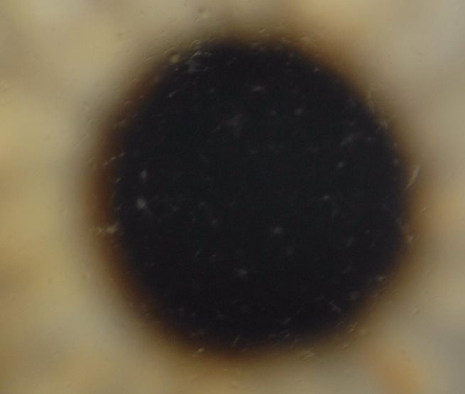
OD                                                                               OS
Figures 1 and 2 show diffuse stellate keratic precipitates in both OD and OS. Thin longer arrows point to the stellate shape of keratic precipitates and
the thick short arrow points to fibrin extension.

stellate keratic precipitates were seen diffusely distributed on                was diagnosed with a recurrent bilateral uveitis flare-
the endothelium of both eyes (See Figures 1 and 2). Grade                       up. Homatropine 5% ophthalmic solution BID OU and
1+ cells were present in the anterior chamber of OD and 2+                      prednisolone acetate 1% ophthalmic suspension every two
cells in OS without posterior synechiae. Mild diffuse iris                      hours OU were initiated. Also at this exam, the patient was
atrophy was noted OU. Goldmann applanation tonometry                            referred to a uveitis specialist to determine the appropriate
(GAT) revealed intraocular pressures of 9 mm Hg OD and                          management for this persistent and recurrent uveitis.
11 mm HG OS. Gonioscopy confirmed no peripheral anterior
synechiae. Dilated fundus examination was negative for                          The patient was seen by the uveitis specialist one week later
vitritis, but revealed mild nuclear sclerotic cataracts OU, with                and his diagnosis was determined to be bilateral Fuch’s
healthy, small cupping and an unremarkable peripheral exam.                     uveitis syndrome. The diagnosis was supported by stellate
                                                                                keratic precipitates, iris atrophy and no posterior synechiae.
His systemic medical history was only noteworthy for                            This confirmed why his past systemic work-ups had been
hyperlipidemia. His ocular history was extensive and dated                      negative and why the uveitis was persistent, despite treatment.
back to 1982 when he was first diagnosed with chronic                           The uveitis specialist recommended a taper schedule over
bilateral non-granulomatous iritis of unknown etiology.                         the next few weeks which consisted of taking prednisolone
He reported a full uveitis work-up in 1982 which had been                       acetate 1% ophthalmic suspension every two hours OU x 10
completed in the private sector. Copies of the results were                     days, then four time a day x 2 weeks, followed by three times
obtained and reviewed when the patient transferred his eye                      a day x 1week, once a day x 1 week, and then once every
care to this facility. The results were unrevealing for systemic                other day for week. He recommended switching the patient
disease (see Table 1 for lab tests and results of the uveitis                   to a maintenance dose once daily OU of fluorometholone
work-up).                                                                       0.1% ophthalmic suspension. The more mild steroid was
                                                                                recommended to limit secondary complications of steroid
In 2006 -2007 the patient was diagnosed with two bilateral                      use. The uveitis specialist reported that the goal of treatment
recurrent uveitis flare-ups at annual routine eye appointments.                 was to control symptoms as there is no curative treatment for
In both episodes he was treated acutely with prednisolone                       this type of uveitis. The current management would consist
acetate 1% ophthalmic suspension OU and tapered over                            of monitoring the patient for complications of FUS such as
the course of 2-3 months. With both recurrent episodes, the                     cataracts and glaucoma and keeping the patient comfortable.
patient never returned to the clinic for follow up appointments                 Currently this patient is managed with flurometholone
once he was tapered off of the medication.                                      0.1% ophthalmic suspension once daily OU and remains
                                                                                comfortable. IOPS remain in the low teens with mild nuclear
Given the patient’s extensive history of bilateral non-                         sclerotic cataracts OU and currently no evidence of posterior
granulomatous uveitis, on his June 11 2009 visit, the patient                   subcapsular cataracts.

                                                                                                               Fuch’s Uveitis Syndrome              157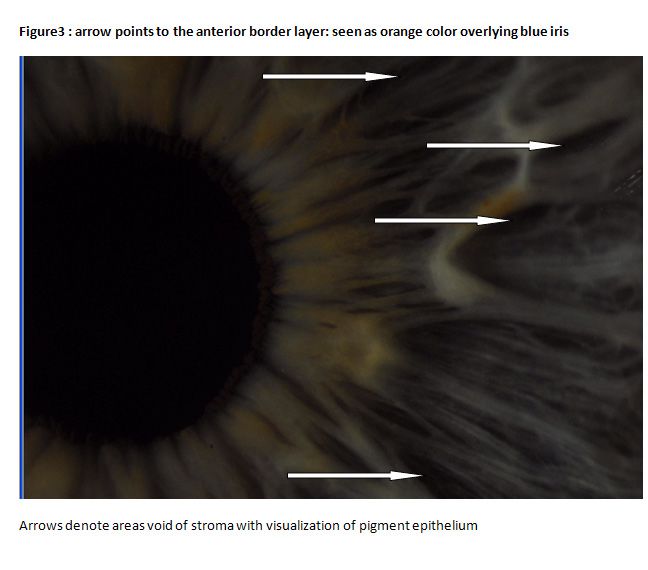
vascular permeability. A defective production of melanin
      Table 2: Differential Diagnosis of FUS
                                                                   leads to iris hypochromia while denervation of the vasculature
    Glaucoma, Pigmentary            Ocular Manifestations of HIV   leads to the leakage of protein and white blood cells into the
    Glaucoma, Uveitis               Posner-Schlossman Syndrome     anterior chamber. Although this theory may explain a select
    Herpes Simplex                  Retinitis, CMV                 few cases it does not account for the majority of cases that
    Herpes Zoster                   Sarcoidosis                    lack the concurrence of both sympathetic disease and FUS.10
    HIV                             Toxoplasmosis
    HLA-B27 Syndromes               Tuberculosis                   At one time, the possibility of hereditary FUS was
    Horner Syndrome                 Uveitis, Intermediate          contemplated, however past studies of FUS in familial cases
                                                                   cannot be substantiated. The small number of familial FUS
                                                                   cases in comparison to the overall number FUS cases weakens
Discussion:                                                        this theory. Furthermore, Loewenfeld and Thompson in 1973,
                                                                   retrospectively reviewed 1500 cases with FUS and found
Epidemiology                                                       only five families with two cases of FUS. 11
FUS accounts for approximately 2-11% of all anterior uveitis
cases. 3 The syndrome occurs more commonly in the 3rd              Additionally, Jones and Read reported a case in which FUS
to 4th decades of life with no sex or race predilection and        developed in only one child of monozygotic twins, disproving
approximately 90% of cases are unilateral.5 FUS is an unusual      the familial association.12
form of uveitis. It’s pathogenesis still remains a mystery with
speculation on a common immunologic pathway triggered              Other theories involving vascular abnormalities secondary to
by a multitude of factors. Recent literature focuses on the        an immune complex vasculitis have been considered in the
infectious rubella virus as a potential etiologic trigger.         role of triggering FUS. It was thought that immune complexes
                                                                   found in the vessel walls of FUS patients were responsible
Pathogenesis                                                       for the vascular abnormalities and chronic inflammation seen
The pathogenic mechanism of FUS remains elusive. Over the          in FUS. 10 It has also been postulated that an autoimmune
years, several theories have been proposed, however, many          reaction against altered uveal tissue secondary to a trigger,
of these cannot be substantiated. More recently, attention has     such as infection, leads to the inflammatory reaction seen in
been focused on the role of the rubella virus as a potential       FUS. However, no anti-uveal antibodies can be repeatedly
etiologic factor for FUS. However, other clinical studies          confirmed across studies.
provide evidence that demonstrate the rubella virus may not
be the only virus or causative factor involved in initiating the   Infectious etiologies have also been considered, most notably
immune response seen in FUS.                                       toxoplasmosis gondii . The coexistence of chorio-retinal
                                                                   lesions characteristic of toxoplasmosis gondii in FUS patients
There are well-documented cases of the coexistence of              was not uncommon. Multiple studies have documented the
FUS and sympathetic syndromes such as Parry-Romberg                simultaneous presence of peripheral lesions and FUS with
(hemi facial atrophy) and Horner’s syndromes. Calmettes            variable frequency ranging from 7.5% to as high as 65%.10
and Makley have confirmed two cases of Horner’s and                Because these scars have attributes of typical toxoplasmosis
FUS developing after sympathetic denervation secondary to          scars; it was originally thought that toxoplasmosis gondii
stellate ganglionectomy.6,7                                        could be the etiologic trigger for FUS. Presently, definitive
                                                                   laboratory evidence has not been established and recent
Furthermore, five cases of concurrent FUS and congenital           studies tend to refute this original association of FUS and
Horner’s syndrome have been reported in a retrospective            toxoplasmosis gondii. 13 Quentin et al., found only two of 16
study by Regenbogen and Naveh-Floman.8 Similarly, La Hey           FHC cases had increased toxoplasmosis antibody production,
and Baarsma documented a concurrent case of progressive            and one of these cases with unilateral FUS had bilateral
Parry-Romberg syndrome and FUS, possibly linking a                 peripheral chorioretinal scars formerly diagnosed as bilateral
common sympathetic defect.9 The neurogenic theory of               toxoplasmosis.3 Similarly, Devissor et al, found chorioretinal
Passow8 assumes the changes noted in FUS are a result of           scars in rubella associated uveitis patients, however not one
injury to the sympathetic nervous system. 8 The coexistence of     of these patients tested positive for the toxoplasmosis genome
FUS and Horner’s is based on the mutual commonality, loss          or for the toxoplasmosis antibody.
of sympathetic innervation. Iris heterochromia and pupillary
changes have been reported as clinical signs of impaired           Chee et al investigated the role of the cytomegalovirus
sympathetic innervation. A lack of sympathetic innervation to      (CMV) in triggering hypertensive anterior uveitis in
both the iris vasculature and stromal melanocytes is believed      immunocompetent patients. They found that 22.8% of patients
to result in defective melanin production and increased            with hypertensive uveitis were positive for CMV DNA. Of

158         Clinical & Refractive Optometry 29.4, 2018
zoster, herpes simplex, and toxoplasmosis. Of significance,
all 52 of their patients with clinically diagnosed FUS had the
presence of the rubella antibody with a statistically significant
AI. Furthermore, 0% of the 83 cases with clinically diagnosed
idiopathic anterior uveitis, toxoplasmosis retinitis, varicella
zoster and herpes simplex iritis did not have a statistically
significant AI for the rubella antibody. Similarly the non-
inflammatory control group with senile cataracts also had no
cases of a statistically significant AI for the rubella virus. 3
Interestingly, the rubella antibody was found intraocularly in
73% of multiple sclerosis (MS) patients with the diagnosis
of uveitis intermedia or periphlebitis retinae. But with MS
patients, this increased rubella antibody synthesis represented
a “polyspecific immune response” instead of a “virus driven
antibody response” as seen in FUS.3 Furthermore in the
subset of MS patients, increased antibody synthesis was also
seen for the measles, herpes simplex virus, varicella zoster
virus, and toxoplasmosis gondii indicating the polyspecific
response was the cause for increased antibody synthesis.3 A
seven fold increase in the AI was seen when evaluating FUS
versus MS, and of even more significance, the actual rubella
antibody fraction in FUS compared to MS was approximately
40 times higher. Thus, signifying the viral driven response is
specific for rubella in FUS.3
Quentin and Reiber also analyzed the AI from both the
unaffected fellow eye and the cerebrospinal fluid (CSF) of
a select few Fuchs’ patients. It was found that these select
patients had a normal AI in the unaffected eye and CSF,
indicating that FUS is a local process specific to the eye and
in most cases is unilateral.3
these patients, 75% had been clinically diagnosed with Posner
Schlossman syndrome and 20.8% diagnosed with FUS. All Similarly de Groot-Mijnes in their study found 13 out
of their treated eyes responded favorably to the anti-viral, 14 of their FUS patients had the presence of intraocular
gancyclovir. There were relapses with discontinuation of the immunoglobulin G production against the rubella virus and in
antiviral, but a good clinical response with re-initiation of the these same patients, antibody production for herpes simplex
anti- viral medication. Thus, their results showed CMV may virus, varicella zoster virus, or toxoplasmosis gondii was
play a role in select cases of FUS, as well as other previously undetected. 16
diagnosed idiopathic uveitis cases. 14
As for the intraocular persistence of the rubella virus itself in
Labalette et al, through their research confirmed CD8-positive FUS patients, Quentin et al. discovered the presence of the
T cells in the aqueous humor of Fuchs’ patients signifying an viral genome via method, polymerase chain reaction (PCR),
antigenic triggering process.15 Quentin and Reiber , expanded in 18% of FUS patients.3 However, if FUS patients less than
upon this and have provided quantitative data of aqueous 40yrs of age were isolated, this percentage increased to 56%.3
antibodies in both acute and chronic intraocular inflammation. This signifies the predilection of the persistent rubella virus
Conclusive evidence from their works have suggested that in the younger population but the duration of this persistence
an intraocular immune response against the rubella virus is is still unclear. De Groot and associates provided insight
involved in the pathogenesis for FUS. They have focused for why the viral genome and antibody synthesis may not
on analyzing the Antibody Index (AI) which represents the always correlate in infectious uveitis cases. They studied and
“relative value for the quantity of intraocularly synthesized compared both viral load and antibody synthesis production
specific antibodies.”3 Their study measured the AI in the in other infectious uveitis etiologies such as herpes simplex
aqueous humor of many eyes and determined the intraocular virus, varicella zoster virus, or toxoplasmosis gondii. They
antibody synthesis for the diseases of measles, rubella, varicella used the information provided by these laboratory tests to
Fuch’s Uveitis Syndrome 159confirm the diagnosis of the suspected uveitis infectious increase in foreign born patients seen by the uveitis clinic at
agent. The results of these tests have provided some insight as the University of Illinois. 18
to why confirmed infectious uveitis cases showed differences
between the PCR results and antibody synthesis production. Additionally, Siemerick et al have documented a case of
It was postulated that both antibody synthesis and viral load clinical FUS with positive rubella specific intraocular antibody
may vary depending on the stage the disease was in when production in a non-vaccinated 13- year- old boy. The patient
the diagnostic tests were performed. After initial insult, the showed all of the characteristic clinical signs for FUS and was
pathogen is either eliminated or the antigen load is reduced positive for rubella virus antibody synthesis in two aqueous
to an undetectable level and thus this could explain why the humor samples, taken at two different times. The aqueous
active virus is not detected with all FUS, especially older humor sample was negative for the other potential antigens
patients.17 such as herpes simplex virus, varicella zoster virus and for
toxoplasma gondii. Intraocular inflammation coupled with
Although the exact presence and role of active virus in positive rubella antibody synthesis has provided evidence to
FUS remains elusive, we can conclude that detection of a confirm the role of the rubella virus in triggering the isolated
local persistent rubella virus provides information on the uveitis seen in FUS. 19
pathomechanism for the cause of FUS. 3 Furthermore, the
role of a persistent rubella virus is supportive evidence that Although the most convincing evidence for the cause of FUS
corticosteroid therapy is not a viable treatment option for appears to be the rubella virus, in a few cases of FUS there
FUS as seen with other intraocular inflammatory conditions. still remains ambiguity. De Visser et al, in 2008, demonstrated
with their works a relationship between the clinical signs and
The theory of FUS being driven by the rubella virus has been symptoms of positive rubella associated uveitis and FUS.13
bolstered by the epidemiologic association which has shown
decreasing FUS cases occurring with the implementation According to them, 77% of rubella virus positive patients
of the rubella vaccination program in the United States. had met 3 or more of the criteria for clinical FUS, implying
Birnbaum et al, analyzed the percentages of patients with a causal relationship in a substantial number of cases 13
FUS, idiopathic chronic iridocyclitis, and idiopathic chronic However, 15% of rubella negative associated uveitis met
granulomatous iridocyclitis born over the periods from 1919- the criteria for clinical FUS, despite lack of rubella antibody
1998. They grouped these 3 subsets of patients based on the production.13 This would indicate that although the rubella
year they were born and studied the percentage of cases per virus may be the cause for the majority of the cases, it is not
decade over an eighty year span. From 1919 to 1958, the the only etiologic factor.
average number of patients seen with FUS made up 4.48% of
all uveitic cases seen at the University of Illinois. Starting in Furthermore, the occurrence of both Horner’s and FUS after
the year 1969, after the implementation of the rubella vaccine, stellate ganglionectomy would give the sympathetic theory
the number of FUS cases decreased dramatically. Between credibility in a few select cases. FUS may have more than
1969-1978, the percentage of FUS dropped to 1.18% and in one cause, and these triggers lead to “release of potent
the next decade following, dropped even further to 1% of autoantigens resulting in a common pathway of secondary
all uveitic cases whose etiology was attributed to FUS. This autoimmune uveitis that becomes self perpetuating.” 10
is significant when compared with prior decades before the
initiation the rubella vaccine. The trends for the percentages Viruses are increasingly being linked to what has previously
in the two other subsets, idiopathic chronic iridocyclitis has been considered idiopathic ocular inflammations.14 It is
and idiopathic chronic granulomatous iridocyclitis, did not important to realize that the clinical manifestations seen in
show a statistically significant change in cases per decade response to the viral antigens is most likely not specific to the
as with FUS. Interestingly, Birbaum et al also looked at the virus itself but to how the host’s immune system responds.
percentages of cases for this same period of time comparing The genetic make-up of the individual and the specific pattern
US born versus non US born with FUS. The proportion of FUS response to a particular virus will determine what is clinically
patients born outside the US appears to have increased. Most manifested by the patient.14 Whether, FUS is triggered by
of the countries representing these patients did not implement rubella virus, herpes simplex virus, cytomegalovirus or even
the rubella vaccine program until the 1980’s or later. While a sympathetic defect, the uveal tissue responds in a limited
the FUS increased over the more recent years for foreign way, which results in the clinical spectrum that is diagnosed
born FUS cases, it has remained stable for foreign born cases as FUS.10,14 Other patients, affected by the same viruses
of idiopathic chronic iridocyclitis, and idiopathic chronic may manifest a very different presentation, possibly Posner
granulomatous iridocyclitis. This negates the possibility that Schlossman syndrome or even a corneal endotheliitis. 14
foreign born increase in FUS cases is a result of an overall
160 Clinical & Refractive Optometry 29.4, 2018FUS may be part of a stereotypical response to multitude are diffusely dispersed over the entire corneal endothelium,
factors. Currently the diagnosis of FUS is made on clinical whereas in other etiologic uveitic cases, the predilection is for
observation and not laboratory testing. However, with the inferior portion of the endothelium. Franceschetti, using slit
implication of the role of viruses as a causative factor bolsters lamp biomicroscopy described these keratic precipitates as
the need for confirmatory routine laboratory testing. Whether round or star-shaped with fine filaments between the keratic
or not there is a specific FUS antigen or a multitude of precipitates. 21 Likewise, Jones described characteristic
triggers, confirming potential triggers will, hopefully, lead to keratic precipitates as “stellate with fibrillary extension,
better management. and tiny interspersed fibrils.”1 Labbe’ et al, using the higher
resolving technique of In vivo-confocal microscopy (ICVM)
Clinical Characteristics characterized keratic precipitates in 13 diagnosed FUS
Stereotypical patients are young adults that present with visual patients as dendritic in shape with a small central body and
symptoms, heterochromia and unilateral clinical findings. numerous thin pseudopodia.22 Furthermore, some of these
20
According to Velilla et.al. studied 27 eyes of 26 patients pseudopodic extensions were found to make connections
with diagnosed FUS, the most common presenting symptom between different keratic precipitates. There was great
reported was visual deterioration. Similarly, Jones studied consistency among all 13 FUS patients studied with ICVM,
103 patients with clinically diagnosed FUS and decreased all demonstrating, these characteristic stellate precipitates.22
vision was the most common symptom reported by the Similarly, Mocan et al., evaluated 14 patients with known FUS
patients at initial presentation.1 The visual deterioration was and their depiction of the keratic precipitates, “dendritiform”
either described as a mild disturbance of vision or as reduced paralleled the findings of Labbe and associates.23 Interestingly,
visual acuity. 1 The visual acuity levels recorded by Vellila et other ICVM studies looking at other infectious causes of
al were 20/40 or better and the primary cause for the reduced uveitis tend to find this similar characteristic infiltrating and
acuity was usually cataracts. Other reported symptoms by dendritic keratic precipitate. Whereas non-infectious uveitis
patients but less common was discomfort, floaters, and the keratic precipitates tend to be described as smooth, round,
awareness of heterochromia.20 The symptom of floaters and and globular. This may suggest that FUS represents “a true
its occurrence in FUS is not consistent depending on study inflammatory component, related to a triggering infectious
being reviewed. In Jones’s study, the symptoms of floaters was origin.”22 Furthermore, ICVM endothelial analysis of
found in approximately one third of FUS patients indicating unilateral FUS patients confirmed endothelial changes only
it may be more common than noted in comparative studies.1 in the involved eye, which further supports an infectious
precipitating trigger.22
Currently no diagnostic criterion for diagnosis of FUS has
been universally accepted. However, the coexistence of Heterochromia, Iris Atrophy
several common clinical features allows for diagnosis. Heterochromia has been considered an important feature
This rare form of uveitis can vary clinically however some of FUS and accounts for its incorporation into many of the
common clinical signs can aid in the diagnosis. Patients often name variations of FUS. Only more recently has the literature
have little or no ciliary flush, white and stellate diffuse keratic moved from integrating heterochromia in the syndrome’s
precipitates, iris atrophy with or without heterochromia, name. Heterochromia in FUS denotes the lighter involved
posterior subcapsular lens opacities, and vitreal cells.20 Vellila eye. However, this feature is very variable and depends on
et al studied 27 eyes with diagnosed FUS found that 100% several factors such as the initial iris color, the intensity of
of patients presented at their initial exam with diffuse keratic anterior stromal atrophy and the amount of pigment in the iris
precipitates.20 Jones’s study found 83.8% of patients to have pigmented epithelium.5 In all cases of FUS, the involved eye
keratic precipitates at initial exam, most of which were will have iris changes, however, heterochromia is not always
diffusely distributed across the corneal endothelium. Less obvious or always present. For instance, heterochromia
commonly the precipitates were distributed centrally and is typically absent in dark color irides. Also, reverse
infrequently inferiorly. 1 Other common findings were lens heterochromia can exist.5 This is when the inflamed eye has
opacities (77.8%), Heterochromia (70.4%) and an anterior the darker iris because stromal atrophy exposes a substantial
chamber reaction (66.6%). Other findings occurring with amount of the iris pigment epithelium.5 These variations and
less frequency were iris stromal atrophy (14.8%), glaucoma subtleties can complicate diagnosis. Therefore, it is critical
(14.8%), vitreal opacities (14.8%), and iris nodules (7.4%).20 the clinician be aware of the nature and course of the iris
changes seen in FUS as they can often be overlooked.
Keratic Precipitates
Characteristic endothelial keratic precipitates have been Iris findings associated with FUS may include heterochromia,
described in FUS and can provide useful insight, aiding stromal atrophy, patchy atrophy of iris pigment epithelium,
diagnosis. In most cases of FUS, the keratic precipitates increased visibility of iris vasculature, loss of iris detail, less
Fuch’s Uveitis Syndrome 161commonly iris nodules, and rarely neovascularization of the atrophy of this layer is seen as transillumination defects and
iris or irido-corneal angle. Some of these findings may be loss of the pupillary pigment ruff. Predilection for pupillary
very subtle and thus full awareness of the wide spectrum of involvement is specific in FUS but not pathognomonic.
iris presentations in FUS is critical. Transillumination defects in FUS are variable and typically
occur after atrophy of the other layers. 1 Iris atrophy and
Vast differences in iris structure and pigmentation exist among depigmentation is a common and critical feature of FUS with
patients, however, common changes among FUS patients will all layers of the iris being affected. Clinically, however, atrophy
allow for identification in an attuned clinician. Jones fully is appreciated first in the anterior border layer followed by the
depicted the changes that occur in FUS by distinguishing stroma and later pigment epithelium. The cause of this is not
the degree of atrophy in three separate layers of the iris: due to chronological order but is the result of the ability to
1-anterior border layer, 2- stroma, and 3- pigment epithelium visualize each layer’s loss by slit lamp examination.1
in 103 patients diagnosed with FUS.1 The anterior border
layer of the iris is affected early on in the disease process and Iris nodules have been reported in the literature to exist in
depigmentation of this layer is usually responsible for causing FUS patients. Rothova et al from their study implicated
heterochromia.1 This anterior border layer is described by that iris nodules without synechiae may be encountered as
Jones as an anterior stromal condensation densest in the part of FUS and may be important in the identification of
collarette region of the iris. This layer is easier to visualize FUS especially in black patients.24 In their small sample
in the light iris and will typically appear as an orange blush. of black patients diagnosed with FUS, they found the
Often this layer is indistinguishable in patients with dark common characteristic of unilateral multiple transparent iris
irides (Figure 3).1 nodules diffusely scattered across the whole surface of the
iris (Bussaca nodules) with an increase in density toward
The iris stromal layer and its ability to be visualized by the the pupillary margin (Koeppe nodules). 24 Jones found in
clinician will depend on the amount of pigmentation present. his study that 16.2% of FUS patients had iris nodules, and
The denser the stromal pigmentation the less well defined they were not associated with more severe inflammation. 1
the stroma will appear in slit lamp examination. A light iris Synechiae formation is not a typical feature in FUS, and lack
will usually show radial fibrillary architecture with stromal of its presence actually aids in diagnosis. However, should
vasculature evident and a distinct sphincter pupillae. Direct it arise, it occurs transitorily in the area of Koeppe nodules
visualization of the deeper pigment epithelium layer can be and will typicalIy present as radial pigmented lines on the
seen through crypts in the stromal layer. Crypts represent anterior lens capsule. 5 Similarly, Jones’s study although not
absolute defects of the stroma and thus a direct view to the revealing of posterior synechiae associated with Koeppe
iris pigment epithelium (Figure 3). With progressive loss nodules, did show radial stripes of pigment deposition on the
of stromal atrophy and visibility of the deeper pigment anterior lens capsule.1 This finding may suggest a previous
epithelium, a deeper blue hue to the affected iris may occur. site of adhesion. Even though the lack of posterior synechiae
This phenomenon is called reverse heterochromia and can is an important feature of FUS, these patients are not guarded
appear in those with light blue irides.1 On the contrary, a from its occurrence after cataract surgery. In Jones’s study,
dark iris will have a large supply of pigment cells resulting 4 patients following cataract surgery demonstrated posterior
in a smooth featureless appearance. A large degree of stromal synechiae secondary to uveitis. 1
atrophy must occur in dark irides before macroscopic
heterochromia becomes evident. This is why a critical slit The appearance of abnormal iris vessels in FUS has
lamp examination with bilateral iris comparison is necessary. been described in many prior publications. However, the
The first signs of stromal atrophy in dark irides are usually ambiguity and importance of these vessels still needs to be
noted as the revelation of iris detail. The architecture becomes elucidated upon. It is known that rubeotic- like vessels and
more prominent as stromal pigment and volume are lost. Thus normal radial iris vessels can be found in FUS. The cause
stromal vessels, deep excavations, and the sphincter papillae for their appearance is believed to be secondary to iris
become apparent.1 atrophy.25 Normal iris vasculature will naturally become
more apparent with atrophy and depigmentation of the iris
In general, stromal atrophy is harder to appreciate than itself. However, subclinical changes in iris angiography have
anterior border loss and therefore is typically recognized on also been reported in the past literature. 1 Such vessels have
slit lamp examination in a later stage of FUS. It is critical for shown leakage on fluorescein angiography and speculation
the clinician to compare both the affected and unaffected eyes is that these vessels may be the responsible for some of
when trying to evaluate the degree of stromal loss in FUS. 1 the aqueous flare seen in FUS. 25,26 In addition, filiform
hemorrhages following applanation tonometry, paracentesis,
The pigment epithelium is also affected in FUS. Typically or after cataract surgery have also been documented in the
162 Clinical & Refractive Optometry 29.4, 2018literature in FUS patients.1,27 Furthermore, confirmed iris Careful anterior segment evaluation is needed to look for signs
neovascularization in association with glaucoma following suggestive of herpetic disease. A dilated fundus examination
intracapsular cataract surgery was noted in a few select FUS is paramount for ruling out toxoplasmosis retinochoroiditis
patients in Jones’s study. 1 Overall, the interpretation of many or other posterior involvement. Even in cases where clinical
of these studies on abnormal vessels is unclear and, hopefully, examination is suggestive of FUS, mimickers of this disease
future studies will further elucidate their presence and role in (sarcoidosis, herpetic, and toxoplasmosis) should be ruled out
FUS. with the proper laboratory testing. The differential diagnosis
of FUS would include any conditions that can cause uveitis,
Pupil changes in FUS are not typical but may occur. Most pan-uveitis, iris heterochromia, as well as conditions that can
pupils in FUS patients are anatomically round and react cause chorioretinal scars (see Table 2).
normally. But in select cases, atrophy of the dilator or sphincter
pupillae, or partial loss of the pigment frill, may result in an Treatment
irregular pupillary response to light. Also, the affected pupil Generally, topical prednisolone acetate 1% is initiated for
could appear physiologically larger or smaller depending on treatment of the uveitis. If symptoms improve and all ancillary
the pattern of atrophy. 1 Vellila et al reported cataracts in 77.8% and laboratory testing is negative, then a full course of steroid
of FUS eyes at presentation. 20 Posterior subcapsular cataract with taper is warranted.
(PSC) was the typical type of cataract found in FUS patients.
25
Cataract was also the primary cause of visual deterioration If, however, symptoms do not improve or wax and wane then
at the time of presentation. 20 The appearance of cataracts is an oral anti-viral medication is needed to rule out herpetic
probably related to the duration of the disease. 1 In any case, etiology. If both of these treatments do not yield favorable
FUS should be high on the differentials for a young patient results and examination findings are consistent with FUS,
who presents with a unilateral cataract, especially PSC, and then the topical steroid should be tapered slowly and the
without a history of trauma or steroid use. patient should be managed for the potential complications of
FUS such as cataracts, ocular hypertension, and glaucoma,
Vitritis can also occur in FUS. Its prevalence varies across as these conditions may require additional medical and/or
studies. In Jones’, 66.6% of patients had some degree of surgical treatment.
vitreal opacification.1 Velilla et al reported in their study
14.8% of their FUS patients having vitreal involvement Unlike most uveitis syndromes, FUS typically does not
at presentataion.20 According to Mohamed et al, vitritis respond to most corticosteroid treatment. As this is the case,
is a common finding, however, it is usually mild and not most uveitis specialists will avoid the longstanding use
associated with retinal vasculitis. 25 In cases of FUS where of corticosteroids in FUS. With that in mind, therapeutic
vitreal opacification is severe, misdiagnosis for intermediate intervention may still be needed in certain cases in order
or posterior uveitis is possible.25 to address acute flare-ups, significant vitritis, ocular
hypertension, and cataract formation. Following such
But the absence of cystoid macula edema differentiates FUS treatment, palliative therapies are suggested and close
from other chronic vitritis conditions.25 monitoring is recommended.
Glaucoma has been associated in FUS and its presence Conclusion
depending on the study varies from 15%-59% of FUS patients In sum, FUS is an atypical form of uveitis that has a variable
1,28
According to Jones, 26.2% of FUS patients were treated clinical spectrum making diagnosis difficult. It is considered
for glaucoma during some stage of the disease process. 1 to be an under-diagnosed syndrome, likely also because it
Typically the glaucoma is a chronic open angle form. Several lacks universal clinical criteria. The complaint of floaters
factors have contributed to this secondary glaucoma, such as and/or vision deterioration in a young adult with a unilateral
trabeculitis, neovascularization of the iris stroma and angle, uveitis in conjunction with a relatively quiet eye, should alert
induced from steroid treatment, and induced from cataract the clinician to the possibility of FUS.20 Vellila et. al. found
surgery. 20 that delay in diagnosis can range from several days to 24
years.20 Cunningham and Baglivo noted that the mean time
Diagnosis and testing for diagnosis is 3 years, during which time approximately
At this time, there are no laboratory tests that can render the two thirds of patients received anti-inflammatories including
diagnosis of FUS. Rather, the diagnosis of FUS is a clinical immunosuppressive treatments.2 In the current case, the
one. The clinician should take a detailed history and perform patient was diagnosed 27 years following his first incident
a complete comprehensive examination, including dilation. of uveitis. Thus, this case demonstrates the importance of
Fuch’s Uveitis Syndrome 163early proper diagnosis as it is vital for prevention of side 19. Siemerink MJ, Sijssens KM, de Groot-Mijnes JDF, de Boer
effects caused from unnecessary long-term corticosteroid use JH. Rubella virus-associated uveitis in a non-vaccinated child.
leading to unnecessary mortality. Am J Ophthalmol 2007;143:899-900.
20. Velilla S, Dios E, Herreras JM, Calonge M. Fuchs’s
heterochromic iridocyclitis: A review of 26 cases. Ocular
References:
Immunology and Inflammation 2001;9:169-175.
1. Jones NP. Fuchs’ heterochromic uveitis: a reappraisal of the
21. Franceschetti A. Heterochromic cyclitis: Fuchs’ syndrome. Am
clinical spectrum. Eye 1991;5: 649-661.
J Ophthalmol 2005;39:50-8.
2. Cunnigham ET, Baglivo E. Fuchs heterochromic iridocyclitis-
22. Labbe’ A, Dupas B, Offret H, Baudouin C, Labetoulle.
syndrome, disease, or both. Am J Ophthalmol 2009;148:479-481.
Evaluation of keratic precipitates and corneal endothelium in
3. Quentin CD, Reiber H. Fuch’s heterochromic cyclitis: rubeela
Fuchs’ heterochromic cyclitis by in vivo confocal microscopy.
virus antibodies and genome in aqueous humor.
Br J Ophthalmol 2009;93:673-7.
Am J Ophthalmol 2004;138:46-54.
23. Mocan MC, Kadayifcilar S, Irkec M. Keratic precipitate
4. Rothova A. The riddle of Fuchs heterochromic uveitis.
morphology in uveitic syndromes including Behet’s disease as
Am J Ophthalmol 2007;144:447-8.
evaluated with in vivo confocal microscopy. Eye. Published
5. Bonfiolo AA, Curi ALL, Orefice F. Fuchs’ heterochromic
Online First: 25 July 2008. doi: 10.1038/eye.2008.239
cyclitis. Seminars in Ophthalmology 2005;20:143-146.
24. Rothova A, La Hey E, Baarsma GS, Breebaart AC. Iria nodules
6. Calmettes ML, Deodati R, Amalric P. UN cas d’association
in Fuchs’ heterochromic uveitis. Am J Ophthalmol
d’heterochromic de Fuch’s et de syndrome de Claude Bernard.
1994;118:338-342.
Rev Oto-Neuro-Ophthalmol 1953;25:399-400.
25. Mohamed Q, Zamir E. Update on Fuchs’ uveitis syndrome.
7. Makley TA, Abbot K. Neurogenic heterochromia:report of an
Curr Opin Ophthalmol 2005;16:356-363.
interesting case. Am J Ophthalmol 1965;59:927-8.
26. Norrselll K, Holmer AK, Jacobson H. Aqueous flare in patients
8. Regenbogen L, Naveh-Floman N. Glaucoma in Fuch’s
with monocular iris atrophy and uveitis: a laser flare and iris
heterochromic cyclitis associated with congential Horner’s
angiography study. Acta Ophthalmol Scand 1998;76(4):405-412.
syndrome. Br J Ophthalmol 1987;71:844-9.
27. Jones NP. Extracapsular cataract surgery with and without
9. La Hey E, Baarsma G. Seerp. Fuch’s heterochromic cyclitis and
intraocular lens implanatation in Fuchs’ heterochromic uveitis.
retinal vascular abnormalities in progressive hemifacial atrophy.
Eye 1990;4:145-150.
Eye 1993;7:426-8.
28. Jones NP. Glaucoma in Fuchs’ heterochromic uveitis: etiology,
10. La Hey E, De Jong P T V M, Kijlstra A. Fuchs’ heterochromic
management, and outcome. Eye 1991;5:649-661.
cyclitis:review of the literature on the pathogenetic mechanisms.
Br J Ophthalmol 1994;78:307-12.
11. Loewenfeld IE, Thompson HS. Fuch’s heterochromic cyclitis:
a critical review of the literature. II. Aetiology and mechanism.
Surv Ophthalmol 1973;17:2-61.
12. Jones AP, Read AP. Is there a genetic basis for Fuch’s
heterochromic uveitis? Discordance in monozygotic twins. Br J
Ophthalmol 1992;76:22-4.
13. De Visser L, Braakenburg A, Rothova A, De Boer JH. Rubella
virus-associated uveitis: clinical manifestations and visual
prognosis. . Am J Ophthalmol 2008;146:292-7.
14. Chee S-P, Bacsal K, Jap A, Se-Thoe S-Y, Li Cheng CL, Tan
BH. Clinical features of cytomegalovirus anterior uveitis in
immunocompetent patients. Am J Ophthalmol 2008;145:834-840.
15. Labalette P, Caillau D, Grutzmacher D, Dessaint J-P, Labalette
M. Highly focused clonal composition of CD8(+) CD28(neg)
T-cells in aqueous humor of Fuchs’ heterochromic cyclitis. Exp
Eye Res 2002;75:317-325.
16. De Groot-Mijnes JDF, de Visser L, Rothova A, Schuller M, van
Loon AM, Weersink AJL. Rubella virus is associated with
Fuchs heterochromic iridocycllitis. Am J Ophthalmol
2006;141:212-4.
17. De Groot-Mijnes JDF, Rothova A, Van Loon Am, et al.
Polymerase Chain reaction and Goldmann-Witmer coefficient
analysis are complimenatary for the diagnosis of infectious
uveitis. Am J Ophthalmol 2006;141:313-8.
18. Birnbaum AD, Tessler HH, Schultz KL, et al. Epidemiologic
relationship between Fuchs heterochromic iridocyclitis and
the United States rubella vaccination program. Am J Ophthalmol
2007;144:424-8.
164 Clinical & Refractive Optometry 29.4, 2018INSTRUCTIONS FOR 1 HOUR OF CE CREDIT
COPE APPROVED CE-CREDIT POST COURSE TEST QUESTIONNAIRE
This course is valid for 1 hour of COPE CE Credit, if the post-course test shown below is taken and
submitted online for grading no later than February 15, 2022, and a score of 70% or more is achieved.
CLICK HERE TO TAKE AND SUBMIT THIS POST-COURSE TEST ONLINE
Please Note: That in order for you to be able to take this (or any other) CRO Online post-course test, you
must first have registered on the CRO website at www.crojournal.com to open a Personal Dashboard
Account. This will allow you to enroll, complete and submit this or any of our other CRO Online CE
Credit Courses for grading. The registration process is quick and simple and only needs to be done once.
Following your taking and submitting a completed post course test with a score of 70% or more, a
personalized COPE CE Credit Certificate will automatically be made available for you to download from
your Personal Dashboard Account.
For answers to any questions about either the registration or post course test processes, or to report a
specific problem, please contact CRO Support at info@crojournal.com.
IDENTIFICATION
Name: First______________________________ Last___________________________________
Address:________________________________________________________________________
Number Street Suite
_______________________________________________________________________________
City Province Postal Code
Professional License Number:_______________ Office Phone: ( ) _______________________
E-mail: ________________________________________________________________________
QUESTIONNAIRE
Fuch’s Uvietis Syndrome: A Variable Clinical Spectrum
Theresa Zerilli-Zavgorodni, OD FAAO Tam Nguyen, OD MS FAAO Sharon Bisighini, OD FAAO
1 Which statement describing the diagnosis of Fuch’s Uveitis Syndrome (FUS) is incorrect?
q FUS is currently defined as a syndrome recognized by a triad of clinical characteristics
q All the most common clinical characteristics of FUS may not always be present at the same time.
q Diagnostic laboratory testing should be used to make and/or confirm a diagnosis of FUS
q Currently FUS lacks a well characterized pathophysiological mechanism.
2 Which percentage of all anterior uveitis cases are confirmed to be Fuch’s Uveitis Syndrome?
q 1-11%
q 12-22%
q 23-33%
q 34-44%
3 According to Velilla et. al. what is the most common presenting symptom of FUS?
q Persistent headache
q Transient vision loss
q Visual deterioration
q Tunnel visionINSTRUCTIONS FOR 1 HOUR OF CE CREDIT
COPE APPROVED CE-CREDIT POST COURSE TEST QUESTIONNAIRE
This course is valid for 1 hour of COPE CE Credit, if the post-course test shown below is taken and
4 Which characteristic of endothelial keratic precipitates is incorrect?
submitted online for grading no later than February 15, 2022, and a score of 70% or more is achieved.
q In most cases of FUS the keratic precipitates are dispersed evenly over the entire corneal endothelium
q In some cases of FUS the keratic precipitates are dispersed unevenly over the inferior position of the corneal
CLICK HERE TO TAKE AND SUBMIT THIS POST-COURSE TEST ONLINE
endothelium.
q The precipitates associated with FUS cases have been described as being dendritic in shape with numerous
Please Note: That in order for you to be able to take this (or any other) CRO Online post-course test, you
thin pseudopodia.
qmustThefirst have registered
precipitates onwith
associated the CRO website
cases of at www.crojournal.com
non-infective to open aasPersonal
uveitis have been described Dashboard
being smooth, round
and globular.
Account. This will allow you to enroll, complete and submit this or any of our other CRO Online CE
Credit Courses for grading. The registration process is quick and simple and only needs to be done once.
5 Which incidence rate of the following clinical characteristics for diagnosed cases of FUS is incorrect?
q Lens-opacities 77.8%
Following your taking and submitting a completed post course test with a score of 70% or more, a
q Heterochromia 70.4%
personalized COPE CE Credit Certificate will automatically be made available for you to download from
q Anterior chamber reaction 66.6%
qyour Personal
Diffuse Dashboard
keratic Account.
precipitates 24.3%
6ForWhich
answers
iris to any questions
finding aboutassociated
is not typically either thewith
registration
FUS? or post course test processes, or to report a
specific problem,
q Heterochromia please contact CRO Support at info@crojournal.com.
q Stromal atrophy
q Distended iris vasculature
q Patchy atrophy of iris pigment epithelium IDENTIFICATION
Name: First______________________________
7 Which Last___________________________________
characteristic of pigment epithelium affected by FUS is incorrect?
q Typically atrophy of the pigment epitthelium is seen as transillumination defects
Address:________________________________________________________________________
and loss of pupillary pigment
Number
q Predilection for pupillary Street and not pathognomonic.
involvement is specific in FUS Suite
_______________________________________________________________________________
q Transillumination defects in FUS are variable and typically occur after atrophy of the other layers
q These defects City Province progresses.
occur in a chronologic order as the syndrome Postal Code
Professional License Number:_______________ Office Phone: ( ) _______________________
8 Which statement about cataracts associated with FUS is not true?
E-mail:
q They________________________________________________________________________
are usually posterior subcapsular cataracts in young patients
q The cataracts are not usually associated with a history of trauma or steroid use
q The cataracts are usually bilateral QUESTIONNAIRE
q The cataracts are usually the primary cause of reported visual deterioration
9 Which procedure is not necessary inorder to diagnose FUS?
q A complete CBC workup including C-Reactive Protein levels
q A detailed Medical History and comprehensive physical examination
q A careful anterior segment evaluation to look for signs of herpetic disease.
q A dilated fundus exam to rule out posterior involvement and FUS mimickers
10Which option is not recommended for treating FUS?
q Treatment usually begins with the topical administration of 1% prednisolone acetate for the uveitis
q If the uveitis symptoms improve then the topical medication should be replaced with oral steroids
q If following the start of oral steroids there is no improvement then the steroids should be tapered off and
replaced with an oral anti-viral medication for herpetic disease
q If following the start of oral steroids there is no improvement then the steroids should be continued and an
oral anti-viral medication should be added for herpetic disease
166 Clinical & Refractive Optometry 29.4, 2018You can also read