Case Report Spinal Cystic Schwannoma: A Rare Case Report - Bangladesh Journals Online
←
→
Page content transcription
If your browser does not render page correctly, please read the page content below
DOI: https://doi.org/10.3329/bjns.v10i2.53779
Case Report
Spinal Cystic Schwannoma: A Rare Case Report
Das S1, Khan AH2, Sarker AC3, Ghosh D4
Abstract
Conflict of interest: There is no conflict Spinal cystic Schwannomas are very rare tumors and have been reported in only a few
of interest relevant to this paper to case reports in literature. Our case, Mrs. Halima, a 48-year-old female was admitted in
disclose.
Neurosurgery department of Dhaka Medical College Hospital with the complaints of
Funding Agency : was not funded by
low back pain more at night occasionally radiating to right buttock and numbness and
any institute or any group.
tingling of the right lower limb for 2 years. MRI of Lumbo-sacral spine revealed a large
Contribution of Authors : Principal
Investigator- Dr. Sukriti Das circumscribed and well delineated intradural extramedullary space occupying lesion
Manuscript preparation- Professor Dr. of iso to hypointense appearance in T1WI with ring enhancement on administration of
Akhlaque Hossain Khan gadolinium. Per-operative observation and post-operative histopathological
Data collection- Professor Dr. Asit examination revealed schwannoma. Contrast enhanced MRI was very important for
Chandra Sarker
the diagnosis of this rare type of tumor and surgical planning. Surgical removal was
Editorial formatting- Dr. Dipankar Ghosh
complete and patient was recovered uneventfully.
Copyright: @2020bang.BJNS published
by BSNS. This article is published under Key Words: Cystic schwannoma, Magnetic Resonance Imaging, Intradural-
the creative commons CC-BY-NC extramedullary.
license. This license permits use
distribution (https://creativecommons. Bang. J Neurosurgery 2021; 10(2): 223-225
orgf/licences/by-nc/4-0/)reproduction in
any medium, provided the original work
is properly cited and is not used for
commercial purposes.
Received: 10.09.2020
Accepted: 12.11.2020
Introduction pain more at night occasionally radiating to right
Spinal schwannomas are benign tumors arising from buttock with mild difficulty in walking for same duration.
the spinal nerve root sheaths and are the commonest She did not give any history of trauma, fever, night
intradural extramedullary spinal tumors 1 . sweats, cough, hemoptysis, chest pain, melena etc.
Schwannomas are mostly solid or heterogeneously On examination, she was found mid-aged female with
solid tumors. Though cystic changes in solid average body built, non-anemic, anicteric and all the
vital signs were within normal limit. On lower limbs
schwannomas are well described, predominantly
examination, she had motor weakness in right lower
cystic schwannomas are uncommon4,5. Differentiating limbs (MRC grade 4+/5 at the knee and ankle),
these cystic lesions from similar cystic lesions in the decreased sensations below the level of L2
intradural extramedullary space is important, and dermatome. No abnormal deep tendon reflexes or
Magnetic Resonance Imaging (MRI) plays a vital role pathological reflexes were found to be exist. The spinal
in this context. radiographs revealed no endplate destruction,
enlargement of spinal canal, or deformity of spinal
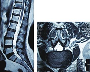
Case Report: structures. MRI of the lumbar spine revealed an
A 48-year-old female was admitted in the Neurosurgery intradural cystic mass occupying the L2-L4 space,
Department of Dhaka Medical College Hospital with which appeared the iso to hypointense appearance in
complaints of numbness and tingling of the right lower T1WI with hyperintensity in T2WI. The adjacent cauda
limb for 2 years. She also complained of low back equina were displaced by the lesion. After intravenous
1. Dr. Sukriti Das, Associate Professor, Department of Neurosurgery, Bangabandhu Sheikh Mujib Medical University.
2. Professor Dr. Akhlaque Hossain Khan, Professor, Department of Neurosurgery, Bangabandhu Sheikh Mujib Medical University.
3. Professor Dr. Asit Chandra Sarker, Head- Department of Neurosurgery, Dhaka Medical college & Hospital.
4. Dr. Dipankar Ghosh, Resident (Phase-B), Department of Neurosurgery, Dhaka Medical College & Hospital
Address of Correspondence: Dr. Sukriti Das, MBBS, FCPS (Surgery), MS (Neurosurgery), FRCS (Edin), Associate Professor,
Department of Neurosurgery, Bangabandhu Sheikh Mujib Medical University. Cell phone: +8801711676848, e-mail: sukriti66@yahoo.com.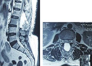
Bangladesh Journal of Neurosurgery                                                     Vol. 10, No. 2, January 2021

gadolinium administration, the cystic mass showed         The cyst was decompressed and clear CSF-like fluid
a marginal enhancement (ring enhancement).                came out from the lesion. Then the lesion was excised
                                                          maintaining the arachnoid plane. The patient had an
The patient underwent laminectomy at L2-L4level with
                                                          uneventful recovery and her motor and sensory
total excision of the lesion. Intraoperatively, after
                                                          impairment improved to near normal post-operatively.
durotomy, a cystic lesion spontaneously came out.
                                                      Table-I
              Differential diagnosis of cystic spinal schwannomas: Analysis on MRI findings2,4,7
Lesion              Salient features             T1WI             T2WI               Contrast enhanced T1WI
Cystic              Mostly seen in the           Iso-to           Hyperintense       Intense ring
schwannoma          cervical and lumber spine.   hypointense                         enhancement,
                    Dorsal or lateral                                                Irregular walls,
                    to spinal cord. Extradural                                        Septation.
                    extension with dumb-bell
                    appearance.
Arachnoid           Mostly seen in the           Hypointense,     Hyperintense,      No contrast
cyst                thoracic spine. Situated     Smooth wall      Smooth wall        enhancement.
                    dorsal to cord. No
                    restriction on DWI.

Fig.-1: Pre-operative MRI                                 Fig.- 2: Per-operative photographs

Fig.-3: Post-operative MRI                                Fig.-4: Post-operative patient condition

224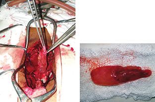
Spinal Cystic Schwannoma: A Rare Case Report                                                                         Das S et al.

Discussion:                                                 Hemi-laminectomy or laminectomy at the level of
Schwannomas are the most common primary spinal              lesion, followed by total excision was followed in
cord tumors, accounting for approximately one-third         treating these lesions at our institute, and good
of cases, usually occurring as solitary, well-              postoperative outcome was achieved in all patients.
circumscribed, encapsulated, solid or heterogeneously
solid, eccentrically located in intradural extramedullary   Conclusion:
space of spinal nerve roots mostly in the cervical and      Cystic spinal schwannomas are uncommon lesions,
lumbar region1,6.                                           presenting as intradural extramedullary lesions mainly
                                                            in the cervical and lumbar spine. Contrast MRI is the
Schwannomas are usually entirely solid or                   investigation of choice and plays a major role in
heterogeneously solid tumors1. Predominantly cystic         predicting these lesions preoperatively. Thick irregular
spinal schwannomas are uncommon lesions and may             walls with septations, which intensely enhance on
pose a preoperative diagnostic dilemma4. Various            contrast injection, can accurately predict cystic
theories have been proposed to explain the cystic           multiloculated schwannomas. Uniloculated cystic
changes occurring in schwannomas. Degeneration of           schwannomas can be considered in cases of purely
the Antoni B portion of a schwannoma can result in          cystic lesions with enhancement of the thin wall.
cyst formation, which may then progress to form a           However, it is unlikely that all such cases can be
larger cyst4,5. This hypothesis explains the formation      predicted preoperatively on radiology. It is important
of totally cystic (uniloculated) schwannomas. Central       to detect these lesions at surgery, as total excision
ischemic necrosis/hemorrhage can be caused by               is possible and almost always results in good long-
tumor growth resulting in cyst formation within the         term neurological outcome.
tumor4,5. Another theory attributes cystic change in
schwannomas to mucinous degeneration3,5. This               References:
                                                            1.   Conti P, Pansini G, Mouchaty H, Capuano C, Conti R. Spinal
theory may hold good for the multiloculated cystic
                                                                 neurinomas: Retrospective analysis and long-term
schwannomas.                                                     outcome of 179 consecutively operated cases and review
The peak incidence of spinal schwannomas is in the               of the literature. Surg Neurol. 2004; 61:35–44.

fourth and fifth decades of life and they do not exhibit    2.   Friedman DP, Tartaglino LM, Flanders AE. Intradural
                                                                 schwannomas of the spine: MR findings with emphasis
any predilection to a particular sex. Cystic tumors
                                                                 on contrast-enhancement characteristics. AJR Am J
have a high risk of causing progressive worsening of             Roentgenol. 1992; 158:1347–50.
symptoms as a result of cyst expansion3. MRI is the
                                                            3.   Karatas A, Merih IS, Yildirim U, Akyuz F, Gezen F. Thoracic
preferred imaging modality for establishing diagnosis.           intradural cystic schwannoma: A case report. Turk
Schwannomas generally have low-to-intermediate                   Neurosurg. 2007; 17:193–6.
signal intensity on T1WI. On T2WI, they may be              4.   Parmar H, Patkar D, Gadani S, Shah J. Cystic lumbar nerve
heterogenous with focal areas of hyper-or                        sheath tumours: MR features in five patients. Australas
hypointensity. Focal areas of intense hyperintensity             Radiol. 2001; 45:123–7.
on T2WI often correspond to cystic portions, whereas        5.   Shiono T, Yoshikawa K, Iwasaki N. Huge lumbar spinal
hypointensity may represent hemorrhage, dense                    cystic neurinomas with unusual MR findings. AJNR Am J
cellularity, or collagen deposition2.                            Neuroradiol. 1995; 16(4 Suppl):881–2.
                                                            6.   Van Goethem JW, van den Hauwe L, Ozsarlak O, De
The treatment of cystic schwannomas involves total
                                                                 Schepper AM, Parizel PM. Spinal tumors. Eur J Radiol.
excision of the lesion. Total excision is recommended            2004; 50:159–76.
because inadequate removal has a risk of recurrence.        7.    Beall DP, Googe DJ, Emery RL, Thompson DB, Campbell SE,
In many large series, it has been confirmed that                 Ly JQ, et al. Extramedullary intradural spinal tumors: A pictorial
recurrence occurred in all cases in subtotal excision1.          review. Curr Probl Diagn Radiol. 2007; 36:185–98.

                                                                                                                             225
You can also read