Case Report Cauda Canis: Variation of a Tinel's Sign for a Sciatic Nerve Tumor
←
→
Page content transcription
If your browser does not render page correctly, please read the page content below
Hindawi
Case Reports in Neurological Medicine
Volume 2020, Article ID 8822866, 4 pages
https://doi.org/10.1155/2020/8822866

Case Report
Cauda Canis: Variation of a Tinel’s Sign for a Sciatic Nerve Tumor

          Keith George ,1 Shane Burke ,1 Knarik Arkun ,1,2 and Ron Riesenburger1
          1
              Department of Neurosurgery, Tufts Medical Center, 800 Washington Street, Boston 02111, MA, USA
          2
              Department of Pathology, Tufts Medical Center, 800 Washington Street, Boston 02111, MA, USA

          Correspondence should be addressed to Keith George; keith.george@tufts.edu

          Received 25 September 2020; Revised 24 November 2020; Accepted 8 December 2020; Published 15 December 2020

          Academic Editor: Mehmet Turgut

          Copyright © 2020 Keith George et al. This is an open access article distributed under the Creative Commons Attribution License,
          which permits unrestricted use, distribution, and reproduction in any medium, provided the original work is properly cited.

          A patient with a prior history of intradural schwannoma and disc herniation presented with radicular pain after being hit in the
          thigh by a dog’s tail. She was worked up and found to have a tumor of her right sciatic nerve. The tumor was resected and histology
          was consistent with schwannoma. The dog’s tail acted as a Tinel’s sign maneuver and led to timely identification of her peripheral
          nerve tumor. Peripheral nerve schwannomas can present in unusual forms, and Tinel’s maneuver may be a useful tool
          in diagnosis.

1. Introduction                                                         sat and improved with activity. She did not endorse a
                                                                        personal history or family history of neurofibromatosis.
Radicular pain in patients undergoing neurosurgical eval-               Physical exam was notable for 4/5 strength in her right
uation can be attributed to a variety of causes. A majority of          anterior tibialis, 4/5 right extensor hallucis longus, decreased
these causes include pathology at the spinal level, principally         sensation to the lateral aspect of the right ankle, and positive
disc herniation, spinal stenosis, and degenerative changes              Tinel’s sign on the posterior thigh. An MRI of the right thigh
leading to bone spurs [1]. However, alternative peripheral              showed a tumor along the right sciatic nerve, 3 cm in size
causes of radicular pain should also be considered part of the          (Figure 1).
full workup. We present an interesting case and the first of its             A preoperative ultrasound was obtained to help localize
kind, where a woman was found to have a tumor of her                    the tumor for surgery. Ultrasound showed the center of the
sciatic nerve after she was hit in her thigh by a dog’s tail            mass 17 cm rostral to the skin crease of the popliteal fossa,
(cauda canis translates to dog’s tail in Latin) and subse-              with the caudal section of the mass 16 cm from the crease
quently developed radicular pain.                                       and the rostral portion 18.5 cm from the crease.
                                                                            During the procedure, the patient was placed prone, and
2. Case Presentation                                                    an intraoperative ultrasound confirmed that the tumor was
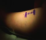
                                                                        18 cm from the popliteal fossa (Figure 2). An incision was
A 57-year-old female, with a prior history of intradural L2             made along the right posterior thigh, and muscle was dis-
lumbar schwannoma that was resected 7 years ago, pre-                   sected until the sciatic nerve tumor was exposed. Intra-
sented with right leg pain that radiated into her lateral leg,          operative electromyography (EMG) monitoring was used to
down to her ankle and big toe. A year prior to her current              delineate tumor versus intact nerve. A section of tumor was
visit, she was found to have an L4-L5 herniated disc with               sent for pathology, and the tumor was histologically con-
right-sided radiculopathy and deferred surgical interven-               firmed to be a schwannoma (Figures 3 and 4). Debulking was
tion, and improved with conservative measures. After some               avascular, and the tumor was excised from the nerve, with
time, she was hit in the back of her right thigh by the tail of a       minimal fascicle loss (Figure 5). The patient woke without a
Labrador dog, and she buckled and noticed severe electric               deficit. The patient recovered well postoperatively with no
pain shooting down her leg, akin to a Tinel’s sign finding.              complications. She was followed up for six weeks after the
The pain occasionally awakened her and was worse when she               surgery and was found to have an improvement in
2 Case Reports in Neurological Medicine
Figure 1: MRI axial showing right sciatic nerve tumor, 4 cm from the skin.
(a) (b)
Figure 2: (a) Intraoperative ultrasound localizing tumor. (b) Planned incision along the right posterior thigh.
impinging on the sciatic nerve. Other cases of sciatic nerve
schwannomas associated with Tinel’s sign have been de-
scribed in the literature [2–4]. Kralick et al. describe a similar
case where a patient shared similar symptoms and had
difficulty sitting or lying in bed and also had a positive Tinel’s
sign on physical exam. Initial radiographic studies could not
identify a source for the radicular pain, and as a result of the
Tinel’s sign, a lower extremity MRI was obtained that
captured the tumor, allowing the patient to be adequately
treated. In our case, the patient’s presentation could have
initially been attributed to a recurrence of her prior L4-L5
Figure 3: Gross specimen of schwannoma.
disc herniation. Were it not for the positive Tinel’s sign as a
result of the dog’s tail, there could have been a delay in
symptoms, with good healing of her surgical incision and 5/5 identifying the true cause of the patient’s symptoms.
strength bilaterally in her lower extremities. Histological analysis showed the tumor to be a schwan-
noma, with characteristic Antoni A and Antoni B patterns,
3. Discussion along with the presence of Verocay bodies. Schwannomas are
encapsulated nerve sheath tumors of Schwann cells. They are
This is a unique case of an underlying tumor being suspected the most common form of peripheral nerve tumor, accounting
through a patient’s physical contact with a dog’s tail (cauda for about 35% of all benign peripheral nerve sheath tumors
canis sign), and the first of such a case to our knowledge. The [5–7]. Despite this, involvement of the sciatic nerve is still rare,
dog’s tail whipping against the posterior thigh and the re- typically less than 1% of cases [8].
sultant electric radiating pain the patient experienced can be Given her history of a prior schwannoma that was
related to a positive Tinel’s sign, which was reproducible on intradural, the patient could have an underlying pathology.
exam. The radicular pain likely arises from the tumor A population-based study found that 90% of schwannomasCase Reports in Neurological Medicine 3
(a) (b)
Figure 4: (a) H&E high magnification (X200) of Antoni A area with Verocay bodies formation. (b) H&E low magnification (X40) of
biphasic spindle cell lesion with cellular Antoni A areas and numerous Verocay bodies formation on the right, along with many hyalinized
vessels and fresh hemorrhage, characteristic of schwannoma. Hypocellular area with looser stroma or Antoni B pattern is on the left.
Pigment laden macrophages are present around vessels at the bottom of the image, representing evidence of old microbleed.
(a) (b)
Figure 5: (a) View of the surgical field with tumor along the sciatic nerve. (b) Tumor excised with sciatic nerve left intact (yellow arrow).
are sporadic, while 3% occurred in patients with neurofi- Conflicts of Interest
bromatosis type 2 (NF2) and 2% in patients with schwan-
nomatosis [9]. NF2 is an autosomal dominant disease that is The authors declare that there are no conflicts of interest.
linked to mutations of the NF2 gene, a tumor suppressor
gene, located on chromosome 22. Inactivation of this gene Acknowledgments
predisposes a patient to developing various tumors in the
nervous system [10]. Patients with schwannomatosis are also The authors thank Walter Dent, Department of Neuro-
susceptible to tumor spread along the nervous system, but surgery, Tufts Medical Center, for acquisition of photos and
the genes implicated in pathogenesis are distinct from the other media related to the case.
NF2 gene [11]. Differentiating between NF2 and schwan-
nomatosis is difficult, as diagnosis is determined by clinical
criteria [12]. The patient’s diagnosis will depend on gene References
testing and follow-up MRI. If there is an absence of a [1] E. Patel and M. Perloff, “Radicular pain syndromes: cervical,
vestibular tumor on MRI and no evidence of an NF2 gene lumbar, and spinal stenosis,” Seminars in Neurology, vol. 38,
mutation, this patient will qualify for a diagnosis of no. 6, pp. 634–639, 2018.
schwannomatosis. She will be followed up by both neuro- [2] F. Kralick and R. Koenigsberg, “Sciatica in a patient with
surgery and neuro-oncology. Family testing will also be unusual peripheral nerve sheath tumors,” Surgical Neurology,
recommended. vol. 66, no. 6, pp. 634–637, 2006.
[3] A. Rhanim, R. El Zanati, M. Mahfoud, M. S. Berrada, and
Data Availability M. El Yaacoubi, “A rare cause of chronic sciatic pain:
schwannoma of the sciatic nerve,” Journal of Clinical Or-
Data on the case report are restricted by patient privacy. thopaedics and Trauma, vol. 4, no. 2, pp. 89–92, 2013.4 Case Reports in Neurological Medicine
[4] K. Mezian, J. Vacek, L. Navrátil, L. Özçakar, and L. Navrátil,
“Bilateral rectus femoris muscle rupture following statin
medication,” American Journal of Physical Medicine & Re-
habilitation, vol. 96, no. 7, pp. e138–e140, 2017.
[5] A. K. Bhattacharyya, R. Perrin, and A. Guha, “Peripheral
nerve tumors: management strategies and molecular in-
sights,” Journal of Neuro-Oncology, vol. 69, no. 1–3,
pp. 335–349, 2004.
[6] O. Godkin, P. Ellanti, and G. O’Toole, “Large schwannoma of
the sciatic nerve,” BMJ Case Reports, vol. 2016, Article ID
bcr2016217717, 2016.
[7] D. H. Kim, J. A. Murovic, R. L. Tiel, G. Moes, and D. G. Kline,
“A series of 397 peripheral neural sheath tumors: 30-year
experience at Louisiana State University health sciences
center,” Journal of Neurosurgery, vol. 102, no. 2, pp. 246–255,
2005.
[8] S. J. Omezzine, B. Zaara, M. Ben Ali, F. Abid, N. Sassi, and
H. A. Hamza, “A rare cause of non discal sciatica: schwan-
noma of the sciatic nerve,” Orthopaedics & Traumatology:
Surgery & Research, vol. 95, no. 7, pp. 543–546, 2009.
[9] J. Antinheimo, R. Sankila, O. Carpén, E. Pukkala, M. Sainio,
and J. Jääskeläinen, “Population-based analysis of sporadic
and type 2 neurofibromatosis-associated meningiomas and
schwannomas,” Neurology, vol. 54, no. 1, p. 71, 2000.
[10] D. R. Evans, “Neurofibromatosis type 2 (NF2): a clinical and
molecular review,” Orphanet Journal of Rare Diseases, vol. 4,
no. 1, p. 16, 2009.
[11] A. Piotrowski, J. Xie, Y. F. Liu et al., “Germline loss-of-
function mutations in LZTR1 predispose to an inherited
disorder of multiple schwannomas,” Nature Genetics, vol. 46,
no. 2, pp. 182–187, 2014.
[12] M. MacCollin, E. A. Chiocca, D. G. Evans et al., “Diagnostic
criteria for schwannomatosis,” Neurology, vol. 64, no. 11,
pp. 1838–1845, 2005.You can also read


