Case Presentation: 62 Year Old Man Presents with Dysarthria and Weakness - Jacob Garrell MS3 - UConn ...
←
→
Page content transcription
If your browser does not render page correctly, please read the page content below
Case Presentation: 62 Year Old Man
Presents with Dysarthria and Weakness

            Jacob Garrell MS3
Patient Presentation
• 62 year old man presented to the ED at 2 PM with chief
  complaint of dysarthria and weakness. Patient reported a
  1 month history of increasing weakness and generalized
  malaise leading to this morning. Upon waking up, he
  experienced significant exhaustion, but no specific
  symptoms. After attempting to engage in golf, he reported
  that he was too fatigued to play. Around 11 AM, he
  reported that he began to struggle with his speech. He
  was transported to the ED by EMS as a stroke code.
Code Stroke called to ED
• Patient is rushed to imaging in the ED with the chief
  complaint of dysarthria.
• What is the initial imaging examination used in the
  evaluation of this patient?
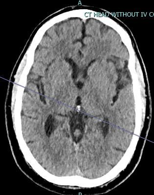
CT of the Head without Intravenous Contrast
CT of the Head without Intravenous Contrast
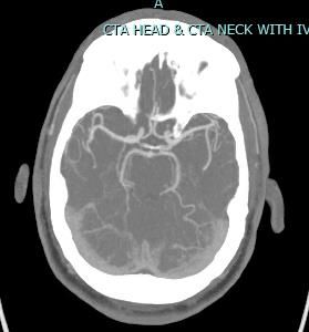
CT Angiography of the Head and Neck
Initial Imaging Impression
CT of the Head w/o Contrast:
  –   No evidence of hemorrhage
  –   No dense vessel sign
  –   No evidence of a previous infarction
  –   Ventricles non-dilated

CT Angiography of the Head and Neck
  – Internal Carotid Arteries are patent
  – Circle of Willis is patent
Physical Exam
•   General: patient lying in bed, NAD. Of note, patient visibly yawning, speaking quietly and flat affect.
•   NIHSS Scoring:
     – Alert – 0
     – Answers Questions – 0
     – Follows Commands – 0
     – Gaze – 0
     – Visual Fields – 0
     – Facial droop – Minor paralysis of L face, 1 point
     – Motor – No drift in all extremities, 0
     – Cerebellar – No ataxia, 0
     – Sensory – Normal in all extremities and face, 0
     – Best language – No aphasia, 0
     – Dysarthria – Mild, 1 point
     – Extinction – No abnormality, 0
     Total NIHSS: 2 points, Classification is mild stroke
Clinical Questions • Is tPA indicated in this patient? What are the contraindications? • Should this patient receive tPA?
Question 1: Is tPA indicated in this patient?1
• Indications:
   – Patient is having a minor stroke with an NIHSS of 2
   – Patient is within the 3 to 4.5 hour window
   – Patient is >18 yo
• Contraindications:
   –   Intracranial hemorrhage on CT – None present
   –   Clinical presentation suggests subarachnoid hemorrhage – None present
   –   Neurosurgery, head trauma, or stroke within 3 months – None
   –   HTN > 185/110 – Patient is normotensive
   –   Hx of intracranial hemorrhage - None
   –   No anticoagulation – Patient is not being treated with anticoagulation
Question 2:
• Should the patient elect to receive tPA?
  – Not a clear cut decision, patient does not have contraindications
  – Risk of bleeding is up to 6% with a NNT at 1 in 10
  – Low score with minor manifestations of stroke

  Patient ultimately ended up discussing with family and given low
  severity of symptoms and risk, decided to not move forward with tPA
  administration
Next steps:
• Is this a stroke?
– Patient has a 1 month history of general malaise with
increasing symptoms starting early in the day prior to onset of
dysarthria
– Possible etiologies explaining fatigue must be evaluated
• Depression, Hypothyroid, Anemia, etc
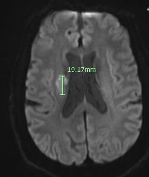
How would we confirm if this was a stroke?MRI - DWI
MRI - ADC
MRI - Flair
Diagnosis and Work Up
• Acute vs. Subacute infarction in the right corona radiata
   – DWI is focally hyperintense, suggesting acute or subacute stroke
   – ADC – Lesion is not hypointense, questions acute versus subacute
      • An acute stroke would be hypointense on the ADC
          – You would correlate this to DWI where the area of suspected ischemia would be hyperintense
      • Possibly explains the patients prolonged symptoms with an acute presentation
• What caused this stroke?
   – Key possible etiologies that require investigation
      • Carotid duplex for stenosis
      • TTE – Looking for thrombis / endocarditis
      • EKG – Looking for Atrial fibrillation
Stroke of the Corona Radiata
• Clinical features of the syndrome
– Varies greatly given location
– Pure hemiplegia, sensory loss in face and arm, dysphagia,
hemi neglect, dysarthria-clumsy hand syndrome
• The site is vulnerable to infarcts
– Lenticulostriate artery junction with the long medullary
penetrating arterial branches of the middle cerebral arteriesCitations 1) TPA contraindications for ischemic stroke. (n.d.). Retrieved April 01, 2021, from https://www.mdcalc.com/tpa- contraindications-ischemic-stroke 2) Corona radiata. (n.d.). Retrieved April 01, 2021, from https://www.sciencedirect.com/topics/neuroscience/corona- radiata
You can also read