Borrelia miyamotoi: A Comprehensive Review - MDPI
←
→
Page content transcription
If your browser does not render page correctly, please read the page content below
pathogens
Review
Borrelia miyamotoi: A Comprehensive Review
Dawn W. Cleveland                   , Cassidy C. Anderson and Catherine A. Brissette *

                                         Department of Biomedical Sciences, University of North Dakota, Grand Forks, ND 58202, USA
                                         * Correspondence: catherine.brissette@und.edu; Tel.: +1-701-777-6412

                                          Abstract: Borrelia miyamotoi is an emerging tick-borne pathogen in the Northern Hemisphere and is
                                          the causative agent of Borrelia miyamotoi disease (BMD). Borrelia miyamotoi is vectored by the same
                                          hard-bodied ticks as Lyme disease Borrelia, yet phylogenetically groups with relapsing fever Borrelia,
                                          and thus, has been uniquely labeled a hard tick-borne relapsing fever Borrelia. Burgeoning research has
                                          uncovered new aspects of B. miyamotoi in human patients, nature, and the lab. Of particular interest
                                          are novel findings on disease pathology, prevalence, diagnostic methods, ecological maintenance,
                                          transmission, and genetic characteristics. Herein, we review recent literature on B. miyamotoi, discuss
                                          how findings adapt to current Borrelia doctrines, and briefly consider what remains unknown about
                                          B. miyamotoi.

                                          Keywords: Borrelia miyamotoi disease (BMD); Ixodes; Lyme disease; relapsing fever; reservoir species;
                                          tick-borne disease; vector

                                          1. Introduction
                                               The genus Borrelia contains Gram-negative, obligate extracellular parasitic bacteria
                                          with a unique spiral morphology. Known as spirochetes, Borrelia owe their shape and
                                          spiraling motility to their periplasmic flagella [1]. Borrelia can be divided into two well-
                                          defined groups: the Lyme disease (LD) group, vectored by hard ticks, and the relapsing
                                          fever (RF) group, vectored by soft ticks and lice. Classically, these groups were highly
                                          distinct with separate vector genera, unique transmission dynamics, and differing disease
Citation: Cleveland, D.W.; Anderson,      outcomes [2,3]. Yet, the discovery of Borrelia miyamotoi, phylogenetically grouped with RF
C.C.; Brissette, C.A. Borrelia            spirochetes, but vectored by the same hard ticks as LD spirochetes, disturbed this archetype
miyamotoi: A Comprehensive Review.        and fueled research [4].
Pathogens 2023, 12, 267. https://              In this review, we discuss novel B. miyamotoi research in recent publications and
doi.org/10.3390/pathogens12020267         examine how new data fit into existing knowledge.
Academic Editors: Charles Pavia and
                                          2. Background
Gary P. Wormser
                                                Ticks are capable of vectoring numerous pathogens, with the most prevalent vector-
Received: 26 December 2022                borne illness in the Northern Hemisphere being Lyme disease. Also known as Lyme
Revised: 31 January 2023
                                          borreliosis, LD is the result of infection with species in the Borrelia burgdorferi sensu lato
Accepted: 2 February 2023
                                          (Bbsl) complex, which are vectored by hard-bodied Ixodes ticks. The primary species
Published: 7 February 2023
                                          responsible for human LD are B. burgdorferi, B. afzelii, and B. garinii [2]. Lyme disease is
                                          commonly characterized by the formation of an erythema migrans skin lesion, often at the
                                          bite location, in conjunction with flu-like symptoms of headache, fatigue, muscle aches,
Copyright: © 2023 by the authors.
                                          and fever along with joint pain and swelling. Bbsl spirochetes can disseminate beyond the
Licensee MDPI, Basel, Switzerland.        bite site to additional tissues. This colonization can result in serious clinical manifestations
This article is an open access article    of the joints, heart, and central nervous system (CNS), known as Lyme arthritis, carditis,
distributed under the terms and           and neuroborreliosis, respectively [5–8]. The treatment for LD is an antibiotic regimen;
conditions of the Creative Commons        however, symptoms can persist, resulting in post-treatment Lyme disease syndrome [5,7].
Attribution (CC BY) license (https://           Another borreliosis found in the Northern Hemisphere, Africa, and Central America
creativecommons.org/licenses/by/          is relapsing fever. Relapsing fever is the result of infection by several species of Borrelia
4.0/).                                    vectored primarily by soft-bodied argasid ticks, the exception being B. recurrentis vectored

Pathogens 2023, 12, 267. https://doi.org/10.3390/pathogens12020267                                     https://www.mdpi.com/journal/pathogens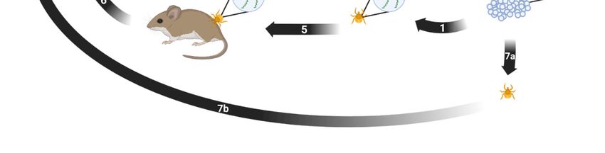
Pathogens 2023, 12, 267                                                                                           2 of 21

                          by lice. Clinical presentations of RF typically consist of a high fever for a few days,
                          followed by an approximately week-long period of well-being, followed by another relapse.
                          Relapses can repeat multiple times without antibiotic treatment. Additional RF symptoms
                          are headache, body aches, and abdominal pain. Historically, outbreaks are caused by
                          louse-borne B. recurrentis and occur during wartime, periods of poor hygienic conditions,
                          and refugee crowding [3].
                               Borrelia miyamotoi, a new tick-borne Borrelia species, was discovered in Japan in 1995
                          when spirochetes were isolated from Ixodes persulcatus midguts and Apodemus argenteus
                          blood [4]. Despite being vectored by hard ticks, B. miyamotoi was found to be genetically
                          distinct from the Bbsl complex and was more closely related to RF species. At the time,
                          there were no cases of RF in the region; this led researchers to speculate that this new
                          Borrelia species was a tick endosymbiont maintained by an enzootic transmission cycle [4].
                               The pathogenicity of B. miyamotoi was realized in Russia in 2011 when 51 patients with
                          suspected tick bites presented with a nonspecific febrile illness. The patients were found
                          to be infected with B. miyamotoi by B. miyamotoi-specific polymerase chain reaction (PCR)
                          of the blood or anti-borreliae immunoglobulins in the serum. The patients’ symptoms
                          were somewhat dissimilar to RF with fever, headache, chills, fatigue, and myalgia. One
                          patient had a relapse, which may have been prevented in other patients due to antibiotic
                          treatment [9]. The RF-like illness caused by B. miyamotoi, now known as B. miyamotoi
                          disease (BMD), has since been widely diagnosed and is considered an emerging public
                          health threat [10].

                          3. Disease
                                Human infection with B. miyamotoi can result in BMD. Symptoms vary depending
                          on the constitution of the patient. Immunocompetent, and otherwise healthy, patients
                          present with milder, flu-like symptoms: fever, fatigue, sleepiness, chills, muscle and joint
                          stiffness, aches and pains, and nausea [11–13]. While uncommon, relapses of febrile
                          episodes can occur [11]. It is possible that many patients with BMD do not seek medical
                          attention due to short-lived and mild symptoms. Similar to the mechanism of relapses in
                          other RF species, B. miyamotoi exhibits antigenic variation, allowing spirochetes to evade
                          adaptive immune responses [14–17]. In the blood, B. miyamotoi has numerous mechanisms
                          to evade complement-mediated killing, allowing for the rapid growth and multiplication
                          of spirochetes that result in the symptoms of BMD [11,14–16].
                                The adaptive immune response is important for controlling B. miyamotoi infection [15].
                          Studies of RF species B. hermsii show that infection clearance is antibody mediated [16].
                          Borrelia miyamotoi activates dendritic cells which phagocytose the bacteria and produce
                          interleukins IL-8, IL-6, and IL-12, as well as tumor necrosis factor alpha (TNF-α) [18]. These
                          cytokines further stimulate and signal immune activation for inflammation, recruitment of
                          additional immune cells, infection clearance, host-defense, and T-cell differentiation [18].
                          Immunocompetent mouse models of BMD only develop transient infections, whereas
                          immunodeficient mice mouse models develop persistent infections [19]. This suggests
                          that healthy patients infected with B. miyamotoi may be able to clear the infection without
                          medical intervention.
                                Borrelia miyamotoi infection in immunocompromised patients can be much more se-
                          vere [20]. In combination with generalized flu-like symptoms, immunocompromised
                          patients often exhibit reduced cognition, disturbed gait, memory deficits, confusion, and
                          other neurological deficiencies resultant of meningoencephalitis [21]. Additionally, hearing
                          loss, weight loss, uveitis, iritis, neck stiffness, and photophobia have been reported [20].
                          The designation “immunocompromised” is broad; however, hospitalization reports of
                          severe BDM are frequently seen in patients prescribed B-cell depletion therapies, such
                          as rituximab, other cancer immunotherapeutics, or immunosuppressants for rheumatoid
                          arthritis [20–25]. While these medications are often seen in conjunction with meningoen-
                          cephalitis and other serious symptoms, there is no finite list of medications, treatments, or
                          immunodeficiencies that can give rise to severe BMD.
Pathogens 2023, 12, 267 3 of 21
In patients lacking a complete immune response, the austerity of symptoms is possibly
due to unchecked growth of B. miyamotoi in the blood. Similar to B. hermsii, it can be
speculated that the humoral immune response is necessary to control B. miyamotoi infec-
tion [16]. This suggests that defects in antibody production allow spirochetes to disseminate
from the blood and colonize farther tissues, resulting in severe BMD symptoms. It can be
inferred that BMD-associated meningoencephalitis is the result of B. miyamotoi colonizing
the CNS, as microscopic analysis of cerebrospinal fluid (CSF) collected from BMD patients
with meningoencephalitis shows visible spirochetes [21–23]. However, the dynamics of
host–pathogen interactions leading to inflammation are unknown.
There are no official clinical guidelines for the treatment of diagnosed or suspected
BMD. Thereby, treatment falls to the guidelines for LD and RF: an antibiotic regimen,
commonly doxycycline and ceftriaxone. Ampicillin, azithromycin, and vancomycin, or a
combination thereof, have also been used to treat BMD. Antibiotic treatment engenders a
full recovery with rare cases of persistent fatigue [20,26]. A systematic review and meta-
analysis by Hoornstra et al. affirmed that doxycycline is the preferred treatment for adults
presenting with no neurological complications, and ceftriaxone is the preferred treatment
for adults with BMD-associated meningoencephalitis [26]. Research has confirmed in vitro
that both clinical and tick isolates of B. miyamotoi are susceptible to doxycycline, ceftriaxone,
and azithromycin, but show resistance to amoxicillin [27]. Albeit rarely, Jarisch-Herxheimer
reactions have occurred with antibiotic treatment of BMD [22].
4. Diagnosis
Diagnosis of BMD and B. miyamotoi infection is possible using microscopy, PCR, and
serodiagnosis, either as single tests or in combination, with the latter two methods being
the most common [11]. However, there is no official standardized diagnostic technique [28].
4.1. Microscopy
Spirochetes can be visualized in blood and CSF using dark-field microscopy, or with
Giemsa staining or acridine orange staining [21–23]. Microscopy can confirm a Borrelia
species infection; however, it cannot be used to distinguish between species and has
low sensitivity [29]. In order to see spirochetes in a blood smear or CSF sample, the
density needs to be greater than 104 cells/mL [11,26]. During the acute phase of BMD,
usually within a few days of symptom onset, spirochete density in the blood ranges from
101 –105 cells/mL [30]. The presence of B. miyamotoi in the CSF is poorly understood. Thus,
there is no guarantee that spirochete density will be high enough for visualization.
Borrelia miyamotoi can be cultured from a patient’s whole blood, serum, or CSF into
specialized media, and organisms can then be visualized. However, as spirochetes may
take longer than two weeks to grow to a visible density and culturing Borrelia species is
notoriously tricky, it is neither timely nor practical to use culturing for diagnosis [31].
The visualization of spirochetes in the blood or CSF confirms Borrelia species infection,
but a lack of visible spirochetes does not rule out infection. Microscopy can support a BMD
diagnosis but should be used in combination with other methods.
4.2. PCR
Diagnosis of BMD often uses PCR assays performed on whole blood, serum, or CSF
and typically targets glpQ and/or flaB [11,22]. It is best to use PCR to diagnose BMD when
spirochetes are most likely to be detectable during symptom presentation and prior to
antibiotic treatment [20,30]. Human borrelioses all have detectable periods of spirochetemia,
albeit brief for Bbsl, and overlapping geographical regions [32–34]. Thus, a PCR assay
must be sensitive enough to detect B. miyamotoi and specific enough to distinguish between
B. miyamotoi, Bbsl, and RF species.
A clinically relevant assay was developed to detect and differentiate between B. miyamotoi,
louse-borne RF, and soft tick-borne RF species. The semi-multiplex real-time PCR assay
is highly sensitive with a lower detection limit of ten genome copies and uses a nestedPathogens 2023, 12, 267 4 of 21
approach to first broadly amplify and detect RF species and B. miyamotoi. A series of probes
then differentiate between groups [35]. If implemented in a clinical setting, this assay
would readily test for RF and BMD in acute phase patients.
4.3. Serodiagnosis
Borrelia miyamotoi infection results in the production of immunoglobulins to spirochete
proteins, and serodiagnosis utilizes the presence of these reactive antibodies to diagnose
a past or present infection. The quantity and variety of antibodies produced fluctuates
depending on the route of inoculation, whether via tick bite or needle stick, if antigenic
variation occurs, and the duration and dissemination of the infection [14–16,36,37]. Im-
munoglobulins IgM and IgG are produced in response to B. miyamotoi infection, with
IgM peaking around one week post infection and IgG peaking around three weeks post
infection. While IgG is highly specific for the antigen against which it is produced, IgM is
not and is more prone to higher background and cross-reactivity [38,39].
Timing is important when utilizing serology for a diagnosis. At the early onset of
BMD symptoms, IgM may be present at low levels, while IgG will not be present [38,39].
Absent or undetectable antibodies could result in a false negative test. Microscopy paired
with PCR is more suited for acute diagnosis [20–23,30]. Patients presenting with chronic
BMD symptoms multiple weeks after disease onset, or for retrospective studies, are better
suited to diagnosis via serology. The indefatigable nature of anti-B. miyamotoi antibodies is
not fully understood; however, the persistence of Borrelia-specific antibodies is correlated
to the duration and dissemination of the infection [36]. As antibody production is integral
for serodiagnosis, the ability to diagnose immunocompromised patients via serology may
be limited due to complications surrounding reduced humoral responses to B. miyamotoi.
Glycerophosphodiester phosphodiesterase (GlpQ) is widely used for serodiagnosis,
but its reliability as a serological indicator of BMD has recently been called into ques-
tion [13,15,40,41]. For PCR, glpQ is reliable as it directly detects the bacteria. However, as
serodiagnosis indirectly detects infection by measuring reactive antibodies to a particular
antigen or antigens, cross-reactivity can potentially occur between similar proteins from
different pathogens [42].
Anti-B. miyamotoi antibodies can be detected using conventional assay systems such
as an ELISA or a Western blot [43,44]. Ideally, the best candidate antigens for serodiagnosis
should be highly conserved and detectable in both early and late infection with low cross-
reactivity to other tick-borne and common bacterial pathogens. Reactivity to GlpQ is often
used in combination with flagellin and p66 proteins; however, neither of these are species
specific [20]. Despite widespread use for serodiagnosis, GlpQ has been shown to be neither
highly sensitive nor specific [41]. Fortunately, researchers have strove to uncover methods
to improve GlpQ accuracy and identify additional reliable markers.
Assaying for GlpQ reactivity in conjunction with variable major proteins (Vmps),
particularly variable large protein (Vlp)-15/16, improves the sensitivity and specificity
of B. miyamotoi detection [37]. This coalition leans heavily on cooperation as Vmps alone
are prone to cross-react with orthologous Bbsl protein Vmp-like sequence expressed
(VlsE) [45,46]. This dual-marker technique using both GlpQ and Vlp-15/16 was found to
be an excellent diagnostic option as IgM against GlpQ and Vmps peaks between 11 and
20 days and IgG peaks between 21 and 50 days. This combination increased specificity to
100% and sensitivity to 79% on days 11–20, and increased specificity to 98.3% and sensitivity
to 86.7% on days 21–50 [37]. Thus, testing for seroreactivity to both Vmps and GlpQ can
improve BMD diagnostic accuracy.
Whereas GlpQ and Vlp-15/16 test best in combination, Borrelia miyamotoi membrane
antigen (BmaA) is a solitary candidate for serodiagnosis. This protein is an externally
located cellular putative lipoprotein of B. miyamotoi, and anti-BmaA antibodies can be
detected in the serum of confirmed BMD patients between 38 and 100 days after disease on-
set [47]. Results after 100 days were inconclusive and additional testing is necessary. BmaA
has little to no observable reactivity in LD patients or with RF species B. turicate, indicatingPathogens 2023, 12, 267 5 of 21
that BmaA may be species specific [47]. Beyond BmaA, more than 400 immunoreactive
peptides, most of which mapped to Vmps, were recently identified as potential targets for
future serology studies [48]. Hence, while GlpQ may not be ideal for serodiagnosis, there
are multiple other options.
5. Human Cases and Infection Prevalence
The National Institute of Allergy and Infectious Diseases classifies B. miyamotoi as an
emerging infectious pathogen and BMD as an emerging infectious disease [10]. Even as
B. miyamotoi becomes more prominent, the ambiguity and often self-limiting nature of BMD
symptoms suggest that many cases are likely subclinical. Patients who do seek medical
treatment may be misdiagnosed with other tick-borne diseases (TBD), particularly LD.
This mistaken serodiagnosis of BMD as LD can be due to the C6 peptide of the Bbsl VlsE
protein. Reactivity to the C6 peptide is widely used for LD serodiagnosis, and infection
with B. miyamotoi leads to cross-reactive antibodies, which may result in a false-positive LD
test [49]. As antibiotic treatment for LD is successful in treating BMD, an accurate diagnosis
of BMD may never be reached. All of this, combined with low awareness among physicians
and the general public, indicates that the known prevalence of BMD may not be wholly
accurate. The most updated and thorough conglomeration of reported diagnoses of BMD
and retrospective studies of banked human sera reactivity is by Hoornstra et al. in their
systematic review and meta-analysis [26].
Including the first cases of BMD diagnosed in Russia, there have been 561 total
diagnoses: 367 in Russia, 101 in the United States, 57 in France, 30 elsewhere in Asia,
and 6 elsewhere in Europe [26,50–54]. The higher number of cases in Russia may be due
to increased awareness of B. miyamotoi prompting more testing, increased transmission
abilities of I. persulcatus ticks, or greater virulence of Russian strains, leading to more
hospitalizations. As there are no official diagnostic guidelines for BMD, criteria for BMD
diagnosis may vary between institutions.
The reactivity of banked sera to B. miyamotoi recombinant proteins, primarily GlpQ
and FlaB, can be measured using ELISA and Western blots, either singly or in conjunction
as a two-tier test. Across these retrospective studies, 45,608 samples were tested from
North America, Europe, and Asia and separated into varying categories depending on
risk, confirmed infection, and cross-reactivity. Seroprevalence was 4.6% in the high risk for
B. miyamotoi infection group, 4.8% in the suspected LD group, 11.9% in the suspected TBD
group, and 1.3% in healthy controls [26]. Prevalence across all groups could indicate that
B. miyamotoi infection is far more common than previously thought, detection methods are
prone to false positives and cross-reactivity, or coinfections with other tick-borne pathogens
(TBP) produce convoluted results [11].
6. Ticks
The geographic distribution of B. miyamotoi in ticks is far greater than that of BMD
in humans. This may be an insight on transmission capabilities, or it may be that more
surveys have been conducted on ticks. Borrelia miyamotoi is vectored by I. scapularis in the
northeastern and northcentral United States and Canada, I. pacificus in the western United
States, I. ricinus in Europe, I. persulcatus in Europe and Asia, and I. ovatus, I. nipponensis
and I. pavoloskyi in Asia [26,50,55]. Borrelia miyamotoi has been detected in all known
vectors of Bbsl [43]. There are reports of B. miyamotoi in other tick species, including
Dermacentor reticulatus in Russia and Haemaphysalis species in China and Slovakia [56–59].
These species are not competent vectors for Bbsl; however, it is unclear if they are capable
of successfully vectoring B. miyamotoi [60,61]. Vector competence is the ability of a tick
to serve as a disease vector. Vector competence is influenced by environmental factors,
such as vector density, longevity, and fitness, and genetic factors, such as host preference
and duration of attachment [62]. Table 1 has an updated list of countries with reports of
ticks infected with B. miyamotoi. The systematic review and meta-analysis by Hoornstra
et al. analyzed B. miyamotoi prevalence in ticks, which is outlined in Table 2. The overallPathogens 2023, 12, 267 6 of 21
prevalence of B. miyamotoi in questing Ixodes ticks is 1.1%, but varies by region and dominant
tick species in that region [26].
A survey of Ixodes ticks in the United States from 2013 to 2019 screened nine Ixodes
species, but only detected B. miyamotoi in I. pacificus (14/1497, 0.94%) and I. scapularis
(594/34,621, 1.72%). The average infection prevalence in nymphs and adults was similar;
there was no mention of larval infection. Borrelia miyamotoi was found in ticks in 19 states,
with infection prevalence being 0.5–3.2%. Ticks tested in 20 additional states were all
negative [63]. Borrelia miyamotoi has been detected in all Canadian provinces, excluding
Newfoundland, in I. scapularis ticks [34]. There are certain states and provinces in which
B. miyamotoi infection has not been reported, which may be due to a paucity of competent
vector species in that region, spirochetes present but below detection limits, or simply that
B. miyamotoi is not in that region.
Natural coinfections in Ixodes ticks occur, as these arthropods vector a number of
TBP beyond Borrelia species [33]. A survey of Ixodes ticks in the United States found
over half of all B. miyamotoi-infected I. scapularis ticks had concurrent infections (351/594,
59.09%). Half of all B. miyamotoi-infected ticks had a dual infection (293/594, 49.33%)
with either B. burgdorferi (220/594, 37.04%), Anaplasma phagocytophilum (43/594, 7.24%), or
Babesia microti (30/594, 5.05%). Additionally, 52 ticks (8.75%) had triple infections and six
ticks (1.01%) had quadruple infections. No concurrent B. miyamotoi infections were seen
with B. mayonii or Ehrlichia muris-like agent [33]. This could be due to coincidence, the result
of geographic constraints, and/or competitive habitation in the vector ticks. Regardless,
these data indicate that concurrent infections in ticks are common, and physicians should
be aware that tick bite patients may be exposed to multiple TBPs.
In the tick, B. miyamotoi disseminates throughout the body into the acini, salivary
gland ducts, basal lamina of the midgut, epithelium of the Malpighian tubes, female
ovarian tissues, male testes, CNS, and near the mouthparts [64]. During tick colonization,
B. burgdorferi species have outer surface membrane proteins that adhere to receptors in the
midgut lumen and protect spirochetes from the ingested host blood containing complement
and antibodies [62,65]. It is unknown if B. miyamotoi uses similar mechanisms for tick
colonization as Bbsl.
Additionally, ticks infected with B. miyamotoi are found to have significantly higher
numbers of Borrelia cells than those infected with Bbsl [66,67]. This is a proposed evolution-
ary trait to compensate for the lower prevalence of B. miyamotoi in tick populations (~1%),
compared with Bbsl (~12%) [26,67,68]. Furthermore, B. miyamotoi reach higher levels in the
blood than Bbsl species, and thus, ticks ingest more bacteria per blood meal [69]. Other
Borrelia species, including Bbsl and B. hermsii, have been shown to replicate within ticks;
however, it is not known if B. miyamotoi replicates within ticks [70,71].
When transmitted from tick to host, Bbsl uses outer surface protein C (OspC) to bind
tick salivary protein Salp15, which provides protection from the mammalian immune
response [72]. Borrelia hermsii variable tick proteins (Vtp) are homologous to OspC and
convey a similar function [73]. The variable small proteins (Vsp) in B. miyamotoi are homolo-
gous to OspC and Vtp and likely aid in transmission from tick to vertebrate host [14]. There
is still much to learn about the intricacies of B. miyamotoi tick colonization and transmission.
Table 1. Countries with B. miyamotoi-infected ticks.
Country Tick
Austria I. ricinus [74]
Belarus I. ricinus [75]
Belgium I. ricinus [76,77]
Canada I. scapularis [78]
China I. persulcatus [79], H. longicornis [57], H. concinna [58]
Czechia I. ricinus [80]
Denmark I. ricinus [81]
Estonia I. ricinus, I. persulcatus [82]Pathogens 2023, 12, 267 7 of 21
Table 1. Cont.
Country Tick
Finland I. ricinus [83–85]
France I. ricinus [81,86,87]
Germany I. ricinus [76,88]
Ireland I. ricinus [89]
Italy I. ricinus [76]
Japan I. persulcatus [4], I. ovatus [90]
Moldova I. ricinus [91]
Mongolia I. persulcatus [90,92]
Netherlands I. ricinus [76,81,93,94]
Norway I. ricinus [95]
Poland I. ricinus [96,97]
Portugal I. ricinus [98]
Russia I. persulcatus [99]
Serbia I. ricinus [100]
Slovakia I. ricinus [59,101], H. inermis [59]
South Korea I. nipponensis [55]
Spain I. ricinus [102]
Sweden I. ricinus [67,76]
Switzerland I. ricinus [103]
Turkey I. ricinus [104]
Ukraine I. ricinus [105]
United Kingdom I. ricinus [76]
United States I. pacificus [106], I. scapularis [107]
Table 2. Prevalence of B. miyamotoi in ticks a .
Questing Tick Prevalence B. miyamotoi Prevalence
I. scapularis 28.0% 1.1%
I. pacificus 14.8% 0.7%
I. ricinus 52.2% 1.0%
I. persulcatus 5.0% 2.8%
The combined surveys listed in Hoornstra et al., 2022 covered a total of 165,637 questing ticks. Tick species
prevalence is calculated as a proportion of total questing ticks surveyed. B. miyamotoi prevalence was calculated
as proportion of the tick species. a adapted from Hoornstra et al., 2022 [26] under creative commons license CC
BY 4.0.
7. Animal Infections
The majority of animals infected with B. miyamotoi are small mammals, particularly
rodents, with a minority of infections being in larger mammals and birds [108]. An im-
portant distinction needs to be made between animal infections, where the animal tissue
tested positive for B. miyamotoi (Table 3), and animal-associated tick infections, where
the animal tissue was not tested, but the tick(s) attached to the animal tested positive for
B. miyamotoi (Table 4). Borrelia miyamotoi has also been detected in both birds and bird-
associated ticks; however, there has been minimal follow-up research to interpret the role
of birds in B. miyamotoi perpetuation and maintenance (Tables 3 and 4) [94,109,110].
The multiple reservoir species identified for B. miyamotoi, all of which are rodents,
are Peromyscus, Apodemus, and Myodes species [69,108,111,112]. Reservoir rodent species
have been identified carrying B. miyamotoi in both rural and urban environments, posing
an increased public health risk [83–85]. A notable potential new reservoir species in North
America is the jumping mouse, Napaeozapus insignis. In a survey of B. miyamotoi in Atlantic-
Canadian wildlife, N. insignis had a much higher infection prevalence than other known
reservoir species tested in the same survey. Unfortunately, the role of the jumping mouse in
B. miyamotoi transmission is unknown, as specimens were collected via public participation
and transmission studies cannot be conducted post-mortem [113].Pathogens 2023, 12, 267 8 of 21
It has been speculated that white-tailed deer may be a reservoir host for B. miyamotoi,
as a study found that deer-associated ticks had higher infection prevalence than questing
ticks [114]. Unlike B. burgdorferi that is lysed by deer blood complement upon tick ingestion,
it appears that B. miyamotoi remains viable in the tick upon ingestion of deer blood. A study
found that engorged female I. scapularis ticks collected from white-tailed deer produced
infected offspring [114–116]. Reservoir competence has not been characterized in white-
tailed deer.
Reservoir species must be able to be infected with B. miyamotoi and remain infected
long enough to allow naïve vector ticks to acquire infection via feeding. An indirect method
to determine a reservoir species tests both wildlife and ticks in a survey plot for B. miyamotoi
infection. A direct method live-traps animals and tests both the animal and any ticks on
them for B. miyamotoi. Xenodiagnostic ticks are then fed on the animal(s) and tested for
B. miyamotoi to determine transmission capabilities.
It is unclear if animals, particularly reservoir species, remain persistently infected
with B. miyamotoi. Bbsl species are known to cause persistent infections in rodents [117]. A
survey of B. miyamotoi in rodents and their ticks found that there was no correlation between
rodent age, month of infection, and infection presence. Infection rates remained stable
across the survey. In the same study, B. burgdorferi infection rates increased throughout
the survey [118]. Furthermore, while Bbsl species have a strong positive association with
rodent population density, maintenance of B. miyamotoi appears to be independent of
rodent population density [119]. This suggests that B. miyamotoi and Bbsl use different
maintenance strategies [69].
Borrelia miyamotoi has been found in a variety of mammals, birds, and associated ticks.
Discerning how B. miyamotoi is maintained in reservoir species, other hosts, and vector
ticks is critical for surveillance, risk assessment, and mitigation.
Table 3. Borrelia miyamotoi animal infections.
% Positive Countries
Small Mammals
Striped field mouse (Apodemus agrarius) 3.13% (1/32); 13.2% (7/53); 7.0% (11/157) Austria [120], Croatia [121], Poland [112]
Small Japanese field mouse (Apodemus argenteus) 0.7% (1/137) Japan [118]
a
0.7% (1/131); 0.9% (1/102) & 1.5% (1/67)
b ; 3.6% (3/84); 2.0% (1/49); 0.4% (1/251) c Croatia [121], Hungary [111], Poland [112],
Yellow-necked mouse (Apodemus flavicollis) *
Romania [122], Slovenia [123]
& 2.2% (1/46) d
Large Japanese field mouse (Apodemus speciosus) 2.2% (10/446) Japan [118]
Wood mouse (Apodemus sylvaticus) 14.3% (3/21) Netherlands [94]
European hedgehog (Erinaceus europaeus) 5.0% (3/60) Czechia [124]
Common vole (Microtus arvalis) 12.5% (1/8) Netherlands [94], Slovakia [59]
Meadow vole
0.7% (1/146) Canada [113]
(Microtus pennsylvanicus)
Bank vole France [86], Netherlands [94], Romania [122],
5.5% (4/72); 8.8% (3/34); 3.1% (1/32)
(Myodes glareolus) * Switzerland [103]
Grey red-backed vole (Myodes rufocanus) 1.0% (2/195) Japan [118]
Jumping mouse
14.3% (3/21) Canada [113]
(Napaeozapus insignis)
Dusky-footed woodrat (Neotoma fuscipes) 16.7% (1/6) United States [125]
Brush mouse
2.8% (2/71) United States [125]
(Peromyscus boylii)
California mouse (Peromyscus californicus) 16.7% (4/24) United States [125]
6.4% (36/556) d & 2.3% (2/86) e; 0.5%
White-footed mouse (Peromyscus leucopus) * United States [69], Canada [126]
(3/625)
Deer mouse
2.9% (1/34) Canada [113]
(Peromyscus maniculatus)Pathogens 2023, 12, 267 9 of 21
Table 3. Cont.
% Positive Countries
Eastern grey squirrel (Sciurus carolinensis) 25.0% (1/4) Canada [113]
Red squirrel
13.6% (3/22) Czechia [124], Hungary [127]
(Sciurus vulgaris)
Common Shrew
16.7% (1/6) Croatia [121]
(Sorex araneus)
Muller’s giant Sunda rat (Sundamys muelleri) 33.3% (1/3) Malaysia [128]
Eastern chipmunk
15.4% (2/13) Canada [126]
(Tamias striatus)
Large mammals
Père David Deer (Elaphurus davidianus) 2.3% (1/43) China [57]
Birds
European greenfinch (Carduelis chloris) 25% (1/4) Netherlands [94]
Wild turkey (Meleagris gallopavo) 56.0% (35/60) United States [109]
Great tits (Parus major) 50% (1/2) Netherlands [94]
Ostrich (Struthio camelus) 16.7% (1/6) Czechia [110]
a Positive skin samples, Positive spleen samples, Retrospective study, d Prospective study, e Positive blood
b c
samples. * Xenodiagnostic ticks were used to confirm species as a reservoir host.
Table 4. Animal-associated B. miyamotoi tick infections.
Tick Countries
Mid-Sized Mammals
Beech marten (Martes foina) I. ricinus Belgium, Netherlands [129]
European pine marten (Martes martes) I. ricinus Belgium, Netherlands [129]
European polecat (Mustela putorius) I. ricinus Belgium, Netherlands [129]
Large Mammals
Cattle (Bos primigenius tarus) I. ricinus Germany [130]
I. ovatus, I. hexagonus, I. ricinus, I. Germany [131,132], Japan [133], Latvia [134],
Dog (Canis lupus familiaris)
persulcatus, Dermacentor reticulatus Russia [56]
Goat (Capra aegagrus hircus) I. ricinus Germany [130]
Roe deer (Capreolus capreolus) I. ricinus Germany [132], Poland [135], Spain [136]
Red deer (Cervus elaphus) I. ricinus Poland [137]
Raccoon dog (Nyctereutes procyonoides) I. ricinus Denmark [138]
White-tailed deer
I. scapularis United States [114,115,139]
(Odocoileus virginianus)
Wild boar (Sus scrofa) I. ricinus Poland [136]
Birds
Northern cardinal
I. dentatus United States [140]
(Cardinalis cardinalis)
Veery (Catharus fuscescens) I. scapularis Canada [126]
Hermit Thrush
I. scapularis, I. dentatus Canada [126], United States [140]
(Catharus guttatus)
European robin
I. ricinus Netherlands [93], Sweden [67]
(Erithacus rubecula)
Song Sparrow
I. scapularis Canada [126]
(Melospiza melodia)Pathogens 2023, 12, 267 10 of 21
Table 4. Cont.
Tick Countries
Common redstart
I. ricinus Sweden [67]
(Phoenicurus phoeincurus)
Common chiffchaff
I. ricinus Netherlands [93]
(Phylloscopus collybita)
Eurasian wren
I. ricinus Sweden [67]
(Troglodytes troglodytes)
Common blackbird
I. ricinus Moldova [91], Netherlands [93], Poland [135]
(Turdus merula)
American robin
I. dentatus United States [140]
(Turdus migratorius)
Song thrush (Turdus philomelos) I. ricinus Netherlands [93]
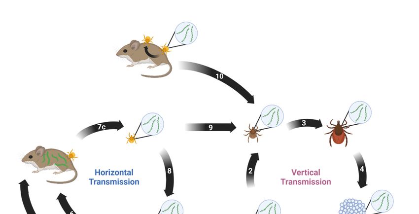
8. Transmission
Borrelia miyamotoi can be transmitted both horizontally and vertically [141]. Horizontal
transmission occurs between a vector and a host or vice versa. Vertical transmission
occurs transovarially between a female tick and her progeny. Both modes are suspected
Pathogens 2023, 12, x FOR PEER REVIEW 12 of 22
to play key roles in persistence of B. miyamotoi in the environment. A proposed enzootic
transmission cycle depicting both transmission methods and their potential interactions is
shown in Figure 1.
Figure 1. Proposed enzootic transmission cycle for Borrelia miyamotoi. (1) Larvae hatch from eggs laid
Figure 1. Proposed enzootic transmission cycle for Borrelia miyamotoi. (1) Larvae hatch from eggs
by a B. miyamotoi-infected female Ixodes tick. Transovarial transmission occurs, resulting in partial
laid by a B. miyamotoi-infected female Ixodes tick. Transovarial transmission occurs, resulting in par-
larval infection.
tial larval infection. (2)larva
(2) The infected takes larva
The infected first blood meal
takes first and
blood molts,
meal remaining
and molts, remaining infected.
infected.(3)
(3)The
The infected nymph takes second blood meal and molts, remaining infected.
infected nymph takes second blood meal and molts, remaining infected. (4) The infected adult female (4) The infected adult
female takes third blood meal, mates, and lays eggs. (5) The infected tick feeds on naïve host. (6)
takes third blood meal,
The host mates,
acquires B. and lays from
miyamotoi eggs.the(5) The infected
infected tick. (7) a)tick
The feeds on naïve
naïve larva b) feedshost. (6)infected
on the The host
acquires B. miyamotoi
host and c) from the B.
acquires infected
miyamotoitick. (7) (a)
via blood The(8)naïve
meal. larva tick
The infected (b) feeds
feeds ononthethe infected
naïve host. (9)host
TheB.
and (c) acquires infected larva takes
miyamotoi blood meal
via blood meal.and(8)
molts
Theinto a nymph,tick
infected remaining
feeds infected. (10) Thehost.
on the naïve naïve(9)
tickThe
acquires B. miyamotoi via co-feeding near an infected tick. Created with BioRender.com (accessed on
infected larva9takes
January blood
2023). meal and molts into a nymph, remaining infected. (10) The naïve tick
acquires B. miyamotoi via co-feeding near an infected tick. Created with BioRender.com (accessed on
9. Phylogenetics
9 January 2023).
The earliest known B. miyamotoi isolates were collected in Japan in 1995: isolate FR64
from A. argenteus field mice and isolates HT24, HT31, Hk004, and NB103/1 from unfed I.
persulcatus ticks [4]. In the decades since, numerous additional B. miyamotoi isolates have
been recovered and described from around the world. Table 5 shows reference genomes
and chromosomes for 36 different strains. These strains have been utilized in multifarious
research objectives including phylogenetics studies.Pathogens 2023, 12, 267 11 of 21
Horizontal transmission of B. miyamotoi from tick to host appears to have varied
success using laboratory models. Mice challenged with B. miyamotoi by horizontally infected
ticks resulted in low infection prevalence in both mice and xenodiagnostic larvae [64]. Yet,
B. miyamotoi can be transmitted from infected ticks to naïve mice during the first 24 h of
feeding. Within this window of time, only 10% of the mice had detectable B. miyamotoi DNA
in their blood. If ticks were allowed to feed to repletion, 73% of the mice had detectable
B. miyamotoi DNA, though no xenodiagnostic ticks were fed on these mice [142]. This
likely indicates that B. miyamotoi in the tick salivary glands enter the host first. Then, as
the tick feeds, spirochetes in the midgut migrate to the salivary glands to contribute to the
infection [3,64].
Tick acquisition of B. miyamotoi via feeding on an infected immunocompetent host is
variable and potentially limited by periods of spirochetemia. Only 1–12% of post-molt ticks
were positive for B. miyamotoi when fed to repletion on infected reservoir species P. leucopus
and infected CD-1 mice [64]. This may be due to B. miyamotoi colonizing the skin at a low
rate combined with brief periods of spirochetemia, or that infection was not sustained
through the tick molting process [69]. All naïve ticks fed on infected severe combined
immunodeficient (SCID) mice with high, persistent spirochetemia became infected, though
infection was frequently lost during the molt [19]. This suggests that spirochetemia may
play a role in horizontal transmission from host to tick and that vertical transmission may be
more important for B. miyamotoi maintenance as horizontal transmission is often inefficient.
Borrelia miyamotoi can be vertically transmitted from the female tick to her offspring.
The rate of transovarial transmission appears to be positively correlated with the maternal
bacterial load. The higher the infection density in the female, the more likely her egg
clutches will be infected with higher bacterial loads. Seven out of ten I. scapularis females
that tested positive for B. miyamotoi produced infected larvae, with the filial infection rate
of infected clutches being 3.3–100%, median 71%. The three females that did not produce
infected clutches had the lowest bacterial loads [64]. Another group saw 10/11 infected
I. scapularis females produce infected clutches, with infection in progeny being 36–100%.
This study also saw similar oviposition rates in B. miyamotoi-infected I. scapularis females
compared with uninfected females, indicating that B. miyamotoi infection does not appear
to impact tick fecundity [115]. These studies support that vertical transmission is relied on
more heavily for B. miyamotoi maintenance than horizontal transmission.
Though there is minimal evidence, additional suggested transmission routes of
B. miyamotoi are vertical transmission in mammals and co-feeding in ticks. A study found a
pregnant jumping mouse to have a fully disseminated infection, resulting in B. miyamotoi
detectable in placental and fetal tissues [113]. It is unclear if the pregnancy had reached
full term if the offspring would have been viable or remained infected. Furthermore, it is
unclear if the pregnancy led to the disseminated infection or if dissemination was due to
unknown factors. Additionally, it has been suggested when both infected and naïve ticks
feed simultaneously on the same naïve host, B. miyamotoi may be transmitted to the naïve
tick, known as co-feeding [64]. Further studies need to be conducted to better understand
these potential transmission routes.
As ticks can be infected with B. miyamotoi prior to their first blood meal, it is important
to note that larval, nymphal, and adult ticks can transmit B. miyamotoi to humans. A
study by Breuner et al. showed that over half (57%) of CD-1 mice exposed to a single
transovarially infected larva had evidence of B. miyamotoi infection [143].
9. Phylogenetics
The earliest known B. miyamotoi isolates were collected in Japan in 1995: isolate FR64
from A. argenteus field mice and isolates HT24, HT31, Hk004, and NB103/1 from unfed
I. persulcatus ticks [4]. In the decades since, numerous additional B. miyamotoi isolates have
been recovered and described from around the world. Table 5 shows reference genomes
and chromosomes for 36 different strains. These strains have been utilized in multifarious
research objectives including phylogenetics studies.Pathogens 2023, 12, 267 12 of 21
Table 5. Borrelia miyamotoi strains with reference genome or chromosome sequences.
Strain Origin Source Accession
LB-2001 North America I. scapularis CP006647
CT13-2396 North America I. scapularis NZ_CP017126
C14D4 North America Human, blood NZ_CP010308
CA17-2241 North America I. pacificus NZ_CP021872
FR64b Japan A. argenteus, blood NZ_CP004217
HT31 Japan I. persulcatus NZ_AP024371
HT24 Japan I. persulcatus NZ_AP024372
Hk004 Japan I. persulcatus NZ_AP024373
NB103/1 Japan A. argenteus, blood NZ_AP024374
MYK1 Japan I. pavlovskyi NZ_AP024375
MYK2 Japan I. persulcatus NZ_AP024391
MYK3 Japan I. persulcatus NZ_AP024392
MYK4 Japan I. persulcatus NZ_AP024393
MYK5 Japan I. persulcatus NZ_AP024394
Y14T1 Japan I. persulcatus NZ_AP024398
Y15T1 Japan I. persulcatus NZ_AP024399
Y14T18 Japan I. ovatus NZ_AP024400
Yekat-1 Russia Human, plasma NZ_CP024333
Yekat-6 Russia Human, plasma NZ_CP024316
Yekat-17 Russia Human, plasma NZ_CP037215
Yekat-18 Russia Human, plasma CP037471
Yekat-19 Russia Human, plasma NZ_CP037058
Yekat-21 Russia Human, plasma NZ_CP036914
Yekat-31 Russia Human, plasma NZ_CP036726
Yekat-76 Russia Human, plasma NZ_CP036557
Izh-4 Russia Human, plasma NZ_CP024390
Izh-5 Russia Human, plasma NZ_CP024205
Izh-14 Russia Human, plasma CP024371
Izh-16 Russia Human, plasma CP024351
NL-IR-1 Europe I. ricinus, eggs NZ_CP044783
NL-IR-2 Europe I. ricinus, eggs NZ_CP044625
CZ-F1E Europe I. ricinus, eggs NZ_CP046389
CZ-F190E Europe I. ricinus, eggs NZ_CP046388
M12C4 Mongolia I. persulcatus NZ_AP024395
M15A8 Mongolia I. persulcatus NZ_AP024396
M20E6 Mongolia I. persulcatus NZ_AP024397
Accession numbers taken from GenBank.
Borrelia miyamotoi isolates collected from Asia, Europe, and North America have high
identity; however, isolates are genetically distinct between continents and can be divided
into three geography-based lineages [144]. Isolates within these large geographic lineages
often are vectored by more than one tick species and cluster based on vector species.
Sequence variation also exists between isolates vectored by the same tick species [144].
The genetic diversity of B. miyamotoi isolates can likely be attributed to genetic drift,
natural selection, and vector tick speciation [145]. This variability has an unknown impact
on population phenotypes [144]. Inferences of isolate infectivity can be made based on
B. miyamotoi infection data from the different regions, but this has not been a focus of
research yet.
10. Laboratory Studies on B. miyamotoi
10.1. Culturing
Borrelia species are known for their fastidious nature regarding in vitro cultivation, and
B. miyamotoi is no exception. The ability to successfully cultivate an organism is at the basis
of research and of utmost importance. Indeed, the development of Barbour-Stoener-Kelly
(BSK) media was groundbreaking for Bbsl research [146]. Unfortunately, B. miyamotoi does
not grow consistently well in BSK media. Several modified versions have been developed
and tested including BSKII, BSK-H, and Modified-Kelly-Pettenkofer (MKP) media, all
of which have different composing concentrations, but use rabbit serum for the serum
source [147–149].Pathogens 2023, 12, 267 13 of 21
Modified-Kelly-Pettenkofer medium was tested with strains HT31 and M1029. Optimal
growth for both strains (maximum growth densities (MGD) 2.5 × 10⁷ and 1 × 10⁷ cells/mL,
respectively) occurred in media containing 50% pooled human serum [150]. These strains
also grew successfully in a modified version of MKP containing 10% fetal calf serum
(MKP-F) [151].
Barbour-Stoener-Kelly-IIB medium was tested with nine North American isolates and
three Japanese isolates. Barbour-Stoener-Kelly-IIB media supported all Japanese isolates
tested, HT31, HT24, and FR64b (MGD 9.0 × 10⁷ , 9.0 × 10⁷ , and 8.5 × 10⁷ cells/mL,
respectively), but only supported the growth of one North American isolate to a lesser
degree, CA17-2241 (MGD 4.2 × 10⁶ cells/mL) [31].
Barbour-Stoener-Kelly-R medium, a diluted version of BSK-II that is supplemented
with Lebovitz’s L-15, mouse serum, and fetal calf serum, was found to support the growth of
all nine North American isolates tested: RI13-2395, CT13-2396, CT15-0838, CT15-0839, CT15-
0840, CT15-0841 (MGD 1.2–1.5 × 10⁸ cells/mL), MN16-2304 (MGD 6.8 × 10⁷ cells/mL),
CA17-2241 (MGD 6.2 × 10⁷ cells/mL), and LB-2001 (MGD 3.6 × 10⁷ cells/mL), and all three
Japanese isolates tested, HT31 (MGD 7.0 × 10⁷ cells/mL), HT24 (MGD 1.5 × 10⁸ cells/mL),
and FR64b (MGD 1.3 × 10⁸ cells/mL). Barbour-Stoener-Kelly-R media also supports the
growth of B. miyamotoi from blood of infected patients and infected tick homogenate [31].
It has also been shown that B. miyamotoi can be isolated from SCID blood if co-cultured in
BSK-R medium with I. scapularis embryonic cells (ISE6) [31].
10.2. Genomics and Pathogenesis
The development of successful B. miyamotoi culturing techniques and complete genome
sequencing of B. miyamotoi has allowed for genetic manipulations of B. miyamotoi.
Borrelia species have a genomic structure of an approximate 1-megabase chromosome
and numerous linear and circular plasmids [152]. A recent study investigated plasmid dif-
ferences between North American isolates LB-2001 and CT13-2396. Both isolates have the
standard chromosome, with plasmid variation: LB-2001 has 12 plasmids, 8 linear and 4 cir-
cular, and CT13-2396 has 14 plasmids, 9 linear and 5 circular. Upon further investigation,
the differences in the plasmid contents were found to be the result of alternative rearrange-
ments [153]. This revealed that B. miyamotoi has the capability for genetic rearrangement
among isolates of the same geographic lineage [154]. The same group also investigated the
genetic stability of B. miyamotoi and found that when passaged over 15 months in vitro,
LB-2001 retained plasmids and infectious phenotype [136].
Conversely, a study found that B. miyamotoi loses complement binding inhibitory
protein (CbiA), an outer surface protein that conveys serum resistance, with prolonged
in vitro passage. As CbiA binds classical and alternative pathway complement factors FH,
C3, C3b, C4b, and C5, the loss decreases serum resistance [155]. It is unclear if the loss
of CbiA can be attributed to plasmid loss, rearrangement, or alternative gene expression.
A caveat of these studies is that plasmid loss was investigated in North American strain
LB-2001, while CbiA loss was investigated in Japanese strain HT31, which may exhibit
differing stability. Furthermore, other Borrelia species, especially Bbsl, demonstrate reduced
genome and plasmid content with extended in vitro passage, often impacting growth and
infectivity [156–158]. This research suggests that plasmid loss in B. miyamotoi may occur.
As genetic manipulation of B. miyamotoi is still developing, heterologous expression
of B. miyamotoi genes in Bbsl species has allowed researchers to characterize in vitro ef-
fects [155,159,160]. This method has identified multiple novel B. miyamotoi proteins that
convey serum resistance including fibronectin binding proteins FbpA and FbpB, BOM1093,
Vlp-15/16, and Vlp18. Fibronectin binding proteins A and B were identified in strains
FR64b and LB-2001 with 85% and 95% similarity, respectively. These proteins are orthologs
of Bbsl protein BBK32 and inhibit the classical complement pathway by binding factor
C1r [161]. Fibronectin binding protein A binds human fibronectin and is capable of restor-
ing serum resistance in a serum-sensitive B. burgdorferi strain, while FbpB does not [161].
Though the role of FbpA in fibronectin binding is unknown, FbpA may be a virulence deter-Pathogens 2023, 12, 267 14 of 21
minant, as fibronectin binding often mediates Borrelia dissemination and colonization [162].
BOM1093 was identified in strain MYK3 and is a vitronectin-binding protein that confers
serum resistance through the inhibition of complement C5b7 complex formation and C9
polymerization. The capability of BOM1093 to bind vitronectin is suspected to convey
additional unknown serum resistance mechanisms [163]. Variable major proteins Vlp-
15/16 and Vlp18 were identified in strain HT31 to inhibit classical and alternative pathway
complement. Additionally, Vlp-15/16 binds plasminogen in an FH-independent manner,
which potentially plays a role in tissue dissemination [17]. All of these proteins exhibiting
redundancy in complement inhibition and serum resistance likely play an important role
in innate immune system evasion.
11. Conclusions
While B. miyamotoi research has rapidly expanded from its discovery in 1995, its
pathogenicity in 2011, to the current classification of BMD as an emerging infectious dis-
ease, much remains unknown. The host–pathogen interactions that result in BMD, with
symptoms ranging from sub-clinical to severe, are mostly speculative. Little is known
of tissue colonization, dissemination, immune evasion, and the related mechanisms in
ticks, animal hosts, and human patients. While herein, we proposed a B. miyamotoi en-
zootic transmission cycle, this is based on inferences and has not been explicitly analyzed.
Borrelia miyamotoi maintenance in nature appears to rely on both horizontal and vertical
transmission; however, the extent to which these processes occur is unclear.
It can be concluded that there is a great need for increased awareness of B. miyamotoi
in the public and in healthcare, and a standardized diagnostic method for B. miyamotoi
infection needs to be developed. Borrelia miyamotoi research still has a great breadth
for expansion.
Author Contributions: Conceptualization, D.W.C., C.C.A. and C.A.B.; writing—original draft prepa-
ration, D.W.C. and C.C.A.; writing—review and editing D.W.C., C.C.A. and C.A.B. All authors have
read and agreed to the published version of the manuscript.
Funding: This research was funded by the National Institutes of Health, grant number R21AI149220,
to C.A.B.
Acknowledgments: The authors would like to thank Tim Casselli, Yvonne Tourand, Becker Lindner,
and Rylee Nelson for their critical reading of the manuscript and continued support.
Conflicts of Interest: The authors declare no conflict of interest. The funders had no role in the design
of the study; in the collection, analyses, or interpretation of data; in the writing of the manuscript; or
in the decision to publish the results.
References
1. Charon, N.W.; Goldstein, S.F. Genetics of Motility and Chemotaxis of a Fascinating Group of Bacteria: The Spirochetes. Annu.
Rev. Genet. 2002, 36, 47–73. [CrossRef] [PubMed]
2. Steere, A.C.; Strle, F.; Wormser, G.P.; Hu, L.T.; Branda, J.A.; Hovius, J.W.R.; Li, X.; Mead, P.S. Lyme Borreliosis. Nat. Rev. Dis. Prim.
2016, 2, 16090. [CrossRef] [PubMed]
3. Lopez, J.; Hovius, J.W.; Bergström, S. Pathogenesis of Relapsing Fever. Curr. Issues Mol. Biol. 2021, 42, 519–550. [CrossRef]
[PubMed]
4. Fukunaga, M.; Takahashi, Y.; Tsuruta, Y.; Matsushita, O.; Ralph, D.; McClelland, M.; Nakao, M. Genetic and Phenotypic Analysis
of Borrelia Miyamotoi Sp. Nov., Isolated from the Ixodid Tick Ixodes Persulcatus, the Vector for Lyme Disease in Japan. Int. J.
Syst. Bacteriol. 1995, 45, 804–810. [CrossRef] [PubMed]
5. Bratton, R.L.; Whiteside, J.W.; Hovan, M.J.; Engle, R.L.; Edwards, F.D. Diagnosis and Treatment of Lyme Disease. Mayo Clin. Proc.
2008, 83, 566–571. [CrossRef]
6. Halperin, J.J. Nervous System Lyme Disease. Curr. Infect. Dis. Rep. 2015, 17, 445. [CrossRef]
7. Wormser, G.P.; Dattwyler, R.J.; Shapiro, E.D.; Halperin, J.J.; Steere, A.C.; Klempner, M.S.; Krause, P.J.; Bakken, J.S.; Strle,
F.; Stanek, G.; et al. Erratum: The Clinical Assessment, Treatment, and Prevention of Lyme Disease, Human Granulocytic
Anaplasmosis, and Babesiosis: Clinical Practice Guidelines by the Infectious Diseases Society of America (Clinical Infectious
Diseases (2006) 43, (1089–1134)). Clin. Infect. Dis. 2007, 45, 941. [CrossRef]
8. Marques, A. Lyme Neuroborreliosis. Contin. (Minneap Minn) 2015, 21, 1729–1744. [CrossRef]Pathogens 2023, 12, 267 15 of 21
9. Platonov, A.E.; Karan, L.S.; Kolyasnikova, N.M.; Makhneva, N.A.; Toporkova, M.G.; Maleev, V.V.; Fish, D.; Krause, P.J. Humans
Infected with Relapsing Fever Spirochete Borrelia Miyamotoi, Russia. Emerg. Infect. Dis. 2011, 17, 1816–1823. [CrossRef]
10. NIAID Emerging Infectious Diseases/Pathogens. Available online: https://www.niaid.nih.gov/research/emerging-infectious-
diseases-pathogens (accessed on 9 January 2023).
11. Krause, P.J.; Fish, D.; Narasimhan, S.; Barbour, A.G. Borrelia Miyamotoi Infection in Nature and in Humans. Clin. Microbiol. Infect.
2015, 21, 631–639. [CrossRef]
12. Delaney, S.L.; Murray, L.A.; Aasen, C.E.; Bennett, C.E.; Brown, E.; Fallon, B.A. Borrelia Miyamotoi Serology in a Clinical
Population with Persistent Symptoms and Suspected Tick-Borne Illness. Front. Med. 2020, 7, 567350. [CrossRef]
13. Molloy, P.J.; Telford, S.R.; Chowdri, H.R.; Lepore, T.J.; Gugliotta, J.L.; Weeks, K.E.; Hewins, M.E.; Goethert, H.K.; Berardi, V.P.
Borrelia Miyamotoi Disease in the Northeastern United States a Case Series. Ann. Intern. Med. 2015, 163, 91–98. [CrossRef]
14. Barbour, A.G. Multiple and Diverse vsp and Vlp Sequences in Borrelia Miyamotoi, a Hard Tick-Borne Zoonotic Pathogen. PLoS
ONE 2016, 11, e0146283. [CrossRef]
15. Wagemakers, A.; Koetsveld, J.; Narasimhan, S.; Wickel, M.; Deponte, K.; Bleijlevens, B.; Jahfari, S.; Sprong, H.; Karan, L.S.;
Sarksyan, D.S.; et al. Variable Major Proteins as Targets for Specific Antibodies against Borrelia Miyamotoi. J. Immunol. 2016, 196,
4185–4195. [CrossRef]
16. Crowder, C.D.; Langeroudi, A.G.; Estabragh, A.S.; Lewis, E.R.G.; Marcsisin, R.A.; Barbour, A.G. Pathogen and Host Response
Dynamics in a Mouse Model of Borrelia Hermsii Relapsing Fever. Vet. Sci. 2016, 3, 19. [CrossRef]
17. Barbour, A.G.; Hayes, S.F. Biology of Borrelia Species. Microbiol. Rev. 1986, 50, 381–400. [CrossRef]
18. Mason, L.M.K.; Koetsveld, J.; Trentelman, J.J.A.; Kaptein, T.M.; Hoornstra, D.; Wagemakers, A.; Fikrig, M.M.; Ersoz, J.I.; Oei, A.;
Geijtenbeek, T.B.H.; et al. Borrelia Miyamotoi Activates Human Dendritic Cells and Elicits T Cell Responses. J. Immunol. 2020,
204, 386–393. [CrossRef]
19. Lynn, G.E.; Breuner, N.E.; Eisen, L.; Hojgaard, A.; Replogle, A.J.; Eisen, R.J. An Immunocompromised Mouse Model to Infect
Ixodes Scapularis Ticks with the Relapsing Fever Spirochete, Borrelia Miyamotoi. Ticks Tick. Borne Dis. 2019, 10, 352–359.
[CrossRef]
20. Gandhi, S.; Narasimhan, S.; Workineh, A.; Mamula, M.; Yoon, J.; Krause, P.J.; Farhadian, S.F. Borrelia Miyamotoi Meningoen-
cephalitis in an Immunocompetent Patient. Open Forum Infect. Dis. 2022, 9, ofac295. [CrossRef]
21. Hovius, J.W.R.; De Wever, B.; Sohne, M.; Brouwer, M.C.; Coumou, J.; Wagemakers, A.; Oei, A.; Knol, H.; Narasimhan, S.;
Hodiamont, C.J.; et al. A Case of Meningoencephalitis by the Relapsing Fever Spirochaete Borrelia Miyamotoi in Europe. Lancet
2013, 382, 658. [CrossRef]
22. Gugliotta, J.L.; Goethert, H.K.; Berardi, V.P.; Telford, S.R. Meningoencephalitis from Borrelia Miyamotoi in an Immunocompro-
mised Patient. N. Engl. J. Med. 2013, 368, 240–245. [CrossRef] [PubMed]
23. Boden, K.; Lobenstein, S.; Hermann, B.; Margos, G.; Fingerle, V. Borrelia Miyamotoi-Associated Neuroborreliosis in Immunocom-
promised Person. Emerg. Infect. Dis. 2016, 22, 1617–1620. [CrossRef] [PubMed]
24. Henningsson, A.J.; Asgeirsson, H.; Hammas, B.; Karlsson, E.; Parke, Å.; Hoornstra, D.; Wilhelmsson, P.; Hovius, J.W. Two Cases
Of Borrelia miyamotoi Meningitis, Sweden, 2018. Emerg. Infect. Dis. 2019, 25, 2017–2020. [CrossRef] [PubMed]
25. Mukerji, S.S.; Ard, K.L.; Schaefer, P.W.; Branda, J.A. Case 32-2020: A 63-Year-Old Man with Confusion, Fatigue, and Garbled
Speech. N. Engl. J. Med. 2020, 383, 1578–1586. [CrossRef] [PubMed]
26. Hoornstra, D.; Azagi, T.; van Eck, J.A.; Wagemakers, A.; Koetsveld, J.; Spijker, R.; Platonov, A.E.; Sprong, H.; Hovius, J.W.
Prevalence and Clinical Manifestation of Borrelia Miyamotoi in Ixodes Ticks and Humans in the Northern Hemisphere: A
Systematic Review and Meta-Analysis. Lancet Microbe 2022, 3, e772–e786. [CrossRef] [PubMed]
27. Koetsveld, J.; Manger, A.; Hoornstra, D.; Draga, R.O.; Oei, A.; Kolyasnikova, N.M.; Toporkova, M.G.; Sarksyan, D.S.; Wagemakers,
A.; Platonov, A.E.; et al. In Vitro Antimicrobial Susceptibility of Clinical Isolates of Borrelia Miyamotoi. Antimicrob. Agents
Chemother. 2018, 62, e00419–e00518. [CrossRef]
28. Rodino, K.G.; Theel, E.S.; Pritt, B.S. Tick-Borne Diseases in the United States. Clin. Chem. 2020, 66, 537–548. [CrossRef]
29. Telford, S.R.; Goethert, H.K.; Molloy, P.J.; Berardi, V. Blood Smears Have Poor Sensitivity for Confirming Borrelia Miyamotoi
Disease. J. Clin. Microbiol. 2019, 57, e01468–e01518. [CrossRef]
30. Karan, L.; Makenov, M.; Kolyasnikova, N.; Stukolova, O.; Toporkova, M.; Olenkova, O. Dynamics of Spirochetemia and Early
PCR Detection of Borrelia Miyamotoi. Emerg. Infect. Dis. 2018, 24, 860–867. [CrossRef]
31. Replogle, A.J.; Sexton, C.; Young, J.; Kingry, L.C.; Schriefer, M.E.; Dolan, M.; Johnson, T.L.; Connally, N.P.; Padgett, K.A.; Petersen,
J.M. Isolation of Borrelia Miyamotoi and Other Borreliae Using a Modified BSK Medium. Sci. Rep. 2021, 11, 1926. [CrossRef]
32. Sage, K.M.; Johnson, T.L.; Teglas, M.B.; Nieto, N.C.; Schwan, T.G. Ecological Niche Modeling and Distribution of Ornithodoros
Hermsi Associated with Tick-Borne Relapsing Fever in Western North America. PLoS Negl. Trop. Dis. 2017, 11, e0006047.
[CrossRef]
33. Xu, G.; Luo, C.-Y.; Ribbe, F.; Pearson, P.; Ledizet, M.; Rich, S.M. Borrelia Miyamotoi in Human-Biting Ticks, United States,
2013–2019. Emerg. Infect. Dis. 2021, 27, 3193–3195. [CrossRef]
34. Dibernardo, A.; Cote, T.; Ogden, N.H.; Lindsay, L.R. The Prevalence of Borrelia Miyamotoi Infection, and Co-Infections with
Other Borrelia Spp. in Ixodes Scapularis Ticks Collected in Canada. Parasites Vectors 2014, 7, 183. [CrossRef]
35. Dietrich, E.A.; Replogle, A.J.; Sheldon, S.W.; Petersen, J.M. Simultaneous Detection and Differentiation of Clinically Relevant
Relapsing Fever Borrelia with Semimultiplex Real-Time PCR. J. Clin. Microbiol. 2021, 59, e0298120. [CrossRef]You can also read