BONE AND SOFT TISSUE PATHOLOGY - (24-73) - Nature
←
→
Page content transcription
If your browser does not render page correctly, please read the page content below
VOLUME 35    |   SUPPLEMENT 2   |   MARCH 2022

MODERN PATHOLOGY

ABSTRACTS                                      (24-73)
BONE AND SOFT TISSUE PATHOLOGY

                                       2022
                USCAP 111TH ANNUAL MEETING

            MARCH 19-24, 2022 LOS ANGELES, CALIFORNIA
ABSTRACTS | PLATFORM AND POSTER PRESENTATIONS
                                                 EDUCATION COMMITTEE
                           Rhonda K. Yantiss                                  William C. Faquin
                                  Chair                                        Karen J. Fritchie
                           Kristin C. Jensen                                Jennifer B. Gordetsky
                       Chair, CME Subcommittee
                                                                 Levon Katsakhyan, Pathologist-in-Training
                            Laura C. Collins
             Chair, Interactive Microscopy Subcommittee                       Melinda J. Lerwill
                              Yuri Fedoriw                                   M. Beatriz S. Lopes
                        Short Course Coordinator                   Julia R. Naso, Pathologist-in-Training
                              Ilan Weinreb                                    Liron Pantanowitz
         Chair, Subcommittee for Unique Live Course Offerings                Carlos Parra-Herran
                               Carla L. Ellis                                   Rajiv M. Patel
                        Chair, DEI Subcommittee
                                                                            Charles “Matt” Quick
                                                                              David F. Schaeffer
                         Adebowale J. Adeniran                                 Lynette M. Sholl
                           Kimberly H. Allison                                Olga K. Weinberg
                              Sarah M. Dry                                    Maria Westerhoff

                                             ABSTRACT REVIEW BOARD
         Benjamin Adam               Julie C. Fanburg-Smith        Stephen M. Lagana               Kurt B. Schaberg
          Oyedele Adeyi                   Gelareh Farshid              Keith K. Lai                Qiuying (Judy) Shi
     Mariam Priya Alexander                Michael Feely                 Goo Lee                     Wonwoo Shon
         Daniela Allende                Susan A. Fineberg             Michael Lee                 Pratibha S. Shukla
         Catalina Amador                Dennis J. Firchau          Vasiliki Leventaki                 Gabriel Sica
Vijayalakshmi Ananthanarayanan         Gregory A. Fishbein            Madelyn Lew                     Alexa Siddon
           Tatjana Antic                   Agnes B. Fogo                Faqian Li                     Anthony Sisk
            Manju Aron                    Andrew L. Folpe                Ying Li                 Kalliopi P. Siziopikou
         Roberto Barrios                 Danielle Fortuna             Chieh-Yu Lin               Stephanie Lynn Skala
         Gregory R. Bean              Billie Fyfe-Kirschner         Mikhail Lisovsky               Maxwell L. Smith
          Govind Bhagat                    Zeina Ghorab              Lesley C. Lomo                Isaac H. Solomon
       Luis Zabala Blanco             Giovanna A. Giannico              Fang-I Lu                       Wei Song
         Michael Bonert                    Anthony J. Gill             aDeqin Ma                    Simona Stolnicu
         Alain C. Borczuk              Tamar A. Giorgadze           Varsha Manucha                   Adrian Suarez
        Tamar C. Brandler               Alessio Giubellino      Rachel Angelica Mariani             Paul E. Swanson
        Eric Jason Burks                   Carolyn Glass          Brock Aaron Martin            Benjamin Jack Swanson
          Kelly J. Butnor          Carmen R. Gomez-Fernandez      David S. McClintock                  Sara Szabo
        Sarah M. Calkins                   Shunyou Gong              Anne M. Mills                 Gary H. Tozbikian
           Weibiao Cao                      Purva Gopal           Richard N. Mitchell               Gulisa Turashvili
      Wenqing (Wendy) Cao                   Abha Goyal             Hiroshi Miyamoto                  Andrew T. Turk
      Barbara Ann Centeno             Christopher C. Griffith        Kristen E. Muller               Efsevia Vakiani
         Joanna SY Chan                  Ian S. Hagemann            Priya Nagarajan                Paul VanderLaan
        Kung-Chao Chang                 Gillian Leigh Hale          Navneet Narula                   Hanlin L. Wang
             Hao Chen                  Suntrea TG Hammer            Michiya Nishino                 Stephen C. Ward
             Wei Chen                   Malini Harigopal              Maura O’Neil                  Kevin M. Waters
           Yunn-Yi Chen                Kammi J. Henriksen         Scott Roland Owens               Jaclyn C. Watkins
           Sarah Chiang                 Jonas J. Heymann          Burcin Pehlivanoglu                    Shi Wei
           Soo-Jin Cho                Carlo Vincent Hojilla       Deniz Peker Barclift               Hannah Y. Wen
          Shefali Chopra                  Aaron R. Huber           Avani Anil Pendse                Kwun Wah Wen
        Nicole A. Cipriani                  Jabed Iqbal                Andre Pinto                   Kristy Wolniak
         Cecilia Clement                    Shilpa Jain            Susan Prendeville                   Deyin Xing
           Claudiu Cotta                    Vickie Y. Jo        Carlos N. Prieto Granada                  Ya Xu
        Jennifer A. Cotter                    Ivy John                 Peter Pytel                  Shaofeng N. Yan
        Sonika M. Dahiya                     Dan Jones              Stephen S. Raab                   Zhaohai Yang
      Elizabeth G. Demicco              Ridas Juskevicius           Emilian V. Racila             Yunshin Albert Yeh
           Katie Dennis                   Meghan E. Kapp            Stanley J. Radio                  Huina Zhang
        Jasreman Dhillon                    Nora Katabi         Santiago Ramon Y Cajal               Xuchen Zhang
          Anand S. Dighe                 Francesca Khani           Kaaren K Reichard                  Bihong Zhao
        Bojana Djordjevic               Joseph D. Khoury           Jordan P. Reynolds                   Lei Zhao
       Michelle R. Downes                  Benjamin Kipp             Lisa M. Rooper
       Charles G. Eberhart             Veronica E. Klepeis       Andrew Eric Rosenberg
        Andrew G. Evans                Christian A. Kunder            Ozlen Saglam
             Fang Fan                     Stefano La Rosa           Ankur R. Sangoi

          To cite abstracts in this publication, please use the following format: Author A, Author B,
       Author C, et al. Abstract title (abs#). In “File Title.” Modern Pathology 2022; 35 (suppl 2): page#
24      NF1 Mutant Spindle Cell Neoplasms: A Selected Series Diagnosed at a Cancer Center
        Oluwadamilare Ajayi1, Marilyn Bui1, Jane Messina1, Daryoush Saeed-Vafa1
        1
        H. Lee Moffitt Cancer Center & Research Institute, Tampa, FL

Disclosures: Oluwadamilare Ajayi: None; Marilyn Bui: Consultant, AstraZeneca; Consultant, Visiopharm; Consultant,
ContextVision; Speaker, Physicians' Education Resources; Speaker, Gather-Ed; Jane Messina: None; Daryoush Saeed-Vafa:
None

Background: Spindle cell neoplasms are a diagnostically challenging entity, and often morphology and immunohistochemistry
(IHC) are insufficient to definitively classify them. Next generation sequencing (NGS) offers the potential to refine diagnoses as well
as therapeutic options. Somatic NF1 mutations have been reported in numerous spindle cell malignancies. We present our
institutional experience with diagnostically challenging spindle cell neoplasms in which NF1 mutations were identified.

Design: An IRB-approved retrospective review of our institutional database from 2018-2021 was performed, identifying all cases
submitted with a potential diagnosis of sarcoma in which an NF1 truncating mutation was detected via Foundation One testing or a
DNA/RNA based NGS assay. Clinical and pathologic features of each case were collected.

Results: Of a total of 248 cases tested, 18 (7.2%) were identified as having NF1 truncating mutations. Final integrated diagnoses
included 4 malignant peripheral nerve sheath tumor (22%), 3 pleomorphic undifferentiated sarcoma (22%), 3 desmoplastic
melanoma (DM) (16.7%), 2 malignant phyllodes tumor (11%), 2 pleomorphic dermal sarcoma (11%), 1 neurofibroma, 1 high-grade
myxofibrosarcoma, 1 pleomorphic rhabdomyosarcoma, and 1 unclassified. In 3 cases, the NGS data changed the final diagnosis
compared to the original diagnosis of high-grade sarcoma. All 3 cases were ultimately classified as DM; each possessed >1
somatic NF1 truncating mutation along with an ultraviolet (UV) signature, suggestive of desmoplastic melanoma. Two of these
three cases were negative for S-100, Sox10 and BRAF by IHC.

Conclusions: NGS is an extremely powerful and potentially underused tool in the pathologist’s toolbox. When conventional
methods of IHC and morphologic analysis fail to render a diagnosis, NGS may provide the final piece of the diagnostic puzzle. Here
we identified three cases for which the NGS results significantly altered the primary pathologic diagnosis leading to a more
accurate diagnosis, treatment plan, and ultimately the most optimal patient care possible.

25      ATM Loss in High-Grade Sarcomas
        Doaa Alqaidy1, Ingram Ingram2, Jeffrey Cloutier2, Khalida Wani2, Alexander Lazar2, John Livingston2,
        Wei-Lien (Billy) Wang2
        1
          University of California San Francisco, San Francisco, CA, 2The University of Texas MD Anderson Cancer Center,
        Houston, TX

Disclosures: Doaa Alqaidy: None; Ingram Ingram: None; Jeffrey Cloutier: None; Khalida Wani: None; Alexander Lazar: None;
John Livingston: Grant or Research Support, REPARE Therapeutics; Wei-Lien (Billy) Wang: None

Background: Sarcomas are a heterogeneous group of rare tumors that are challenging to manage, particularly when widely
metastatic. Several studies including The Cancer Genome Atlas (TCGA) Sarcoma project have identified ATM, Ataxia
Telangiectasia Mutated, mutated in some aggressive sarcomas. ATM is a protein kinase that plays a critical role in DNA damage
repair during cell replication and tumors which have loss maybe more sensitive to DNA damaging chemotherapy. As such, several
trials are underway to identify ATM deficient tumors. One way to detect tumors with inactivating ATM mutations is by examining for
complete protein loss by immunohistochemistry. We assessed the prevalence of ATM loss in various aggressive sarcomas by
using protein loss as an indicator for ATM mutation.

Design: Unstained slides prepared from tissue microarrays covering common aggressive sarcoma subtypes including
leiomyosarcoma(uterine(UtLMS) and soft tissue(StLMS)),undifferentiated pleomorphic sarcoma(UPS) and angiosarcoma(AS) of
various stages(primary, recurrent and metastatic). Immunohistochemical studies performed using an anti-ATM monoclonal
antibody (Y170,1:250, Abcam) and was previously validated on tumors with known deleterious ATM mutations. Nuclear staining
extent was assessed as follows: complete loss (0%), low ATM loss (1-10%), heterogeneous loss (10%-50%) and diffusely retained
(>50%). Staining intensity was recorded as high, medium, and weak.

Results: Complete loss was seen in 17%(N=93/535) of sarcomas and was most frequent in LMS(StLMS N=50/191, 26%, UtLMS
N=27/122,22%), followed by UPS(N=13/130, 10%) and AS(N=3/92,3%). Low loss(weak intensity) was seen in 10%(N=55/535) of

                                                                                                                                     22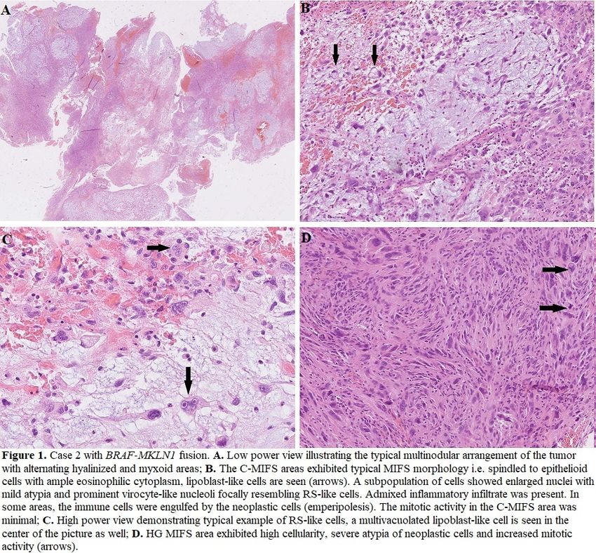
samples(St-LMS N=27/191,14%;UtLMS,N=11/122,9%;UPS,N=13/130,10%;AS,N=4/92,4%). Heterogenous loss(weak moderate
intensity) was seen in 22%(N=116/535) of cases (StLMS N=39/191,20%;UtLMS
N=25/122,21%;UPS,N=44/130,34%;AS,N=8/92,9%). The remaining cases (N=271/535,50%) exhibited moderate strong diffuse
labeling. Amongst stage, loss trended lower with higher stage in StLMS (28%,primary vs 25%,recurrent vs.23%, metastasis) and
UtLMS(33%vs.26%vs. 17%),while increasing prevalence of loss higher with stage in UPS(8%vs. 11%vs.17%) and in
AS(2%vs.0%vs.13%).

Conclusions: ATM loss can be detected in aggressive sarcomas by immunohistochemistry, suggesting some patients maybe
amendable to targeted therapies. Loss of ATM varied by subtype with LMS having the highest loss and may vary by stage.
Heterogenous patterns can be seen and needs further investigation.

26      Osteoid Osteoma of the Short Tubular and Small Bones of Hands and Feet: A Series of 33
        Cases
        Fatimah Alruwaii1, Scott Kilpatrick1, John Reith1, Karen Fritchie1
        1
         Cleveland Clinic, Cleveland, OH

Disclosures: Fatimah Alruwaii: None; Scott Kilpatrick: None; John Reith: None; Karen Fritchie: None

Background: Osteoid osteoma typically arises in the long bones of extremities or the spine. Patients often report pain relieved by
NSAIDS, and radiographic findings are usually sufficient for diagnosis. The clinicopathologic features of these tumors involving the
short tubular and small bones of the hands and feet have been less well clarified.

Design: Our institutional and consultation archives were searched for all cases of pathologically confirmed osteoid osteoma over
the past 30 years. Clinical presentation, radiographic and morphologic features, and follow-up data were recorded.

Results: A total of 220 cases (83 females, 137 males) were retrieved. 33 (15%) arose in the bones of the hands (n= 15) and feet
(n=18). Age ranged from 6-64 years (median 29 years). Specific anatomic sites were distal phalanx (n=13, 40%; including 8 in the
fingers and 5 in the toes) , talus (n=6, 8%), proximal phalanx (n=4,12%; all in the fingers), metatarsal (n=4,12%), metacarpal
(n=2,6%), cuneiform (n=2,6%), calcaneus (n=1,3%) and middle phalanx (n=1, 3%; finger). All cases with available clinical data (31
of 33) presented with pain and significant swelling. The clinical and radiographic impressions were available for 31 (of 33) cases
and included: osteoid osteoma (n=10, 30%), osteomyelitis (n=9, 27%), fracture (n=3, 9%), enchondroma (n=3; 9%), glomus tumor
(n=2, 6%), post-traumatic (n=1; 3%), avascular necrosis (n=1, 3%), ligament tear (n=1, 3%) and ankle instability (n=1, 3%).
Histologically, all cases showed characteristic histologic features including anastomosing woven bony trabeculae lined by
osteoblasts with intervening fibrovascular stroma with no aggressive features seen. Follow-up was available in 13 cases (2-216
months, median 60 months); 5 cases presented with clinically local recurrence/residual disease within a year; all were initially
thought to have osteomyelitis and were treated by debridement.

Conclusions: Osteoid osteoma involving the small and short tubular bones of the hands and feet is rare. These lesions often
present with a broad differential diagnosis and frequently are initially confused with osteomyelitis clinically. Although follow-up was
limited, morphology and behavior seems to parallel osteoid osteomas arising at more conventional sites. Awareness that this entity
may present in the hands and feet will help pathologists and clinicians accurately classify these tumors.

27      Molecular Characterization of Dedifferentiated Solitary Fibrous Tumor
        William Anderson1, Adrián Mariño Enríquez2, Christopher Fletcher2
        1
          Brigham and Women's Hospital, Harvard Medical School, Boston, MA, 2Brigham and Women's Hospital, Boston, MA

Disclosures: William Anderson: None; Adrián Mariño Enríquez: None; Christopher Fletcher: None

Background: Solitary fibrous tumor (SFT) is a fibroblastic tumor with distinctive histologic features, molecularly defined by the
presence of NAB2-STAT6 fusions. Most cases are biologically low-grade, and the overall recurrence rate is 10-30%. However, very
rare examples show transition to a high-grade morphologically non-distinctive sarcoma (dedifferentiated SFT, DDSFT) and
are associated with a very poor prognosis. Such cases are genomically unstable and harbor more frequent alterations
of TP53 and TERT, although the full spectrum of molecular alterations in DDSFT is unknown. We studied the genetics of DDSFT to
identify clinically actionable molecular alterations.

                                                                                                                                    23
Design: DDSFT (n=12) were retrieved from departmental and consultation files. Immunohistochemistry was performed for STAT6.
Targeted next-generation sequencing, using a clinically validated platform (Oncopanel) covering the coding regions of 447 cancer-
associated genes and 191 frequently rearranged genomic regions, was performed in each case in the region of dedifferentiation.

Results: Our cohort comprised 12 patients (8 females, 4 males), with a median age of 59 years (range 31-80). Tumors had a
median size of 10.0 cm (range 3.0-23.8) and involved the abdominal cavity (5/12), thorax (3/12), vertebrae (2/12), thigh (1/12) and
trunk (1/12). For each tumor, separate areas of well-differentiated SFT were verified in the same (n=11) or a prior (n=1) specimen.
Dedifferentiated areas were histologically diverse, with epithelioid, spindled, pleomorphic, or round cell morphology, and occasional
heterologous components. STAT6 expression was lost or reduced in 50% (6/12) of cases. Molecular findings are summarized in
Figure 1. All tumors harbored NAB2-STAT6 fusions, predominantly truncating STAT6 (75%; 9/12). Median tumor mutational burden
was 4.2 mutations/Mb. Frequently altered genes were TP53 (67%; 8/12), TERT (58%; 7/12), RB1 (17%; 2/12), and PTEN (17%;
2/12). One case that lacked these alterations showed IGF2 amplification.

                                                           Figure 1 - 27

Conclusions: The diagnosis of DDSFT may be challenging due to reduced or absent STAT6 expression and requires extensive
sampling to identify a concurrent or previous well-differentiated component. DDSFT is enriched for fusions
truncating STAT6 and TERT promoter mutations, and shows frequent inactivation of tumor suppressor genes, most
commonly TP53, as well as RB1 and PTEN.

28      Superficial CD34-Positive Fibroblastic Tumor: A Clinicopathologic Analysis of 52 Cases
        William Anderson1, Fredrik Mertens2, Jason Hornick1, Christopher Fletcher3
        1
         Brigham and Women's Hospital, Harvard Medical School, Boston, MA, 2University Hospital Lund, Lund, Sweden, 3Brigham
        and Women's Hospital, Boston, MA

Disclosures: William Anderson: None; Fredrik Mertens: None; Jason Hornick: None; Christopher Fletcher: None

Background: Superficial CD34-positive fibroblastic tumor (SCD34PFT) is a rare mesenchymal neoplasm that was recently
established as a distinct entity in the 2020 World Health Organization Classification. Around 60 cases have now been described
and although it appears to have a very good prognosis, with only one reported metastasis, long-term follow-up data are limited. In
addition, SCD34PFT shows considerable clinical and morphologic overlap with soft tissue tumors
harboring PRDM10 rearrangement, which are known to overexpress CADM3; this relationship is incompletely understood. We

                                                                                                                                  24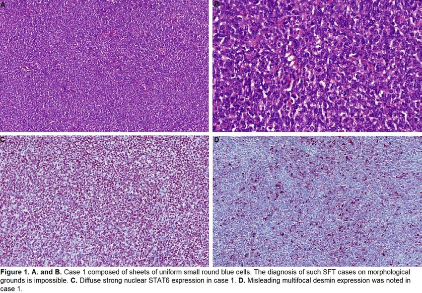
present a large series of SCD34PFT to further characterize its clinicopathologic features and clinical behavior. We also evaluate
the diagnostic utility of CADM3 immunohistochemistry in our cohort.

Design: Fifty-two SCD34PFTs were retrieved from departmental and consultation archives. Immunohistochemistry for CD34 and
CADM3 was performed; AE1/AE3 and desmin were performed in a subset of cases. Clinical follow-up data were obtained.

Results: Our cohort comprised 52 patients (27 males, 25 females), with a median age of 40 yrs (range: 14-85). SCD34PFT most
commonly arose in the lower limbs (71%) followed by the trunk (13%), upper limbs (10%), and head and neck (6%). The median
tumor size was 2.9 cm (range 1.0-9.0 cm). Tumors were composed of sheets of spindled, polygonal or epithelioid tumor cells with
abundant often glassy eosinophilic cytoplasm, including many cells showing striking nuclear pleomorphism (Figure 1). Mitoses
were very rare. Prominent stromal histiocytes were often present. All tumors expressed CD34. CADM3 was also frequently positive
(96%; 49/51). There was more limited AE1/AE3 (76%; 13/17) and desmin (38%; 18/48) expression. Follow-up in 13 patients so far
(median duration: 21 months) revealed that all patients are currently alive with no evidence of disease. However, one patient had
suffered both a local recurrence and regional lymph node metastasis, which were completely excised.

                        Table 1. Clinical characteristics of 52 superficial CD34-positive fibroblastic tumors.

                                     Age
                                      Median (range)                   40 (14 - 85) years
                                     Sex
                                      Male                             27
                                      Female                           25
                                      Male-to-female ratio             1.1:1
                                     Anatomic site [%]
                                      Lower limb                       37 (71%)
                                        Thigh                          23 (44%)
                                        Leg                            14 (27%)
                                      Upper limb                       5 (10%)
                                        Arm                            4 (8%)
                                        Forearm                        1 (2%)
                                      Trunk                            7 (13%)
                                      Head / neck                      3 (6%)
                                     Depth (n=41) [%]
                                      Subcutis only                    28 (69%)
                                      Subcutis and dermis              12 (29%)
                                      Dermis only                      1 (2%)
                                      Deep/subfascial                  0
                                     Tumor size (cm)
                                      Median (range)                   2.9 (1.0 - 9.0)
                                                            Figure 1 - 28

                                                                                                                                    25
Conclusions: Our series contributes further understanding to the clinicopathologic spectrum of SCD34PFT. Follow up data
obtained thus far corroborate that the majority are indolent, but that rare cases have the potential to metastasize to regional lymph
nodes. Finally, our initial results indicate that CADM3 is a highly sensitive marker, which may be used to support the diagnosis.

29      Myxofibrosarcoma in Adolescents and Young Adults: A Clinicopathologic Study of 34 Cases
        Karen Arispe Angulo1, Steven Billings2, Shruti Agrawal1, Karen Fritchie3
        1
          Cleveland Clinic Foundation, Cleveland, OH, 2Cleveland Clinic, Lerner College of Medicine, Cleveland, OH, 3Cleveland
        Clinic, Cleveland, OH

Disclosures: Karen Arispe Angulo: None; Steven Billings: None; Shruti Agrawal: None; Karen Fritchie: None

Background: Myxofibrosarcoma (MFS) is a locally aggressive fibroblastic malignancy that characteristically arises in the proximal
extremity of patients in the sixth and seventh decade of life. Cases arising in adolescence and young adulthood are rare, potentially
leading to diagnostic challenges.

Design: Our institutional and consultation archives were searched for ‘myxofibrosarcoma’ and ‘myxoid malignant fibrous
histiocytoma’ arising in patients 40 years or younger over the last 29 years. Clinicopathologic data was collected.

Results: 34 cases of myxofibrosarcoma arising in patients < 40 years were identified (23 females and 11 males; 14 to 40 years;
median 34 years). Tumor sizes ranged from 1.5 to 31 cm (median 5 cm). Anatomic sites included: proximal extremity (17; 3 thigh),
distal extremity (8), trunk (5), head/neck (3), intra-abdominal (1). Among tumors in the trunk and extremities where depth was
known, 13 were superficial and 9 were deep-seated. Using the FNCLCC grading system, 9 cases were grade 1, 13 grade 2, and 12
grade 3. 3 cases harbored predominantly epithelioid morphology while 7 additional cases had foci with epithelioid features. FISH
for MDM2 amplification was negative for all cases tested (4/4), including the intraabdominal mass. NGS sarcoma fusion panel
performed on a single case failed to show gene fusions and a comprehensive cancer profiling performed on another patient did not
reveal any actionable variants. Available follow up information revealed that one patient experienced local recurrence after 13
months of diagnosis, while 2 developed distant metastasis (both patients had large (>20 cm) intramuscular tumors). Sites of
metastasis included lung, bone and lymph nodes. Three patients were alive without disease (46 to 53 months; median 50 months).

Conclusions: Our study confirmed that myxofibrosarcoma arising in adolescents and young adults are rare but appear to show
similar aggressive behavior to those tumors arising in their older adult counterparts. Interestingly, nearly one-third of cases in this
age group showed some degree of epithelioid features, potentially resulting in diagnostic difficulty.

30      Genetic and Genomic Landscape of Myxoid Liposarcoma and their Association with Round
        Cell Phenotypes
        Davsheen Bedi1, Azfar Neyaz1, Bao Riyue1, Svetlana Yatsenko2, Karen Fritchie3, Arivarasan Karunamurthy1, Ivy John1
        1
          University of Pittsburgh Medical Center, Pittsburgh, PA, 2University of Pittsburgh, Pittsburgh, PA, 3Cleveland Clinic,
        Cleveland, OH

Disclosures: Davsheen Bedi: None; Azfar Neyaz: None; Bao Riyue: None; Svetlana Yatsenko: None; Karen Fritchie: None;
Arivarasan Karunamurthy: None; Ivy John: None

Background: Limited studies have been published on myxoid liposarcoma (MLS) genomics. Herein, we explore the genetic and
genomic landscape of MLS and their association with high grade features.

Design: We performed karyotype and whole-exome sequencing (WES) analysis on 19 MLS tumors. WES was performed on DNA
isolated from unmatched tumor samples (n=19) on an Illumina NextSeq500 instrument using Agilent SureSelect Human All Exon
V6 for exome capture and output reads were mapped to GRCh38 followed by duplicate removal and base quality score
recalibration. Somatic variants were called using GATK4-MuTect2 with rigorous filtering to remove germline contamination,
sequence or alignment artifacts, and variants of low quality. Total tumor mutational burden (TMB) was calculated using protein-
changing mutations; synonymous mutations were removed.

Results: Complex karyotypes were identified in 10 of 19 MLS, of which high-grade features were seen in 7 of 11 complex tumors.
A total of 762 somatic mutations were detected by WES, including 162 protein-changing mutations. The average TMB is 8.10

                                                                                                                                      26
(ranging from 2 to 19) per exome, suggesting MLS is low-TMB cancer. Somatic mutational signature (v3.2) analysis revealed
SBS86 (C>G predominant, chemotherapy treatment) enriched across all cases regardless of chemotherapy status. Higher TMB
was observed in tumors with complex karyotypes compared to those with isolated chromosome aberrations (9.20±1.48 vs.
6.89±1.14, mean±S.E.M), and in high-grade tumors compared to low-grade (8.90±1.53 vs. 7.22±1.13).
Activating PIK3CA mutations (p.H1047R, p.H1047L, p.G1049R, p.E545K) were detected in 6/19 (32%) cases. Five out of
six PIK3CA mutant tumors carry complex karyotypes, indicating a strong association between PIK3CA activation and genomic
instability (5/10 (50%) vs. 1/9 (11%) in tumors with karyotype containing complex vs. isolated chromosome abnormality; P=0.14,
odds ratio=7.15, two-sided Fisher’s exact test). Three of six PIK3CA mutant tumors showed high-grade morphology. Other protein-
changing mutations were present in ATM (1/19 cases), EP300 (1/19), FANCA (1/19), KMT2A/KMT2D (3/19), PTEN (1/19),
and TP53 (1/19) genes.

Conclusions: PIK3CA mutations show a strong association with MLS with complex karyotypes. Increased mutational load
(relatively higher TMB) is seen in MLS tumors with higher-grade features. MLS harbor highly enriched C>G transversions; while
some could be related to chemotherapy, additional studies are required to evaluate their genetic etiopathogenesis.

31      Diagnostic Value of MDM2 RNA In Situ Hybridization for Low Grade Osteosarcoma:
        Consistency Comparison of RNA In Situ Hybridization with Fluorescence In Situ Hybridization
        and Immunohistochemistry
        Chen Chen1, Xin He1, Min Chen1, Tianhai Du1, Wenyi Jing1, Hongying Zhang1
        1
         West China Hospital, Sichuan University, Chengdu, China

Disclosures: Chen Chen: None; Xin He: None; Min Chen: None; Tianhai Du: None; Wenyi Jing: None; Hongying Zhang: None

Background: Detection of MDM2 gene amplification via fluorescence in situ hybridization (FISH) and MDM2
immunohistochemistry (IHC) have been recognized as useful auxiliary tools for the diagnosis of low grade osteosarcoma (LGOS).
MDM2 RNA in situ hybridization (RNA-ISH) has been recommended as an alternative diagnostic assay for well-differentiated
liposarcoma/dedifferentiated liposarcoma recently. The aim of this study was to evaluate the diagnostic value of MDM2 RNA-ISH in
LGOSs, compare it with MDM2 FISH and IHC and explore its feasibility in decalcified samples.

Design: Twenty-three LGOSs and 49 control samples of bone and soft tissue were included. MDM2 RNA-ISH, FISH, IHC were
performed on all undecalcified and formalin-fixed paraffin-embedded tissue. Nineteen out 23 LGOSs were evaluated
by MDM2 RNA-ISH and FISH in both decalcified and undecalcified samples.

Results: In undecalcified samples, two LGOSs were failed for MDM2 FISH and a total of 20/21 (95.2%) LGOSs showed positive
results. 19 decalcified samples were failed for FISH. All control cases (n=49) were negative for MDM2 FISH. In 21 LGOSs with
interpretable FISH results, 21(100%) cases were positive for RNA-ISH. One LGOS without MDM2 amplification was positive for
RNA-ISH. The result of one FISH failed case was uninterpretable and the other showed no staining. In the control cases, only one
undifferentiated pleomorphic sarcoma showed positivity for MDM2 RNA-ISH and the remaining cases were negative. The
diagnostic sensitivity and specificity of MDM2 RNA-ISH in LGOSs were 100.0% and 96.2%, respectively. There was a great
consistency between MDM2 RNA-ISH and FISH (P<0.05, kappa=0.932). In 19 decalcified samples, one was positive for RNA-ISH
and the other showed no staining. In 21 LGOSs with interpretable FISH results, 76.3% (16/21) were positive for IHC. Two FISH
failed cases showed focal positivity and no staining, respectively. All control cases except for one fibrous dysplasia were negative
for IHC. In MDM2 IHC-positive LGOSs, 94.1% (16/17) were positive for RNA-ISH. There was a great consistency between MDM2
RNA-ISH and IHC (P<0.05, kappa=0.753).

Conclusions: MDM2 RNA-ISH equipped with high value in the diagnosis of LGOS, with consistent accuracy with FISH and higher
sensitivity than IHC. But its specificity needs to be verified in more cases. Its success in decalcified sample indicates that it may
have relatively low requirements on sample quality which needs to be investigated in further study.

                                                                                                                                   27
32     Genome-Wide DNA Methylation Profiling Reveals Differentially Methylated Genes in Clinically
       Distinct Subsets of Solitary Fibrous Tumor
       Jeffrey Cloutier1, Hannah Beird1, Nolan Maloney2, Davis Ingram, Khalida Wani1, Alexander Lazar1, Wei-Lien (Billy) Wang1,
       Elizabeth Demicco
       1
         The University of Texas MD Anderson Cancer Center, Houston, TX, 2Loma Linda University, Loma Linda, CA, 3University
       of Toronto, Toronto, Canada

Disclosures: Jeffrey Cloutier: None; Hannah Beird: None; Nolan Maloney: None; Davis Ingram: None; Khalida Wani: None;
Alexander Lazar: None; Wei-Lien (Billy) Wang: None; Elizabeth Demicco: None

Background: Solitary fibrous tumor (SFT) is a rare mesenchymal neoplasm characterized by a recurrent NAB2-STAT6 fusion and
metastatic progression in 10-30% of cases. The specific molecular alterations leading to aggressive behavior are not well
understood, although TERT promoter mutations and/or TP53 mutations may play a role in some cases. Epigenetic alterations are
pervasive in oncogenesis but are largely unexplored in SFT. We examined genome-wide DNA methylation in a large series of SFT
and demonstrate that biologically distinct SFTs have differential methylation profiles.

Design: A total of 85 SFTs were included in the study, including 75 primary tumors, 6 metastases, and 2 local
recurrences (unknown status in 2). Tumors were located in the thorax (n=25) and extrathoracic soft tissue (n=58) (unknown in
2). 15 patients had a history of metastatic disease. Genome-wide DNA methylation was evaluated using
Illumina MethylationEpic array. Methylation profiles were compared across several variables, including tumor behavior (benign vs
malignant), tumor status (primary vs metastasis), and TERT promoter mutation status.

Results: After data QC and exclusion of X&Y chromosomes to remove bias on the basis of sex chromosomes, principal
component analysis separated data into distinct groups sharing DNA methylation patterns. PC1 and PC2
segregated SFT according to tumor location (thoracic vs extrathoracic) and patient sex, respectively. Comparing tumors from
patients with no history of metastasis vs those with a history of metastasis, 80 significantly differentially methylated gene
probes were identified, including several cancer-associated transcription factor genes. TERT promoter mutation status correlated
with 150 differentially methylated gene probes including genes involved in survival and autophagy. Immunohistochemical analysis
to investigate expression of biologically relevant differentially methylated genes is ongoing.

Conclusions: The biological and behavioral heterogeneity of SFT is likely influenced by a combination of genetic
and epigenetic alterations. We found that DNA methylation profiles are associated with distinct clinical features in SFT. DNA
methylation patterns correlate with anatomic localization, suggesting that thoracic and extrathoracic tumors are epigenetically
distinct. Tumors associated with metastatic behavior showed differential methylation of several cancer-associated genes. Together,
these findings suggest that epigenetic alterations can influence tumor behavior in SFT.

33     Chromosome 8 Gain is a Frequent Copy Number Variation in Soft Tissue Sarcomas
       Carina Dehner1, Robert Bell1, Kevin He1, John Chrisinger1, Amy Armstrong1, John Shern2, Marielle Yohe3, Angela Hirbe1
       1
        Washington University School of Medicine, St. Louis, MO, 2Center for Cancer Research, National Cancer Institute,
       Bethesda, MD, 3Center for Cancer Research, National Cancer Institute, National Institutes of Health, Bethesda, MD

Disclosures: Carina Dehner: None; Robert Bell: None; Kevin He: None; John Chrisinger: None; Amy Armstrong: None; John
Shern: None; Marielle Yohe: None; Angela Hirbe: Consultant, Springworks Therapeutics; Advisory Board Member,
AstraZeneca; Grant or Research Support, Tango Therapeutics

Background: Soft tissue sarcomas are rare mesenchymal neoplasms that frequently show complex chromosomal aberrations
such as amplifications or deletions of DNA sequences or even whole chromosomes. We recently found that gain of Chromosome 8
is associated with worse overall survival in soft tissue sarcomas as a group and plays a role in high-grade transformation of
malignant peripheral nerve sheath tumor (MPNST). Herein, we evaluated the role of chromosome 8 gain in a cohort of 7 different
types of non-fusion driven soft tissue sarcomas using fluorescent-in-situ hybridization (FISH) testing.

Design: The departmental archive was searched for cases of undifferentiated pleomorphic sarcoma (UPS), well- and
dedifferentiated liposarcoma (WDLPS and DDLPS), leiomyosarcoma (LMS), Neurofibromatosis 1 (NF1)-MPNST, non-NF1
MPNST, pleomorphic rhabdomyosarcoma (PRMS) and embryonal rhabdomyosarcoma (ERMS) with available tumor. Centromeric-
enumerating (Vysis CEP8 D8Z2) was performed and 200 cells per tumor were counted. Average of Chr8 gain, percentage of cells
with Chr8 gain and highest copy number of Chr8 were assessed.

                                                                                                                                   28
Results: Sixty-five samples underwent Chr8 FISH: 13 cases of ERMS, 8 cases of PRMS, 8 cases of LMS, 8 cases of WDLPS and
their 8 corresponding DDLPS, 10 cases of NF1-MPNST and 10 cases of non-NF1 MPNST. The highest Chr8 gain was noted in
UPS with an average copy number of 3.71, followed by ERMS with 2.98 copies. UPS showed Chr8 gain in more than half of the
counted cells per sample (average: 59%), followed closely by ERMS (average: 56%) (28-59%). The highest individual copy number
per cells was seen in UPS with an average of 15.1 copies, followed by 12 copies in LMS (range: 6.5-15.1). There was no difference
in copy number gain between WDLPS and its corresponding DDLPS (p = 0.7, 0.47 and 0.40, respectively).

                    Type of sarcoma       Average CEP8        Gain of Chr8 in %      Highest copy number of 8
                    UPS                   3.714               59.2                   15.125
                    WDLPS                 2.325               28.6                   8.000
                    DDLPS                 2.454               36.3                   6.571
                    LMS                   2.964               44.9                   12.00
                    PRMS                  2.766               40.5                   8.625
                    ERMS                  2.975               56.2                   7.385
                    Non-NF1 MPNST         2.716               42.9                   9.188
                    NF1-MPNST             2.743               43.12                  9.50
                                                            Figure 1 - 33

Conclusions: The pathogenesis of soft tissue sarcomas is poorly understood and many of them are associated with poor outcome
supporting the need of new treatment approaches with better risk stratification. In our pilot study we show that Chr8 gain is
commonly seen in soft tissue sarcomas, in particular in UPS, ERMS and LMS. Additionally, Chr8 gain does not appear to play a
role in the dedifferentiation of retroperitoneal liposarcoma. Future studies looking at the outcome of these tumors in correlation with
Chr8 and comparison to epithelial neoplasms are necessary to determine its prognostic relevance.

34      Molecular Characterization of Multifocal Granular Cell Tumors
        Carina Dehner1, Molly Schroeder2, John Chrisinger1
        1
         Washington University School of Medicine, St. Louis, MO, 2Washington University in St. Louis, St. Louis, MO

Disclosures: Carina Dehner: None; Molly Schroeder: None; John Chrisinger: None

Background: Granular cell tumor (GrCT) is a distinctive neoplasm with neuroectodermal differentiation composed of cells with
abundant granular cytoplasm and a predilection for the tongue, and superficial tissues of the head and neck. The prognosis is
usually excellent, though malignant GrCT are extremely aggressive. The majority of GrCT present as solitary lesions, however
multiple tumors occur in 10-15% of cases and are not thought to be associated with increased risk of aggressive behavior. Recent

                                                                                                                                     29
data has shown that inactivating mutations in H+-ATPase components, most commonly ATP6AP1 or ATP6AP2 are likely
tumorigenic drivers. Herein we investigate the molecular alterations of multifocal GrCT to determine if there is evidence to suggest
a clonal relationship versus molecularly distinct tumors.
Design: The pathology departmental archive was searched from 1988-2019 for patients with multifocal GrCT (≥ 2 tumor sites per
patient) and 13 patients were identified with 2 or more synchronous lesions (range: 2 – 10 locations) leading to a total of 43
samples. Clinical parameters such as gender, tumor sites, follow up and associated syndromes were collected. Targeted next-
generation sequencing for recently described potential driver mutations (ATP6AP1, ATP6AP2, ATP6AP1L, ATP6V0A3,
ATP6V0A4, ATP6V0B, ATP6V0C, ATP6V1A, ATP6V1D and PTEN) was performed.
Results: Histologically none of the tumors showed atypical or malignant features. Sequencing results for 42 samples are shown in
table 1 (one sample failed due to poor read quality). Tumors showed predominately mutually exclusive mutations in 33 of 42
samples. None of the multifocal tumors within a single patient showed the same potential driver alteration. Median follow up time
was 18 years (7-29 years) and aggressive behavior was not observed. Syndromic associations were not noted.
Patient Sample Somatic Variation
P1 S_61 None observed
S_72 ATP6AP2 (c.706delG (p.Ala236fs)) 15% VAF
(F) S_53 ATP6AP1 (c.225_226delTA (p.Thr76fs)) 19% VAF
S_7 ATP6AP2 (c.978C>A (p.Tyr326*)) 24% VAF
P2 S3 None observed
S32 ATP6AP2 (c.589-2A>G (splice acceptor)) 13% VAF
(F) S22 ATP6AP1 (c.939T>G (p.Tyr313*)) 43% VAF
S11 ATP6AP1 (c.546delC (p.Tyr183fs)) 19% VAF
S42 ATP6AP1 (c.1085_1094delCAGGGCCCAG (p.Gly363fs)) 9% VAF
S36 ATP6AP1 (c.1087_1096delGGGCCCAGCA (p.Gly363fs)) 24% VAF
P3 S76 ATP6AP1 (c.149G>A (p.Trp50*)) 36% VAF
S63 None observed
(M)
P4 S51 ATP6AP1 (c.1167_1168insC (p.Ser391fs)) 14% VAF
S34 ATP6AP1 (c.1242C>A (p.Tyr414*)) 8% VAF
(F)
P5 S54 ATP6AP1 (c.127C>T (p.Gln43*)) 11% VAF
(F)
P6 S52 None observed
S60 None observed
(F)
P7 S13 ATP6AP1 (c.1083_1084delCA (p.Thr362fs)) 6% VAF
S59 None observed
(F)
P8 S71 ATP6AP2 (c.508delC (p.Leu170fs)) 11% VAF
S59 None observed
(F) S57 ATP6AP1 (c.685-2A>T (splice acceptor)) 24% VAF
S50 ATP6AP1 (c.364-2A>G *splice acceptor)) 15% VAF
S38 ATP6AP1 (c.50delC (p.Gln18fs)) 7% VAF
S42 ATP6AP1 (c.1192C>T (p.Gln398*)) 26% VAF
S24 None observed
S18 ATP6AP2 (c.751delA (p.Met251fs)) 20% VAF
S45 ATP6AP2 (c.805_806delTT (p.Phe269fs)) 31% VAF
S33 ATP6AP1 (c.1208A>C (p.Gln403Pro)) 12% VAF
ATP6V0A4 (c.1155C>T (p.Val385Val)) 100% VAF Highest MAF = 15%
P9 S58 ATP6AP1 (c.414G>A (p.Trp138*)) 15% VAF
S43 ATP6AP1 (c.1299C>T (p.Ser433Ser)) 18% VAF
S19 ATP6AP2 (c.485delA (p.Asn162fs)) 27% VAF
(M)
P10 S30 ATP6AP2 (c.24_28delGGCGT (p.Ala9fs)) 6% VAF
S36 ATP6AP1 (c.500_509delTGAAGCTCAA (p.Lys168fs)) 6% VAF
(F)
P11 S32 ATP6AP1 (c.1032_1036delCCTCG (p.Leu345fs)) 8% VAF
S56 None observed
(F)
S25 ATP6V0A4 (c.1662delC (p.Phe554fs)) 100% VAF
30P12 S44 None observed
S24 ATP6AP1 (c.929C>T (p.Ser310Leu)) 7% VAF
(F)
P13 S15 ATP6AP1 (c.414delG (p.Trp138fs)) 6% VAF
(M) ATP6AP2 (c.165delA (p.Glu56fs)) 16% VAF
S18 None observed
S55 ATP6AP2 (c.752delT (p.Met251fs)) 48% VAF
± Variants in trans
Table legend: Genomic variation detected in tumor-only sequencing of a small gene panel consisting of ATP6AP1, ATP6AP2,
ATP6AP1L, TCRIG1 (ATP6V0A3), ATP6V0A4, ATP6V0B, ATPV0C, ATP6V1A, ATP6B1D, and PTEN.
“Apparently germline variation” is defined as that observed at variant allele frequency (VAF) between 45-55% in all samples from a
patient. “Highest MAF” value is the greatest population minor allele frequency observed in aggregated gnomAD data.
Somatic variation is absent from gnomAD and COSMIC databases unless a frequency for their presence is noted.
Two somatic variants in ATP6V0A4 (in S33 and S25) were observed at 100% VAF, suggesting the possibility of a loss of
heterozygosity event in this region.
Transcripts:
ATP6V0C: NM_001694.4
ATP6AP2: NM_005765.2
ATP6AP1: NM_001183.6
TCIRG1 (ATP6V0A3): NM_006019.4
ATP6V0A4: NM_020632.3
ATP6V0B: NM_004047.5
ATP6V1A: NM_001690.4
PTEN: NM_ 000314.7
Conclusions: Benign appearing multifocal GrCT are not uncommonly encountered and our data suggests that many are
genetically independent lesions. It remains unclear why certain patients show a propensity for these tumors in a non-syndromic
setting; however, multifocal lesions do not appear to be associated with increased malignant potential.
35 Comprehensive Genomic Profiling of EWSR1/FUS-CREB Family of Translocation-Associated
Tumors Uncovers Prognostically Significant Recurrent Genetic Alterations and Methylation-
Transcriptional Correlates
Josephine Dermawan1, Fabio Vanoli1, Lei Zhang1, Memorial Center1, Tejus Bale1, Brendan Dickson2, Cristina Antonescu1
1
Memorial Sloan Kettering Cancer Center, New York, NY, 2Mount Sinai Health System, Toronto, Canada
Disclosures: Josephine Dermawan: None; Fabio Vanoli: None; Lei Zhang: None; Memorial Center: None; Tejus Bale: None;
Brendan Dickson: None; Cristina Antonescu: None
Background: Recurrent gene fusions involving EWSR1/FUS with members of the cAMP response element binding protein (CREB)
family (ATF1, CREB1 and CREM) are shared amongst multiple tumor-types spanning a wide clinicopathologic spectrum.
Design: To elucidate the mechanisms underlying the divergent clinicopathologic spectrum of EWSR1/FUS-CREB family of
translocation-associated tumors, we performed a comprehensive genomic analysis of fusion transcript variants, recurrent genetic
alterations (mutations, copy number alterations), gene expression and methylation profiles across a large cohort of tumor types
(Table 1).
Results: The distribution of the EWSR1/FUS fusion partners—ATF1, CREB1, and CREM—and exon involvement was significantly
different across different tumor types (Figure 1). Our targeted sequencing showed that secondary genetic events are associated
with tumor type rather than fusion type. Of the 39 cases that underwent targeted NGS testing, 18 (46%) had secondary OncoKB
mutations or copy number alterations (29 secondary genetic events in total), of which 15 (52%) were recurrent. Recurrent,
but mutually exclusive, TERT promoter and CDKN2A mutations were identified only in clear cell sarcoma (CCS) and associated
with worse overall survival. CDKN2A/B homozygous deletions were recurrent in angiomatoid fibrous histiocytoma (AFH) and
restricted to metastatic cases (Figure 2). mRNA upregulation of MITF, CDH19, PARVB, and PFKP was found in CCS compared to
AFH and correlated with a hypomethylated profile. In contrast, S100A4 and XAF1 were differentially upregulated and
hypomethylated in AFH but not CCS. A sarcoma methylation classifier was able to accurately match 100% of CCS cases to the
correct methylation class; however, it was suboptimal when applied to other histologies.
31Table 1. Study cohort demographics
Diagnosis Female, n Male, n Mean age in years Total
(%) (%) (range)
Angiomatoid fibrous histiocytoma (AFH) 18 (51) 17 (49) 28.8 (2-79) 36
Clear cell odontogenic carcinoma (CCOC) 1 (100) 0 57.0 1
Clear cell sarcoma (CCS) 16 (40) 24 (60) 32.9 (7-71) 40
Clear cell sarcoma-like tumor of the gastrointestinal tract (GICCS) 14 (70) 6 (30) 39.0 (18-76) 20
Hyalinizing clear cell carcinoma of salivary gland (HCCC) 8 (80) 2 (20) 67.6 (55-86) 10
Malignant epithelioid neoplasm with predilection for mesothelial- 7 (50) 7 (50) 37.2 (9-63) 14
lined cavities (ME)
Mesothelioma (Meso) 5 (62) 3 (38) 39.0 (15-78) 8
Myxoid mesenchymal tumor (MMT) 3 (60) 2 (40) 27.0 (12-48) 5
Primary pulmonary myxoid sarcoma (PPMS) 3 (100) 0 59.0 (43-82) 3
Total 74 60 36.9 (5-86) 137
Figure 1 - 35
32Figure 2-35
Conclusions: In conclusion, our comprehensive genomic profiling of EWSR1/FUS-CREB family of translocation-associated
tumors uncovered mostly histotype rather than fusion-type associated correlations in transcript variants, prognostically significant
recurrent secondary genetic alterations, and gene expression and methylation patterns.
36 The Incidence and Significance of Calcium Pyrophosphate Dihydrate Deposits (pseudogout)
in Revision Arthroplasty Specimens
Josephine Dermawan1, Fatimah Alruwaii2, John Reith2, Scott Kilpatrick2
1
Memorial Sloan Kettering Cancer Center, New York, NY, 2Cleveland Clinic, Cleveland, OH
Disclosures: Josephine Dermawan: None; Fatimah Alruwaii: None; John Reith: None; Scott Kilpatrick: None
Background: We recently evaluated the incidence and significance of calcium pyrophosphate dihydrate deposits (CPPD) in hip,
knee, and shoulder primary total arthroplasty specimens, concluding that CPPD was more common in older patients and in the
knees and shoulders when compared to the hips (PMID: 33720299). Given these findings, we now sought to determine the
incidence and significance of CPPD in revision arthroplasties and whether such deposits might be correlated with periprosthetic
joint infection (PJI), issues which have not been thoroughly evaluated in the literature.
Design: We retrospectively reviewed the clinicopathologic characteristics, especially focusing on CPPD, among consecutive hip,
knee, shoulder, and elbow joint revision arthroplasties (n=1111) seen at our institution and signed out by the authors. Specific sites
included 628 hips, 379 knees,101 shoulders, and 3 elbow specimens. Significant acute inflammation was defined as 5 or more
neutrophils in 5 or more high power fields, and this was considered a "positive" result for periprosthetic joint infection (PJI), per
Musculoskeletal Infection Society (MSIS) criteria. 190 cases (17.1%) were "positive" pathologically and thought to have PJI, more
specifically 106 hips (16.9%), 74 knees (19.5%), 9 shoulders (8.9%), and 1 elbow (33.0%).
Results: Forty-seven cases (4.2%) had CPPD deposits in at least one section from one specimen of the revision arthroplasty case,
more specifically 29 knees (7.7% of knees), 15 hips (2.4% of hips), 2 shoulders (2.0%), and 1 elbow (33.0%). The overwhelming
majority (44, 93.6%) of cases of CPPD occurred in specimens which were negative for PJI, lacking significant acute inflammation;
only 3 cases of CPPD, all associated with the knee, were found in patients considered positive for PJI at revision. The age range
33among all patients in this series was 27-97 years (mean 65.1); those with CPPD were only slightly older, range 47-88 years (mean
68.6).
Conclusions: The distribution of CPPD among hip and knee revision arthroplasty specimens is similar to our experience with
primary arthroplasties, with knees being significantly more frequently affected than the hips. However, shoulders were less
commonly involved by CPPD than the hips or knees and also less often showed histologic evidence of PJI. CPPD appears more
likely to be found in patients with aseptic loosening than in patients with PJI. The exact reasons behind CPPD being associated
with an absence of PJI is unclear but deserves further investigation.
37 TMEFF2 Protein Expression and Clinicopathologic Profiles in Ewing Sarcoma
Jing Di1, Subhasree Basu2, Mike Russell2, Tom Jorfi2, Jackson Wong2, Shaozhou Tian2, Shadi Qasem3
1
University of Kentucky College of Medicine, Lexington, KY, 2Janssen R&D, LLC, Raritan, NJ, 3University of Kentucky
Healthcare, Lexington, KY
Disclosures: Jing Di: Primary Investigator, Janssen R&D, LLC; Subhasree Basu: Employee, The Janssen Pharmaceutical
Companies of Johnson & Johnson; Mike Russell: Employee, Janssen R&D; Tom Jorfi: Employee, Janssen R&D Inc.; Jackson
Wong: Employee, Janssen R&D; Shaozhou Tian: Employee, Janssen R&D; Shadi Qasem: Primary Investigator, Janssen
Pharmaceuticals; Primary Investigator, Janssen Pharmaceuticals
Background: TMEFF2 is a conserved cell-membrane-bound proteoglycan that is often expressed at high levels in cells of prostatic
ductal epithelial origin from normal early development through carcinogenesis. Bioinformatical analysis of major public cancer
mRNA registry databases demonstrates TMEFF2 transcripts are present in other tumors such as Ewing sarcoma. The direct
biological significance of mRNA expression is limited, and immunohistochemistry (IHC) studies are required to determine protein
expression. In this study, we aim to define TMEFF2 distribution in Ewing sarcoma and evaluate TMEFF2 expression with
clinicopathological findings.
Design: An extensive animal immunization campaign was conducted against specific regions of the TMEFF2 protein to generate a
tool reagent (antibody) suitable for IHC studies. A proprietary TMEFF2 IHC assay was developed and utilized on 41 FFPE tumor
materials of 32 Ewing sarcoma cases. Clinicopathological variables recorded included age, gender, and tumor site. H-scores were
determined by 2 pathologists. One-way ANOVA, Chi-square, and Bartlett's test were used with pFigure 1 - 37
Conclusions: To our knowledge, this TMEFF2 IHC study was the first to demonstrate TMEFF2 protein expression in Ewing
sarcoma. In addition, higher levels of TMEFF2 expression may be present in recurrent and metastatic tumors. The biological,
prognostic, and therapeutic significance of our findings needs to be confirmed with additional studies with more robust tumor sets.
38 Droplet Digital (dd) PCR as a Novel Technology in Detecting CTNNB1 Mutations in Desmoid
Fibromatosis
Jatin Gandhi1, Erica Kao2, Jose Mantilla Arango1, Robert Ricciotti1, Anshu Bandhlish3, Yu Wu1, Yajuan Liu1, Eleanor Chen1
1
University of Washington, Seattle, WA, 2San Antonio Uniformed Services Health Education Consortium, San Antonio,
TX, 3University of Washington - Pathology, Seattle, WA
Disclosures: Jatin Gandhi: None; Erica Kao: None; Jose Mantilla Arango: None; Robert Ricciotti: None; Anshu Bandhlish: None;
Yu Wu: None; Yajuan Liu: None; Eleanor Chen: None
Background: Desmoid fibromatosis (DF) is a locally aggressive soft tissue neoplasm characterized by infiltrative growth and
frequent recurrences. It can mimic a range of benign and malignant processes, posing a diagnostic challenge in some instances.
Molecularly, DF is characterized by alterations in the Wnt/β-Catenin pathway, with the majority showing sporadic mutations
in CTNNB1 while others have germline mutations in APC (Gardner syndrome). IHC staining for β-Catenin is often difficult to
interpret due to non-specific reactivity and can be negative in up to 30% of cases. Prior studies have shown that some DFs lacking
nuclear expression of β-Catenin may still carry activating CTNNB1 mutations. ddPCR has been used effectively in detecting gene
mutations in FFPE samples of various cancer types, suggesting its clinical utility. In this study we assess the diagnostic utility of
ddPCR to detect CTNNB1 mutations in DF with and without β-Catenin positivity, as well as in challenging cases.
35Design: We identified 30 cases of DF, including 28 with nuclear β-Catenin expression by IHC and 2 without nuclear b-Catenin, as
well as 7 (myo)fibroblastic lesions not classified as DF but for which DF was in the differential. Genomic DNA extracted from FFPE
tissue blocks was subjected to ddPCR using primers for the most common point mutations in exon 3 of CTNNB1 (S45F, S45P,
T41A). Results were compared with IHC stains for β-Catenin. Clinicopathologic and demographic parameters were recorded.
Results: The patient age for DF ranged from 18-72 years (median, 44.5) with female predominance (F:M::1.8:1). Anatomic
locations included neck/shoulder (n=9), extremities (n=6), chest wall (n=4), abdominal wall (n=4), mesentery (n=3), back (n=1) and
breast (n=1). Tumor size ranged from 1.7 to 23 cm (median; 8.5 cm). 25 of 28 cases with nuclear β-Catenin on IHC showed
a CTNNB1 mutation by ddPCR (89%). The most frequent mutation was T41A (n=14; 50%) followed by S45F (n=8; 33%) and S45P
(n=3;12%). The 2 cases without nuclear β-Catenin staining were negative for the three mutations tested. 2 of the 7 cases not
previously classified as DF exhibited CTNNB1 mutation (one each, S45F and S45P).
Conclusions: ddPCR is a sensitive and cost-efficient methodology to detect hotspot CTNNB1 mutations in DF, especially
diagnostically challenging cases. This method can also be applied as a targeted mutation assay to detect disease- defining point
mutations in various tumor types.
39 Clinicopathologic Spectrum of NTRK fusion-positive Tumors Detected by Clinical RNA
Sequencing Panel
Annie Garcia1, Jacquelyn Reuther2, Horatiu Voicu2, Carrie Mohila3, Adekunle Adesina2, Deborah Schady3, Wei-Lien (Billy)
Wang4, Alexander Lazar4, Dolores Lopez-Terrada5, Frank Lin2, Rajkumar Venkatramani2, Sharon Plon2, Williams Parsons2,
Kevin Fisher2, Angshumoy Roy1
1
Baylor College of Medicine, Houston, TX, 2Baylor College of Medicine/Texas Children's Hospital, Houston, TX, 3Texas
Children's Hospital, Houston, TX, 4The University of Texas MD Anderson Cancer Center, Houston, TX, 5Texas Children's
Hospital, Baylor College of Medicine, Houston, TX
Disclosures: Annie Garcia: None; Jacquelyn Reuther: None; Horatiu Voicu: None; Carrie Mohila: Stock Ownership, Johnson and
Johnson; Adekunle Adesina: None; Deborah Schady: None; Wei-Lien (Billy) Wang: None; Alexander Lazar: None; Dolores Lopez-
Terrada: None; Frank Lin: None; Rajkumar Venkatramani: None; Sharon Plon: None; Williams Parsons: None; Kevin Fisher: None;
Angshumoy Roy: None
Background: Gene fusions involving neurotrophic receptor tyrosine kinase (NTRK) genes are actionable targets approved for
targeted therapy in any solid tumor. While generally infrequent in cancers, NTRK fusions are common in certain classic rare tumors
where they are recurrently reported with ~100 different gene partners. However, overlapping clinical and histopathological features
between classic and emerging NTRK fusion-positive entities pose practical challenges in developing a screening strategy to test for
these fusions. Here we illustrate this by describing the varied clinical and histopathological features of a series of NTRK fusion-
positive tumors detected in the clinical laboratory by targeted RNA sequencing.
Design: Solid tumors referred to the clinical laboratory for testing with a targeted fusion panel (Archer FusionPlex) were searched
for NTRK1, NTRK2 or NTRK3 fusions. Electronic records were reviewed for clinical and laboratory findings. An NTRK fusion was
considered an expected event if a classic fusion-positive entity, based on histopathology and/or immunohistochemistry (IHC), was
provided as test indication.
Results: Eleven patients (6 males, 5 females; age 2 weeks – 30 years) were identified with diverse NTRK fusion-positive tumors
in varied anatomical sites (Table 1). Fusions involved NTRK1 (n=5), NTRK2 (n=1) and NTRK3 (n=5) with five different 5’ gene
partners. ETV6/NTRK3 was the most common fusion (n=5) followed by TPM3/NTRK1 (n=3), one tumor each with infrequent
fusions (LMNA/NTRK1, KANK1/NTRK2) and a novel DYNC1I2/NTRK1 fusion. Prior to fusion testing, clinical, histopathology and
IHC features suggested an expected NTRK fusion-positive entity in 7/11 cases, including 4 infantile fibrosarcoma (IFS) cases, 1
glioblastoma, and 2 pan-TRK IHC-positive soft tissue tumors. Notably, the canonical ETV6/NTRK3 fusion in IFS was detected in
only 2/4 IFS tumors (cases 6, 7), with two others (cases 4, 11) harboring non-canonical/novel NTRK fusions (Table 1). Significantly,
in 4/11 (36.3%) cases, the NTRK fusion was an unexpected finding (Table 1; cases 1, 5, 8, 10).
36Case Age Sex Location Tumor (Initial diagnosis) Pan-TRK Fusion panel results
IHC
Fusion 5' partner Diagnostic
frequency significance
(any tumor)
01 9 M Supra-renal Round blue cell tumor NP TPM3:NTRK1 Common Unexpected
(suggestive of Ewing)
02 21 M Para- Spindle cell Positive LMNA:NTRK1 Infrequent Expected
vertebral mesenchymal tumor of (diffuse)
uncertain malignant
potential
03 30 F Uterine Atypical spindle cell Positive TPM3:NTRK1 Common Expected
Cervix proliferation; a low-grade
sarcoma cannot be
excluded
04 4Mo M Forearm Congenital infantile NP TPM3:NTRK1 Common Expected*
fibrosarcoma
05 14 F Nasal ala Fibrohistiocytic NP ETV6:NTRK3 Common Unexpected
proliferation
06 10Mo M Orbit Infantile fibrosarcoma Positive ETV6:NTRK3 Common Expected
(focal)
07 2W M Back Infantile fibrosarcoma NA ETV6:NTRK3 Common Expected
VS myofibroma
08 2 F Kidney Undifferentiated NP ETV6:NTRK3 Common Unexpected
Sarcoma
09 6 F Brain, Epithelioid glioblastoma, NP ETV6:NTRK3 Common Expected
frontal lobe recurrent
10 6 M Brain Anaplastic ependymoma NP KANK1:NTRK2 Infrequent Unexpected
11 3 F Retro- Infantile fibrosarcoma NP DYNC1/2:NTRK1 Novel Expected*
peritoneum
F, female; IHC, Immunohistochemical stain; M, male; Mo, month; NP, not performed; NA, not available; W, week; *non-canonical
Conclusions: NTRK fusion-positive entities presented with a wide clinical and histologic spectrum, in a third of which the finding
was unexpected. Uncommon/novel NTRK fusions may be present in classic entities negative for canonical fusions by targeted
testing and comprehensive fusion detection should be pursued in such cases. Positivity for pan-TRK IHC, diffuse or focal, may be
helpful in uncommon clinical presentations.
40 Copy Number Alterations Outside 12q13-15 in Well-Differentiated/ Dedifferentiated
Liposarcomas
Nicolas Giraldo Castillo1, Chad Vanderbilt2
1
Memorial Sloan Kettering Cancer Center, Baltimore, MD, 2Memorial Sloan Kettering Cancer Center, New York, NY
Disclosures: Nicolas Giraldo Castillo: None; Chad Vanderbilt: Consultant, Paige AI
Background: Well-differentiated liposarcoma (WDLS) and dedifferentiated liposarcomas (DDLS) are rare malignant, locally
aggressive mesenchymal neoplasms characterized by amplification of chromone 12q13-15, to include genes like MDM2, CDK4,
and HMGA2. Previous studies in small cohorts of WDLS /DDLS suggest that there might be areas outside 12q13-15 with copy-
number alterations (CNA) in these tumors. However, due to the limited sample size of these studies, the characterization of
37recurrent CNA and their association with patient’s clinical outcome remains unknown. In this study, we perform a large-scale
genomic analysis of WDLS/DDLS to identify recurrent somatic mutations and CNA events associated with poor prognosis.
Design: We studied the prevalence of somatic mutations, CNA, and structural variants in n=81 WDLS and n=248 DDLS samples
sequenced by MSK-IMPACT panel using the cBioPortal platform. We then assessed the prognostic impact of the most common
genomic events using Cox Proportional-Hazards modeling for overall survival (OS.) Relevant findings were validated in an
additional cohort of n=167 DDLS from the TCGA.
Results: Preliminary analysis of n=329 WDLS/DDLS showed recurrent amplifications in 6q21 (including TNFAIP3 in 18%, IFNGR1
in 12%, and LATS1 in 12%), 5p15.33 (including TERT in 12%, and TRIP13 in 10%), and 1q21-23 (including SDHC in 8% and
MCL1 in 7%) regions. In addition, amplification of JUN was seen in 12% of cases. A small fraction of cases showed focal deletions,
including the 9p21 region (MTAP 4%, CDKN2AP14ARF 4%, and CDKN2AP16INK4A 3%) and ATRX (3%.) Somatic mutations
were relatively uncommon and included ATRX (4%), TP53 (3%), and ERBB4 (2%.) Although JUN and 5p15.33 amplification events
were independent, tumors with alteration in any of these two regions were enriched in dedifferentiated histology (chi-squared
p=0.001 and 0.01, respectively) and associated with shorter OS (p=0.008 and 0.01, respectively.) Analysis from additional n=167
TCGA DDLS cases confirmed that both JUN and 5p15.33 amplification were associated with poor prognosis (p=0.002 and
p=0.002, respectively.)
Conclusions: This large-scale genomic analysis reveals genomic events potentially associated with progression of WDLS into
DDLS and identified TERT/TRIP13 and JUN CNA as potential prognostic biomarkers in these two entities.
41 Primary Inflammatory Myofibroblastic Tumor of the Liver: Clinicopathologic and Genetic
Study of 10 Cases Including a Subset of ETV6-NTRK3 Fusion
Qianqian Han1, Xin He1, Min Chen1, Xiaojun Pang2, Zhang Zhang1, Hongying Zhang1
1
West China Hospital, Sichuan University, Chengdu, China, 2Mianyang Hospital of Traditional Chinese Medicine,
Mianyang, China
Disclosures: Qianqian Han: None; Xin He: None; Min Chen: None; Xiaojun Pang: None; Zhang Zhang: None; Hongying Zhang:
None
Background: Inflammatory myofibroblastic tumor (IMT) is a borderline myofibroblastic tumor, most frequently arising in mesentery,
retroperitoneum, pelvis and lung, and rarely in the liver. The purpose of this study was to analyze the clinicopathological and
molecular characteristics of primary liver IMT.
Design: 10 primary liver IMTs were identified, accounting for 2.6% of all diagnosed IMTs (10/384) over the past 10 years at our
medical centre. The clinicopathological data was collected and analyzed by immunohistochemistry, fluorescence in situ
hybridization (FISH), reverse transcription polymerase chain reaction, next generation sequencing (NGS) and Sanger sequencing.
Results: 4 males and 6 females were reviewed, age ranged from 1 year to 48 years (median 35 years, including 3 children). The
main morphological features included compact fascicular spindle cell proliferation, extensive myxoid stroma, and sparse tumor cells
arranged in a hyaline stroma. Tumors of 9 cases showed mild to moderate cellular atypia, 1 case exhibited obvious atypia with
large and round nuclei with prominent nucleoli. Mitotic figures (0~2/10HPF) were observed. Positive SMA staining was shown in 9
cases (90%), ALK cytoplasmic expression in 6 cases (6/10, 60%) and pan-TRK nuclear reactivity in 3 cases (3/10,
30%). ALK gene rearrangement was detected in 3 of 5 ALK-positive cases undergone FISH and/or NGS detection, of which one
case contained ALK double fusion transcripts (TFG-ALK and FCHSD2-ALK). All 3 pan-TRK-positive cases exhibited ETV6-
NTRK3 fusion confirmed by NGS and/or Sanger sequencing, but FISH only determined positive results in 1 case. Unavailable
immunostaining or gene rearrangement was detected in 1 case (1/10, 10%). All tumors undergone completely resection survived
without disease (follow-up time was 3 to 96 months), except one biopsy case died of tumor 12 months later.
Conclusions: Primary liver IMT is extremely rare and should be differentiated from metastatic IMT and inflammatory pseudotumor.
ALK overexpression/ALK rearrangement IMTs was the majority, consistent with classical IMTs; Notably, an ALK double fusion
IMT was detected, which had not been reported so far. Secondly, the proportion of ETV6-NTRK3-rearranged IMTs was higher than
that of IMT in other anatomical location. And we should be aware of the possibility of false-negative results by FISH detection. Early
detection and complete resection with negative margins are very important for the favourable prognosis.
38You can also read