Association with HLA-DRβ1 position 37 distinguishes juvenile dermatomyositis from adult-onset myositis
←
→
Page content transcription
If your browser does not render page correctly, please read the page content below
Human Molecular Genetics, 2022, Vol. 00, 00, 1–11
                                                                                                                          https://doi.org/10.1093/hmg/ddac019
                                                                                                                Advance access publication date 31 January 2022
                                                                                                                                 Association Studies Article

Association with HLA-DRβ1 position 37 distinguishes
juvenile dermatomyositis from adult-onset myositis

                                                                                                                                                                  Downloaded from https://academic.oup.com/hmg/advance-article/doi/10.1093/hmg/ddac019/6517526 by guest on 04 March 2022
                         1 ,2 ,3 ,
Claire T. Deakin                     *, John Bowes4 , 5 , Lisa G. Rider6 , Frederick W. Miller6 , Lauren M. Pachman7 , Helga Sanner8 , 9 ,
Kelly Rouster-Stevens10 , Gulnara Mamyrova11 , Rodolfo Curiel11 , Brian M. Feldman12 , Adam M. Huber13 , Ann M. Reed14 ,
Heinrike Schmeling15 , Charlotte G. Cook1 , Lucy R. Marshall1 , 2 , 3 , Meredyth G. Ll. Wilkinson1 , 2 , 3 , Stephen Eyre4 , 5 ,
Soumya Raychaudhuri4 , 5 , 16 , 17 , Lucy R. Wedderburn1 , 2 , 3 and on behalf of the Juvenile Dermatomyositis Cohort and Biomarker Study,
the Childhood Myositis Heterogeneity Study Group, and the Myositis Genetics Consortium (MYOGEN)
1 Infection, Immunity and Inflammation Research and Teaching Department, UCL Great Ormond Street Institute of Child Health, London, UK
2 Centre  for Adolescent Rheumatology Versus Arthritis at UCL, UCL Hospital and Great Ormond Street Hospital, London, UK
3 NIHR Biomedical Research Centre at Great Ormond Street Hospital, London, UK
4 Centre for Genetics and Genomics Versus Arthritis, Centre for Musculoskeletal Research, Manchester Academic Health Science Centre, The University of

Manchester, Manchester, UK
5 National Institute of Health Research Manchester Biomedical Research Centre, Manchester Academic Health Science Centre, Manchester University NHS

Foundation Trust, Manchester, UK
6 Environmental Autoimmunity Group, Clinical Research Branch, National Institute of Environmental Health Sciences, National Institutes of Health, Bethesda, MD,

USA
7 Ann & Robert H. Lurie Children’s Hospital of Chicago, Feinberg School of Medicine, Northwestern University, Chicago, IL, USA
8 Department of Rheumatology, University of Oslo, Oslo, Norway
9 Oslo New University College, Oslo, Norway
10 Emory University School of Medicine, Atlanta, GA, USA
11 Division of Rheumatology, George Washington University School of Medicine and Health Sciences, Washington, DC, USA
12 Division of Rheumatology, Department of Pediatrics, The Hospital for Sick Children, Toronto, ON, Canada
13 IWK Health Centre and Dalhousie University, Halifax, NS, Canada
14 Pediatrics, Duke University, Durham, NC, USA
15 Department of Pediatrics, Alberta Children’s Hospital, University of Calgary, Calgary, AB, Canada
16 Department of Medicine, Brigham and Women’s Hospital and Harvard Medical School, Boston, MA, USA
17 Program in Medical and Population Genetics, Broad Institute of MIT and Harvard, Cambridge, MA, USA

*To whom correspondence should be addressed at: Infection, Immunity and Inflammation Research and Teaching Department, UCL Great Ormond Street
Institute of Child Health, 30 Guilford Street, London WC1N 1EH, UK. Tel: +442079052671; Email: c.deakin@ucl.ac.uk
† The   authors wish it to be known that, in their opinion, the final two authors should be regarded as joint Senior Authors.

Abstract
Juvenile dermatomyositis (JDM) is a rare, severe autoimmune disease and the most common idiopathic inf lammatory myopathy
of children. JDM and adult-onset dermatomyositis (DM) have similar clinical, biological and serological features, although these
features differ in prevalence between childhood-onset and adult-onset disease, suggesting that age of disease onset may inf luence
pathogenesis. Therefore, a JDM-focused genetic analysis was performed using the largest collection of JDM samples to date. Caucasian
JDM samples (n = 952) obtained via international collaboration were genotyped using the Illumina HumanCoreExome chip. Additional
non-assayed human leukocyte antigen (HLA) loci and genome-wide single-nucleotide polymorphisms (SNPs) were imputed. HLA-
DRB1∗ 03:01 was confirmed as the classical HLA allele most strongly associated with JDM [odds ratio (OR) 1.66; 95% confidence interval
(CI) 1.46, 1.89; P = 1.4 × 10⁻¹⁴ ], with an independent association at HLA-C∗ 02:02 (OR = 1.74; 95% CI 1.42, 2.13, P = 7.13 × 10⁻⁸ ). Analyses
of amino acid positions within HLA-DRB1 indicated that the strongest association was at position 37 (omnibus P = 3.3 × 10⁻¹⁹ ), with
suggestive evidence this association was independent of position 74 (omnibus P = 5.1 × 10⁻⁵ ), the position most strongly associated
with adult-onset DM. Conditional analyses also suggested that the association at position 37 of HLA-DRB1 was independent of some
alleles of the Caucasian HLA 8.1 ancestral haplotype (AH8.1) such as HLA-DQB1∗ 02:01 (OR = 1.62; 95% CI 1.36, 1.93; P = 8.70 × 10⁻⁸ ), but
not HLA-DRB1∗ 03:01 (OR = 1.49; 95% CR 1.24, 1.80; P = 2.24 × 10⁻⁵ ). No associations outside the HLA region were identified. Our findings
confirm previous associations with AH8.1 and HLA-DRB1∗ 03:01, HLA-C∗ 02:02 and identify a novel association with amino acid position
37 within HLA-DRB1, which may distinguish JDM from adult DM.

Received: September 10, 2021. Revised: January 14, 2022. Accepted: January 17, 2022
© The Author(s) 2022. Published by Oxford University Press.
This is an Open Access article distributed under the terms of the Creative Commons Attribution License (https://creativecommons.org/licenses/by/4.0/), which
permits unrestricted reuse, distribution, and reproduction in any medium, provided the original work is properly cited.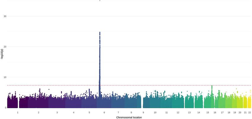
2 | Human Molecular Genetics, 2022, Vol. 00, No. 00
Introduction necrotising myopathy (15,16). To date, the rarity of JDM
has meant that candidate gene studies in JDM have been
Juvenile dermatomyositis (JDM) is a rare, severe autoim-
small and subgroup analyses of JDM in genome-wide
mune disease and the most prevalent idiopathic inflam-
studies have had limited statistical power relative to
matory myopathy with proximal muscle weakness
other myositis phenotypes. The aim of this research was
and skin rash as typical features. Clinical features are
to identify novel genetic loci associated with JDM using a
heterogeneous and can include serious complications
larger cohort of patients.
such as calcinosis, ulceration, treatment-resistant rash
and involvement of major organs, including gut, lungs
and brain. Although some patients achieve remission Results
Downloaded from https://academic.oup.com/hmg/advance-article/doi/10.1093/hmg/ddac019/6517526 by guest on 04 March 2022
following standard disease management, which consists Samples and genotyping quality control
of long-term immunosuppression with glucocorticoids, Samples of Caucasian ancestry (n = 952) were obtained
methotrexate and other medications, others respond via international collaboration including samples from
poorly. the UK Juvenile Dermatomyositis Cohort & Biomarker
While JDM and adult-onset dermatomyositis (DM) Study, the Childhood Myositis Heterogeneity Study Group
share similar clinical and biological features, there are and the Myositis Genetics Consortium (Table 1). Many of
differences in prevalence of clinical features (1). The these cases have contributed to previous analyses (10,17).
incidence of JDM is approximately one-tenth of the Demographic features are described in Supplementary
incidence of DM (2). DM can be associated with cancer, Material, Table S1. After genotyping and stringent qual-
but this has not been reported in JDM. Conversely, ity control (QC), n = 178 164 single-nucleotide polymor-
calcinosis is a major cause of morbidity in JDM but has phisms (SNPs) remained (Supplementary Material, Table
a lower prevalence in DM. The prevalence of myositis- S2) on n = 851 JDM samples (Supplementary Material,
specific autoantibodies (MSAs), which are linked to Tables S3 and S4). The proportion of phenotypic variance
different clinical features of disease, also differs between explained by these markers was estimated as 0.18 (0.02).
the adult and juvenile forms of the disease. Anti-nuclear
matrix protein-2 is one of the more abundant MSAs in HLA-DRB1∗ 03:01 confirmed as allele most
JDM (reported in 20–25% of patients (3–5)) but has a strongly associated with JDM
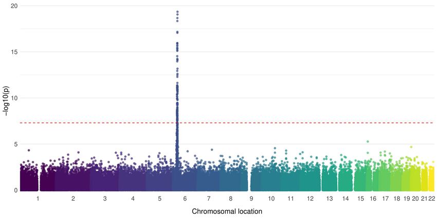
lower prevalence in DM (reported in 1.6–17% of different Case–control analysis of assayed SNPs confirmed that
patient populations) (6–8). The most prevalent MSA in the strongest association with JDM was within the HLA
DM, anti-histidyl tRNA synthetase (anti-Jo-1), is rare in region (Fig. 1; Supplementary Material, Fig. S1). Analysis
JDM. These differences in the distribution of MSA and of imputed markers within the HLA region indicated
clinical features suggest an inf luential role for age of dis- that the strongest association was with SNP rs3117103
ease onset in the pathogenesis of disease. However, little [odds ratio (OR) = 1.87; 95% confidence interval (CI)
is known at the mechanistic level about the influence 1.64, 2.13; P = 1.79 × 10⁻²⁰ ] and the classical allele HLA-
of age on JDM phenotypes and pathogenesis. Knowledge DRB1∗ 03:01, consistent with previous reports (9,10). The
about how disease mechanisms differ between patient OR for HLA-DRB1∗ 03:01 itself was 1.66 (95% CI 1.46, 1.89;
subgroups and interact with patient age to result in P = 1.4 × 10⁻¹⁴ ; Supplementary Material, Table S5).
different complications may enable targeting of novel Conditioning on this allele, an independent association
molecular pathways, more accurate modelling of life- with HLA-C∗ 02:02 was identified (OR = 1.74; 95% CI 1.42,
long risk and more stratified therapeutic approaches to 2.13, P = 7.13 × 10⁻⁸ ) as reported previously (10). The next
address this risk. most associated allele was HLA-B∗ 44:02, but the P-value
Candidate gene and genome-wide studies of myositis for this association was above the threshold for statistical
have established the strongest genetic association within significance (OR = 1.31, 95% CI 1.15, 1.50; P = 6.04 × 10⁻⁵ ).
the Caucasian 8.1 ancestral haplotype (AH8.1; HLA A1- No alleles within HLA-A were associated with JDM (all P-
B8-DR3-DQ2) of the major histocompatibility complex values above 0.08).
(MHC), also associated with many other immune-
mediated diseases (9–13). Distinct human leukocyte Analysis of amino acid positions within
antigen (HLA) alleles have been identified as associated HLA-DRB1 identifies position 37 as the most
with serological subphenotypes of myositis in different significantly associated with JDM
ethnic populations. Most notably associations between Since the strongest association with JDM was identified
the development of anti-Jo-1 autoantibodies and HLA- within HLA-DRB1, further analysis sought to resolve
DRB1∗ 03:01, HLA-DQB1∗ 02:01 and HLA-B∗ 08, consistent this association at the level of amino acid positions.
with AH8.1, have been identified in Caucasian and Position 37 was the most strongly associated with
African-American patients (11,12,14). In adult myosi- disease (omnibus P = 3.3 × 10⁻¹⁹ ; Fig. 2 and Table 2).
tis, gene–environment interactions have been found Residues at position 37 are located within the P9
between HLA-DRB1∗ 03, smoking and the presence of anti- pocket of the antigen-binding groove (Supplementary
Jo-1 autoantibodies, and between HLA-DRB1∗ 11:01 and Material, Fig. S2) (18,19). Relative to Tyr-37, Ser-37 was
anti-3-hydroxy-3-methylglutaryl-CoA reductase (anti- the most significantly associated residue at this position
HMGCR)-positive statin-induced immune-mediated (OR = 0.65; 95% CI 0.57, 0.75; P = 7.34 × 10⁻¹⁰ ), followedHuman Molecular Genetics, 2022, Vol. 00, No. 00 | 3
Table 1. Sources of samples from Caucasian patients with JDM
Country Source Genotyped samples (n = 952) Genotyped samples after quality
control (n = 851)
UK UK Juvenile Dermatomyositis Cohort & 365 326
Biomarker Study
USA National Institute of Environmental Health 262 224
Sciences
Northwestern University 140 116
Emory University 41 31
George Washington University 32 30
Downloaded from https://academic.oup.com/hmg/advance-article/doi/10.1093/hmg/ddac019/6517526 by guest on 04 March 2022
Mayo Clinic 13 12
Canada The Hospital for Sick Children, Toronto 24 24
IWK Health Centre 16 16
Alberta University 10 9
Norway Oslo University Hospital 49 46
Figure 1. Manhattan plot of the association of assayed SNPs with JDM. SNPs (n = 178 164) were available for n = 851 JDM samples and n = 12 232 controls
of Caucasian origin. Association was tested using logistic regression, with the first 10 principal components included as covariates to account for
population stratification. The red dotted line indicates genome-wide level of significance (5 × 10⁻⁸ ).
by Phe-37 (OR = 0.63; 95% CI 0.54, 0.75; P = 1.14 × 10⁻⁷ ) HLA-DQB1∗ 02:01 within AH8.1 (OR = 1.62; 95% CI 1.36,
(Table 3). Asn-37, which is found on HLA-DRB1∗ 03:01, 1.93; P = 8.70 × 10⁻⁸ ) and HLA-C∗ 02:02 (OR = 1.72; 95% CI
was not associated relative to Tyr-37 (OR = 1.13; 95% CI 1.40, 2.10; P = 1.58 × 10⁻⁷ ). However, the effect for HLA-
1.06, 1.19; P = 0.05). Conditioning on position 37, there was DRB1∗ 03:01 did not meet the threshold for significance
suggestive evidence of possible independent associations (OR = 1.49; 95% CR 1.24, 1.80; P = 2.24 × 10⁻⁵ ). Taken
at positions 74 (P = 5.1 × 10⁻⁵ ) and 26 (P = 5.9 × 10⁻⁵ ), together, these results indicate an effect of position 37
although the P-values for these associations were not that is independent of some of the previously reported
significant (Table 2). effects within AH8.1 and at HLA-C∗ 02:02, but that is not
To evaluate whether position 37 explained the asso- independent of HLA-DRB1∗ 03:01.
ciation within HLA-DRB1 more convincingly than the
classical HLA-DRB1∗ 03:01 allele and AH8.1, condi- Genome-wide imputation identifies possible loci
tional analyses were performed. Conditioning on HLA- associated at a suggestive level of significance
DRB1∗ 03:01, the association of position 37 was above Following genome-wide imputation, there were no
the threshold for significance (omnibus P = 4.42 × 10⁻⁵ ), additional loci identified at the genome-wide level
although there may be weak evidence of an independent of statistical significance (P = 5 × 10⁻⁸ ). Two loci with
association. After conditioning on all residues at position P < 1 × 10⁻⁶ and minor allele frequency (MAF) over 0.05
37, there was evidence of independent effects for were identified (Table 4; Fig. 3). rs6501160 is an intronic4 | Human Molecular Genetics, 2022, Vol. 00, No. 00
Downloaded from https://academic.oup.com/hmg/advance-article/doi/10.1093/hmg/ddac019/6517526 by guest on 04 March 2022
Figure 2. Representation of the P-values for omnibus tests of individual amino acid positions within HLA-DRB1 for association with JDM. Blue circles
represent P-values for amino acid positions tested alone. Green circles represent P-values for amino acid positions after conditioning on position 37. Red
circles represent P-values for 4-digit classical alleles. The grey dashed line represents the threshold for statistical significance (P = 6.8 × 10⁻⁶ ).
Table 2. Analysis of amino acid positions within HLA-DRB1 were identified at P < 5 × 10⁻⁸ (Supplementary Material,
associated with JDMa
Table S6 and Supplementary Material, Fig. S4), these
Effects independent of
had MAF between 0.01 and 0.05 and may represent
position 37
imputation artefacts.
Position χ2 b P-value χ2 P-value
37 89.2 3.3 × 10⁻¹⁹ – –
74 76.7 1.6 × 10⁻¹⁶ 22.5 5.1 × 10−05 Discussion
26 59.5 1.2 × 10⁻¹³ 19.5 5.9 × 10−05 This analysis represents the largest international genetic
10 47.5 5.4 × 10⁻¹²
study of JDM to date. We found that the strongest asso-
67 46.0 1.2 × 10⁻¹¹
−17 41.0 1.5 × 10⁻¹⁰ ciation was in the HLA region within HLA-DRB1∗ 03:01,
−16 37.5 9.1 × 10⁻¹⁰ consistent with previous studies (9,10,17). We confirmed
30 39.5 2.6 × 10⁻⁹ an independent association with HLA-C∗ 02:02 and found
−25 34.4 4.5 × 10⁻⁹ some evidence of a possible additional independent asso-
11 40.2 9.9 × 10⁻⁹
ciation at HLA-B∗ 44:02, which would need to be con-
13 40.0 1.1 × 10−08
233 32.3 1.3 × 10⁻⁸ firmed in a future study. Interestingly, we did not find
71 27.8 1.4 × 10⁻⁷ evidence of an association with HLA-A, even though
−1 26.1 3.2 × 10⁻⁷ associations within this gene are well known for mul-
181 28.9 5.3 × 10⁻⁷ tiple other autoimmune diseases including rheumatoid
57 24.2 8.8 × 10⁻⁷
arthritis (RA), juvenile idiopathic arthritis (JIA), psoriatic
60 25.4 3.1 × 10⁻⁶
arthritis and type I diabetes (20–23).
a Alpha threshold for statistical significance in the MHC region taken as In this analysis focused on juvenile-onset disease, we
6.8 × 10⁻⁶ (47). b Test statistics compared using a chi-squared distribution.
found amino acid position 37 within HLA-DRB1 had the
strongest association with JDM, with Ser-37 and Phe-
37 having protective effects relative to Tyr-37. We also
variant within transmembrane protein 114 (TMEM114), a found evidence that the association with position 37 was
transmembrane protein. rs6892006 is an intronic variant independent of AH8.1, a well-established association
within myocyte enhancer factor 2C antisense RNA 1 (MEF2C- with myositis, but there was only weak evidence of
AS1), an anti-sense RNA gene (Supplementary Material, independence from HLA-DRB1∗ 03:01, and so this needs
Fig. S3). Associations with clinical phenotypes have not to be followed up in future studies with greater numbers
been reported for either variant. Although additional loci of both juvenile-onset and adult-onset patients using theHuman Molecular Genetics, 2022, Vol. 00, No. 00 | 5
Table 3. Association of residues within position 37 with JDM, with effects of classical HLA alleles linked to these residuesa,b
Residue Classical HLA allele Allele frequency Allele frequency OR 95% CI P-value
linked to residue in JDM in controls
Tyr-37 – 0.37 0.31 - - -
Ser-37 – 0.21 0.26 0.65 0.57, 0.75 7.3 × 10⁻¹⁰
– HLA-DRB1∗ 01:01 0.10 0.12 0.76 0.65, 1.46 1.7 × 10⁻³
– HLA-DRB1∗ 15:01 0.11 0.14 0.66 0.57, 0.78 6.5 × 10⁻⁷
Phe-37 – 0.11 0.15 0.63 0.54, 0.75 1.1 × 10⁻⁷
– HLA-DRB1∗ 07:01 0.09 0.13 0.67 0.57, 0.79 2.8 × 10⁻⁶
– HLA-DRB1∗ 14:01 0.02 0.02 0.86 0.60, 1.24 0.42
Downloaded from https://academic.oup.com/hmg/advance-article/doi/10.1093/hmg/ddac019/6517526 by guest on 04 March 2022
Asn-37 – 0.31 0.24 1.13 1.06, 1.19 0.05
– HLA-DRB1∗ 03:01 0.19 0.12 1.66 1.46, 1.89 1.4 × 10⁻¹⁴
a Summary statistics for individual residues derived from the omnibus model for position 37, with Tyr-37 used as the reference residue. Summary statistics for
classical four-digit HLA alleles derived from case–control analysis of imputed HLA data. Alleles linked to these residues at position 37 but not represented were
filtered out during QC. b Alpha threshold for statistical significance in the MHC region taken as 6.8 × 10⁻⁶ (47).
Table 4. Genome-wide imputed loci displaying potential association with JDM, with P-value cut-offs of 1 × 10⁻⁶ and MAF cut-off of
0.01a
Rsidb Chromo- Position Allele A Allele B MAF OR 95% CI P-value Imputation Information Association
some R2 c measured information
measuree
rs6501160 16 8 607 139 G A 0.27 0.72 0.65, 0.81 5.8 × 10⁻⁸ 0.91 0.91 0.91
rs6892006 5 88 450 541 T G 0.13 0.68 0.58, 0.79 8.7 × 10⁻⁷ 0.87 0.87 0.89
a Lociwith P-values below a suggestive level of significance (1 × 10⁶) are displayed for MAF cut-offs of 0.05 and 0.01; only additional loci captured by the lower
MAF cut-off are shown for that cut-off. b Rsid, Reference SNP cluster ID. There were no assayed markers within 100 000 kb of either of these loci. c Squared
correlation of imputation of genotypes with true unmeasured genotypes, as estimated by Minimac3 during imputation via the Michigan Imputation
Server: https://genome.sph.umich.edu/wiki/Minimac3_Info_File#Rsq. d Impute INFO measure calculated by SNPTEST: https://mathgen.stats.ox.ac.uk/genetics_
software/snptest/snptest.html#info_measures. e Relative information measure about parameters of the model fitted during association testing: https://
mathgen.stats.ox.ac.uk/genetics_software/snptest/snptest.v2.p.
Figure 3. Manhattan plot of the association of imputed SNPs with JDM. Loci were filtered according to MAF of 0.05. Data were imputed for n = 851 JDM
samples and n = 12 232 controls of Caucasian origin. The first 10 principal components were adjusted for during analysis to account for population
stratification. SNPs were weighted by the information score to account for imputation uncertainty. The red dotted line indicates genome-wide level
of significance (5 × 10⁻⁸ ). The degree of transparency of each data-point represents the R2 value for imputation accuracy, with more solid colours
representing higher certainty.
same genotyping chip. Amino acid position 37 is within The previous Immunochip analysis of adult and
the P9 pocket of the antigen-binding groove (18,19). juvenile myositis combined identified the strongest
Substitution of Ser-37 to Tyr-37 has been shown to be association within HLA-DRB1 at amino acid position
sufficient to alter alloantigenicity and stimulate a T-cell 74 (10). Subsequent analyses of that dataset identified
response (24). Tyr-37 promotes a stronger response to position 74 as having an association with anti-Jo1, anti-
streptococcal protein (25). PM/Scl and Anti-cytosolic 5 -nucleotidase 1A (anti-cN1A)6 | Human Molecular Genetics, 2022, Vol. 00, No. 00
autoantibodies (26), which are less prevalent in juvenile- acid positions within HLA-DRB1 such as position 37. Loci
onset disease. We found weak evidence of independent outside the HLA region identified at genome-wide and
association at position 74 after conditioning on position suggestive levels of significance in previous analyses
37, above the threshold for significance and so this (including PTPN22, UBE2L3, CD28, TRAF6, STAT4) were
finding needs to be followed up with greater number of not replicated in this study, although signals at these
patients. Nonetheless, it raises an intriguing possibility loci were not specifically tested for. This may reflect the
that children and adults may have different dominant smaller sample size of this study and the dominance of
autoantigenic peptides being presented in the antigen- adult patients in those analyses whose characteristics
binding groove, although we recognize that multiple may differ from paediatric disease (10). JDM is a het-
antigens with different allelic associations are likely to erogeneous phenotype; however, the small sample size
Downloaded from https://academic.oup.com/hmg/advance-article/doi/10.1093/hmg/ddac019/6517526 by guest on 04 March 2022
be involved in disease. While the Immunochip analyses restricted our ability to analyse more clinically homo-
benefitted from a relatively large cohort (n = 2544), the geneous subgroups, such as autoantibody subgroups. It
number of JDM patients was approximately half that may be that future developments in methodologies for
of our analysis (n = 493). These patients are represented genetic analysis will enable further insights to be derived
in our analysis, along with additional cases. It may be from this dataset. Many of the patients in this study over-
that the effects of juvenile-onset disease were obscured lapped with a previous analysis, which identified asso-
by larger number of adult-onset patients and other ciations with major MSA subtypes, including anti-Jo-1,
phenotypes present in the combined cohort. Future work anti-PM/Scl, anti-cN1A, anti-Mi-2 and anti-TIF1γ (26). As
and greater numbers are required to better dissect the more patients become available for inclusion in genetic
genetic inf luence on age effects in juvenile and adult- studies, analyses of further MSA subtypes may become
onset disease, as well as on clinical and serological possible. It will also be critical to study genetic associa-
heterogeneity. tions with MSA subtypes in different ethnic populations.
Associations between other positions within HLA-DRB1 Analysis following genome-wide imputation identified
and autoimmune disease have been reported. For exam- 2 possible suggestive loci, an intronic variant within
ple, position 11 has the strongest effect in RA, with inde- TMEM114 and an intronic variant within MEF2C-AS1.
pendent effects at positions 71 and 74 (27). In JIA, position TMEM114 is a glycosylated transmembrane protein, and
13 has the strongest effect, although in systemic JIA the knowledge of its cellular function is limited, although
strongest effect is at position 58 (21,28). Positions 71/74 missense mutations in this gene and a chromosomal
have also been implicated in anti-fibrillarin-positive sys- translocation in its promoter are associated with
temic sclerosis, Crohn’s disease, multiple sclerosis, type I congenital and juvenile cataract disorders, respectively
diabetes and Grave’s disease (29–33). In systemic lupus (39). MEF2C-AS1 is a non-coding anti-sense RNA gene
erythematosus (SLE), Ser-1, Phe47 and Ala71 are asso- with no known function. It is unclear how these loci
ciated with disease (34). Position 37 has been found to relate to JDM, but it may be these alleles function as
be associated with primary sclerosing cholangitis, ulcer- epigenetic marks.
ative colitis in Asians, ACPA+ RA in Han Chinese, SLE in In summary, we have confirmed the association
Asians and psoriasis vulgaris in Taiwanese (18,19,35–37). between JDM and HLA-DRB1∗ 03:01 and shown that within
Interestingly, a recent report of Japanese patients with HLA-DRB1, position 37 is most strongly associated with
RA identified an association between position 37 and disease in a population of patients with juvenile-onset
younger age of disease onset (defined as 16–30 years of myositis.
age), but not with older age of onset (defined as over
60 years of age) (38). At present, it is not well understood
how these differing positional effects relate to the spec- Materials and Methods
trum of phenotypes represented in autoimmune disease, Genotyping and genotype calling
and how these effects interact with ethnicity and age of Genotyping of Caucasian JDM samples (n = 952) was per-
disease onset. formed using the Illumina (Cambridge, UK) HumanCore-
The major limitation of our study is limited statistical Exome chip across three batches at a single centre (Uni-
power for identifying associations outside the HLA versity College London). This cohort included the major-
region, which is a practical challenge for rare disease ity of JDM cases included in previous analyses, as well as
research. Although our combined cohort represents the additional cases recruited subsequent to those analyses
largest-ever assembled international cohort of Cau- including cases from more centres (10,17). GenomeStudio
casian patients with JDM, it is a relatively small sample 2.0 (version 2.0.4 of the Genotyping Module and the
size for a genetic study. As such, the findings of sug- GenTrain 3.0 Cluster Algorithm) was used for genotype
gestive associations will need to be confirmed in future clustering and calling for each separate batch. Samples
studies with greater number of patients. Nonetheless, we with less than 90% call rate were excluded, and genotype
were able to confirm the findings of previous studies in clustering and calling were repeated, before data were
a larger cohort dedicated to JDM. Future studies with exported in PLINK format for QC.
greater numbers may better define the relationship Data on healthy individuals (n = 12 474) of European
between the associations at HLA-DRB1∗ 03:01 and amino ancestry who had also been genotyped using theHuman Molecular Genetics, 2022, Vol. 00, No. 00 | 7
HumanCoreExome chip as part of the International Analysis of HLA and amino acid positions
Age-Related Macular Degeneration Genetic Consortium Case–control analysis of all imputed HLA was performed
were obtained from dbGaP (dbGAP Study Accession assuming an additive model using logistic regression in
phs001039.v1.p1). PLINK, also with adjustment for the first 10 PCs, to iden-
tify the most strongly associated locus. Clinical covari-
Quality control ates were not adjusted for. For HLA analyses, P-values
QC of markers and samples was performed as described below 6.8 × 10⁻⁶ were considered statistically significant,
previously (22). The following steps were done separately using a Bonferroni correction for the number of imputed
for each of the three batches of JDM data and the markers as reported previously (47). The two- and four-
digit classical alleles identified by this locus were sub-
Downloaded from https://academic.oup.com/hmg/advance-article/doi/10.1093/hmg/ddac019/6517526 by guest on 04 March 2022
control data using PLINK 1.07 (40). Mitochondrial and
Y chromosome SNPs were excluded. SNPs were also sequently conditioned on in further logistic regression
excluded if they had elevated missing rates (over 2%), had analyses to identify any independent associations.
a MAF below 1% and deviated significantly from Hardy– Amino acid positions were interrogated using likeli-
Weinberg equilibrium (HWE; threshold of P < 0.0001). hood ratio tests (LRTs) in R as follows. At each position,
Samples with elevated missing genotypes (over 5%) and logistic regression models were fitted with all residues in
outlying heterozygosity rates (above or below 5 standard the model, except the residue that was most prevalent
deviations from the mean rate). Data were aligned to the in the controls and served as the reference. The first
Haplotype Reference Consortium (HRC) before merging 10 PCs were adjusted for in each of these models. To
for further QC using the HRC checking tool (41). evaluate whether amino acid positions were associated
Duplicated or related individuals in the merged with disease, LRTs were performed to compare the model
dataset were identified using identity-by-descent, per- that was fitted for the residues at each position against
formed on a linkage disequilibrium (LD)-pruned dataset a null model, which comprised the first 10 PCs only.
of 65 862 SNPs with MAF over 5%. A PI_HAT threshold The effect of each amino acid position evaluated using
of 0.2 was used, with the individual with the most miss- a LRT is represented by the LRT P-value, but there is no
ing data excluded. Principal component analysis was estimated effect size for the amino acid position gener-
performed using PLINK 1.9 (42), to evaluate population ated by this test. Allele frequencies, odds ratios and P-
stratification in the LD-pruned data merged with the values for amino acid residues at key positions are also
International HapMap 3 data (43). Cases and controls reported. Possible independent effects were identified by
were retained if they were within 10 standard deviations conditional analysis as above.
of the mean value for the first two principal components
(PCs) for the HapMap CEU population (Supplementary Genome-wide imputation and analysis
Material, Fig. S5). Genome-wide imputation of SNPs was performed using
Following QC, there were 178 164 SNPs for n = 851 JDM the Michigan Imputation Server (version 1.2.4) and the
cases and n = 12 232 controls (Supplementary Material, HRC reference panel (version r1.1; Supplementary Mate-
Tables S1–S4). rial, Fig. S6) (41,48). Imputed SNPs with MAF below 0.01 or
imputation information score below 0.5 were filtered out.
Analysis of assayed markers Imputed SNPs were analysed using SNPTEST version
2.5.4-beta3 (49), with adjustment for the first 10 PCs
Autosomal markers were analysed for association with
and weighting for the imputation information score to
JDM using logistic regression in PLINK with adjustment
account for imputation uncertainty. Since imputation
for the first 10 PCs to control for population stratification.
artefacts are enriched in rare variants, a stringent MAF
Manhattan plots were generated using R version 6.3 and
threshold of 0.05 was used during analysis, although loci
the ‘ggplot2’ package (version 3.3.2). The proportion of
with MAF 0.01–0.05 are also reported in Supplementary
phenotypic variance explained by the SNPs and a stan-
Material.
dard error for that estimated proportion were estimated
using the genome-based restricted maximum likelihood
(GREML) method using GTAC version 1.93.3, assuming an Supplementary Material
approximate prevalence of 0.00004 for JDM (2,44,45).
Supplementary Material is available at HMG online.
Imputation of HLA loci
Classical HLA alleles, amino acids and SNPs within the Acknowledgements
HLA region were imputed using SNP2HLA (version 1.0.3) C.T.D. would like to thank the study administrators
and the Type 1 Diabetes Genetics Consortium reference and research assistants at each international centre
panel (n = 5225) (46). QC of imputed markers used the who assisted with extraction of clinical data and
following criteria: imputation information score (R2 ) over preparation and shipment of DNA samples, including
0.9, MAF over 0.01 and significant departures from HWE Dr Terry O’Hanlon and Payam Noroozi Farhadi (NIH),
in controls (P < 0.001). Imputed markers were coded as Siri Tennebø Flåm (University of Oslo); Gabrielle Mor-
present or absent. gan (Northwestern University); Dwaraka Veeramreddy8 | Human Molecular Genetics, 2022, Vol. 00, No. 00
(Alberta Children’s Hospital); Hayyah Clairman (Hospital Caitlin Clifford and Ms Linda Suffield (University College
for Sick Children, Toronto). C.T.D. would also like to London Hospital, London); Ms Helen Lee, Ms Sam Leach,
thank Dr Socrates Varakliotis (Centre for Adolescent Ms Helen Smith, Dr Anne-Marie McMahon, Ms Heather
Rheumatology Versus Arthritis at UCL, UCLH and GOSH) Chisem, Jeanette Hall, Ruth Kingshott and Maxine
for setting up remote access to a Linux desktop computer Mutten (Sheffield’s Children’s Hospital, Sheffield); Dr
which enabled analyses to continue during the COVID-19 Nick Wilkinson, Ms Emma Inness, Ms Eunice Kendall,
pandemic. C.T.D. thanks Drs Michael Ombrello and Elaine Mr David Mayers, Ruth Etherton, Danielle Miller and Dr
Remmers for useful comments on the manuscript. Kathryn Bailey (Oxford University Hospitals, Oxford); Dr
The Juvenile Dermatomyositis Research Group would Jacqui Clinch, Ms Natalie Fineman, Ms Helen Pluess-
like to thank all of the patients and their families who Hall and Suzanne Sketchley (Bristol Royal Hospital for
Downloaded from https://academic.oup.com/hmg/advance-article/doi/10.1093/hmg/ddac019/6517526 by guest on 04 March 2022
contributed to the Juvenile Dermatomyositis Cohort Children, Bristol); Dr Joyce Davidson, Margaret Connon
& Biomarker Study & Repository. We thank all local and Ms Lindsay Vallance (Royal Aberdeen Children’s
research coordinators and principal investigators who Hospital); Dr Kirsty Haslam, Charlene Bass-Woodcock,
have made this research possible. The JDRG members Trudy Booth and Ms Louise Akeroyd (Bradford Teaching
were as follows: Hospitals); Dr Alice Leahy, Amy Collier, Rebecca Cutts,
Dr Kate Armon, Mr Joe Ellis-Gage, Ms Holly Roper, Ms Emma Macleod, Dr Hans De Graaf, Dr Brian Davidson,
Vanja Briggs and Ms Joanna Watts (Norfolk and Norwich Sarah Hartfree, Danny Pratt, Elizabeth Fofana and Lorena
University Hospitals); Dr Liza McCann, Mr Ian Roberts, Dr Caruana (University Hospital Southampton) and all the
Eileen Baildam, Ms Louise Hanna, Ms Olivia Lloyd, Susan Children, Young people and their families who have
Wadeson and Michelle Andrews (The Royal Liverpool contributed to this research.
Children’s Hospital, Alder Hey, Liverpool); Dr Phil Riley, The Childhood Myositis Heterogeneity Study Group
Ms Ann McGovern, Verna Cuthbert (Royal Manchester is supported by the Intramural Research Program of
Children’s Hospital, Manchester); Dr Clive Ryder, Mrs the NIH, National Institute of Environmental Health
Janis Scott, Mrs Beverley Thomas, Professor Taunton Sciences. The Childhood Myositis Heterogeneity Study
Southwood, Dr Eslam Al-Abadi and Ruth Howman Group thanks the following contributors to this study:
(Birmingham Children’s Hospital, Birmingham); Dr Sue Daniel Albert (Dartmouth-Hitchcock Medical Center),
Wyatt, Mrs Gillian Jackson, Dr Mark Wood, Dr Tania Kathy Amoroso (Carilion Roanoke Memorial Hospital),
Amin, Dr Vanessa VanRooyen and Ms Deborah Burton, Bita Arabshahi (Fairfax Inova Medical Center), Edsel
Louise Turner & Sarah Hanson (Leeds General Infirmary, Arce-Hernandez (Valley Children’s Healthcare), Alan
Leeds); Dr Joyce Davidson, Dr Janet Gardner-Medwin, Baer (Johns Hopkins University School of Medicine),
Dr Neil Martin, Ms Sue Ferguson, Ms Liz Waxman and Susan Ballinger (Riley Children’s Hospital, Indiana
Mr Michael Browne, Ms Roisin Boyle, Ms Emily Blyth University), Lilliana Barillas (Albany Medical Center),
(The Royal Hospital for Sick Children, Yorkhill, Glasgow); Mara Becker (Children’s Mercy, Kansas City), Catherine
Dr Mark Friswell, Professor Helen Foster, Mrs Alison April Bingham (Penn State Children’s Hospital), John
Swift, Dr Sharmila Jandial, Ms Vicky Stevenson, Ms Bohnsack (University of Utah), Jon M Burnham (Chil-
Debbie Wade, Dr Ethan Sen, Dr Eve Smith, Ms Lisa Qiao, dren’s Hospital of Philadelphia), Ruy Carrasco (Child
Mr Stuart Watson and Ms Claire Duong (Great North Neurology Consultants of Austin), Victoria Cartwright
Children’s Hospital, Newcastle); Dr Helen Venning, Dr (Legacy Emanuel Medical Center), Gail Cawkwell (Johns
Rangaraj Satyapal, Mrs Elizabeth Stretton, Ms Mary Hopkins All Children’s Hospital), Wendy De La Pena
Jordan, Dr Ellen Mosley, Ms Anna Frost, Ms Lindsay Crate, (Loma Linda University), Fatma Dedeoglu (Boston
Dr Kishore Warrier, Stefanie Stafford, Kelly Sandhu & Children’s Hospital), Barbara Anne Eberhard (Northwell
Tracey Dandy (Queens Medical Centre, Nottingham); Health), Kaleo Ede (Phoenix Children’s Hospital), Barbara
Professor Lucy Wedderburn, Dr Clarissa Pilkington, Dr Edelheit (University of Connecticut), Andrew Eichenfield
Nathan Hasson, Dr Muthana Al-Obadi, Dr Giulia Varnier, (New York-Presbyterian Hospital), Terri Finkel (University
Dr Sandrine Lacassagne, Mrs Sue Maillard, Mrs Lauren of Colorado), Robert Fuhlbrigge (Boston Children’s
Stone, Ms Elizabeth Halkon, Ms Virginia Brown, Ms Hospital), Abraham Gedalia (Children’s Hospital New
Audrey Juggins, Dr Sally Smith, Mrs Sian Lunt, Ms Elli Orleans), Stephen George (University of Maryland), Harry
Enayat, Mrs Hemlata Varsani, Miss Laura Kassoumeri, Gewanter (Virginia Commonwealth University), Ellen
Miss Laura Beard, Miss Katie Arnold, Mrs Yvonne Goldmuntz (Children’s National Medical Center), Donald
Glackin, Ms Stephanie Simou, Dr Beverley Almeida, Dr Goldsmith (St. Christopher’s Hospital for Children, Drexel
Kiran Nistala, Dr Raquel Marques, Dr Claire Deakin, Dr University), Beth Gottlieb (Northwell Health), Brent Gra-
Parichat Khaosut, Ms Stefanie Dowle, Dr Charalampia ham (Vanderbilt University), Thomas Griffin (Carolinas
Papadopoulou, Dr Shireena Yasin, Dr Christina Boros, Dr Medical Center), Hilary Haftel (University of Michigan),
Meredyth Wilkinson, Dr Chris Piper, Mrs Cerise Johnson- Melissa Hawkins-Holt (University of Maryland), Theresa
Moore, Ms Lucy Marshall, Ms Kathryn O’Brien, Ms Emily Hennon (John R. Oishei Children’s Hospital, University
Robinson (Great Ormond Street Hospital, London); Dr of Buffalo), Michael Henrickson (Cincinnati Children’s
Kevin Murray (Princess Margaret Hospital, Perth, Western Hospital), Aimee Hersh (University of Utah), Gloria
Australia); Dr Coziana Ciurtin, Dr John Ioannou, Mrs Higgins (Nationwide Children’s Hospital), Lisa ImundoHuman Molecular Genetics, 2022, Vol. 00, No. 00 | 9
(Columbia University), Christi Inman (University of Funding
Utah), Anna Jansen (NIEHS, NIH), Rita Jerath (Medical
TMA Fellowship and a Cure JM grant (to C.T.D.). UK
College of Georgia), Olcay Y Jones (Walter Reed National
JDM Cohort and Biomarker study has been provided by
Military Medical Center), Lawrence Jung (Children’s
generous grants from the Wellcome Trust UK (085860);
National Medical Center), Ildy Katona (Uniformed
Action Medical Research UK (SP4252); the Myositis
University of the Health Sciences), Philip Kahn (New
Support Group UK, Arthritis Research UK now Versus
York University), Hanna Kim (NIAMS, NIH), Susan
Arthritis (14518, 20164, 21593); the Henry Smith Charity
Kim (University of California, San Francisco), Yukiko
and Great Ormond Street Children’s Charity (V1268);
Kimura (Hackensack Meridian School of Medicine),
Tiny Hearts Society, the Myositis Association, Cure
Daniel Kingsbury (Legacy Emanuel Medical Center), W
Downloaded from https://academic.oup.com/hmg/advance-article/doi/10.1093/hmg/ddac019/6517526 by guest on 04 March 2022
JM, the Medical Research Council (MR/N003322/1) and
Patrick Knibbe (St Luke’s Clinic), Bianca Lang (Dalhousie
the National Institute for Health Research (NIHR) via
University), Melissa Lerman (Children’s Hospital of
the Translational Research Collaboration (TRC) Rare
Philadelphia), Carol Lindsley (University of Kansas),
Diseases and the NIHR-Biomedical Research Centre at
Daniel Lovell (Cincinnati Children’s Hospital), Azadeh
GOSH. The views expressed are those of the authors
Majlessi (Loma Linda University), Gulnara Mamyrova
and not necessarily those of the NHS, the NIHR or the
(George Washington University), Paul McCarthy (Yale
Department of Health.
University), Diana Milojevic (Johns Hopkins All Children’s
Hospital), Stephen R. Mitchell (Georgetown University),
Renee Modica (Arnold Palmer Hospital for Children, References
University of Florida), Chihiro Morishima (University of
1. Tansley, S. and Wedderburn, L.R. (2015) Comparing and contrast-
Washington), Simona Nativ (Atlantic Health System), ing clinical and serological features of juvenile and adult-onset
Edward Oberle (Nationwide Children’s Hospital), Kiem myositis. Curr. Opin. Rheumatol., 27, 601–607.
Oen (University of Manitoba), Judyann Olson (Children’s 2. Meyer, A., Meyer, N., Schaeffer, M., Gottenberg, J.-E., Geny, B.
Wisconsin, Medical College of Wisconsin), Elif Oral and Sibilia, J. (2015) Incidence and prevalence of inflammatory
(University of Michigan), Barbara Ostrov (Penn State myopathies: a systematic review. Rheumatology, 54, 50–63.
Children’s Hospital), Lauren M Pachman (Lurie Children’s 3. Tansley, S.L., Betteridge, Z.E., Shaddick, G., Gunawardena, H.,
Hospital, Northwestern University), Murray Passo (The Arnold, K., Wedderburn, L.R. and McHugh, N.J. (2014) Calcinosis
Medical University of South Carolina), Maria Perez in juvenile dermatomyositis is influenced by both anti-NXP2
autoantibody status and age at disease onset. Rheumatology
(Cook Children’s Medical Center), Marilynn Punaro
(Oxford), 53, 2204–2208.
(University of Texas at Dallas), C. Egla Rabinovich (Duke
4. Rider, L.G., Shah, M., Mamyrova, G., Huber, A.M., Rice, M.M., Tar-
University), Linda Ray (University of Mississippi), Robert
goff, I.N. and Miller, F.W. (2013) The myositis autoantibody phe-
Rennebohm (Cleveland Clinic), Rafael Rivas-Chacon notypes of the juvenile idiopathic inflammatory myopathies.
(Nicklaus Children’s Hospital, University of Miami), Medicine, 92, 223–243.
Tova Ronis (Children’s National Hospital), Margalit 5. Espada, G., Maldonado Cocco, J.A., Fertig, N. and Oddis, C.V. (2009)
Rosenkranz (University of Pittsburgh Medical Cen- Clinical and serologic characterization of an Argentine pediatric
ter), Deborah Rothman (Albany Medical College), Sara myositis cohort: identification of a novel autoantibody (anti-mj)
Sabbagh (NIAMS, NIH), Adam Schiffenbauer (NIEHS), to a 142-kda protein. J. Rheumatol., 36, 2547–2551.
Bracha Shaham (Children’s Hospital of Los Angeles, 6. Ichimura, Y., Matsushita, T., Hamaguchi, Y., Kaji, K., Hasegawa,
University of Southern California), Robert Sheets (Rady M., Tanino, Y., Inokoshi, Y., Kawai, K., Kanekura, T., Habuchi, M.
et al. (2012) Anti-NXP2 autoantibodies in adult patients with
Children’s Hospital, University of California at San
idiopathic inflammatory myopathies: possible association with
Diego), Susan Shenoi (Seattle Children’s, University of
malignancy. Ann. Rheum. Dis., 71, 710–713.
Washington), David Sherry (Seattle Children’s, University
7. Fiorentino, D.F., Chung, L.S., Christopher-Stine, L., Zaba, L., Li,
of Washington), Abigail Smukler (The Toledo Clinic), S., Mammen, A.L., Rosen, A. and Casciola-Rosen, L. (2013) Most
Jennifer Soep (University of Colorado), Sangeeta Sule patients with cancer-associated dermatomyositis have antibod-
(Children’s National Medical Center), Robert Sundel ies to nuclear matrix protein NXP-2 or transcription intermedi-
(Boston Children’s Hospital), Stacey Tarvin (Riley Hospital ary factor 1γ . Arthritis Rheum., 65, 2954–2962.
for Children, University of Indiana), Mohamad Tolaymat 8. Ceribelli, A., Fredi, M., Taraborelli, M., Cavazzana, I., Franceschini,
(University of Florida), Cagri Toruner (Texas Children’s F., Quinzanini, M., Tincani, A., Ross, S.J., Chan, J.Y.F., Pauley, B.A.
Hospital), Rita Volochayev (NIEHS, NIH), Jennifer Wargula et al. (2012) Anti-MJ/NXP-2 autoantibody specificity in a cohort
(Seattle Children’s), Peter Weiser (University of Alabama), of adult Italian patients with polymyositis/dermatomyositis.
Arthritis Res. Ther., 14, R97.
Andrew White (Washington University), Patience White
9. Miller, F.W., Chen, W., O’Hanlon, T.P., Cooper, R.G., Vencovsky, J.,
(George Washington University), Andrew Zeft (Cleveland
Rider, L.G., Danko, K., Wedderburn, L.R., Lundberg, I.E., Pachman,
Clinic), Lawrence Zemel (Connecticut Children’s Medical
L.M. et al. (2015) Genome-wide association study identifies HLA
Center). 8.1 ancestral haplotype alleles as major genetic risk factors for
myositis phenotypes. Genes Immun., 16, 470–480.
10. Rothwell, S., Cooper, R.G., Lundberg, I.E., Miller, F.W., Gregersen,
Conf lict of Interest statement. The authors have declared P.K., Bowes, J., Vencovsky, J., Danko, K., Limaye, V., Selva-
that no conf lict of interest exists. O’Callaghan, A. et al. (2016) Dense genotyping of immune-related10 | Human Molecular Genetics, 2022, Vol. 00, No. 00
loci in idiopathic inflammatory myopathies confirms HLA alle- distinct adult inflammatory arthritic diseases. Ann. Rheum. Dis.,
les as the strongest genetic risk factor and suggests different 76, 765–772.
genetic background for major clinical subgroups. Ann. Rheum. 22. Bowes, J., Budu-Aggrey, A., Huffmeier, U., Uebe, S., Steel, K.,
Dis., 75, 1558–1566. Hebert, H.L., Wallace, C., Massey, J., Bruce, I.N., Bluett, J. et al.
11. Arnett, F.C., Targoff, I.N., Mimori, T., Goldstein, R., Warner, N.B. (2015) Dense genotyping of immune-related susceptibility loci
and Reveille, J.D. (1996) Interrelationship of major histocompat- reveals new insights into the genetics of psoriatic arthritis. Nat.
ibility complex class II alleles and autoantibodies in four ethnic Commun., 6, 1–11.
groups with various forms of myositis. Arthritis Rheum., 39, 23. Nejentsev, S., Howson, J.M.M., Walker, N.M., Szeszko, J., Field, S.F.,
1507–1518. Stevens, H.E., Reynolds, P., Hardy, M., King, E., Masters, J. et al.
12. O’Hanlon, T.P., Carrick, D.M., Targoff, I.N., Arnett, F.C., Reveille, (2007) Localization of type 1 diabetes susceptibility to the MHC
Downloaded from https://academic.oup.com/hmg/advance-article/doi/10.1093/hmg/ddac019/6517526 by guest on 04 March 2022
J.D., Carrington, M., Gao, X., Oddis, C.V., Morel, P.A., Malley, J.D. class I genes HLA-B and HLA-A. Nature, 450, 887–892.
et al. (2006) Immunogenetic risk and protective factors for the 24. Kaneko, T. and Obata, F. (1996) Allogeneic recognition of HLA-
idiopathic inflammatory myopathies: distinct HLA-A, -B, -Cw, - DRB1(∗ )0406 by T cells with HLA-DRB1(∗ )0403: role of amino acid
DRB1, and -DQA1 allelic profiles distinguish European American residue 37 on the β sheet in T cell recognition. Immunobiology,
patients with different myositis autoantibodies. Medicine, 85, 195, 261–270.
111–127. 25. Chen, Y.Z., Matsushita, S. and Nishimura, Y. (1997) A single
13. Chinoy, H., Salway, F., Fertig, N., Shephard, N., Tait, B.D., Thom- residue polymorphism at DRβ37 affects recognition of peptides
son, W., Isenberg, D.A., Oddis, C.v., Silman, A.J., Ollier, W.E.R.R. by T cells. Hum. Immunol., 54, 30–39.
et al. (2006) In adult onset myositis, the presence of interstitial 26. Rothwell, S., Chinoy, H., Lamb, J.A., Miller, F.W., Rider, L.G., Wed-
lung disease and myositis specific/associated antibodies are derburn, L.R., McHugh, N.J., Mammen, A.L., Betteridge, Z.E., Tans-
governed by HLA class II haplotype, rather than by myositis ley, S.L. et al. (2019) Focused HLA analysis in Caucasians with
subtype. Arthritis Res. Ther., 8, R13. myositis identifies significant associations with autoantibody
14. O’Hanlon, T.P., Rider, L.G., Mamyrova, G., Targoff, I.N., Arnett, subgroups. Ann. Rheum. Dis., 78, 996–1002.
F.C., Reveille, J.D., Carrington, M., Gao, X., Oddis, C.V., Morel, P.A. 27. Raychaudhuri, S., Sandor, C., Stahl, E.A., Freudenberg, J., Lee, H.-
et al. (2006) HLA polymorphisms in African Americans with S., Jia, X., Alfredsson, L., Padyukov, L., Klareskog, L., Worthington,
idiopathic inflammatory myopathy: allelic profiles distinguish J. et al. (2012) Five amino acids in three HLA proteins explain most
patients with different clinical phenotypes and myositis autoan- of the association between MHC and seropositive rheumatoid
tibodies. Arthritis Rheum., 54, 3670–3681. arthritis. Nat. Genet., 44, 291–296.
15. Chinoy, H., Adimulam, S., Marriage, F., New, P., Vincze, M., 28. Ombrello, M.J., Remmers, E.F., Tachmazidou, I., Grom, A., Foell,
Zilahi, E., Kapitány, A., Gyetvai, A., Ekholm, L., Novota, P. et al. D., Haas, J.P., Martini, A., Gattorno, M., Özen, S., Prahalad, S. et al.
(2012) Interaction of HLA-DRB1∗ 03 and smoking for the devel- (2015) HLA-DRB1∗11 and variants of the MHC class II locus are
opment of anti-Jo-1 antibodies in adult idiopathic inflammatory strong risk factors for systemic juvenile idiopathic arthritis. Proc.
myopathies: a European-wide case study. Ann. Rheum. Dis., 71, Natl. Acad. Sci. U. S. A., 112, 15970–15975.
961–965. 29. Goyette, P., Boucher, G., Mallon, D., Ellinghaus, E., Jostins, L.,
16. Mammen, A.L., Gaudet, D., Brisson, D., Christopher-Stine, L., Huang, H., Ripke, S., Gusareva, E.S., Annese, V., Hauser, S.L. et al.
Lloyd, T.E., Leffell, M.S. and Zachary, A.A. (2012) Increased fre- (2015) High-density mapping of the MHC identifies a shared
quency of DRB1∗ 11:01 in anti-HMG-CoA reductase-associated role for HLA-DRB1∗01:03 in inflammatory bowel diseases and
autoimmune myopathy. Arthritis Care Res., 64, 1233–1237. heterozygous advantage in ulcerative colitis. Nat. Genet., 47,
17. Miller, F.W., Cooper, R.G., Vencovský, J., Rider, L.G., Danko, K., 172–179.
Wedderburn, L.R., Lundberg, I.E., Pachman, L.M., Reed, A.M., 30. Patsopoulos, N.A., Barcellos, L.F., Hintzen, R.Q., Schaefer, C., van
Ytterberg, S.R. et al. (2013) Genome-wide association study of Duijn, C.M., Noble, J.A., Raj, T., Gourraud, P.-A., Stranger, B.E.,
dermatomyositis reveals genetic overlap with other autoim- Oksenberg, J. et al. (2013) Fine-mapping the genetic association
mune disorders. Arthritis Rheum., 65, 3239–3247. of the major histocompatibility complex in multiple sclerosis:
18. Guo, J., Zhang, T., Cao, H., Li, X., Liang, H., Liu, M., Zou, Y., Zhang, HLA and non-HLA effects. PLoS Genet., 9, e1003926.
Y., Wang, Y., Sun, X. et al. (2019) Sequencing of the MHC region 31. Menconi, F., Osman, R., Monti, M.C., Greenberg, D.A., Concepcion,
defines HLA-DQA1 as the major genetic risk for seropositive E.S. and Tomer, Y. (2010) Shared molecular amino acid signa-
rheumatoid arthritis in Han Chinese population. Ann. Rheum. ture in the HLA-DR peptide binding pocket predisposes to both
Dis., 78, 773–780. autoimmune diabetes and thyroiditis. Proc. Natl. Acad. Sci. U. S.
19. Hov, J.R., Kosmoliaptsis, V., Traherne, J.A., Olsson, M., Boberg, A., 107, 16899–16903.
K.M., Bergquist, A., Schrumpf, E., Bradley, J.A., Taylor, C.J., Lie, B.A. 32. Ban, Y., Davies, T.F., Greenberg, D.A., Concepcion, E.S., Osman, R.,
et al. (2011) Electrostatic modifications of the human leukocyte Oashi, T. and Tomer, Y. (2004) Arginine at position 74 of the HLA-
antigen-DR P9 peptide-binding pocket and susceptibility to pri- DR β1 chain is associated with Graves’ disease. Genes Immun., 5,
mary sclerosing cholangitis. Hepatology, 53, 1967–1976. 203–208.
20. Han, B., Diogo, D., Eyre, S., Kallberg, H., Zhernakova, A., Bowes, 33. Gourh, P., Safran, S.A., Alexander, T., Boyden, S.E., Morgan, N.D.,
J., Padyukov, L., Okada, Y., González-Gay, M.A., Rantapää- Shah, A.A., Mayes, M.D., Doumatey, A., Bentley, A.R., Shriner, D.
Dahlqvist, S. et al. (2014) Fine mapping seronegative and seropos- et al. (2020) HLA and autoantibodies define scleroderma sub-
itive rheumatoid arthritis to shared and distinct HLA alleles by types and risk in African and European Americans and suggest
adjusting for the effects of heterogeneity. Am. J. Hum. Genet., 94, a role for molecular mimicry. Proc. Natl. Acad. Sci. U. S. A., 117,
522–532. 552–562.
21. Hinks, A., Bowes, J., Cobb, J., Ainsworth, H.C., Marion, M.C., 34. Langefeld, C.D., Ainsworth, H.C., Graham, D.S.C., Kelly, J.A.,
Comeau, M.E., Sudman, M., Han, B., Becker, M.L., Bohnsack, J.F. Comeau, M.E., Marion, M.C., Howard, T.D., Ramos, P.S., Croker, J.A.,
et al. (2017) Fine-mapping the MHC locus in juvenile idiopathic Morris, D.L. et al. (2017) Transancestral mapping and genetic load
arthritis (JIA) reveals genetic heterogeneity corresponding to in systemic lupus erythematosus. Nat. Commun., 8, 1–18.Human Molecular Genetics, 2022, Vol. 00, No. 00 | 11
35. Han, B., Akiyama, M., Kim, K.K., Oh, H., Choi, H., Lee, C.H., Jung, et al. (2016) A reference panel of 64,976 haplotypes for genotype
S., Lee, H.S., Kim, E.E., Cook, S. et al. (2018) Amino acid position 37 imputation. Nat. Genet., 48, 1279–1283.
of HLA-DRβ1 affects susceptibility to Crohn’s disease in Asians. 42. Chang, C.C., Chow, C.C., Tellier, L.C., Vattikuti, S., Purcell, S.M. and
Hum. Mol. Genet., 27, 3901–3910. Lee, J.J. (2015) Second-generation PLINK: rising to the challenge
36. Molineros, J.E., Looger, L.L., Kim, K., Okada, Y., Terao, C., Sun, of larger and richer datasets. GigaScience, 4, 7.
C., Zhou, X., Raj, P., Kochi, Y., Suzuki, A. et al. (2019) Amino 43. Altshuler, D.M., Gibbs, R.A., Peltonen, L., Schaffner, S.F., Yu, F.,
acid signatures of HLA class-I and II molecules are strongly Dermitzakis, E., Bonnen, P.E., De Bakker, P.I.W., Deloukas, P.,
associated with SLE susceptibility and autoantibody production Gabriel, S.B. et al. (2010) Integrating common and rare genetic
in Eastern Asians. PLoS Genet., 15, e1008092. variation in diverse human populations. Nature, 467, 52–58.
37. Jee, S.-H., Tsai, T.-F., Tsai, W.-L., Liaw, S.-H., Chang, C.-H. and 44. Yang, J., Benyamin, B., McEvoy, B.P., Gordon, S., Henders, A.K.,
HU, C.-Y. (1998) HLA-DRB1∗ 0701 and DRB1∗ 1401 are associated
Downloaded from https://academic.oup.com/hmg/advance-article/doi/10.1093/hmg/ddac019/6517526 by guest on 04 March 2022
Nyholt, D.R., Madden, P.A., Heath, A.C., Martin, N.G., Montgomery,
with genetic susceptibility to psoriasis vulgaris in a Taiwanese G.W. et al. (2010) Common SNPs explain a large proportion of the
population. Br. J. Dermatol., 139, 978–983. heritability for human height. Nat. Genet., 42, 565–569.
38. Oka, S., Furukawa, H., Shimada, K., Hashimoto, A., Komiya, A., 45. Lee, S.H., Wray, N.R., Goddard, M.E. and Visscher, P.M. (2011)
Tsunoda, S., Saisho, K., Tsuchiya, N., Katayama, M., Shinohara, Estimating missing heritability for disease from genome-wide
S. et al. (2019) Association of HLA-DRB1 genotype with younger association studies. Am. J. Hum. Genet., 88, 294–305.
age onset and elder age onset rheumatoid arthritis in Japanese 46. Jia, X., Han, B., Onengut-Gumuscu, S., Chen, W.-M., Concannon,
populations. Medicine, 98, e18218. P.J., Rich, S.S., Raychaudhuri, S. and de Bakker, P.I.W. (2013) Imput-
39. Jamieson, R.V., Farrar, N., Stewart, K., Perveen, R., Mihelec, M., ing amino acid polymorphisms in human leukocyte antigens.
Carette, M., Grigg, J.R., McAvoy, J.W., Lovicu, F.J., Tam, P.P.L. et al. PLoS One, 8, e64683.
(2007) Characterization of a familial t(16;22) balanced transloca- 47. Okada, Y., Han, B., Tsoi, L.C., Stuart, P.E., Ellinghaus, E., Tejasvi,
tion associated with congenital cataract leads to identification T., Chandran, V., Pellett, F., Pollock, R., Bowcock, A.M. et al. (2014)
of a novel gene, TMEM114, expressed in the lens and disrupted Fine mapping major histocompatibility complex associations in
by the translocation. Hum. Mutat., 28, 968–977. psoriasis and its clinical subtypes. Am. J. Hum. Genet., 95, 162–172.
40. Purcell, S., Neale, B., Todd-Brown, K., Thomas, L., Ferreira, M.A.R., 48. Das, S., Forer, L., Schönherr, S., Sidore, C., Locke, A.E., Kwong,
Bender, D., Maller, J., Sklar, P., De Bakker, P.I.W., Daly, M.J. et al. A., Vrieze, S.I., Chew, E.Y., Levy, S., McGue, M. et al. (2016)
(2007) PLINK: a tool set for whole-genome association and Next-generation genotype imputation service and methods. Nat.
population-based linkage analyses. Am. J. Hum. Genet., 81, Genet., 48, 1284–1287.
559–575. 49. Marchini, J., Howie, B., Myers, S., McVean, G. and Donnelly, P.
41. McCarthy, S., Das, S., Kretzschmar, W., Delaneau, O., Wood, A.R., (2007) A new multipoint method for genome-wide association
Teumer, A., Kang, H.M., Fuchsberger, C., Danecek, P., Sharp, K. studies by imputation of genotypes. Nat. Genet., 39, 906–913.You can also read