Artificial intelligence to detect malignant eyelid tumors from photographic images - Nature
←
→
Page content transcription
If your browser does not render page correctly, please read the page content below
www.nature.com/npjdigitalmed

                  ARTICLE                  OPEN

                  Artificial intelligence to detect malignant eyelid tumors from
                  photographic images
                  Zhongwen Li 1,2,7 ✉, Wei Qiang1,7, Hongyun Chen3, Mengjie Pei4, Xiaomei Yu1, Layi Wang1, Zhen Li1, Weiwei Xie1, Xuefang Wu5,
                  Jiewei Jiang 6 ✉ and Guohai Wu 1 ✉

                      Malignant eyelid tumors can invade adjacent structures and pose a threat to vision and even life. Early identification of malignant
                      eyelid tumors is crucial to avoiding substantial morbidity and mortality. However, differentiating malignant eyelid tumors from
                      benign ones can be challenging for primary care physicians and even some ophthalmologists. Here, based on 1,417 photographic
                      images from 851 patients across three hospitals, we developed an artificial intelligence system using a faster region-based
                      convolutional neural network and deep learning classification networks to automatically locate eyelid tumors and then distinguish
                      between malignant and benign eyelid tumors. The system performed well in both internal and external test sets (AUCs ranged from
                      0.899 to 0.955). The performance of the system is comparable to that of a senior ophthalmologist, indicating that this system has
                      the potential to be used at the screening stage for promoting the early detection and treatment of malignant eyelid tumors.
                      npj Digital Medicine (2022)5:23 ; https://doi.org/10.1038/s41746-022-00571-3
1234567890():,;

                  INTRODUCTION                                                                               tumors, particularly malignant ones, which need early detection
                  Eyelid tumors are the most common neoplasm encountered in                                  and prompt referral, are not well investigated. Employment of a
                  daily ophthalmology practice1,2. As eyelids have many tissue                               deep learning algorithm in conjunction with eyelid tumor images
                  types, various benign and malignant tumors can develop3.                                   may realize the early identification of malignant eyelid tumors
                  Malignant eyelid tumors pose a great threat because of their                               with potential benefits including increased accessibility and
                  proximity to the eyeballs, brain, and paranasal sinuses, which may                         affordability for the suspected cases. In addition, for allowing
                  cause cosmetic disfigurement and severe morbidity4,5. Early                                 medical practitioners and suspected patients to proactively track
                  recognition and treatment of malignant eyelid tumors can result                            eyelid tumors and identify malignant ones earlier, the algorithm
                  in the most cosmetically and functionally satisfactory outcomes4–6.                        should be capable of localizing eyelid tumors autonomously
                  In addition, although melanoma and sebaceous gland carcinoma                               within images.
                  (SGC) of the eyelid are rare lesions, they have high mortality7,8.                            In this study, we tried to develop an AI system, which used a
                  However, the estimated 5-year survival rate of these malignant                             faster region-based convolutional neural network (Faster R-CNN)
                  eyelid tumors can be over 99% if they could be detected in their                           and deep learning classification networks, to automatically locate
                  earliest stages (depth of skin invasion ≤0.76 mm)8. Therefore, early                       eyelid tumors and distinguish malignant from benign tumors in
                  detection of these malignant eyelid tumors is considerably critical.                       photographic images captured by ordinary digital cameras.
                     Benign and malignant eyelid tumors sometimes have over-                                 Besides, we separately investigated the performance (dichoto-
                  lapping features, hence differentiation between them can be                                mous diagnosis: malignant, benign) of this system in detecting the
                  challenging for primary care physicians, dermatologists, and                               most frequent malignant and benign eyelid tumors. Moreover, we
                                                                                                             compared the performance of the system to that of ophthalmol-
                  ophthalmologists without sufficient experience4,5. Due to the
                                                                                                             ogists of different levels.
                  intricate anatomy of the eyelid, the diagnosis of eyelid tumors
                  often requires experienced ophthalmologists. However, while over
                  200,000 ophthalmologists worldwide, there is a present and
                  expected future shortfall in the number of ophthalmologists in                             RESULTS
                  both developing and developed countries9. The shortage of                                  Dataset characteristics
                  experienced ophthalmologists may hinder the early detection of                             After excluding 150 photographic images without histopatholo-
                  malignant eyelid tumors, especially in underdeveloped countries                            gical diagnoses, a total of 1,417 images with 1,533 eyelid tumors
                  and remote regions.                                                                        delineated by tight bounding boxes were used to establish and
                     Recently, artificial intelligence (AI) has been reported to attain a                     evaluate an eyelid tumor detection system (ETDS). A total of 1,533
                  high level of accuracy in the automated detection of numerous                              cropped images (1,161 images of benign tumors and 372 images
                  diseases from clinical images10–15. In ophthalmology, a large                              of malignant tumors) created by the ETDS were leveraged to
                  number of studies developed deep learning-based systems that                               develop and assess the deep learning classification system. Details
                  could accurately detect ocular diseases such as diabetic retino-                           on the development set and external test set are described in
                  pathy, retinal detachment, and glaucoma16–21. However, eyelid                              Table 1.

                  1
                   Ningbo Eye Hospital, Wenzhou Medical University, Ningbo 315000, China. 2School of Ophthalmology and Optometry and Eye Hospital, Wenzhou Medical University, Wenzhou
                  325027, China. 3Zunyi First People’s Hospital, Zunyi Medical University, Zunyi 563000, China. 4School of Computer Science and Technology, Xi’an University of Posts and
                  Telecommunications, Xi’an 710121, China. 5Guizhou Provincial People’s Hospital, Guizhou University, Guizhou 550002, China. 6School of Electronic Engineering, Xi’an University of
                  Posts and Telecommunications, Xi’an 710121, China. 7These authors contributed equally: Zhongwen Li, Wei Qiang. ✉email: li.zhw@qq.com; jiangjw924@126.com;
                  haiguowu@126.com

                  Published in partnership with Seoul National University Bundang Hospital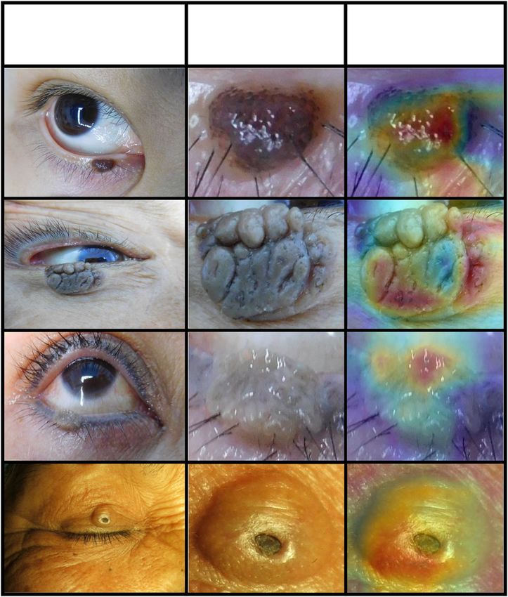
Z. Li et al.
                  2
                       Table 1.   Characteristics of the development set and the external test set.

                                                                                 Development set                                         External test set

                      Total no. of photographic images                           1,258                                                   309
                      Total no. of qualified photographic                         1,151                                                   266
                      imagesa
                      Total no. of cropped images                                1, 248                                                  285
                      No. of patients                                            675                                                     176
                      Mean age/range (years)                                     49.6 (1–100)                                            50.0 (2–87)
                      No. of women (%)                                           421 (62.4)                                              111 (63.1)
                      Institution                                                NEH                                                     JEH and ZFPH
                      Location of institution                                    East of China                                           East and west of China
                      Camera model                                               Canon IXUS-130, Nikon COOLPIX-                          FUJIFILM FinePix-F41, HUAWEI EVA-
                                                                                 S7000                                                   AL00
                                                                  Training set   Validation set                          Internal
                                                                                                                         test set
                      Benign eyelid tumorb                        668/           121/168 (72.0)                          146/197 (74.1) 226/285 (79.3)
                                                                  883 (75.7)
                      Malignant eyelid tumorb                     215/           47/168 (28.0)                           51/197 (25.9)   59/285 (20.7)
                                                                  883 (24.3)
                       NEH Ningbo Eye Hospital, JEH Jiangdong Eye Hospital, ZFPH Zunyi First People’s Hospital.
                       a
                        Qualified photographic images indicate the images with unequivocal histopathological diagnoses.
1234567890():,;

                       b
                         Data are no. of cropped images/total no. (%) unless otherwise indicated.

                         The top three malignant eyelid tumors in our datasets are basal              CI, 84.4–98.6), and a specificity of 79.2% (95% CI, 73.9–84.5) in the
                      cell carcinoma (BCC) (245/372, 65.9%), squamous cell carcinoma                  external test set. Further information encompassing accuracies,
                      (SCC) (50/372, 13.4%), and SGC (66/372, 17.7%). The proportion of               sensitivities, and specificities of these four algorithms is displayed
                      the other malignant eyelid tumors (e.g., melanoma and actinic                   in Table 2. Compared to the ground truth, the unweighted
                      keratosis) is 3.0% (11/372). The top three benign eyelid tumors are             Cohen’s κ coefficients of the optimal algorithm DenseNet121 were
                      squamous cell papilloma (SCP) (104/1161, 9.0%), nevus (385/1,161,               0.613 (95% CI, 0.497–0.730) in the internal test set and 0.560 (95%
                      33.2%), and seborrheic keratosis (106/1161, 9.1%). The proportion               CI, 0.452–0.668) in the external test set.
                      of the other benign eyelid tumors (e.g., hemangioma and                            The distribution of malignancy scores related to the major
                      xanthoma) is 47.4% (566/1161). In total, 116 (7.6%) malignant                   categories determined by the optimal algorithm DenseNet121 in
                      tumors and 630 (41.1%) benign tumors appeared on the upper                      internal and external test sets is shown in Fig. 3. With a
                      eyelid, 238 (15.5%) malignant tumors and 470 (30.7%) benign                     malignancy cutoff at > 0.5, the percentage of correctly classified
                      tumors appeared on the lower eyelid, 11 (0.7%) malignant tumors                 images in malignant tumors was 91.0% (61/67) in BCC, 92.9% (13/
                      and 55 (3.6%) benign tumors appeared in the inner canthus, and 7                14) in SCC, 100% (19/19) in SGC, and 100% (7/7) in melanoma. In
                      (0.5%) malignant tumors and 6 (0.4%) benign tumors appeared in                  benign tumors, the percentage of correctly classified images was
                      the outer canthus.                                                              70.2% (73/104) in nevus, 56.3% (18/32) in seborrheic keratosis,
                                                                                                      69.2% (27/39) in SCP, and 90.0% (27/30) in cyst.
                      Performance of the ETDS and different deep learning                                A total of 36 images (12 malignant tumor images and 24 benign
                      algorithms                                                                      tumor images) were classified into the borderline case group
                      The average precision (AP) scores of the ETDS for locating eyelid               (eyelid tumors of uncertain malignant nature) by the expert. The
                      tumors were 0.801 in the internal test set and 0.762 in the external            optimal algorithm DenseNet121 achieved an accuracy of 77.8%
                      test set. The representative detection results of the Faster-RCNN               (95% CI, 64.2–91.4) with a sensitivity of 83.3% (95% CI, 62.2–100)
                      for eyelid tumors were shown in Fig. 1. Four classic deep learning              and a specificity of 75.0% (95% CI, 57.7–92.3) in differentiating
                      algorithms, DenseNet121, ResNet50, Inception-v3, and VGG16                      malignant eyelid tumors from benign ones on this group. The
                      were used to train models to distinguish malignant eyelid tumors                receiver operating characteristic curve of our system in images of
                      from benign ones. The receiver operating characteristic (ROC)                   eyelid tumors of uncertain malignant nature is shown in
                      curves of these algorithms in internal and external test sets are               Supplementary Fig. 3.
                      shown in Fig. 2, and the corresponding confusion matrices are
                      presented in Supplementary Fig. 1, which indicates that the
                                                                                                      Classification errors of the deep learning system
                      optimal algorithm is the DenseNet121. The t-distributed stochastic
                      neighbor embedding (t-SNE) technique also showed that the                       In total, 87 images (18.0% of the 482 images) from the internal and
                      features of benign and malignant eyelid tumors learned by the                   external test sets had inconsistent findings between the system
                      DenseNet121 were more separable than those of the ResNet50,                     and the ground truth. In the category of malignant eyelid tumors
                      Inception-v3, and VGG16 (Supplementary Fig. 2).                                 (110 images), seven images (6.4%) were misclassified by the
                         In discerning malignant eyelid tumors, the optimal algorithm                 system as benign tumors (false-negative classification). In the
                      DenseNet121 achieved an area under the receiver operating                       category of benign eyelid tumors (372 images), 80 images (21.5%)
                      characteristic curve (AUC) of 0.955 (95% confidence interval [CI],               were misclassified by the system as malignant tumors (false-
                      0.923–0.979), a sensitivity of 96.1% (95% CI, 90.8–100), and a                  positive classification). The details regarding images misclassified
                      specificity of 77.4% (95% CI, 70.6–84.2) in the internal test set, and           by the system are illustrated in Supplementary Fig. 4. Examples of
                      an AUC of 0.899 (95% CI, 0.854–0.934), a sensitivity of 91.5% (95%              incorrectly classified images are shown in Supplementary Fig. 5.

                      npj Digital Medicine (2022) 23                                                  Published in partnership with Seoul National University Bundang Hospital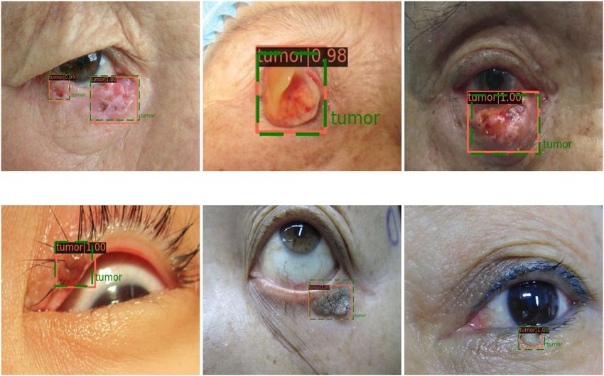
Z. Li et al.
                                                                                                                                                         3
                           a. Malignant eyelid tumor
                            a1                              a2                                  a3

                           b. Benign eyelid tumor
                            b1                              b2                                  b3

Fig. 1 Representative detection results of the Faster-RCNN for eyelid tumors. The green dotted line boxes refer to the ground truth of the
eyelid tumors. The orange solid line boxes refer to the detection results by Faster region-based convolutional neural network (R-CNN). The
orange numerical values indicate confidence scores (range 0–1) which reflect how confident the model is that the box contains an eyelid
tumor. a Malignant eyelid tumor: a1 basal cell carcinomas. a2 squamous cell carcinomas. a3 sebaceous gland carcinoma. b Benign eyelid
tumor: b1 nevus. b2 seborrheic keratosis. b3 squamous cell papilloma.

                                 a. Internal test set                           b. External test set

Fig. 2 Performance of four deep learning algorithms in discerning malignant eyelid tumors. a The receiver operating characteristic (ROC)
curves of the deep learning algorithms in the internal test set. b The ROC curves of the deep learning algorithms in the external test set. AUC
area under the ROC curve. CI confidence interval.

 Table 2.   Performance of four deep learning algorithms in identifying malignant eyelid tumors in the internal and external test sets.

Deep learning               Internal test set                                                External test set
algorithms
                            Sensitivity (95% CI) Specificity (95% CI) Accuracy (95% CI) Sensitivity (95% CI) Specificity (95% CI) Accuracy (95% CI)

DenseNet121                 96.1% (90.8–100)     77.4% (70.6–84.2)    82.2% (76.9–87.6)      91.5% (84.4–98.6)   79.2% (73.9–84.5)   81.8% (77.3–86.2)
ResNet50                    94.1% (87.7–100)     77.4% (70.6–84.2)    81.7% (76.3–87.1)      69.5% (57.7–81.2)   84.5% (79.8–89.2)   81.4% (76.9–85.9)
Inception-v3                90.2% (82.0–98.4)    77.4% (70.6–84.2)    80.7% (75.2–86.2)      74.6% (63.5–85.7)   73.9% (68.2–79.6)   74.0% (68.9–79.1)
VGG16                       86.3% (76.8–95.7)    78.8% (72.1–85.4)    80.7% (75.2–86.2)      66.1% (54.0–78.2)   82.3% (77.3–87.3)   78.9% (74.2–83.7)
 CI, confidence interval.

   The relationship between the classification error rates and                 0% and the classification error rate of the benign eyelid tumors is
predicted probabilities of our system is displayed in Fig. 4, which           about 8%. When the predicted probabilities are less than 0.7, the
denoted that the classification error rate of each category and the            classification error rates of these two categories are both greater
total classification error rate increased with the decrease of the             than 20% and the total classification error rate is over 35%. As our
predicted probabilities. When the predicted probabilities are over            system is a binary classification system, the lowest predicted
0.9, the classification error rate of the malignant eyelid tumors is           probability value of the system’s output is greater than 0.5.

Published in partnership with Seoul National University Bundang Hospital                                              npj Digital Medicine (2022) 23
Z. Li et al.
4
sensitivity of 66.1% (95% CI, 54.0–78.2) and a specificity of
73.9% (95% CI, 68.2–79.6), the senior ophthalmologist achieved an
accuracy of 77.9% (95% CI, 73.1–82.7) with a sensitivity of 74.6%
(95% CI, 63.5–85.7) and a specificity of 78.8% (95% CI, 73.4–84.1),
and the expert achieved an accuracy of 90.2% (95% CI, 86.7–93.6)
with a sensitivity of 94.9% (95% CI, 89.3–100) and a specificity of
88.9% (95% CI, 84.8–93.0), while the system achieved an accuracy
of 81.8% (95% CI, 77.3–86.2) with a sensitivity of 91.5% (95% CI,
84.4–98.6) and a specificity of 79.2% (95% CI, 73.9–84.5). The
sensitivity of the system was superior to that of the junior and
senior ophthalmologists and comparable to that of the expert,
whereas the specificity of the system was only inferior to that of
the expert (Supplementary Table 1). Confusion matrices of these
three ophthalmologists are presented in Supplementary Fig. 6.
DISCUSSION
Our objective in this study was to evaluate the performance of a
deep learning system in distinguishing malignant eyelid tumors
Fig. 3 Malignancy scores (range 0–1) predicted by the deep from benign ones based on photographic images captured by
learning classification system for the major categories of
malignant and benign eyelid tumors. Scores closer to 1 denote a ordinary digital cameras. The findings shown in Fig. 2 demon-
higher probability of malignancy. The upper and lower bounds of strated that deep learning algorithms performed well in discern-
the box refer to the 25th and 75th percentiles, and the line ing malignant eyelid tumors and the algorithm DenseNet121 had
intersection in the box refers to the median. Whiskers refer to the better performance than the other three algorithms. The general-
full range of malignancy scores. BCC basal cell carcinoma, SCC izability of our system was confirmed on the basis of its good
squamous cell carcinoma, SGC sebaceous gland carcinoma, SCP performance (AUC 0.899, sensitivity 91.5%, specificity 79.2%) in
squamous cell papilloma. the external test set, of which images were collected from two
other hospitals. Besides, the agreement between the outputs of
the system and the ground truth was substantial according to the
unweighted Cohen’s κ coefficients, further verifying the reliability
of our system. When compared to the ophthalmologists of
different levels, the system’s sensitivity was higher than that of the
junior and senior ophthalmologists and comparable to that of
the expert, while the system’s specificity is lower than that of the
expert. As a high sensitivity is a prerequisite in a potential
screening tool22, the results implied that our system can
potentially serve as an efficient approach for the early detection
of malignant eyelid tumors, reducing the medical costs and
workload via avoiding the need for the further examination of
evidently benign eyelid tumors.
In both eastern and western countries, the top three malignant
eyelid tumors are BCC, SCC, and SGC, and the top three benign
eyelid tumors are SCP, seborrheic keratosis, and nevus1,2,23–25,
which are consistent with the statistics of our datasets. The
accuracy of our system in detecting these most frequent
malignant tumors is greater than 90%. Although melanoma is a
rare lesion on eyelids, it has considerable potential morbidity and
Fig. 4 Relationship between the classification error rates and mortality4. The early recognition and timely treatment of patients
predicted probability values. The classification error rate is the with melanoma are crucial for improving the prognosis4,5.
fraction of incorrectly classified images in each predicted probability Therefore, we investigated the performance of our system in
interval between the breaking points. BET benign eyelid tumor, MET images of melanoma. Inspiringly, the percentage of correctly
malignant eyelid tumor. classified images by the system in malignant eyelid tumors was
100% in melanoma. These results suggested that our system had
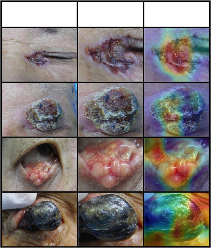
Interpretability of the deep learning system good performance in identifying both frequent and rare
To investigate the interpretability of the system in classifying malignant eyelid tumors.
benign and malignant eyelid tumors, heatmaps were created to Differentiating a malignant eyelid tumor from a benign one can
visualize the regions that contributed most to the system’s be challenging for the examining physician in primary care centers
decisions. We found that heatmaps highlighted the regions of due to the relatively small size, variability in clinical presentation,
benign and malignant eyelid tumors, regardless of the size, and minimal ophthalmologic training in the medical school5,26.
location, and shape of the tumors. Examples (images and Unlike skin tumors in other regions of the body, where physicians
corresponding heatmaps) of malignant and benign eyelid tumors might feel comfortable conducting biopsies, the intricate anatomy
are shown in Figs. 5 and 6, respectively. of eyelids often calls for a referral to an ophthalmologist. Even
oculoplastic ophthalmologists have only 70% accuracy in diag-
nosing eyelid tumors5. Due to the reliable performance, our
Deep learning system versus ophthalmologists system could be utilized both at the screening stage before
For differentiating malignant eyelid tumors from benign ones patients visit the physician and at the disease confirmation stage
based on the external test set, the junior ophthalmologist after the consultation, promoting the early detection of malignant
achieved an accuracy of 72.3% (95% CI, 67.1–77.5) with a eyelid tumors.
npj Digital Medicine (2022) 23 Published in partnership with Seoul National University Bundang Hospital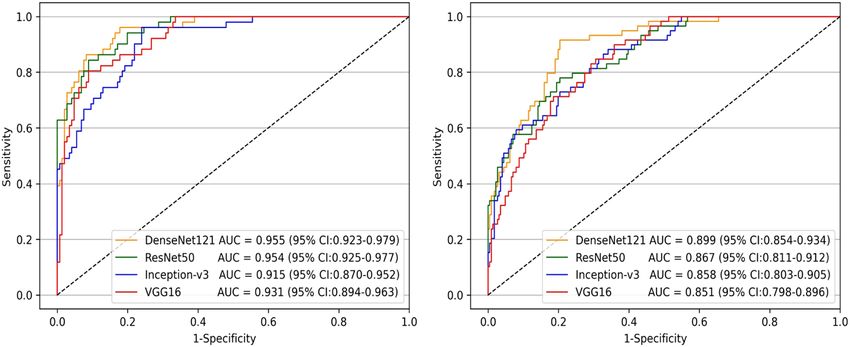
Z. Li et al.
                                                                                                                                                     5

                                  Clinical images              Tumor-centered Tumor-centered
                                                               cropped images   heatmaps
                            a

                            b

                            c

                            d

Fig. 5 Examples of photographic images, cropped images and corresponding heatmaps of malignant eyelid tumors. a Basal cell
carcinomas. b Squamous cell carcinomas. c Sebaceous gland carcinoma. d Melanoma.

   Recently, Adamopoulos et al.27 trained models using a deep              regions which contributed most to the system’s classification. The
learning artificial neural network for classifying patients with            heatmap revealed that the eyelid tumors in images, irrespective of
eyelid BCC and healthy individuals without eyelid tumors based             malignant and benign categories, were identified as the critical
on 143 photographic images obtained from a single clinical                 regions, which further demonstrated the validity of the system
center. The AUC of their best model reached approximately 1.00.            (Figs. 5 and 6). Our system’s interpretability feature could facilitate
As their model was mainly used for detecting eyelid BCC, it may            its application in real-world settings.
not be employed to discern other malignant eyelid tumors. In                  Compared to the ground truth, our system made a few
comparison to their study, our study showed several important              mistakes. For the malignant eyelid tumors incorrectly classified
features. First, we developed a deep learning system that could            as benign ones, 85.7% (6/7) images showed BCC. The BCCs in
distinguish a variety of malignant eyelid tumors from benign ones          these images are relatively small, unclear, and similar to the scar
with AUCs ranging from 0.899 to 0.955. In addition, our system has         and seborrheic keratosis, which might be possible contributors to
the potential to be applied to ordinary digital cameras, which             this misclassification. For benign eyelid tumors misclassified as
would be a convenient and cost-effective approach for promoting            malignant ones, most images showed nevus (31/80, 38.8%),
the early detection of malignant eyelid tumors. Third, our datasets        followed by seborrheic keratosis (14/80, 17.6%), and SCP (12/80,
included 1,417 photographic images collected from three
                                                                           15.0%). These benign tumors, in varying degrees, have similar
different clinical hospitals and thereby were more representative
                                                                           appearances (e.g. irregular shapes, irregular pigmentation, and
of the data in real-world settings. Fourth, the ground-truth label of
each image in this study is based on an unequivocal histopatho-            telangiectasia) to malignant ones. When analyzing the relationship
logical diagnosis.                                                         between the system’s predicted probability and classification error
   While deep learning has great performance in medical image              rate, the results suggested that the higher classification error rate
diagnosis problems, it remains highly criticized for being “a black        is associated with the lower predicted probability. Hence
box”28. This is considered as a major shortcoming in the                   ophthalmologists need to pay more attention to the images with
application of deep learning to high-stakes decisions29. To explore        low predicted probability values. As an ideal intelligent screening
this issue, we generated heatmaps using a Gradient-weighted                system should minimize both false-negative and false-positive
Class Activation Mapping (Grad-CAM) technique to visualize the             errors, further studies are needed to address this issue.

Published in partnership with Seoul National University Bundang Hospital                                        npj Digital Medicine (2022) 23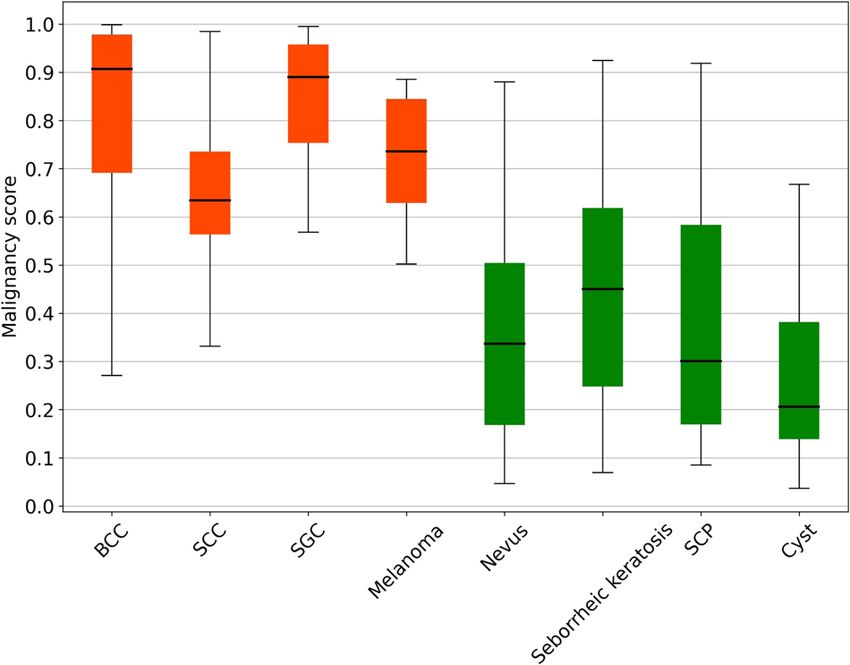
Z. Li et al.
6

                                     Clinical images             Tumor-centered Tumor-centered
                                                                 cropped images   heatmaps
                                 a

                                 b

                                 c

                                d

    Fig. 6 Examples of photographic images, cropped images and corresponding heatmaps of benign eyelid tumors. a Compound nevus. b
    Seborrheic keratosis. c Squamous cell papilloma. d Epidermoid cyst.

       Although the present study proves the potential of the deep            subjects who presented for eye examinations and ophthalmology
    learning system in discriminating between malignant and benign            consultations due to the discovery of eyelid tumors. The images were
    eyelid tumors, the system has several limitations which we wish to        captured between January 2010 and March 2021 using ordinary digital
    address in the near future. First, since our system was developed         cameras. To better confirm the effectiveness and generalizability of the
    solely based on the Chinese Population from several different             deep learning system, an additional dataset including 248 photographic
                                                                              images (129 patients) collected at Jiangdong Eye Hospital (JEH) and 61
    geographic regions, its effectiveness in other racial populations         photographic images (47 patients) collected at Zunyi First People’s
    would need to be further verified. Additional training on various          Hospital (ZFPH) were used to externally assess the system. The images
    clinical and demographic cohorts might further improve the                of the development set and the external test set were taken at various
    performance and clinical utility of the system in a broad range of        locations, such as outpatient clinics, inpatient wards, operating rooms,
    populations. In addition, while our system appears well suited for        hence the lighting and background of the images were not uniform,
    a screening purpose (discerning malignant eyelid tumors), it              indicating the richness and diversity of our datasets. All anonymized,
    cannot provide a specific diagnosis based on images. We expect             unaltered images (size, 0.6–5.5 megabytes per image) were transferred to
    to collect more images of eyelid tumors of each category and then         researchers for inclusion in the study. This study followed the recently
                                                                              published reporting guidelines for the clinical research involving AI: the
    develop an AI system to realize this function.
                                                                              CONSORT-AI extension30.
       In conclusion, the current study demonstrated that our deep
    learning system had roust performance in differentiating malig-
    nant eyelid tumors from benign ones. This AI system has the               Development of an eyelid tumor detection system
    potential to assist medical practitioners and suspected patients to       As one photographic image may show one or more eyelid tumors of
    proactively track eyelid tumors and identify malignant ones               different nature, this study first developed an ETDS using the Faster-RCNN,
    earlier.                                                                  an object detection network depending on region proposal algorithms31,
                                                                              to automatically locate and crop eyelid tumors from photographic images.
                                                                              This step can also remove the background noise around tumors in
                                                                              photographic images for better training the subsequent deep learning-
    METHODS
                                                                              based classification networks. Each eyelid tumor in a photographic image
    Image acquisition                                                         was delineated by a tight bounding box for the training of the Faster-RCNN
    For developing a deep learning system, a total of 1,258 photographic      model. Each cropped image only contains one eyelid tumor. The pipeline
    images (675 patients) were collected at NEH. The NEH dataset included     of the ETDS is described in Supplementary Fig. 7.

    npj Digital Medicine (2022) 23                                            Published in partnership with Seoul National University Bundang Hospital
Z. Li et al.
7
Ground truth and image classification visualize the embedding features of each category learned by the system
Two junior ophthalmologists who both had two-year clinical experience in a two-dimensional space41.
were recruited to annotate cropped images. The label of each cropped To investigate the performance of the system in eyelid tumors without
image was based on an unequivocal histopathological diagnosis which evident malignant features (borderline cases), we recruited an expert with
was considered as the ground truth of this study. Images without sufficient 15 years of clinical experience to read all images from the external test set
diagnostic certainty were excluded from the study. All images with clear and select images of eyelid tumors of uncertain malignant nature by
diagnoses were classified by the study steering committee into two evaluating the appearance of the tumors.
categories: malignant eyelid tumors (including premalignant ones) and
benign eyelid tumors. The malignant eyelid tumors included BCC, SCC, Heatmap generation
SGC, etc. The benign eyelid tumors included SCP, seborrheic keratosis,
The Grad-CAM technique42 was employed to produce visual explanations
nevus, etc.
for the decisions from the system by superimposing a visualization layer at
the end of the CNN model. This method leverages the gradients of any
Development of a deep learning classification system target concepts, flowing into the last layer of the CNN to generate a
The cropped images created by the ETDS using the NEH dataset were localization map highlighting the key regions in the image for predicting
randomly split at a 7:1.5:1.5 ratio for training, validation, and testing of a the concept42. Redder regions denote the more significant features of the
deep learning classification system. No overlap was allowed among system’s prediction. Using this tool, the heatmap was generated to
training, validation, and internal test sets. For acquiring the best deep interpret the rationale of the system on the discrimination between
learning algorithm to identify malignant eyelid tumors, four state-of-the-art malignant and benign eyelid tumors.
CNN architectures (DenseNet121, ResNet50, Inception-v3, and VGG16)
were investigated in this study. The architectural characteristics of these Analysis of misclassified images
four networks were described as follows: In a post hoc analysis, a senior ophthalmologist who was not involved in
(1) DenseNet121: This network has 121 layers that are densely the original analysis reviewed all false-negative and false-positive findings
connected through jointing all preceding layers into subsequent made by the system. To illustrate these discrepancies, the possible reasons
layers to accomplish strengthened feature propagation and alleviate for misclassified images were analyzed and documented on the basis of
a vanishing-gradient issue32. Recently, DenseNet121 has been used the observed characteristics in images. In addition, the relationship
to identify keratitis from slit-lamp images20. between the classification error rates and the system’s predicted
(2) ResNet50: This CNN is a 50-layer network that utilizes skip residual probability was investigated.
connections to bypass signals across layers, allowing for the increase
in layers without compromising the ease of training33. ResNet50 has Performance comparison between the deep learning system
been employed to detect brain abnormality from fluorodeoxyglu- and ophthalmologists
cose positron emission tomography images34.
(3) Inception-v3: This network has 42 layers and consists of 10 inception To evaluate our deep learning classification system in the context of
modules which can decrease the number of parameters to be malignant eyelid tumor detection, we recruited three ophthalmologists
trained and thereby reduce the computational complexity35. who had different levels of clinical experience (a junior ophthalmologist
Inception-v3 has been applied to identify age-related macular with three-year experience, a senior ophthalmologist with seven-year
degeneration from fundus images36. experience, and an expert with 15-year experience). The external test set
(4) VGG16: This network contains 41 layers, of which, 16 layers have was employed to compare the performance of the optimal system to that
learnable weights37. It includes the features of the classical of ophthalmologists with the ground truth. Of note, to reflect the real level
network’s simple structure while expanding the network’s depth of the ophthalmologists in routine clinical practices, they were not
via the flexible use of 3 × 3 convolution37. VGG16 has been used to informed that they competed with the system to avoid bias from the
detect breast cancer from histopathologic images37. competition.
Transfer learning was adopted as it could promote the performance of
the deep learning algorithms in the tasks of medical image classification38. Statistical analysis
Weights pre-trained for ImageNet classification (1,000 object classes) were The performance of the ETDS was evaluated by calculating the AP score
leveraged to initialize the CNN architectures39. Image standardization was using mmdetection 2.10.0. The sensitivity, specificity, accuracy, and AUC
performed as a preprocessing step before training the models. Due to were calculated to assess the performance of the deep learning
using the transfer learning approach, all cropped images were resized to classification system. The 95% CIs of sensitivity, specificity, and accuracy
224 × 224 pixels for the DenseNet121, ResNet50, and VGG16 algorithms were estimated with the Wilson Score approach utilizing a package of
and to 299 × 299 pixels for the Inception-v3 algorithm. Image pixel values Statsmodels 0.11.1, and for AUC, utilizing Empirical Bootstrap with 1000
were normalized within the range of 0 to 1. Data augmentation techniques replicates. The ROC curves were drawn according to the sensitivity versus
were adopted because they were capable of enhancing the robustness of 1–specificity utilizing the packages of Scikit-learn 0.23.2 and Matplotlib
CNN networks40. Random brightness, rotation, and horizontal and vertical 3.3.1. The deep learning provided a malignancy score ranging from 0 to 1
flipping were applied to the images of the training set to augment the with a cutoff over 0.5 for classifying an eyelid tumor as being malignant.
sample size to 6 times larger than the original size (from 883 to 5,298). Unweighted Cohen’s κ coefficients were calculated to compare the
The PyTorch deep learning framework (version 1.6.0) was used to train, classification results of the system with the ground truth. The Kappa result
validate, and test our models. The DenseNet121, ResNet50, Inception-v3, is interpreted as follows: values ≤0 as indicating no agreement, 0.01–0.20
and VGG16 were trained using 4 Nvidia 2080TI graphics processing units. as slight, 0.21–0.40 as fair, 0.41– 0.60 as moderate, 0.61–0.80 as substantial,
The mini-batch size was set at 32 on each GPU to gain 128 images in one and 0.81–1.00 as almost perfect agreement43. The differences in
iteration. The average value of these samples was computed to update the sensitivities, specificities, and accuracies between the deep learning
trainable parameters. A variation of the stochastic gradient descent system and ophthalmologists were analyzed using the McNemar test. All
algorithm, adaptive moment estimation (ADAM) optimizer, was used with statistical tests were 2-sided and the results were considered statistically
an initial learning rate at 0.001, β1 of 0.9, β2 of 0.999, and a weight decay significant at the level of p < 0.05. Statistical analyses were carried out
of 1e-4. Each algorithm was trained for 80 epochs. During the training using Python 3.7.8 (Wilmington, Delaware, USA).
process, accuracy and cross-entropy loss were calculated on the training
and validation sets after each epoch and utilized as a reference for model
selection. Each time the accuracy increased or cross-entropy loss
Reporting summary
decreased, a checkpoint saved the model state and corresponding weight Further information on research design is available in the Nature Research
matrix. The model with the highest validation accuracy was selected for Reporting Summary linked to this article.
use on the internal test set.
The performance of the binary classification model was further
evaluated on an independent external test set. The process of the DATA AVAILABILITY
development and assessment of the deep learning classification system is The main data supporting the results of this study are available in the manuscript and
described in Supplementary Fig. 8. The t-SNE technique was applied to its Supplementary Information. The raw datasets from the Ningbo Eye Hospital,
Published in partnership with Seoul National University Bundang Hospital npj Digital Medicine (2022) 23Z. Li et al.
8
Jiangdong Eye Hospital, and Zunyi First People’s Hospital cannot be made available 24. Silverman, N. & Shinder, R. What’s new in eyelid tumors. Asia Pac. J. Ophthalmol.
due to hospital regulation restrictions and patient privacy concerns. Some (Philos.) 6, 143–152 (2017).
anonymized data may be available for research purposes from the corresponding 25. Yin, V. T., Merritt, H. A., Sniegowski, M. & Esmaeli, B. Eyelid and ocular surface
authors on reasonable request. carcinoma: diagnosis and management. Clin. Dermatol. 33, 159–169 (2015).
26. Noble, J., Somal, K., Gill, H. S. & Lam, W. C. An analysis of undergraduate oph-
thalmology training in Canada. Can. J. Ophthalmol. 44, 513–518 (2009).
CODE AVAILABILITY 27. Adamopoulos, A., Chatzopoulos, E. G., Anastassopoulos, G. & Detorakis, E. Eyelid
The code and example data used in this study can be accessed at GitHub (https:// basal cell carcinoma classification using shallow and deep learning artificial
github.com/jiangjiewei/EyelidTumors-Source). neural networks. Evol. Syst. 12, 583–590 (2021).
28. Thomas, S. M., Lefevre, J. G., Baxter, G. & Hamilton, N. A. Interpretable deep
learning systems for multi-class segmentation and classification of non-
Received: 14 October 2021; Accepted: 4 February 2022; melanoma skin cancer. Med. Image Anal. 68, 101915 (2020).
29. Rudin, C. Stop explaining black box machine learning models for high stakes
decisions and use interpretable models instead. Nat. Mach. Intell. 1, 206–215
(2019).
30. Liu, X., Cruz, R. S., Moher, D., Calvert, M. J. & Denniston, A. K. Reporting guidelines
REFERENCES for clinical trial reports for interventions involving artificial intelligence: the
1. Yu, S. S., Zhao, Y., Zhao, H., Lin, J. Y. & Tang, X. A retrospective study of 2228 cases CONSORT-AI extension. Nat. Med. 26, 1364–1374 (2020).
with eyelid tumors. Int J. Ophthalmol. 11, 1835–1841 (2018). 31. Ren, S., He, K., Girshick, R. & Sun, J. Faster R-CNN: towards real-time object
2. Deprez, M. & Uffer, S. Clinicopathological features of eyelid skin tumors. A ret- detection with region proposal networks. IEEE Trans. Pattern Anal. Mach. Intell. 39,
rospective study of 5504 cases and review of literature. Am. J. Dermatopathol. 31, 1137–1149 (2017).
256–262 (2009). 32. Huang, G., Liu, Z., Van Der Maaten, L. & Weinberger, K. Q. Densely Connected
3. Pe’Er, J. Pathology of eyelid tumors. Indian J. Ophthalmol. 64, 177–190 (2016). Convolutional Networks. 2017 IEEE Conference on Computer Vision and Pattern
4. Huang, Y. Y. et al. Comparison of the clinical characteristics and outcome of Recognition (CVPR). 2261–2269 (2017).
benign and malignant eyelid tumors: an analysis of 4521 eyelid tumors in a 33. Yip, M. et al. Technical and imaging factors influencing performance of deep
tertiary medical center. Biomed. Res. Int. 2015, 453091 (2015). learning systems for diabetic retinopathy. NPJ Digit Med 3, 40 (2020).
5. Leung, C., Johnson, D., Pang, R. & Kratky, V. Identifying predictive morphologic 34. Whi, W. et al. Fully automated identification of brain abnormality from whole-
features of malignancy in eyelid lesions. Can. Fam. Physician 61, e43–e49 body FDG-PET imaging using deep learning-based brain extraction and statistical
(2015). parametric mapping. EJNMMI Phys. 8, 79 (2021).
6. Burgic, M. et al. Clinical characteristics and outcome of malignant eyelid tumors: a 35. Szegedy, C., Vanhoucke, V., Ioffe, S., Shlens, J. & Wojna, Z. Rethinking the
five-year retrospective study. Med Arch. 73, 209–212 (2019). Inception Architecture for Computer Vision. 2016 IEEE Conference on Computer
7. Malhotra, R., Chen, C., Huilgol, S. C., Hill, D. C. & Selva, D. Mapped serial excision Vision and Pattern Recognition (CVPR). 2818–2826 (2016).
for periocular lentigo maligna and lentigo maligna melanoma. Ophthalmology 36. Keel, S. et al. Development and validation of a deep-learning algorithm for the
110, 2011–2018 (2003). detection of neovascular age-related macular degeneration from colour fundus
8. Cook, B. J. & Bartley, G. B. Treatment options and future prospects for the photographs. Clin. Exp. Ophthalmol. 47, 1009–1018 (2019).
management of eyelid malignancies: an evidence-based update. Ophthalmology. 37. Deniz, E. et al. Transfer learning based histopathologic image classification for
108, 2088–2098 (2001). breast cancer detection. Health Inf. Sci. Syst. 6, 18 (2018).
9. Resnikoff, S., Felch, W., Gauthier, T. M. & Spivey, B. The number of ophthalmol- 38. Kermany, D. S. et al. Identifying medical diagnoses and treatable diseases by
ogists in practice and training worldwide: a growing gap despite more than image-based deep learning. Cell 172, 1122–1131 (2018).
200,000 practitioners. Br. J. Ophthalmol. 96, 783–787 (2012). 39. Russakovsky, O. et al. ImageNet large scale visual recognition challenge. Int. J.
10. Lotter, W. et al. Robust breast cancer detection in mammography and digital Comput. Vision. 115, 211–252 (2015).
breast tomosynthesis using an annotation-efficient deep learning approach. Nat. 40. Bloice, M. D., Roth, P. M. & Holzinger, A. Biomedical image augmentation using
Med. 27, 244–249 (2021). Augmentor. Bioinformatics 35, 4522–4524 (2019).
11. Li, Z. et al. A deep learning system for identifying lattice degeneration and retinal 41. van der Maaten, L. & Hinton, G. Visualizing data using t-SNE. J. Mach. Learn. Res. 9,
breaks using ultra-widefield fundus images. Ann. Transl. Med. 7, 618 (2019). 2579–2605 (2008).
12. Zhang, K. et al. Clinically applicable AI system for accurate diagnosis, quantitative 42. Selvaraju, R. R. et al. Grad-CAM: Visual explanations from deep networks via
measurements, and prognosis of COVID-19 pneumonia using computed tomo- gradient-based localization. 2017 IEEE International Conference on Computer
graphy. Cell 182, 1360 (2020). Vision (ICCV). 618–626 (2017).
13. Shi, Z. et al. A clinically applicable deep-learning model for detecting intracranial 43. McHugh, M. L. Interrater reliability: the kappa statistic. Biochem Med (Zagreb) 22,
aneurysm in computed tomography angiography images. Nat. Commun. 11, 276–282 (2012).
6090 (2020).
14. Li, Z. et al. Deep learning from “passive feeding” to “selective eating” of real-world
data. NPJ Digit. Med. 3, 143 (2020). ACKNOWLEDGEMENTS
15. Li, Z. et al. Automated detection of retinal exudates and drusen in ultra-widefield
This study received funding from the Natural Science Foundation of Zhejiang
fundus images based on deep learning. Eye (Lond), (2021). Online ahead of print.
16. Li, Z. et al. Deep learning for automated glaucomatous optic neuropathy Province (grant no. LQ22H120002), Medical Health Science and Technology Project of
detection from ultra-widefield fundus images. Br. J. Ophthalmol. 105, 1548–1554 Zhejiang Province (grant no. 2022RC069), Zhejiang Postdoctoral Science Fund for
(2021). Excellent Project (grant no. ZJ2021088), Ningbo Science & technology program (grant
17. Li, Z. et al. Deep learning for detecting retinal detachment and discerning no. 2021S118), and Ningbo Medical Technology Program (grant no. 2020Y54). The
macular status using ultra-widefield fundus images. Commun. Biol. 3, 15 (2020). funding organization played no role in the study design, data collection and analysis,
18. Ting, D. et al. Development and validation of a deep learning system for diabetic decision to publish, or preparation of the manuscript.
retinopathy and related eye diseases using retinal images from multiethnic
populations with diabetes. JAMA 318, 2211–2223 (2017).
19. Li, Z. et al. Development and evaluation of a deep learning system for screening
retinal hemorrhage based on ultra-widefield fundus images. Transl. Vis. Sci. AUTHOR CONTRIBUTIONS
Technol. 9, 3 (2020). Conception and design: Z.L., J.J., W.Q., and G.W. Funding obtainment: Z.L. Provision of
20. Li, Z. et al. Preventing corneal blindness caused by keratitis using artificial study data: G.W. and H.C. Collection and assembly of data: Z.L., W.Q., H.C., X.Y., L.W.,
intelligence. Nat. Commun. 12, 3738 (2021). Z.L., W.X., and G.W. Data analysis and interpretation: Z.L., J.J., M.P., X.W., and G.W.
21. Li, Z. et al. Comparison of deep learning systems and cornea specialists in Manuscript writing: all authors. Final approval of the manuscript: all authors. Z.L. and
detecting corneal diseases from low-quality images. iScience 24, 103317 (2021). W.Q. contributed equally as first authors.
22. Gulshan, V. et al. Development and validation of a deep learning algorithm for
detection of diabetic retinopathy in retinal fundus photographs. JAMA 316,
2402–2410 (2016).
23. Shields, J. A. & Shields, C. L. Sebaceous adenocarcinoma of the eyelid. Int Oph- COMPETING INTERESTS
thalmol. Clin. 49, 45–61 (2009). The authors declare no competing interests.
npj Digital Medicine (2022) 23 Published in partnership with Seoul National University Bundang HospitalZ. Li et al.
9
ETHICAL APPROVAL Publisher’s note Springer Nature remains neutral with regard to jurisdictional claims
Approval from the institutional review board of Ningbo Eye Hospital (NEH) was in published maps and institutional affiliations.
obtained (identifier, 2021-qtky-44), and the study protocol was performed following
the Declaration of Helsinki principles. Informed consent was waived by the ethics
committee of NEH due to the retrospective nature of the data collection and the use Open Access This article is licensed under a Creative Commons
of de-identified images. Attribution 4.0 International License, which permits use, sharing,
adaptation, distribution and reproduction in any medium or format, as long as you give
appropriate credit to the original author(s) and the source, provide a link to the Creative
ADDITIONAL INFORMATION Commons license, and indicate if changes were made. The images or other third party
material in this article are included in the article’s Creative Commons license, unless
Supplementary information The online version contains supplementary material
indicated otherwise in a credit line to the material. If material is not included in the
available at https://doi.org/10.1038/s41746-022-00571-3.
article’s Creative Commons license and your intended use is not permitted by statutory
regulation or exceeds the permitted use, you will need to obtain permission directly
Correspondence and requests for materials should be addressed to Zhongwen Li,
from the copyright holder. To view a copy of this license, visit http://creativecommons.
Jiewei Jiang or Guohai Wu.
org/licenses/by/4.0/.
Reprints and permission information is available at http://www.nature.com/
reprints © The Author(s) 2022
Published in partnership with Seoul National University Bundang Hospital npj Digital Medicine (2022) 23You can also read