ARGOS Swept Source OCT Biometer - Clinical Science Compendium Summary of peer-reviewed bench and clinical research - Alcon | Medical Affairs
←
→
Page content transcription
If your browser does not render page correctly, please read the page content below

ARGOS
                 ®

Swept Source OCT Biometer
Clinical Science Compendium
Summary of peer-reviewed
bench and clinical research

Medical Affairs North America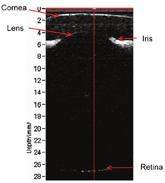
INTRODUCTION

At Alcon, our surgical medical device products, such as the ARGOS®
swept-source optical coherence tomography (SS-OCT) biometer,
are designed, manufactured and marketed with a body of science
developed through rigorous bench research and clinical studies. As
the body of knowledge behind Alcon’s products grows, so does the
challenge of making our customers aware of its depth. Our medical
affairs organization is thus focused on both high-quality data generation
and its communication to the clinical community. High-quality
scientific publications are essential to convey the clinical community’s
knowledge and experience with the latest technology. This clinical
science compendium provides a consolidated view of peer-reviewed
publications for ARGOS®, an industry-leading SS-OCT biometer used to
measure eye parameters for patients before cataract surgery.

In addition to exploring this compendium, we encourage you to visit
Alcon’s Medical Affairs website—AlconScience.com—to learn more
about how medical science matters to us. Beyond scientific publications
relating to Alcon’s portfolio, you will find more information on
independent medical educational grants, teaching facility equipment
placement, and areas of interest for investigator-initiated trials.

   METHODOLOGY

The 16 articles summarized in this compendium were identified
using the PubMed and Google Scholar databases incorporating the
search terms “ARGOS” and “swept-source optical coherence tomography
biometer.” Articles were included when they were published between
January 1, 2009 and July 31, 2020 and contained research relevant
to the ARGOS® SS-OCT biometer and its indication for acquiring
ocular measurements and performing calculations to determine the
appropriate IOL power and type for implantation during IOL placement.
Only manuscripts published in peer-reviewed journals and available in
English were included in this compendium.
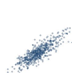
Table of Contents

Bench Studies

Large Coherence Length Swept Source for Axial Length Measurement of the Eye.                                      1
Chong C, Suzuki T, Totsuka K, Morosawa A, Sakai T. Appl Opt. 2009;48:D144-D150.

Clinical Studies

Biometry Measurements Using a New Large-Coherence-Length Swept-Source Optical                                     2
Coherence Tomographer.
Shammas HJ, Ortiz S, Shammas MC, Kim SH, Chong C. J Cataract Refract Surg. 2016;42:50-61.

Changes in the Anterior Segment After Cycloplegia With a Biometer Using Swept-Source                              3
Optical Coherence Tomography.
Higashiyama T, Iwasa M, Ohji M. PLoS One. 2017;12:e0183378.

Comparison of a New Biometer Using Swept-Source Optical Coherence Tomography and                                  4
a Conventional Biometer Using Partial Coherence Interferometry.
Higashiyama T, Mori H, Nakajima F, Ohji M. PLoS One. 2018;13:e0196401.

Comparison of Axial Length Using a New Swept-Source Optical Coherence Tomography-Based                            5
Biometer - ARGOS With Partial Coherence Interferometry- Based Biometer -IOLMaster Among
School Children.
Hussaindeen JR, Mariam EG, Arunachalam S, Bhavatharini R, Gopalakrishnan A, Narayanan A, Agarkar S,
Sivaraman V. PLoS One. 2018;13:e0209356.

Predictive Accuracy of Partial Coherence Interferometry and Swept-Source Optical Coherence                        6
Tomography for Intraocular Lens Power Calculation.
Whang WJ, Yoo YS, Kang MJ, Joo CK. Sci Rep. 2018;8:1373.

Accuracy of Swept-Source Optical Coherence Tomography Based Biometry for Intraocular Lens                         7
Power Calculation: A Retrospective Cross-Sectional Study.
An Y, Kang EK, Kim H, Kang MJ, Byun YS, Joo CK. BMC Ophthalmol. 2019;19:30.

A Comparison of Two Methods to Calculate Axial Length.                                                            8
Cooke DL, Cooke TL. J Cataract Refract Surg. 2019;45:284-292.

Comprehensive Comparison of Axial Length Measurement With Three Swept-Source OCT-Based                            9
Biometers and Partial Coherence Interferometry.
Huang J, Chen H, Li Y, Chen Z, Gao R, Yu J, Zhao Y, Lu W, McAlinden C, Wang Q. J Refract Surg. 2019;35:115-120.

Ocular Measurements of a Swept-Source Biometer: Repeatability Data and Comparison With                            10
an Optical Low-Coherence Interferometry Biometer.
Nemeth G, Modis L Jr. J Cataract Refract Surg. 2019;45:789-797.

Ocular Biometry and Refractive Outcomes Using Two Swept-Source Optical Coherence                                  11
Tomography-Based Biometers With Segmental or Equivalent Refractive Indices.
Omoto MK, Torii H, Masui S, Ayaki M, Tsubota K, Negishi K. Sci Rep. 2019;9:6557.

Comparative Analysis of 2 Swept-Source Optical Coherence Tomography Biometers.                                    12
Sabatino F, Matarazzo F, Findl O, Maurino V. J Cataract Refract Surg. 2019;45:1124-1129.

Clinical Evaluation of a New Swept-Source Optical Coherence Biometer That Uses Individual                         13
Refractive Indices to Measure Axial Length in Cataract Patients.
Tamaoki A, Kojima T, Hasegawa A, Yamamoto M, Kaga T, Tanaka K, Ichikawa K. Ophthalmic Res. 2019;62:11-23.
Table of Contents / Continued

Comparison of Two Swept-Source Optical Coherence Tomography Biometers and a Partial              14
Coherence Interferometer.
Yang CM, Lim DH, Kim HJ, Chung TY. PLoS One. 2019;14:e0223114.

Effects on IOL Power Calculation and Expected Clinical Outcomes of Axial Length Measurements     15
Based on Multiple vs Single Refractive Indices.
Shammas HJ, Shammas MC, Jivrajka RV, Cooke DL, Potvin R. Clin Ophthalmol. 2020;14:1511-1519.

Agreement Between Two Optical Biometers Based on Large Coherence Length SS-OCT and               16
Scheimpflug Imaging/Partial Coherence Interferometry.
Tu R, Yu J, Savini G, Ye J, Ning R, Xiong J, Chen S, Huang J. J Refract Surg. 2020;36:459-465.
Large Coherence Length Swept Source for Axial                                                                                         Axial Length

Length Measurement of the Eye                                                                                                         Repeatability/Reproducibility

Chong et al. Appl Opt. 2009;48:D144-D150

     OVERVIEW

STUDY DESIGN              STUDY SITE(S)            PATIENTS            METHODOLOGY                        BIOMETERS                                  KEY ENDPOINT(S)
Proof of concept study    Single center in Japan   Not applicable;     Illustrate the new                 Experimental SS-OCT                        Optical performance,
to demonstrate the                                 bench study         technology and                     biometer with QPCT and                     AL, repeatability of
measurement of AL                                                      measurement of AL in a             multiple beam expanders                    measurement
with a swept source                                                    pig’s eye                          (Santec, Inc.)
using a quasi-phase
continuous tuning
(QPCT) technique

     ANALYSIS AND CONCLUSIONS

    This study demonstrated a swept source with a large coherence length for IOL measurement with a quasi-phase
    continuous tuning technique as well as multiple beam expanders.
    This experimental SS-OCT system enables the measurement of the AL of a pig’s eye with 20 mm length in physical size.

     STUDY RESULTS

TECHNOLOGY PARAMETERS                                                        Figure 1. (Color online)    Cornea                 U–
                                                                             2D image of the whole
n	The swept source consists of a fiber ring extended cavity laser           pig’s eye.                                          2–
                                                                                                            Lens
   with a diffraction grating and a polygon scanner-based tunable                                                                4–
   filter configuration; the projected beam on the diffraction                                                                                                   Iris
                                                                                                                                 6–
   grating is expanded with a multiple of beam expanders to                                                                      8–
   achieve high finesse of the filter
                                                                                                                                10–
PERFORMANCE PARAMETERS                                                                                                          12–
n	The source demonstrated an 18 nm swept range at 1060 nm                                                                      14–
   wavelength, 28 mm coherence length, and 6.2 MW peak power                                                                    16–
   at a 2.5 kHz swept rate                                                                                                      18–
                                                                                                                   Depth (mm)

n	OCT imaging results showed that a coherence length of 28 mm                                                                  20–
   enables the measurement of the AL of a pig’s eye with 20 mm                                                                  22–
   length in physical size                                                                                                      24–
n Figure 1 shows a tomographic image of the whole eye over
 	                                                                                                                             26–                              Re�na
  5 mm width with 200 A-lines in the transverse direction; the                                                                  28–
  contours of the cornea and iris, lens surface, and retina on the
  far side are all recognized, although the detail of each segment
  is slightly blurred because lateral resolution is only 0.8 mm              Figure 2. 1D signal of one A-line (solid line in Figure 1).
n    igure 2 shows a 1D signal indicating the positions of the
    F
    different parts; peaks at the cornea, lens, and retina are
    apparent, and the distances between them are 3.5 mm and
    27 mm between the cornea and lens and the lens and retina,
    respectively
REPEATABILITY
n	The repeatability of measurement was better than 20 μm,
   which is superior to the performance of commercial IOL
   measurement equipment

                                                                         1
Biometry Measurements Using a New Large- Axial Length
Coherence-Length Swept-Source Optical Biometric Parameters
Coherence Tomographer Repeatability/Reproducibility
Shammas et al. J Cataract Refract Surg. 2016;42:50-61*
OVERVIEW
STUDY DESIGN STUDY SITE(S) PATIENTS METHODOLOGY BIOMETERS KEY ENDPOINT(S)
Prospective Single private practice Sixty-six (66) ARGOS® performance ARGOS® (SS- Repeatability and
observational in the United States consecutive patients and comparison to OCT, Movu, Inc.), reproducibility of ARGOS®
study to evaluate enrolled (41 patients IOLMaster® 500 and IOLMaster® 500 (PCI, measurements; comparison
the repeatability had bilateral LENSTAR LS 900® in Carl Zeiss Meditec of AL, ACD, and average
and reproducibility cataracts), 107 cataractous eyes (same AG), LENSTAR LS anterior corneal radius
of ARGOS® eyes (54 right eyes) day); for the comparison 900® (OLCR, Haag- of curvature (RAV)
measurements and to measured by SS-OCT part, only the first eye of Streit AG) measurement with results
compare them with and OLCR and 91 each patient was used obtained by PCI and OLCR;
IOLMaster® 500 and eyes (46 right eyes) comparison of CCT, aqueous
LENSTAR LS 900® measured by PCI depth, lens thickness, pupil
size, and corneal diameter
with results obtained by OLCR
ANALYSIS AND CONCLUSIONS
This study found that AL measurements obtained with ARGOS® were comparable to those obtained with the
IOLMaster® 500 and LENSTAR LS 900®, with a faster and higher acquisition rate, even in the presence of a dense
nuclear or posterior subcapsular cataract.
The authors believe that this is the first study to report the precision of ARGOS® in measuring all parameters needed for IOL power
calculation in patients having cataract extraction, with good repeatability and reproducibility of measurements. The wide scanning beam
and longer wavelength of SS-OCT may contribute to the higher AL acquisition rate.
* Drs. Ortiz, Kim, and Chong have proprietary interest in the new technology
STUDY RESULTS
REPEATABILITY/REPRODUCIBILITY (N=104 EYES) Table 1. Mean, SD, and range of the variation of the 9 images produced by ARGOS® in 3
consecutive acquisitions.
n Repeatability (intraset) and reproducibility (interset) of ARGOS®
measurements showed comparable values and a low variation rate; Parameter AL CCT AD ACD LT PS CD RAV
data are shown in Table 1 Mean 0.01 0.01 0.01 0.01 0.03 0.10 0.14 0.02
COMPARISON OF BIOMETRIC PARAMETERS (N=56 EYES) Standard deviation 0.02 0.01 0.01 0.01 0.02 0.06 0.06 0.02
0.00, 0.00, 0.00, 0.00, 0.01, 0.02, 0.05, 0.00,
n he patient population in this study included a large number of eyes
T Range
0.11 0.04 0.04 0.03 0.10 0.32 0.38 0.08
with advanced cataracts
All measurements are in millimeters. AD, aqueous depth; CD, corneal diameter; LT, lens thickness; PS, pupil
n RGOS® successfully measured AL in 96% (54/56) of the cases; in
A size; RAV, average anterior corneal radius of curvature
comparison, LENSTAR LS 900® successfully measured 79% (44/56) of
cases (Dense Cataract Measurement mode was not available at the
Table 2. A Summary of the comparison of ARGOS®, IOLMaster® 500 and LENSTAR LS 900®.
study time) and IOLMaster® 500 successfully measured 77% (43/56) of Adapted from Shammas et al. J Cataract Refract Surg. 2016;42:50-61.
cases. Reasons for unsuccessful AL measurement:
- ARGOS®: 2 cases with the mature cataracts discarded because of no Parameter Devices Spearman Mean difference 95% confidence
vs. ARGOS® Correlation (SD) interval
visibility of the retina Coefficient (rs) (mm) (mm)
- IOLMaster® 500: 2 mature cataracts, 2 cases with stage 5 nuclear IOLMaster® 500 1.00 -0.01 (0.05) -0.10, 0.08
AL, mm
sclerosis with posterior subcapsular changes, and 9 cases of stage LENSTAR LS 900® 1.00 0.01 (0.06) -0.10, 0.12
2 to stage 3 nuclear sclerosis with stage 3 posterior subcapsular IOLMaster® 500 0.76 -0.17 (0.20) -0.57, 0.23
ACD, mm
changes LENSTAR LS 900® 0.88 0.08 (0.15) -0.21, 0.37
IOLMaster® 500 0.98 -0.01 (0.05) -0.10, 0.09
- LENSTAR LS 900®: 2 mature cataracts, 3 cases with stage 4 cortical RAV, mm
LENSTAR LS 900® 0.97 0.00 (0.05) -0.09, 0.09
changes, and 7 cases with stage 3 posterior subcapsular changes
CCT, mm LENSTAR LS 900® 0.93 0.00 (0.01) -0.03, 0.02
n able 2 shows comparisons of all measurements for ARGOS®
T AD, mm LENSTAR LS 900® 0.91 0.07 (0.14) -0.20, 0.34
vs IOLMaster® 500 and LENSTAR LS 900®; Spearman correlation LT, mm LENSTAR LS 900® 0.80 -0.22 (0.20) -0.62, 0.18
coefficients of the AL were extremely high and relatively high for ACD
PS, mm LENSTAR LS 900® 0.87 -0.29 (0.53) -1.33, 0.76
and RAV comparisons, but poor for corneal diameter
CD, mm LENSTAR LS 900® 0.41 -0.34 (0.76) -1.83, 1.15
n imilarly, Bland-Altman plots showed excellent agreement for AL and
S
AD, aqueous depth; CD, corneal diameter; LT, lens thickness; n, number of eyes studied for the comparison;
good agreement for ACD and for RAV PS, pupil size; RAV , average anterior corneal radius of curvature
2Changes in the Anterior Segment After Axial Length
Cycloplegia With a Biometer Using Swept-Source Biometric Parameters
Optical Coherence Tomography Refractive Outcomes
Higashiyama et al. PLoS One. 2017;12:e0183378
OVERVIEW
STUDY DESIGN STUDY SITE(S) PATIENTS METHODOLOGY BIOMETERS KEY ENDPOINT(S)
Prospective study to Single center in Ten (10) eyes of 10 Biometric and refractive assessment ARGOS® (Santec, Inc.) AL, CCT, ACD, lens
investigate changes Japan pediatric patients before and after cycloplegia with SS- thickness, spherical and
in the anterior with strabismus OCT. Cyclopentolate hydrochloride cylindrical refraction
segment of the eye or amblyopia who 1% (Cyplegin 1% ophthalmic
after cycloplegia underwent cycloplegia; solution, Santen Pharmaceutical,
as measured by a mean age of 7.2 years; Osaka, Japan) was instilled three
biometer with SS-OCT range: 4 to 14 years times at 10-min intervals. The
measurements were obtained 60
min after the last instillation
ANALYSIS AND CONCLUSIONS
This study demonstrated that ARGOS® was useful for accurately detecting changes in the anterior segment of the
eye after cycloplegia in pediatric patients.
Measurements with ARGOS® showed that ACD was increased and lens thickness was decreased after cycloplegia, and also that ACD was
increased relative to the decrease in lens thickness. No significant differences were detected on AL and CCT.
STUDY RESULTS
BIOMETRIC PARAMETERS Figure 1. Lens thickness before cycloplegia Figure 2. ACD before cycloplegia (left plots) and
(left plots) and after cycloplegia (right plots), after cycloplegia (right plots), showing that mean
After cycloplegia, mean lens thickness
n
showing that mean lens thickness had ACD significantly increased after cycloplegia
significantly decreased (PComparison of a New Biometer Using Swept- Axial Length
Source Optical Coherence Tomography and a Comparative Accuracy
Conventional Biometer Using Partial Coherence
Interferometry
Higashiyama et al. PLoS One. 2018;13:e0196401
OVERVIEW
STUDY DESIGN STUDY SITE(S) PATIENTS METHODOLOGY BIOMETERS KEY ENDPOINT(S)
Retrospective review Single center in Japan Fifty-five (55) eyes Comparison of AL ARGOS® (SS-OCT, Santec, AL and AL acquisition
of medical records in 55 patients measurements with SS- Inc.), IOLMaster® version 5 rate
to compare AL using who underwent OCT and PCI, including (PCI, Carl Zeiss Meditec AG)
a biometer with SS- cataract surgery; subgroup analysis in
OCT versus using a mean age of 72.9 short, intermediate and
conventional biometer years long-AL groups (Comparison of Axial Length Using a New Swept- Axial Length
Source Optical Coherence Tomography-Based Biometric Parameters
Biometer - ARGOS With Partial Coherence Comparative Accuracy
Interferometry- Based Biometer -IOLMaster
Among School Children
Hussaindeen et al. PLoS One. 2018;13:e0209356
OVERVIEW
STUDY DESIGN STUDY SITE(S) PATIENTS METHODOLOGY BIOMETERS KEY ENDPOINT(S)
Prospective, Single center Three hundred Compare biometric ARGOS® (SS-OCT, Santec, AL and corneal
operator-blinded in India (part and seventy-six measurement from Inc.), IOLMaster® (PCI, curvature
study to compare of a school (376) eyes of 188 the right eyes with two version 5, Carl Zeiss measurements
AL measurements vision screening children with best biometers Meditec AG)
obtained by an SS-OCT program) corrected vision of
biometer with a PCI 6/9 or better and
biometer in school without any ocular
children abnormalities;
mean (SD) age of
13.88±1.69 years
ANALYSIS AND CONCLUSIONS
This study demonstrated that AL measurements obtained with ARGOS® and IOLMaster® were well within the clinically
agreeable limits among pediatric population, with comparable measurements for shorter and intermediate AL.
The authors concluded that data from this study can be used as a reference for pediatric AL measurements, and that ARGOS® can be
recommended for use in a pediatric population due to its speed of acquisition and improved resolution rates.
STUDY RESULTS
AXIAL LENGTH/CORNEAL CURVATURE
n The mean (SD) AL was 23.93± 1.02 mm and 23.82 ± 1.05 mm with n An additional analysis was conducted in eyes stratified by AL
ARGOS® and IOLMaster®, respectively measurements: short (Predictive Accuracy of Partial Coherence Axial Length
Interferometry and Swept-Source Optical Biometric Parameters
Coherence Tomography for Intraocular Lens Comparative Accuracy
Power Calculation
Whang et al. Sci Rep. 2018;8:1373
OVERVIEW
STUDY DESIGN STUDY SITE(S) PATIENTS METHODOLOGY BIOMETERS KEY ENDPOINT(S)
Retrospective study to Single center in One hundred and Use the 3-month post- ARGOS® (SS-OCT, Movu, Biometric
compare the predictive South Korea fifty-three (153) op refractive outcomes Inc.), IOLMaster® (PCI, measurements;
accuracy of IOL eyes of 153 patients and pre-op biometric version 5, Carl Zeiss comparison of
calculations made with who underwent measurement from Meditec AG) predictive accuracy for
PCI and SS-OCT uncomplicated two biometers to IOL power calculations
conventional compare the IOL power using the Barret-
cataract surgery; prediction error; PCI Universal II, Haigis,
mean age was 64.84 biometer based lens Hoffer Q, SRK/T, and T2
years (range: 47 to constants were used formulas
81 years)
ANALYSIS AND CONCLUSIONS
In this study, the predictive accuracies of ARGOS® and IOLMaster® were nearly the same, except in the case of
medium-long eyes, for which the predictive accuracy of ARGOS® was higher.
The investigators noted that to the best of their knowledge, this was the first study to evaluate the predictive accuracy of ARGOS® in conjunction with
the commonly used IOL power calculation formulas (Barret-Universal II, Haigis, Hoffer Q, SRK/T, and T2); personalized IOL constants were used.
STUDY RESULTS
AL/CORNEAL CURVATURE/ACD Figure 1. Bland-Altman plots for the AL. The limits of agreement were set at
±1.96 × standard deviation (SD).
AL measurement with ARGOS® (24.62 ± 2.29 mm) were significantly
n
shorter than with IOLMaster® (24.65 ± 2.35 mm, PAccuracy of Swept-Source Optical Coherence Axial Length
Tomography Based Biometry for Intraocular Biometric Parameters
Lens Power Calculation: A Retrospective Cross- Comparative Accuracy
Sectional Study
An et al. BMC Ophthalmol. 2019;19:30
OVERVIEW
STUDY DESIGN STUDY SITE(S) PATIENTS METHODOLOGY BIOMETERS KEY ENDPOINT(S)
Retrospective Single center and Four hundred Biometric performance ARGOS® (SS-OCT, Movu, Failure rate of AL
observational study same surgeon for all and thirty-one from SS-OCT, PCI, and Inc.), IOLMaster® version measurement
to evaluate the cataract surgeries in eyes (431) of 431 A-scan ultrasonography; 5.4 (PCI, Carl Zeiss Meditec according to cataract
accuracy of biometric South Korea patients underwent prediction error using AG), Axis NanoTM (A-scan type and severity;
measurements with complicated cataract 2-month post-op US, Quantel Medical) in comparison of mean
SS-OCT for IOL power surgeries; mean age manifest refraction combination with OM-4 absolute error (MAE)
calculation 66.7 years (range of spherical equivalent and (manual keratometry, and percentage of eyes
23 to 87 years) measurement from 3 TOPCON Corp) with a prediction error
biometers using SRK/T (PE) of ±0.50 D
formula
ANALYSIS AND CONCLUSIONS
This study showed that use of biometry with advanced OCT is more effective in obtaining biometric measurements
in eyes with posterior subcapsular cataract and predictable refraction results than conventional devices.
The investigators concluded that ARGOS® was useful in clinical practice; it was more effective in obtaining AL in eyes with posterior
subcapsular cataract and provided accurate measurements for IOL power calculation regardless of cataract type and severity.
STUDY RESULTS
AXIAL LENGTH MEASUREMENT PREDICTION ACCURACY (MAE AND PE)
n Among 431 eyes the AL measurement failure rate was 0.00% (0 eyes) for Axis n There was no difference in MAE between ARGOS® and IOLMaster®,
NanoTM, 2.32% (10 eyes) for ARGOS®, and 15.31% (66 eyes) for IOLMaster® but both showed significantly lower MAE compared with Axis NanoTM
in Group A (PA Comparison of Two Methods to Calculate Axial Length
Axial Length Comparative Accuracy
Cooke et al. J Cataract Refract Surg. 2019;45:284-292
OVERVIEW
STUDY DESIGN STUDY SITE(S) PATIENTS METHODOLOGY BIOMETERS KEY ENDPOINT(S)
Case series to compare Single center in the One thousand four Predictions developed LENSTAR LS 900® Mean absolute error
prediction accuracy United States hundred and forty- for 9 formulas, grouped (Haag-Streit AG); sum-of- (MAE); formulas
with the AL calculation two (1442) eyes (54 into those derived with segments AL method used ranked by MAE in short
method of the short eyes and 67 long ultrasound (SRK/T, Holladay by ARGOS® was employed eyes (traditional AL
LENSTAR® biometer eyes) of 1070 patients 1 and 2, Hoffer Q, Haigis) with measurement from 26.0
that of the ARGOS® small incision optical biometry (Barrett, measurements with the mm), and all eyes
biometer (sum-of- phacoemulsification OKULIX, Olsen from ARGOS®
segments AL) cataract surgery PhacoOptics®, and Olsen
from LENSTAR®)
ANALYSIS AND CONCLUSIONS
This study found that using sum-of-segments AL instead of traditional AL improved predictions for formulas designed
on ultrasound data (SRK/T, Holladay 1, Holladay 2, Hoffer Q, and Haigis), although it worsened the Barrett and Olsen
formulas; OKULIX ranked first based on MAE.
Limitation of the study: only one optical biometer, ARGOS®, uses sum of segments AL. However, this study did not use ARGOS® for actual measurements.
STUDY RESULTS
PREDICTION ACCURACY Figure 1. Ultra-sound derived formulas using (A) traditional and (B) sum-of-segments AL.
n For PE calculations, optimized lens constants, A B
which remained the same for Holladay
1, Holladay 2, Barrett, and both Olsen
formulas, were used for each formula
n Compared with using traditional AL, the
sum-of segments AL methods improved the
predictive accuracy of US-derived formulas,
especially in short and long eyes (Figure 1)
n Similar comparison showed that the sum-
of segments AL method decreased the
predictive accuracy of optical biometry-
derived formulas, especially in short and
long eyes (Figure 2)
n In all eyes, the best formulas, in general, Figure 2. Optical-biometry-derived formulas using (A) traditional and (B) sum-of-segments AL.
were those designed using optical biometry,
as long as traditional AL was used A B
n When using sum-of-segments AL instead of
traditional AL, Holladay 2 improved the most
and Olsen PhacoOptics® worsened the most
n Overall, the top two formulas, when ranked
by MAE, were as follows:
- Short eyes: OKULIX (sum-of-segments AL),
then Olsen PhacoOptics® (traditional AL)
- Long eyes: Haigis (sum-of-segments AL),
then Olsen LENSTAR® (traditional AL)
- All eyes: OKULIX (sum-of-segments AL),
then OKULIX (traditional AL) The 1442 eyes are divided into 12 AL bins. Each bin has data from at least 45 eyes, except for the bin with the shortest eyes, which has data from
only 9 eyes and the longest bin, which has data from only 36 eyes.
8Comprehensive Comparison of Axial Length Axial Length
Measurement With Three Swept-Source Repeatability/Reproducibility
OCT-Based Biometers and Partial Coherence Comparative Accuracy
Interferometry
Huang et al. J Refract Surg. 2019;35:115-120
OVERVIEW
STUDY DESIGN STUDY SITE(S) PATIENTS METHODOLOGY BIOMETERS# KEY ENDPOINT(S)
Study to compare axial Single center in China One hundred AL measured with four ARGOS® (SS- AL measurements and
length measurements seventy-one biometers (SS-OCT OCT, Movu, Inc.), failure rates; intraobserver
(and failure rate) of (171) eyes of 119 and PCI) in a random IOLMaster® 700 repeatability (within-
three SS-OCT-based patients scheduled order; determination (SS-OCT, Carl Zeiss subject standard
biometers to those for cataract of success rates, Meditec AG), OA- deviation [Sw], test-retest
provided by a PCI- surgery; mean intraobserver 2000 (SS-OCT, Tomey repeatability [TRT],
based optical biometer age of 68.87 years repeatability and Corp.), IOLMaster® coefficient of variation
(range: 38 to 88 agreement assessment version 5.4 (PCI, Carl [CoV], intraclass correlation
years) Zeiss Meditec AG) coefficients [ICCs]);
agreement assessment
(Bland-Altman plots)
ANALYSIS AND CONCLUSIONS
In this study, the SS-OCT–based biometers (ARGOS®, IOLMaster® 700) demonstrated superiority in terms of the
acquisition rate of AL measurements in comparison to the PCI-based biometer IOLMaster® version 5.4.
The Bland-Altman 95% limits of agreement were as narrow as 0.09 mm, indicating excellent agreement among the SS-OCT biometers and
the PCI biometer.
STUDY RESULTS
AXIAL LENGTH Table 1. Intraobserver repeatability outcomes for AL measurements.
n Out of 171 eyes, AL measurements were successfully Device Eyes (n) Mean ± SD (mm) Sw TRT COV (%) ICC (95% CI)
measured in 166 eyes (97.08%) with IOLMaster® 700, 170 IOLMaster 700
®
166 23.24 ± 1.02 0.01 0.03 0.04 1.000 (1.000 to 1.000)
eyes (99.42%) with ARGOS®, and 138 eyes (80.70%) with
ARGOS® 170 23.22 ± 0.99 0.02 0.05 0.07 1.000 (1.000 to 1.000)
IOLMaster® version 5.4
SD, standard deviation; Sw , within-subject standard deviation; TRT, test–retest repeatability (2.77 Sw ); COV,
n Chi-square analysis indicated a significant difference in AL within-subject coefficient of variation; ICC, intraclass correlation coefficient
measurement success rates between the SS-OCT-based
biometers and the PCI-based biometer (POcular Measurements of a Swept-Source Biometer: Biometric Parameters
Repeatability Data and Comparison With an Optical Repeatability/Reproducibility
Low-Coherence Interferometry Biometer
Nemeth et al. J Cataract Refract Surg. 2019;45:789-797
OVERVIEW
STUDY DESIGN STUDY SITE(S) PATIENTS METHODOLOGY BIOMETERS KEY ENDPOINT(S)
Evaluation of diagnostic Single center in Ninety-six One eye of each ARGOS® (SS-OCT, Movu, Biometric parameters
technique to assess the Hungary (96) eyes (50 patient was examined Inc.), ALADDIN (OLCI, and cross-cylinder power
repeatability of a swept- phakic and 46 with SS-OCT and OLCI Topcon Medical Systems vector components
source biometer in pseudophakic) biometers; assessment Inc.) of astigmatism;
phakic and pseudophakic of 96 patients of biometric parameters, intrasession repeatability
patients, and to compare (mean age of cross-cylinder power (within-subject standard
measurement data with 69.22 years and vector components deviation, intraclass
those obtained by an 71.14 years, of astigmatism, correlation coefficients
optical low-coherence respectively) repeatability and [ICCs]); agreement
interferometry (OLCI) agreement assessment (Bland-
method Altman plots)
ANALYSIS AND CONCLUSIONS
This study showed that the repeatability assessment regarding the ARGOS® biometer was excellent for all
parameters except astigmatism in both the phakic and pseudophakic groups and ACD in the pseudophakic group.
Only limited agreement was observed between ARGOS® and ALADDIN in both phakic and pseudophakic patients, except for AL and ACD in
the phakic group and AL in the pseudophakic group; therefore, the devices are not interchangeable in clinical practice.
STUDY RESULTS
MEASURED PARAMETERS AND AGREEMENT Table 1. ICC in the phakic and pseudophakic groups for the measured parameters
derived from ARGOS® based on three consecutive measurements.
n In the phakic group, the two devices showed a significant
difference regarding astigmatism (measured larger by ARGOS®,
P=0.03) and corneal diameter (CD) (measured larger by ARGOS®, Phakic group Pseudophakic group
POcular Biometry and Refractive Outcomes Biometric Parameters
Using Two Swept-Source Optical Coherence Refractive Outcomes
Tomography-Based Biometers With Segmental or
Equivalent Refractive Indices
Omoto et al. Sci Rep. 2019;9:6557
OVERVIEW
STUDY DESIGN STUDY SITE(S) PATIENTS METHODOLOGY BIOMETERS KEY ENDPOINT(S)
Retrospective chart Single center in Japan One hundred Comparison of biometric ARGOS® (segmental Biometric
review to compare and six (106) measurements from indices, Movu, Inc.), measurements
measurement data eyes (80 right two SS-OCT biometers IOLMaster® 700 and postoperative
and the postoperative eyes) of 106 and refractive prediction (equivalent refractive refractive outcomes;
refractive outcomes patients with error using 4 IOL indices, Carl Zeiss subgroup analysis for
using two SS-OCT cataracts; mean formulas; lens constants Meditec AG) eyes with medium
biometers age of 67.0 for IOL optimized for (22.00 ≤ AL < 26.00 mm,
years; range: 43 ZCB00 with IOLMaster® n=76 eyes) and long ALs
to 91 years measurements (AL ≥ 26.00 mm,
n=30 eyes )
ANALYSIS AND CONCLUSIONS
This study demonstrated that the measured parameters obtained from ARGOS® and IOLMaster® 700 differed
statistically significantly with overall good agreement, while the refractive outcomes were comparable between
devices and clinically acceptable.
The refractive outcomes using segmental refractive indices (i.e. ARGOS®) showed a significant hyperopic trend and less arithmetic
prediction errors compared with those using equivalent refractive index (i.e. IOLMaster® 700), especially in eyes with long axial lengths.
STUDY RESULTS
MEASURED PARAMETERS REFRACTIVE OUTCOMES
n The mean AL, CCT, ACD, and Rm, but n he percentages of eyes within ±0.50 and ±1.00 diopter of the predicted error (PE) did not differ
T
not LT, differed significantly (P0.05) with IOLMaster® 700 (71.1 and 68.4) compared with ARGOS® (67.1 and 61.8)
with the IOLMaster® 700 compared
-F
or medium ALs, two formulas (Haigis and SRK/T) provided higher percentages of eyes with
with ARGOS® (Table 1)
arithmetic PE of ±0.50 D or less when the calculations were derived from IOLMaster® 700 compared
- Significant differences were seen with ARGOS®
for the same measured parameters
- For long ALs, all formulas provided higher percentages of eyes with arithmetic PE of ±0.50 D or less
when eyes were stratified by medium
when the calculations were derived from ARGOS® vs IOLMaster® 700 (Barrett; 70.0 vs 63.3, Haigis;
(n=76) and long axial lengths (n=30)
73.3 vs 63.3, Hoffer Q; 36.7 vs 30.0, SRK/T; 66.7 vs 46.7)
n Excellent agreement between the two n The overall median arithmetic PE were closer to zero with the ARGOS® than with the IOLMaster® 700
biometers (Bland-Altman plot) was
for all four formulas (PComparative Analysis of 2 Swept-Source Optical Axial Length
Coherence Tomography Biometers Biometric Parameters
Sabatino et al. J Cataract Refract Surg. 2019;45:1124-1129
Repeatability/Reproducibility
Comparative Accuracy
OVERVIEW
STUDY DESIGN STUDY SITE(S) PATIENTS METHODOLOGY BIOMETERS KEY ENDPOINT(S)
Retrospective case Single center in the Two hundred Inter-instrument ARGOS® (segmental Biometric parameters;
series to report the United Kingdom and eighteen comparative analysis; indices, Movu, Inc.), astigmatism power
level of agreement, (218) eyes of 112 each patient assessed IOLMaster® 700 (equivalent vectorial analysis (J0
repeatability, and patients from a using both SS-OCT refractive indices, Carl Zeiss and J45); intraoperator
correlation of biometric cataract clinic; biometers; subgroup Meditec AG) repeatability (intraclass
measurements of two median age of 67.9 analysis of right and left correlation coefficient
SS-OCT biometers years; range: 29 to eyes was conducted [ICC]); agreement
87 years assessment (Bland-
Altman plots)
ANALYSIS AND CONCLUSIONS
This study found a statistically significant difference between ARGOS® and IOLMaster® 700 in all measurements
except axial length, ARGOS® provided good agreement and repeatability compared to IOLMaster® 700.
Differences in mean keratometry and lens thickness were found to be statistically significant, but the authors noted that these differences
probably did not have a significant impact on IOL power calculation.
STUDY RESULTS
BIOMETRIC PARAMETERS Table 1. Biometric variables and statistical differences between data acquired with the 2
biometers.
n AL as successfully acquired in 213 of 218 eyes (97.7%), with
neither biometer able to acquire AL measurement in 2 eyes Both eyes Right eye Left eye
(0.92%); no information on the stage of cataract Mean ± SD/ Mean ± SD/ Mean ± SD/
Parameter P-value P-value P-value
Median (IQR) Median (IQR) Median (IQR)
n There was a statistically significant difference between
ARGOS® and IOLMaster® 700 for all biometric parameters AL (mm)
IOLMaster®700 23.79 (1.30)* 0.07§ 23.79 (1.27)* 0.543§ 23.80 (1.34)* 0.04§
(P0.90), Mean K (D) -0.09* -0.54, 0.36
but only low correlation was observed for corneal diameter CCT (µm) 4.58* -12.71, 21.87
ACD (mm) -0.12* -0.34, 0.10
n For vector components of astigmatism, mean differences
CD, corneal diameter; LoA. limits
between IOLMaster® 700 and ARGOS® were 0.01 D for J0 and LT (mm) -0.06* -0.71, 0.59
of agreement; LT, lens thickness.
0.05 D for J45; differences were not statistically significant CD (mm) -0.31†
-1.52, 0.93 *Mean. †Median
12Clinical Evaluation of a New Swept-Source Axial Length
Optical Coherence Biometer That Uses Individual Refractive Outcomes
Refractive Indices to Measure Axial Length in Comparative Accuracy
Cataract Patients
Tamaoki et al. Ophthalmic Res. 2019;62:11-23
OVERVIEW
STUDY DESIGN STUDY SITE(S) PATIENTS METHODOLOGY BIOMETERS# KEY ENDPOINT(S)
Retrospective study to Japan Six hundred and Biometric assessment ARGOS® (Santec, Inc.), AL acquisition rate;
compare the ARGOS® twenty-two (622) with SS-OCT; IOLMaster® 700 (Carl Zeiss comparison of biometry
biometer, which uses eyes of 622 patients postoperative refractive Meditec AG), OA-2000 measurement values;
individual refractive who had undergone error evaluated using (Tomey) refractive outcomes;
indices to measure AL, biometry with the the Haigis formula with analyses in subgroups
with the IOLMaster® 700 three biometers before lens constants optimized of short, medium, and
and OA-2000 biometers, cataract surgery; mean for IOLMaster® 500 long AL
which use an equivalent age of 71.95 years (n=158 eyes)
refractive index
ANALYSIS AND CONCLUSIONS
The AL acquisition rate was significantly higher for ARGOS® than for IOLMaster® 700. In eyes with long axial length, refractive
prediction error was slightly more myopic when using ARGOS® compared to IOLMASTER® 700.
The authors suggest this occurred because the lens constants in the Haigis formula were optimized using measurements of axial length based
on the equivalent refractive index.
STUDY RESULTS
AXIAL LENGTH Figure 1. Comparison of AL acquisition rate. In all patients (A) and in those with Grade IV or
higher cataract (B), the AL acquisition rate was compared between biometers.
n AL acquisition rate was significantly higher for ARGOS (PComparison of Two Swept-Source Optical Axial Length
Coherence Tomography Biometers and a Partial Biometric Parameters
Coherence Interferometer Refractive Outcomes
Yang et al. PLoS One. 2019;14:e0223114
Comparative Accuracy
OVERVIEW
STUDY DESIGN STUDY SITE(S) PATIENTS METHODOLOGY BIOMETERS KEY ENDPOINT(S)
Retrospective study Single center in South One hundred Biometric assessments ARGOS® (Movu, Inc.), Biometric parameters
to compare three Korea forty-six (146) eyes with 3 biometers IOLMaster® 700 (Carl (AL, ACD, white-
biometers on ocular of 83 patients in a random order; Zeiss Meditec AG), PCI : to-white distance);
biometry, success rate who underwent comparing prediction IOLMaster® version 5.4 refractive outcomes;
of AL measurement, ocular biometric error using Haigis (Carl Zeiss Meditec AG) agreement assessment
and prediction measurements in formula in eyes (Bland-Altman plots);
of postoperative preparation for implanted with Alcon predictive errors
refractive outcomes cataract surgery; SN60WF IOL (106 eyes) (PE) one month after
mean age of 64.23 surgery
years
ANALYSIS AND CONCLUSIONS
AL measured by ARGOS® showed a significant difference compared with the two IOLMaster® biometers, and both
SS-OCT devices (ARGOS® and IOLMaster® 700) were superior in successfully performing measurements compared
with PCI device (IOLMaster® version 5.4).
The authors emphasized two benefits of SS-OCT: it has a high success rate of AL measurement (making it useful in cases where AL has not
been measured with PCI), and it has a low refractive PE (making it useful for accurate refractive correction).
STUDY RESULTS
AXIAL LENGTH Figure 1. Bland-Altman plot of axial length measurements for each device. The mean difference
is indicated by the dashed lines, and 95% LoA is indicated by the solid line. Comparison of
n The success rate of AL measurements for IOLMaster® version 5.4 IOLMaster® version 5.4 and IOLMaster® 700 (A); IOLMaster® version 5.4 and ARGOS® (B); and
was 88.4% (129/146 eyes); the success rate with the two SS-OCT IOLMaster® 700 and ARGOS® (C).
devices (IOLMaster® 700 and ARGOS®) was 97.9% (143/146 eyes)
A B C
n The Pearson correlation coefficients of AL were high (IOLMaster® ® ® ® ® ® ®
version 5.4 vs. IOLMaster® 700: r = 0.999; IOLMaster® version 5.4
vs. ARGOS®: r = 0.999; IOLMaster® 700 vs. ARGOS®: r = 0.9996)
n The AL measurements of IOLMaster® version 5.4 and IOLMaster®
700 were not statistically different (P=0.162), whereas ARGOS®
showed a statistically significant difference compared with the
other two devices (P26.0 mm (PEffects on IOL Power Calculation and Expected Axial Length
Clinical Outcomes of Axial Length Measurements Refractive Outcomes
Based on Multiple vs Single Refractive Indices
Shammas et al. Clin Ophthalmol. 2020;14:1511-1519*
OVERVIEW
STUDY DESIGN STUDY SITE(S) PATIENTS METHODOLOGY BIOMETERS KEY ENDPOINT(S)
Single-arm, non- Single site in the Five hundred 1) Evaluation of the AL from ARGOS® (SS-OCT using AL, mean prediction
interventional, non- United States ninety-five ARGOS® SS-OCT biometer multiple indices of errors (MPE), mean
randomized retrospective (595) eyes of using specific refractive refraction, Movu, Inc.) absolute prediction
chart review to compare 595 patients index for each segment of errors (MAE);
axial length measurements based on undergoing the eye and the simulated percentages of eyes
multiple specific refractive indices for cataract surgery AL using a single refractive with an absolute
each segment of the eye (ALmultiple) to with Alcon index, the same methods prediction error (AE)
those obtained using a single refractive SN60WF IOL used in LENSTAR biometry; 2) ≤0.25 D, ≤0.50 D, ≤0.75
index for the entire eye (ALsingle), and to implantation compare the postoperative D and ≤1.00 D
evaluate the subsequent effects on IOL prediction errors with two ALs
power calculation using different IOL formulas
ANALYSIS AND CONCLUSIONS
This study found that differences were found between ALs calculated using a single refractive index and multiple
refractive indices, mainly in short and long eyes.
These differences had some effect on IOL power calculation, and the investigators concluded that such effects may become increasingly
important as the precision of formulas increases.
*This study was financially supported by Alcon.
STUDY RESULTS
AXIAL LENGTH PREDICTION ERRORS
n Differences between the AL determined in the single and n In nearly all cases, the average MAE and median absolute prediction error (MedAE)
multiple groups ranged from +0.28 mm to -0.14 mm, in the multiple group was lower than that for the single group across all ALs and
with a significant correlation between the difference in formulas
AL and average AL (r2 = 0.73, PAgreement Between Two Optical Biometers Based Axial Length
on Large Coherence Length SS-OCT and Scheimpflug Biometric Parameters
Imaging/Partial Coherence Interferometry Comparative Accuracy
Tu et al. J Refract Surg. 2020;36:459-465
OVERVIEW
STUDY DESIGN STUDY SITE(S) PATIENTS METHODOLOGY BIOMETERS KEY ENDPOINT(S)
Prospective study Single center in China One hundred Measurements with the ARGOS® (SS-OCT, Movu, AL, CCT, ACD, mean
to evaluate the forty-five (145) two biometers (ARGOS® Inc.), Pentacam® AXL (a K, J0 and J45 vectors,
agreement between eyes of 145 and Pentacam® AXL) rotating Scheimpflug and corneal diameter;
measurements patients; mean were conducted in camera combined with a agreement assessed
obtained with two age of 37.55 triplicate per instrument PCI, Oculus Optikgeräte with Bland-Altman
biometers years in a random order by GmbH) method
the same examiner
ANALYSIS AND CONCLUSIONS
This study found excellent agreement between the measurements provided by the ARGOS® biometer based on SS-
OCT and the optical biometer using Scheimpflug imaging and PCI, except for corneal diameter.
The authors noted that more investigations are to be undertaken to elucidate the agreement between the two optical methods in their
application in the diagnosis of a wide range of ocular diseases.
STUDY RESULTS
BIOMETRIC PARAMETERS Table 1. Mean difference, paired t test, and 95% LOA for differences between Pentacam® AXL and ARGOS®.
The differences between the Pentacam® AXL
n
Parameter Mean ± SD P-value 95% LoA ICC
Scheimpflug imaging biometer and the ARGOS®
AL (mm) -0.02 ± 0.05 0.125 -0.11 to 0.07 0.999
biometer were as follows: -0.02 ± 0.05 mm for AL,
1.15 ± 5.79 µm for CCT, -0.04 ± 0.04 mm for ACD, CCT (μm) 1.15 ± 5.79Abbreviations
ACD, anterior chamber depth
AD, aqueous depth
AL, axial length
CCT, central corneal thickness
ICC, intraclass correlation coefficients
IOL, intraocular lens
K, keratometry
LT, lens thickness
LOA, limits of agreement
MAE, mean absolute error
ME, mean error
MedAE, median absolute error
OLCI, optical low-coherence interferometry
OLCR, optical low-coherence reflectometry
PCI, partial-coherence interferometry
PE, prediction error
PS, pupil size
RAV, average anterior corneal radius of curvature
SS-OCT, swept-source optical coherence tomography
17References
An Y, Kang EK, Kim H, Kang MJ, Byun YS, Joo CK. Accuracy of Swept-Source Optical Coherence Tomography Based Biometry for Intra-
ocular Lens Power Calculation: A Retrospective Cross-Sectional Study. BMC Ophthalmol. 2019;19:30.
Chong C, Suzuki T, Totsuka K, Morosawa A, Sakai T. Large Coherence Length Swept Source for Axial Length Measurement of the Eye.
Appl Opt. 2009;48:D144-D150.
Cooke DL, Cooke TL. A Comparison of Two Methods to Calculate Axial Length. J Cataract Refract Surg. 2019;45:284-292.
Higashiyama T, Iwasa M, Ohji M. Changes in the Anterior Segment After Cycloplegia With a Biometer Using Swept-Source Optical
Coherence Tomography. PLoS One. 2017;12:e0183378.
Higashiyama T, Mori H, Nakajima F, Ohji M. Comparison of a New Biometer Using Swept-Source Optical Coherence Tomography
and a Conventional Biometer Using Partial Coherence Interferometry. PLoS One. 2018;13:e0196401.
Huang J, Chen H, Li Y, Chen Z, Gao R, Yu J, Zhao Y, Lu W, McAlinden C, Wang Q. Comprehensive Comparison of Axial Length Measure-
ment With Three Swept-Source OCT-Based Biometers and Partial Coherence Interferometry. J Refract Surg. 2019;35:115-120.
Hussaindeen JR, Mariam EG, Arunachalam S, Bhavatharini R, Gopalakrishnan A, Narayanan A, Agarkar S, Sivaraman V. Comparison of
Axial Length Using a New Swept-Source Optical Coherence Tomography-Based Biometer - ARGOS With Partial Coherence Inter-
ferometry- Based Biometer -IOLMaster Among School Children. PLoS One. 2018;13:e0209356.
Nemeth G, Modis L Jr. Ocular Measurements of a Swept-Source Biometer: Repeatability Data and Comparison With an Optical
Low-Coherence Interferometry Biometer. J Cataract Refract Surg. 2019;45:789-797.
Omoto MK, Torii H, Masui S, Ayaki M, Tsubota K, Negishi K. Ocular Biometry and Refractive Outcomes Using Two Swept-Source Opti-
cal Coherence Tomography-Based Biometers With Segmental or Equivalent Refractive Indices. Sci Rep. 2019;9:6557.
Sabatino F, Matarazzo F, Findl O, Maurino V. Comparative Analysis of 2 Swept-Source Optical Coherence Tomography Biometers.
J Cataract Refract Surg. 2019;45:1124-1129.
Shammas HJ, Ortiz S, Shammas MC, Kim SH, Chong C. Biometry Measurements Using a New Large-Coherence-Length Swept-Source
Optical Coherence Tomographer. J Cataract Refract Surg. 2016;42:50-61.
Shammas HJ, Shammas MC, Jivrajka RV, Cooke DL, Potvin R. Effects on IOL Power Calculation and Expected Clinical Outcomes of
Axial Length Measurements Based on Multiple vs Single Refractive Indices. Clin Ophthalmol. 2020;14:1511-1519.
Tamaoki A, Kojima T, Hasegawa A, Yamamoto M, Kaga T, Tanaka K, Ichikawa K. Clinical Evaluation of a New Swept-Source Optical
Coherence Biometer That Uses Individual Refractive Indices to Measure Axial Length in Cataract Patients. Ophthalmic Res.
2019;62:11-23.
Tu R, Yu J, Savini G, Ye J, Ning R, Xiong J, Chen S, Huang J. Agreement Between Two Optical Biometers Based on Large Coherence
Length SS-OCT and Scheimpflug Imaging/Partial Coherence Interferometry. J Refract Surg. 2020;36:459-465.
Whang WJ, Yoo YS, Kang MJ, Joo CK. Predictive Accuracy of Partial Coherence Interferometry and Swept-Source Optical Coherence
Tomography for Intraocular Lens Power Calculation. Sci Rep. 2018;8:1373.
Yang CM, Lim DH, Kim HJ, Chung TY. Comparison of Two Swept-Source Optical Coherence Tomography Biometers and a Partial
Coherence Interferometer. PLoS One. 2019;14:e0223114.
18Notes
19© 2020 Alcon Inc. 11/20 PUCS-0001
You can also read