Acute ischemic stroke triggers a cellular senescence associated secretory phenotype - Nature
←
→
Page content transcription
If your browser does not render page correctly, please read the page content below
www.nature.com/scientificreports
OPEN Acute ischemic stroke triggers
a cellular senescence‑associated
secretory phenotype
Coral Torres‑Querol1,6, Pascual Torres2,6, Noemí Vidal3, Manel Portero‑Otín2,7,
Gloria Arque1,2,7 & Francisco Purroy1,4,5,7*
Senescent cells are capable of expressing a myriad of inflammatory cytokines and this pro-
inflammatory phenomenon is known as senescence-associated secretory phenotype (SASP). The
contribution of this phenomenon in brain ischemia was scarce. A mouse model of transient focal
cerebral ischemia by compressing the distal middle cerebral artery (tMCAo) for 60 min was used.
SASP, pro-inflammatory cytokines and cell cycle mRNAs levels were quantified at 30-min and 72 h
post-surgery. Immunohistochemistry in paraffin embedded human brain slides and mouse brain tissue
was performed. Our results showed an increase of both p16 and p21 mRNA restricted to the infarct
area in the tMCAo brain. Moreover, there was an induction of Il6, Tnfa, Cxc11, and its receptor Cxcr2
mRNA pro-inflammatory cytokines with a high positive correlation with p16/p21 mRNA levels. The p16
was mainly shown in cytoplasm of neurons and cytoplasm/membrane of microglial cells. The p21 was
observed in membrane of neurons and also it showed a mixed cytoplasmic and membranous pattern in
the microglial cells. In a human stroke patient, an increase of P16 in the perimeter of the MCA infarct
area was observed. These suggest a role of SASP in tMCAo mouse model and in human brain tissue.
SASP potentially has a physiological role in acute ischemic stroke and neurological function loss.
Abbreviations
SASP Senescence-associated secretory phenotype
MCA Middle cerebral artery
IL Interleukin
tMCAO Transient middle cerebral artery occlusion
rCBF Regional cerebral blood flow
RT-qPCR Real time quantitative polymerase chain reaction
CXCL1 Chemokine (C-X-C motif) ligand 1
CXCR2 Chemokine (C-X-C motif) receptor 2
NF-κB Nuclear factor kappa-light-chain-enhancer of activated B cells
An ischemic event is caused by the occlusion within an arterial vessel supplying blood to an area of the brain,
resulting in a corresponding loss of neurological f unction1. The local lack of oxygen and nutrients leads to a dis-
arrangement of cellular homeostasis and, eventually, to cell death within the original infarct area. The ischemic
microenvironment shows a high vulnerability of neurons and some resistance in glial cells. Although many
damaging processes have been described as fundamental on the pathophysiology of an ischemic event, biological
aging might also play a role1,2. Further, age is associated to a worse p
rognosis3,4.
Cellular senescence is defined as a biological limit in cell replication, but senescent cells can survive longer
times. In a late stage, senescent cells are capable of expressing a myriad of NF-κB-regulated inflammatory
cytokines5, like interleukin 6 (IL-6), IL-8, IL-1ß, TNF-α, interferon-γ, and TGF-β6. This condition is known as
1
Clinical Neurosciences Group, Institut de Recerca Biomèdica de Lleida (IRBLleida), Lleida, Spain. 2Experimental
Medicine Department, Universitat de Lleida, Institut de Recerca Biomèdica de Lleida (IRBLleida), Lleida,
Spain. 3Department of Pathology, Hospital Universitari de Bellvitge, L’Hospitalet de Llobregat, Barcelona,
Spain. 4Stroke Unit, Department of Neurology, Hospital Universitari Arnau de Vilanova, Medicine Department,
Clinical Neurosciences Group IRBLleida, Universitat de Lleida, Avda Rovira Roure 80, 25198 Lleida, Spain. 5Medicine
Department, Universitat de Lleida, Institut de Recerca Biomèdica de Lleida (IRBLleida), Lleida, Spain. 6These
authors contributed equally: Coral Torres-Querol and Pascual Torres. 7These authors jointly supervised this work:
Manel Portero-Otín, Gloria Arque and Francisco Purroy. *email: fpurroygarcia@gmail.com
Scientific Reports | (2021) 11:15752 | https://doi.org/10.1038/s41598-021-95344-5 1
Vol.:(0123456789)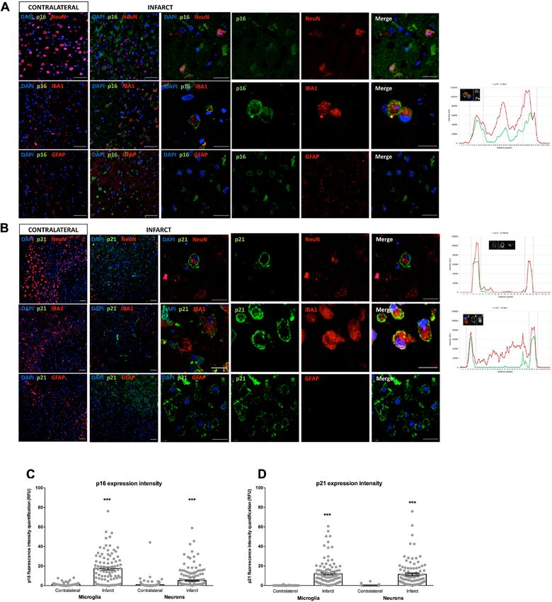
www.nature.com/scientificreports/
senescence-associated secretory phenotype (SASP) and it occurs in different cell lines and in neurodegenerative
diseases like Alzheimer’s disease7, Parkinson’s d
isease8 and amyotrophic lateral s clerosis9. SASP factors modulate
the surrounding cellular microenvironment by activating the immune system and also by affecting neighboring
non-senescent cells promoting malignant transformation via paracrine s ignaling10. Activation of senescence is
modulated by two main pathways: the Cdkn2a (p16) and Cdkn1a (p21) signaling cascades, which promote an
irreversible cell cycle arrest and cause changes in cell morphology, chromatin organization and in mitochondrial
and lysosomal function11,12. However, whether SASP factors contribute to ischemic brain injury within the insult
area or the cellular phenotype in this biological context is still unknown.
Here, we have investigated the expression of senescence markers in the brain of a preclinical mouse model of
ischemic stroke by mRNA gene quantification and immunohistological approach to determine whether cellular
stress in stroke is associated with activation of senescence pathways in the brain, specifically determining neu-
ronal and glial expression for cellular senescence-associated proteins, and further describe them in post-mortem
brain tissue of ischemic stroke donors.
Experimental procedures
Murine stroke model of focal ischemia. Transient middle cerebral artery occlusion (tMCAO) surgi-
cal procedure was previously described and detailed surgical procedure is included in the section Extended
Methods13. Briefly, a glass micropipette was used to compress the MCA for 60 min in CD1 male mice of 12 weeks
of age (n = 11). The compression was applied to achieve a steady decrease in regional cerebral blood flow (rCBF)
below 75–80% of baseline. After recanalization, animals whose rCBF did not reach to 80% of the baseline were
excluded. tMCAO animals were sacrificed 30 min and 72 h after surgery. The animal experiments followed
ARRIVE guidelines14. Cervical dislocation was the method performed to euthanize mice before extracting the
brain for the gene expression analysis.
Mice under anesthetic agent (ketamine/xylazine) were euthanized by exsanguination/cardiac perfusion to
obtain perfused brain tissue and perform immunohistochemical characterization.
All procedures conformed to the National Guidelines on Animal Protection and were approved by the Animal
Ethics Committee of Universitat de Lleida (CEEA 02-07/17).
Human samples. Paraffined human brain sections from two ischemic stroke patients coming from the
“Banco de Cerebros” of biobank HUB-ICO-IDIBELL (registration number: B.0000609) were analyzed. The
biobank HUB-ICO-IDIBELL follows the Comite de Ética de la Investigación (CEIC) del Hospital Univesitari
de Bellvitge (Barcelona). Written informed consent was obtained before autopsy. All procedures followed the
guidelines of the Declaration of Helsinki developed by the World Medical Association (WMA) regarding ethical
principles for medical research involving human subjects. Patient #1 was 79 years old male with a left middle
cerebral artery infarct and mortality was due to cirrhosis decompensation. Patient #2 was 47 years old male with
a lacunar right Globus pallidus infarct and mortality was due to sepsis. Both patients had a stroke more than
15 days before death.
Gene expression of SASP and cell cycle markers. Brain samples from tMCAO mice (n = 3 for 30 min;
n = 5 for 72 h) were dissected on core, ipsilateral (the area adjacent to the core) and contralateral area. On those
three areas, RNA was isolated according to detailed protocol described on Extended Methods. Then cDNA was
synthesized with random hexamer primers using the High-Capacity cDNA Reverse Transcription kit (Applied
Biosystems, Thermo Fisher Scientific, Spain). Principal transcriptional components of senescence (p16 and p21),
SASP (Il6, Cxcl1, Tnfα, Tgfb2, Cxcr2) and cell cyle (Cdk4) were analyzed. Real time quantitative polymerase
chain reaction (RT-qPCR) experiments were performed using a CFX96 instrument (Bio-Rad, Hercules, Cali-
fornia, USA) with SYBR Master Mix (Thermo Fisher Scientific, #4472908). Primers sequences are included in
Table S1. All samples were run in duplicates. 2 ΔΔCt method was used to quantify mRNA levels of expression and
fold changes were calculated relative to Actb gene as an endogenous reference gene.
Immunohistochemical characterization of p16 and p21. Animals (n = 3) were transcardially per-
fused with 4% paraformaldehyde (PFA) 72 h after surgery and brain frozen coronal sections obtained (16 μm).
Immunohistochemistry was performed against mouse anti-p16 (1:200; Abcam, Cat. No. ab54210), mouse anti-
p21 (1:200; Santa Cruz Biotechnology, Cat. No. sc-817), rabbit anti-GFAP (1:200; Abcam, Cat. No. ab7260), rab-
bit anti-Iba1 (1:200; FUJIFILM Wako, Cat. No. 019-19741), rabbit anti-NeuN (1:200; Abcam, Cat.No.ab177487),
rabbit anti-NF-κB p65 (1:200; Proteintech, Cat. No. 10745-1-AP), chicken anti-Iba1 (1:200; Synaptic Systems,
Cat No. 234 006), and rabbit gamma H2AX [p Ser139] antibody (EP854(2)Y, 1:200; NOVUS Biologicals, Cat.
No. NB100-79967) primary antibodies and followed by appropriated secondary antibodies. Samples were cap-
tured using a laser scanning confocal microscope (Olympus FV1000). Adequate controls (secondary antibody
only) gave no specific signals. Images were analyzed in a double-blinded fashion. NF-κB colocalization with
Iba1-positive cells and cellular expression of individual p16 and p21-positive cells were determined with Fiji
software15. Quantification of protein intensity was performed by CellProfiler s oftware16.
Statistical analysis. Data were analyzed using the Statistical Package for the Social Sciences (version
21.0, SPSS Inc.). mRNA expression studies (cellular senescence and cell cycle markers) and protein expression
intensity were defined by either one‐way ANOVA or paired Student’s t‐test as appropriate. Data are expressed
as means ± s.e.m. A p value of < 0.05 was considered statistically significant. Data was plotted using GraphPad
Prism 5.04 (GraphPad Software, Inc., La Jolla, CA, USA) and MetaboAnalyst17.
Scientific Reports | (2021) 11:15752 | https://doi.org/10.1038/s41598-021-95344-5 2
Vol:.(1234567890)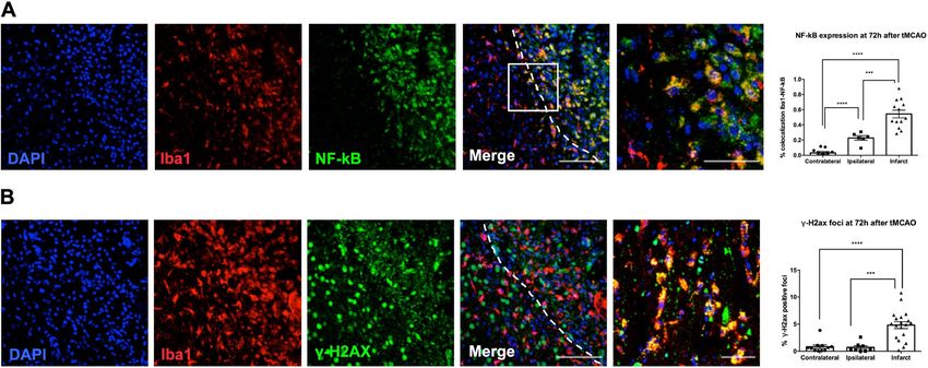
www.nature.com/scientificreports/

                                  Figure 1.  mRNA levels of a set of SASP and inflammatory markers. mRNA levels of a set of cellular senescence
                                  markers (fold change over contralateral) are increased and correlated in infarct core at 72 h after tMCAO.
                                  (A) qRT-PCR analyses of p16, p21, Il6, Tnfa, Tgfb2, Cxcl1, Cxcr2 and Cdk4 comparing infarct, ipsilateral and
                                  contralateral regions of MCAO mouse model at 30 min after tMCAO. (B) qRT-PCR analyses of p16, p21,
                                  Il6, Tnfa, Tgfb2, Cxcl1, Cxcr2 and Cdk4 comparing infarct, ipsilateral and contralateral regions of MCAO
                                  mouse model at 72 h after tMCAO. (C) Heatmap of analyzed regions, average sample expression. (D) Variable
                                  importance in projection (VIP) plot.

                                  Ethics approval. Human tissue was obtained from biobank HUB-ICO-IDIBELL (registration number:
                                  B.0000609). All procedures performed in studies involving animals were in accordance with the ethical stand-
                                  ards of the animal Ethics Committee of Universitat de Lleida (CEEA-020220).

                                  Results
                                  mRNA expression of SASP and cell cycle markers in tMCAO mouse model.                               At 30 min post-
                                  surgery, there was an increase of p16, p21, Il6, tnfa, Cxcl1 and Cdk4 on the ipsilateral region compared to the
                                  contralateral but no significant differences were found. Only the chemokine receptor Cxcr2 was significantly
                                  increased on the ipsilateral region. The expression of Tnfa mRNA was higher on the infarct area, nor differences
                                  did not reach statistical significance (Fig. 1A).
                                      The p16, p21, Cxcl1 and Cxcr2 mRNA expression increased significantly on the infarct area of the cerebrum
                                  at 72 h after injury, whereas the other transcriptional components were not differently expressed on the analyzed
                                  regions (6.09-fold increase for p16, 4.63-fold increase for p21, 4.07-fold increase for Cxcl1 and 3.65-fold increase
                                  for Cxcr2 over contralateral; p < 0.05) (Fig. 1B). The Il6, Tnfa, Tgfb2 and Cdk4, showed a trend to increase in the
                                  infarct area but differences did not reach statistical significance. Interestingly, the contralateral area showed a
                                  similar expression pattern for all genes and the ipsilateral and infarct areas showed a clear clustering expression.
                                  The Il6 and Tnfa correlate positively on the ipsilateral area whereas p16, p21, Cxcl, Cxcr2 and Tgfb2 correlate
                                  positively on the infarct area. The expression profile of these components clustered the different regions as it
                                  was shown in the heatmap at 72 h after tMCAO (Fig. 1C). VIP score analysis (> 1) identified Il6, Tnfa and Tgfb2
                                  genes that most contributed to the variation of the model at 72 h after tMCAO (Fig. 1D).

                                  p16, p21, p65 NF‑κB and γH2AX cellular distribution in tMCAO mouse model.                       To determine
                                  whether senescence markers (p16 and p21) are expressed in specific cell types, antibodies recognizing neurons
                                  (NeuN marker), microglia (Iba1 marker) and astroglia (GFAP marker) were used. The p16 and p21 positive cells
                                  were found in the infarct area among neurons and microglia at 72 h after tMCAO injury. However, p16 and p21

Scientific Reports |   (2021) 11:15752 |               https://doi.org/10.1038/s41598-021-95344-5                                                   3

                                                                                                                                               Vol.:(0123456789)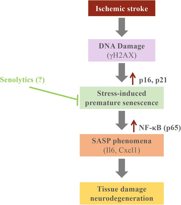
www.nature.com/scientificreports/
were not expressed in the contralateral area of the brain. Strikingly, the expression of p16 and p21 positive cells
in neurons and microglia showed a cytoplasmic and membranous pattern (Fig. 2A,B and S1).
The subcellular intensity localization of p16 and p21 was analyzed in individual neurons and microglia.
A higher p16 and p21 expression in the infarct area was observed, and p16 showed a granular cytoplasmatic
expression in microglia and neurons, while p21 showed a membrane expression in neurons. The total expression
intensity of p16 (Fig. 2C) and p21 (Fig. 2D) in those cells was higher in the infarct area (p16 microglia: p < 0.0001;
p16 neurons: p < 0.0001; p21 microglia: p < 0.0001; p21 neurons: p < 0.0001).
To determine the activation of an important pro-inflammatory pathway, related to SASP and neuroinflamma-
tion, we analyzed the expression of p65 NF-κB in microglia. An increase in the correlation coefficient (Rcoloc)
of this marker and Iba1 (microglia) was observed in infarct and ipsilateral area (Fig. 3A). To analyze upstream
pathways elements of stress-induced senescence, we determined the expression of DNA reparation foci (γH2AX
marker) in microglia. This marker was significantly increased in infarct area and remains unchanged between
contralateral and ipsilateral area (Fig. 3B).
Senescence‑associated‑β‑galactosidase (SA‑β‑gal) staining in tMCAO mouse model. SA-β-
gal activity, a common marker of cellular senescence, was used in tMCAO mice tissue brains to identify senes-
cent cells at 72 h after surgery. No SA-β-gal positive cells were observed in the infarct area nor in the contralateral
area of each brain (Fig. S2).
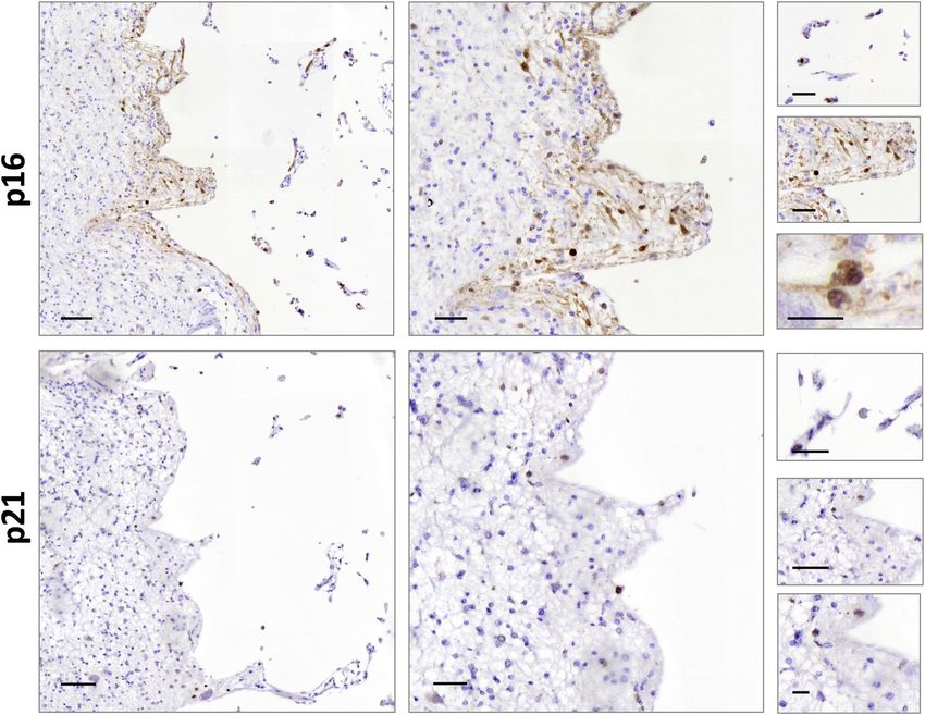
P16 and P21 immunohistochemistry in post‑mortem human stroke samples. As cellular senes-
cence-associated secretory phenotype was triggered by an ischemic event in a preclinical model, we investigated
whether this phenotype would also be observed in human brains. Post-mortem brain samples of stroke donor
with a left middle cerebral artery infarct were analyzed. Immunohistochemistry against P16 and P21 was per-
formed and the adjacent area of the ischemic core showed a positive immunoreactivity for P16 and P21, with a
nuclear and cytoplasmic expression pattern and extracellular aggregated, respectively. A higher number of P16
positive cells were observed in the perimeter of the infarct area (60.32% P16 positive cells in the perimeter of
the infarct area vs 7.79% in the rest of the section) (Fig. 4). Samples of a second stroke donor with globus pal-
lidus infarct were also analyzed, but P16 and P21 immunoreactivity was weaker within the infarct area (Fig. S3).
Discussion
Recently, a concept which connects cellular senescence and age-related tissue dysfunction has emerged18. Indeed,
in the central nervous system, involvement of senescence in neurons, astrocytes and microglia is increasingly rec-
ognized in the development of brain aging and/or age-related neurological disorders19. However, whether cellular
senescence impacts ischemic stroke has not been well established. In this study, we have described for the first
time the SASP phenomenon in ischemic stroke. We have showed that the gene expression of senescence mark-
ers (p16 and p21) and pro-inflammatory cytokines (Il6, Cxcl1, Tnfa and Cxcr2) in the brain of tMCAO mouse
is restricted to the dissected infarct area 72 h after the ischemic insult. Moreover, p16 and p21 are expressed in
neurons and microglial cells within the infarct area with both cytoplasmic and membranous expression pattern
in tMCAO mouse brain, in contrast with the expected nuclear distribution. Finally, an increase of P16 in the
perimeter of the infarct area was observed in post-mortem human brain tissue of ischemic stroke donor.
Senescence and SASP has been described in several age-related pathologies20. However, it has not been
described in ischemic stroke. Our results are compatible with SASP transcriptional profile, characterized by an
increase of cell cycle inhibitors (p16 and p21) and pro-inflammatory c ytokines21. Although previous authors
have described a neuroinflammatory profile in ischemic stroke, our novelty is the description of stress-induced
senescence triggering a neuroinflammatory profile, which is compatible with SASP phenomenon. These changes
related to gene synthesis are restricted to ischemic core, where cytokines are synthesized and probably diffused
across the tissue in a paracrine manner, and those results are not related with cell cycle roles of the genes. The role
of p16 and p21 expression in senescence and inflammatory process is controversial. While both p16 and p21 are
described as the main inducers of senescence22 and therefore participating in SASP and chronic inflammation6,
some authors demonstrate the anti-inflammatory and pro-resolution role of p16 and p21 in a complex interaction
with CDK and NF-κB pathway (reviewed in23), which is increased concomitantly with p16 and p21 in microglial
cells in our mouse model. Moreover, a high DNA reparation focus in infarct is compatible with the upregula-
tion of p21 expression within DNA Damage Response. This regulation is important to maintain the survival of
senescent cells under stress conditions15. In contrast to canonical senescence associated with aging, which implies
a long period of time and the persistence of chronic inflammation, p16 and p21 pro-resolution effects are seen
in acute stress models. This may reflect an important role of the time regulating the functions of p16 and p21 in
senescence and inflammation and could have implications in ischemic stroke. In transient forebrain ischemia,
elevated p21 might be a response to the ischemic insult to protect the neurons by arresting them in first steps of
the cell cycle. The p21 expression is enhanced in surviving neurons surrounding dead a rea24. Protein subcellular
location is also relevant for p16 function. The cytoplasmic immunoreactivity of p16 appears to be an unfavorable
prognostic indicator in high-grade astrocytoma p atients25. Moreover, expression of P16 and P21 in glia suggests
senescence induction, but neuronal p21 expression might reflect a more general mechanism of age‐related cell
cycle dysregulation in the frontal association cortex of amyotrophic lateral sclerosis patients26. Several cell cycle
independent functions of p16 and p21 have been described. Interestingly, p16 membrane localization regulates
cytoskeleton in endothelial c ells27 and its depletion causes defects in motility, morphogenesis and cytoskeletal
organization28. Herein, microglia migration and phagocytosis could be regulated by similar mechanism after
ischemic insult.
Scientific Reports | (2021) 11:15752 | https://doi.org/10.1038/s41598-021-95344-5 4
Vol:.(1234567890)
www.nature.com/scientificreports/
Figure 2. p16 and p21 subcellular location in a preclinical animal model of stroke. (A) Double
immunofluorescence for co-localization of p16 (green), NeuN/IBA1/GFAP (red) and DAPI (blue). Intensity
expression plot showed the cellular distribution of p16 in microglia. (B) Double immunofluorescence for
co-localization of p21 (green), NeuN/IBA1/GFAP (red) and DAPI (blue). p16 and p21 immunostained cells
could be observed in the infarct area at 3 days after tMCAO. Intensity expression plot showed the cellular
distribution of p21 in microglia and neurons. (C) p16 expression intensity in microglia and neurons on
contralateral and infarct areas. (D) p21 expression intensity in microglia and neurons on contralateral and
infarct areas. Scale bar contralateral and infarct = 50 µm. Scale bar infarct zoom = 20 µm.
A recent study shows a similar SASP transcriptional profile in trauma brain injury (TBI) mouse m odel29. In
contrast with our results, it has observed an increase in SA-β-gal activity which increases at 4, 7- and 14-days
post-stroke. This would explain the lack of SA-β-gal activity in our model (3 days after ischemic stroke) due to
a shorter post-ischemic time window.
Scientific Reports | (2021) 11:15752 | https://doi.org/10.1038/s41598-021-95344-5 5
Vol.:(0123456789)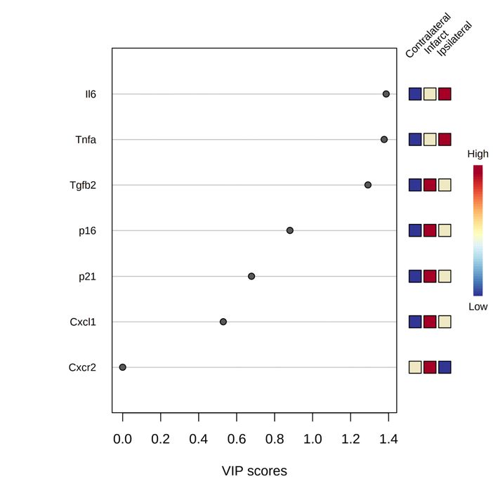
www.nature.com/scientificreports/

                                            Figure 3.  Microglial cells of the ischemic area showed a higher expression of NF-kB and γ-H2AX. (A)
                                            Immunofluorescence NF-kB expression showed an infarct area location, delimited by white dotted line. Plot
                                            represented the NF-kB expression by areas (contralateral vs infarct area: p < 0.0001; ipsilateral vs infarct:
                                            p = 0.0009; contralateral vs ipsilateral: p < 0.0001). Scale bar = 100 µm; insert scale bar = 40 µm. (B) Nuclear
                                            expression of γ-H2AX in microglial cells. Plot represented the γ-H2AX positive foci in contralateral, ipsilateral
                                            and infarct areas (contralateral vs infarct: p < 0.0001; ipsilateral vs infarct: p = 0.0004). Scale bar = 100 µm; insert
                                            scale bar = 50 µm.

                                            Figure 4.  P16 and P21 subcellular location in MCA ischemic stroke patient’s brain tissue. Scale bar left
                                            images = 100 µm, middle images = 50 µm and right images = 20 µm.

          Scientific Reports |   (2021) 11:15752 |                https://doi.org/10.1038/s41598-021-95344-5                                                           6

Vol:.(1234567890)
www.nature.com/scientificreports/
Figure 5. Graphical summary of the results.
The SASP hypothesis, in which the senescent cells that accumulate in body tissues over time contribute to
inflammation progression and functional decay in the elderly has recently been p roposed30. However, how is
it related to stroke progression remains unclear. Ischemic stroke is characterized by the disruption of cerebral
blood flow, which produces a central core of dead neurons surrounded by a penumbra of damaged but partially
functional neurons. As we have observed, during the first 30 min after ischemic stroke, cellular senescent markers
were increased in the penumbra area and over time were higher in the ischemic core. This would be related with
stroke progression. If an ischemic event would accelerate the SASP phenomena in a cellular environment, that
would be related with the hypoxia as an apoptotic mechanism contributing to the senescence phenotype and it
would also be related with further neurodegeneration of alive cells and selective loss of neurological function.
We have demonstrated the role of P16 in human MCA stroke but not in lacunar stroke. This could be explained
by the notorious differences in the pathology of lacunar and non-lacunar s trokes1.
As for the limitations of the present work, we are aware that a longer period of time after tMCAO would have
shown different phenomena and that the number of analyzed human samples is minimal. We should also consider
that age constraints could play a role in this pathway, as the mice evaluated are young. Further, experimental
senolytic treatments, such as quercetin and navitoclax, among others, it could have led to functional improve-
ment. The evaluation of its translational impact, and the study of ageing phenomena, it is envisaged in the near
future. However, doubts on whether these treatments should be given in a preventive scheme (i.e. anticipating
the hypoxic environment) or rather, they should be administered in a more clinically amenable manner (i.e.
short after the insult) are present and both experimental designs would be beneficial.
In conclusion, there is an increase of senescence markers highly correlated with SASP cytokines restricted
to the ischemic core in the tMCAO preclinical mouse model (Fig. 5). Furthermore, there is an increase of P16
positive cells surrounding the ischemic core in a human case of MCA stroke. These results would open new thera-
peutic opportunities for the emerging senolytic drugs, which selectively kill senescence cells. Further research
would contribute to define the cellular phenotype of SASP in ischemic stroke and new therapeutic approaches
would be determine for ischemic stroke patients.
Data availability
Requests for access to the data reported in this paper will be considered by the Lead contact.
Received: 8 February 2021; Accepted: 19 July 2021
References
1. Campbell, B. C. V. et al. Ischaemic stroke. Nat. Rev. Dis. Primers 5, 70. https://doi.org/10.1038/s41572-019-0118-8 (2019).
2. Iadecola, C. & Anrather, J. Stroke research at a crossroad: Asking the brain for directions. Nat. Neurosci. 14, 1363–1368. https://
doi.org/10.1038/nn.2953 (2011).
3. Purroy, F. et al. Age- and sex-specific risk profiles and in-hospital mortality in 13,932 Spanish stroke patients. Cerebrovasc. Dis.
https://doi.org/10.1159/000500205 (2019).
Scientific Reports | (2021) 11:15752 | https://doi.org/10.1038/s41598-021-95344-5 7
Vol.:(0123456789)www.nature.com/scientificreports/
4. Vena, A. B. et al. Clinical evolution of elderly adults with ischemic stroke. J. Am. Geriatr. Soc. 64, 2167–2170. https://doi.org/10.
1111/jgs.14426 (2016).
5. Chien, Y. et al. Control of the senescence-associated secretory phenotype by NF-kappaB promotes senescence and enhances
chemosensitivity. Genes Dev. 25, 2125–2136. https://doi.org/10.1101/gad.17276711 (2011).
6. Coppé, J. P. et al. Senescence-associated secretory phenotypes reveal cell-nonautonomous functions of oncogenic RAS and the
p53 tumor suppressor. PLoS Biol. 6, 2853–2868. https://doi.org/10.1371/journal.pbio.0060301 (2008).
7. Musi, N. et al. Tau protein aggregation is associated with cellular senescence in the brain. Aging Cell 17, e12840. https://doi.org/
10.1111/acel.12840 (2018).
8. Chinta, S. J. et al. Cellular senescence is induced by the environmental neurotoxin paraquat and contributes to neuropathology
linked to Parkinson’s disease. Cell Rep. 22, 930–940. https://doi.org/10.1016/j.celrep.2017.12.092 (2018).
9. Vazquez-Villaseñor, I. et al. Expression of p16 and p21 in the frontal association cortex of ALS/MND brains suggests neuronal
cell cycle dysregulation and astrocyte senescence in early stages of the disease. Neuropathol. Appl. Neurobiol. https://doi.org/10.
1111/nan.12559 (2019).
10. Sedaghat, Z., Kadkhodaee, M., Seifi, B. & Ahghari, P. Hepatoprotective effects of remote perconditioning during renal ischemia.
Bratisl. Lek. Listy 115, 675–679 (2014).
11. Coppe, J. P. et al. Tumor suppressor and aging biomarker p16(INK4a) induces cellular senescence without the associated inflam-
matory secretory phenotype. J. Biol. Chem. 286, 36396–36403. https://doi.org/10.1074/jbc.M111.257071 (2011).
12. Fitzgerald, A. L. et al. Reactive oxygen species and p21Waf1/Cip1 are both essential for p53-mediated senescence of head and neck
cancer cells. Cell Death Dis. 6, e1678. https://doi.org/10.1038/cddis.2015.44 (2015).
13. Cambray, S. et al. Metabolomic estimation of the diagnosis and onset time of permanent and transient cerebral ischemia. Mol.
Neurobiol. 55, 6193–6200. https://doi.org/10.1007/s12035-017-0827-5 (2018).
14. Percie du Sert, N. et al. The ARRIVE guidelines 2.0: Updated guidelines for reporting animal research. BMC Vet. Res. 16, 242.
https://doi.org/10.1186/s12917-020-02451-y (2020).
15. Schindelin, J. et al. Fiji: An open-source platform for biological-image analysis. Nat. Methods 9, 676–682. https://doi.org/10.1038/
nmeth.2019 (2012).
16. McQuin, C. et al. Cell Profiler 3.0: Next-generation image processing for biology. PLoS Biol. 16, e2005970. https://d oi.o
rg/1 0.1 371/
journal.pbio.2005970 (2018).
17. Pang, Z., Chong, J., Li, S. & Xia, J. MetaboAnalystR 3.0: Toward an optimized workflow for global metabolomics. Metabolites
https://doi.org/10.3390/metabo10050186 (2020).
18. LeBrasseur, N. K., Tchkonia, T. & Kirkland, J. L. Cellular senescence and the biology of aging, disease, and frailty. Nestle Nutr. Inst.
Workshop Ser. 83, 11–18. https://doi.org/10.1159/000382054 (2015).
19. Yamazaki, Y. et al. Vascular cell senescence contributes to blood-brain barrier breakdown. Stroke 47, 1068–1077. https://doi.org/
10.1161/STROKEAHA.115.010835 (2016).
20. Childs, B. G., Durik, M., Baker, D. J. & van Deursen, J. M. Cellular senescence in aging and age-related disease: From mechanisms
to therapy. Nat. Med. 21, 1424–1435. https://doi.org/10.1038/nm.4000 (2015).
21. Herranz, N. & Gil, J. Mechanisms and functions of cellular senescence. J. Clin. Investig. 128, 1238–1246. https://doi.org/10.1172/
JCI95148 (2018).
22. Serrano, M., Lin, A. W., McCurrach, M. E., Beach, D. & Lowe, S. W. Oncogenic ras provokes premature cell senescence associated
with accumulation of p53 and p16INK4a. Cell 88, 593–602. https://doi.org/10.1016/s0092-8674(00)81902-9 (1997).
23. Schmitz, M. L. & Kracht, M. Cyclin-dependent kinases as coregulators of inflammatory gene expression. Trends Pharmacol. Sci.
37, 101–113. https://doi.org/10.1016/j.tips.2015.10.004 (2016).
24. Schmidt-Kastner, R. et al. Differential changes of bax, caspase-3 and p21 mRNA expression after transient focal brain ischemia in
the rat. Brain Res. Mol. Brain Res. 79, 88–101. https://doi.org/10.1016/s0169-328x(00)00104-2 (2000).
25. Arifin, M. T. et al. Cytoplasmic, but not nuclear, p16 expression may signal poor prognosis in high-grade astrocytomas. J. Neu-
rooncol. 77, 273–277. https://doi.org/10.1007/s11060-005-9037-5 (2006).
26. Vazquez-Villasenor, I. et al. Expression of p16 and p21 in the frontal association cortex of ALS/MND brains suggests neuronal
cell cycle dysregulation and astrocyte senescence in early stages of the disease. Neuropathol. Appl. Neurobiol. 46, 171–185. https://
doi.org/10.1111/nan.12559 (2020).
27. Alhaja, E. et al. Anti-migratory and anti-angiogenic effect of p16: A novel localization at membrane ruffles and lamellipodia in
endothelial cells. Angiogenesis 7, 323–333. https://doi.org/10.1007/s10456-005-0368-9 (2004).
28. Kan, C. Y. et al. Endothelial cell dysfunction and cytoskeletal changes associated with repression of p16(INK4a) during immor-
talization. Oncogene 31, 4815–4827. https://doi.org/10.1038/onc.2011.645 (2012).
29. Tominaga, T., Shimada, R., Okada, Y., Kawamata, T. & Kibayashi, K. Senescence-associated-β-galactosidase staining following
traumatic brain injury in the mouse cerebrum. PLoS ONE 14, e0213673. https://doi.org/10.1371/journal.pone.0213673 (2019).
30. van Deursen, J. M. The role of senescent cells in ageing. Nature 509, 439–446. https://doi.org/10.1038/nature13193 (2014).
Acknowledgements
We are very grateful to Dr. M. Encinas (IRBLleida-UdL) for Tnfa, Cxcl1 and Tgfb2 antibodies, to Dr. M. Santacana
from Immunohistochemistry Core Facility at IRBLleida for her help and to all members of Clinical Neuroscience
group at IRBLleida for scientific discussions and instrumental help.
Author contributions
P.T., M.P.O. conceived the study. C.T.-Q., P.T., G.A. designed experiments. C.T.-Q., P.T., G.A. sample processing,
data analysis and data interpretation. C.T.-Q., G.A., F.P. draft the manuscript. M.P.O. and F.P. procured funding.
All authors critically revised and approved the final version of the manuscript.
Funding
This study was supported by the Government of Catalonia-Agència de Gestió d’Ajuts Universitaris i de Recerca
(FP: 2017 SGR 1628), Instituto de Salud Carlos III and co-funded by European Union (ERDF/ESF, “Investing
in your future”) (FP: Project PI17-01725; MP-O: PI17-00134 and PI20-0155) and the INVICTUS plus Research
Network (Carlos III Health Institute). CT-Q was supported by a grant from Contratos predoctorales de formación
en investigación en salud (PFIS; FI18/00319). PT was supported by a grant from Ministerio de Ciencia, Inno-
vación y Universidades (FPU16/01446). This study has been co-financed by FEDER funds from the European
Union ("A way to build Europe").
Competing interests
The authors declare no competing interests.
Scientific Reports | (2021) 11:15752 | https://doi.org/10.1038/s41598-021-95344-5 8
Vol:.(1234567890)www.nature.com/scientificreports/
Additional information
Supplementary Information The online version contains supplementary material available at https://doi.org/
10.1038/s41598-021-95344-5.
Correspondence and requests for materials should be addressed to F.P.
Reprints and permissions information is available at www.nature.com/reprints.
Publisher’s note Springer Nature remains neutral with regard to jurisdictional claims in published maps and
institutional affiliations.
Open Access This article is licensed under a Creative Commons Attribution 4.0 International
License, which permits use, sharing, adaptation, distribution and reproduction in any medium or
format, as long as you give appropriate credit to the original author(s) and the source, provide a link to the
Creative Commons licence, and indicate if changes were made. The images or other third party material in this
article are included in the article’s Creative Commons licence, unless indicated otherwise in a credit line to the
material. If material is not included in the article’s Creative Commons licence and your intended use is not
permitted by statutory regulation or exceeds the permitted use, you will need to obtain permission directly from
the copyright holder. To view a copy of this licence, visit http://creativecommons.org/licenses/by/4.0/.
© The Author(s) 2021
Scientific Reports | (2021) 11:15752 | https://doi.org/10.1038/s41598-021-95344-5 9
Vol.:(0123456789)You can also read