Acanthamoeba Keratitis: A Single-Institution Series of Four Cases With Literature Review - Cureus
←
→
Page content transcription
If your browser does not render page correctly, please read the page content below
Open Access Case
                               Report                                                 DOI: 10.7759/cureus.21112

                                                Acanthamoeba Keratitis: A Single-Institution
                                                Series of Four Cases With Literature Review
Review began 12/17/2021
                                                Clarissa Smith 1 , Nida Ashraf 2 , Megan Haghnegahdar 3 , Kenneth Goins 3 , Jessica R. Newman 2
Review ended 01/04/2022
Published 01/11/2022                            1. Department of Internal Medicine, University of Kansas Medical Center, Kansas City, USA 2. Department of Internal
© Copyright 2022                                Medicine, Division of Infectious Diseases, University of Kansas Medical Center, Kansas City, USA 3. Department of
Smith et al. This is an open access article     Ophthalmology, University of Kansas Medical Center, Kansas City, USA
distributed under the terms of the Creative
Commons Attribution License CC-BY 4.0.,
                                                Corresponding author: Jessica R. Newman, jnewman@kumc.edu
which permits unrestricted use, distribution,
and reproduction in any medium, provided
the original author and source are credited.

                                                Abstract
                                                Acanthamoeba species are free-living protozoa found pervasively in water and soil, which can cause
                                                infections of the central nervous system, skin, and eye. Amoebic keratitis (AK) is a vision-threatening, often
                                                chronic infection that is associated with the use of soft contact lenses due to corneal microtrauma and
                                                improper cleaning and storage. Although AK infections are rare, they cause significant morbidity including
                                                vision loss due to the diagnostic and therapeutic challenges they pose.

                                                The clinical course is determined by the organism’s inherent pathogenicity, delay of diagnosis, and the
                                                paucity of data on effective therapeutic regimens. The case series and review of literature that follows
                                                examine current latest best practices in AK diagnosis including in vivo confocal microscopy (IVCM) and
                                                therapeutic interventions including miltefosine.

                                                Categories: Ophthalmology, Infectious Disease, Therapeutics
                                                Keywords: corrective contact lens, confocal microscopy, miltefosine, protozoal keratitis, acanthamoeba keratitis

                                                Introduction
                                                Acanthamoeba species are free-living protozoa found ubiquitously in soil and water and are implicated in
                                                serious infections of the central nervous system, skin, and eye [1]. Their life cycle is notable for being
                                                biphasic, with a trophozoite form capable of attaching to host epithelium and invading to deeper structures,
                                                as well as a cystic form that can endure adverse conditions including treatment attempts [1]. Human studies
                                                of serum antibodies to Acanthamoeba show that most individuals have been exposed to the organism;
                                                however, clinically significant infections are of relatively low incidence and frequently affect contact lens
                                                users and the immunocompromised [2,3].

                                                Amoebic keratitis (AK) is a progressive, vision-threatening infection that is strongly associated with soft
                                                contact lenses due to contamination related to improper cleaning and storage with infested tap water.
                                                Additionally, corneal microtrauma and epithelial changes are known to occur with chronic contact use,
                                                predisposing the user to infection [4]. It is common for these infections to be chronic in nature due to the
                                                durability of the cystic form and the possibility for reinfection, which is at least partially attributed to the
                                                immunologically privileged nature of this anatomic site. Treatment is challenging due to the diagnostic
                                                delay related to initial misdiagnosis, nonspecific presentation of the disease, and the need for specialized
                                                diagnostic measures. Additionally, Acanthamoeba infection is oftentimes polymicrobial, including
                                                coinfections with fungi and bacteria. Without treatment of coinfections, a symbiotic relationship between
                                                the two organisms persists, causing continued damage to the cornea despite treatment of the Acanthamoeba.
                                                Untreated and suboptimally treated infections have been observed to spread contiguously into the deeper
                                                structures of the eye and central nervous system manifesting as granulomatous amoebic encephalitis [5].
                                                While AK infections are relatively uncommon, they are associated with significant morbidity and vision loss
                                                due to the diagnostic and therapeutic challenges they pose.

                                                The series of cases that follow highlight common presenting features, discuss diagnostic assays, and report
                                                on experiences of multidrug oral treatment strategies. Rapid referral to an ophthalmologist trained in
                                                confocal microscopy can be key to making an early diagnosis of AK. If topical treatments fail, systemic
                                                therapies including miltefosine, a pharmacologic agent with recent approval for refractory AK, and
                                                voriconazole may be of benefit.

                                                Case Presentation
                                                Case 1
                                                A previously healthy 38-year-old female was referred to the ophthalmology clinic for three months of
                                                pruritis and photosensitivity in her left eye associated with intermittent left-sided headaches and decreased
                                                visual acuity. She failed to improve with empiric treatment for herpes simplex virus (HSV) keratitis. Exposure
                                                history was significant for use of corrective soft contact lenses. There was a history of a positive antinuclear

                               How to cite this article
                               Smith C, Ashraf N, Haghnegahdar M, et al. (January 11, 2022) Acanthamoeba Keratitis: A Single-Institution Series of Four Cases With Literature
                               Review. Cureus 14(1): e21112. DOI 10.7759/cureus.21112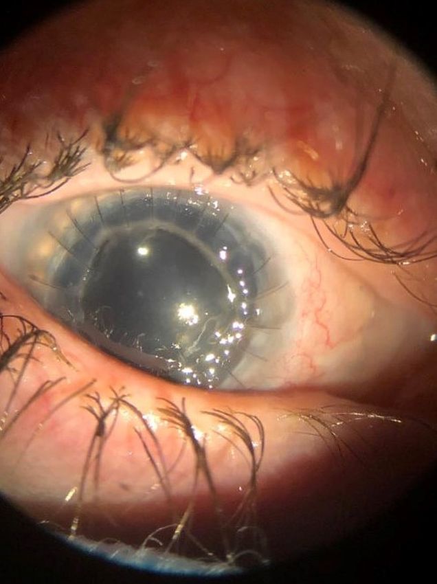
antibody, and oral hydroxychloroquine had been started by her primary physician without improvement.

                                                 Slit-lamp examination demonstrated diffuse corneal haze, central corneal epithelial defect, and diffuse
                                                 conjunctival injection (Figure 1). Confocal microscopy revealed branching hyphae (Figure 2) and multiple
                                                 amoebic cysts (Figure 3). Nucleic acid amplification of corneal scrapings was positive for Acanthamoeba. Her
                                                 condition continued to worsen, and she developed Acanthamoeba scleritis (Figure 4) despite topical
                                                 chlorhexidine, voriconazole, moxifloxacin, and oral valacyclovir and fluconazole; therefore, she was treated
                                                 with a two-week course of IV pentamidine.

                                                    FIGURE 1: Slit-lamp examination of the left eye at presentation with
                                                    diffuse conjunctival injection, central corneal epithelial defect, and
                                                    diffuse corneal haze.

2022 Smith et al. Cureus 14(1): e21112. DOI 10.7759/cureus.21112                                                                                                2 of 20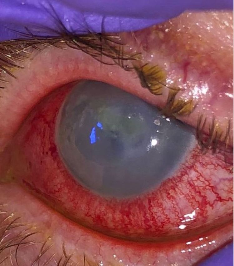
FIGURE 2: Confocal microscopy of the left cornea, 504 microns deep,
                                                    with faint branching figures, consistent with either filamentous fungi or
                                                    chains of bacteria (infectious crystalline keratopathy).

2022 Smith et al. Cureus 14(1): e21112. DOI 10.7759/cureus.21112                                                                3 of 20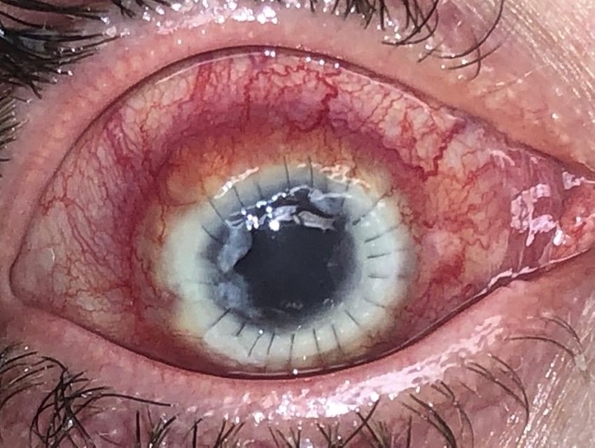
FIGURE 3: Confocal microscopy of the left cornea, 477 microns deep,
                                                    showing multiple double-walled Acanthamoeba cysts.

2022 Smith et al. Cureus 14(1): e21112. DOI 10.7759/cureus.21112                                                          4 of 20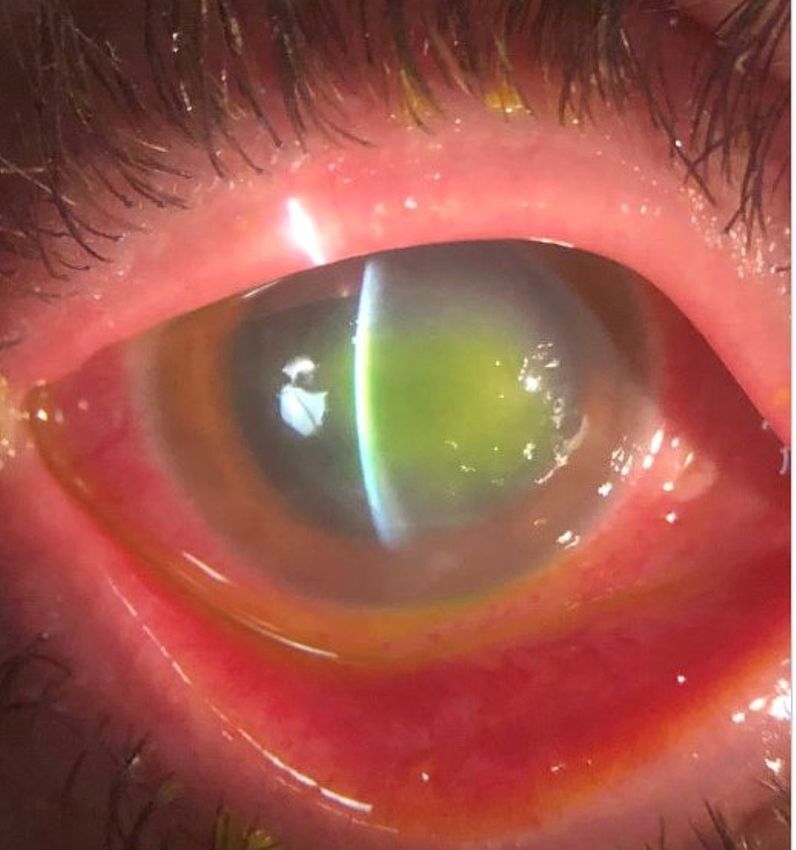
FIGURE 4: Slit-lamp examination of the left eye showing the increasing
                                                    size of stromal infiltrate, anterior chamber hypopyon, and inferior
                                                    scleral nodule at 7:00.

                                                 With further progression of the disease, she underwent penetrating keratoplasty (PKP). Pathology of
                                                 Descemet’s membrane revealed severe inflammation with numerous cysts and possible trophozoites. Post-
                                                 transplantation, an aggressive regimen of oral miltefosine, voriconazole, and
                                                 trimethoprim/sulfamethoxazole was added for six months. The patient continued to improve after surgery,
                                                 tolerated the medications with monitoring, and remained free of disease at three years follow-up (Figure 5).
                                                 Cataract surgery was required to restore best-corrected vision.

2022 Smith et al. Cureus 14(1): e21112. DOI 10.7759/cureus.21112                                                                                                5 of 20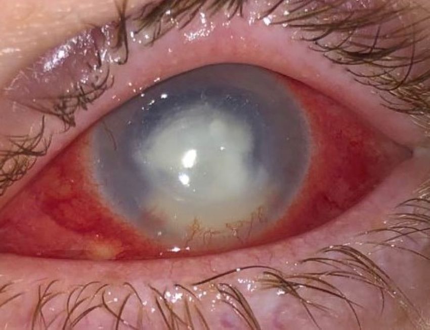
FIGURE 5: External photograph, bridge of both eyes after corneal
                                                    transplantation of the left eye. There is evidence of scleromalacia (blue
                                                    pigmentation of the sclera) in the left eye after resolution of
                                                    Acanthamoeba sclerokeratitis. Iris heterochromia is noted due to loss of
                                                    pigment related to the inflammatory process and following cataract
                                                    surgery. The best-corrected vision of the left eye is 20/30 with
                                                    spectacles.

                                                 Case 2
                                                 A 59-year-old female with an unremarkable past medical history presented to the ophthalmology clinic for
                                                 an evaluation of approximately one month duration of left eye pain and photosensitivity. She had been
                                                 treated previously for presumptive HSV keratitis and corneal abrasion without improvement. Symptoms
                                                 progressed to include pain with extraocular movements, reduced visual acuity, and sinus pain despite
                                                 appropriate topical therapy. Exposure history was significant for use of soft contact lenses, which she
                                                 changed daily and denied wearing to sleep.

                                                 On physical examination, she had a large central epithelial defect, patchy infiltrates, satellite lesions, and a
                                                 hypopyon. There was initial suspicion for fungal keratitis; therefore, she was started on oral voriconazole.
                                                 Topical moxifloxacin was added, and oral acyclovir was continued. A confocal examination was performed
                                                 and demonstrated bright, round double-walled cystic structures, consistent with Acanthamoeba (Figure 6).
                                                 Topical chlorhexidine was then added. Corneal scrapings were found to be positive for Acanthamoeba by
                                                 nucleic acid amplification.

2022 Smith et al. Cureus 14(1): e21112. DOI 10.7759/cureus.21112                                                                                                    6 of 20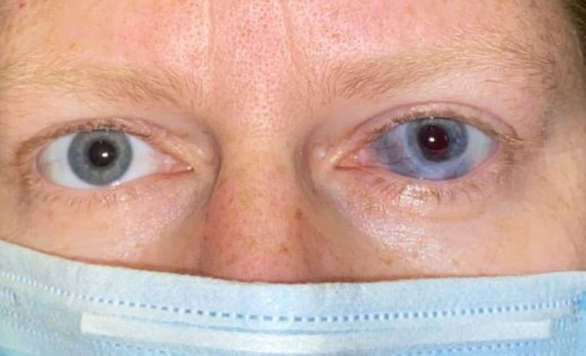
FIGURE 6: Confocal microscopy within the anterior cornea, at the level
                                                    of Bowman’s layer, showing bright, round double-walled cystic
                                                    structures, consistent with Acanthamoeba.

                                                 She continued to worsen despite topical and systemic therapy. Repeat confocal microscopy demonstrated
                                                 persistence of cysts, with a deeper spread into the corneal stroma (Figure 7). The decision was made to
                                                 proceed with deep anterior lamellar keratoplasty (DALK) with anterior chamber washout. Miltefosine was
                                                 added to the regimen for approximately one month prior to this procedure. The corneal tissue sent to
                                                 pathology was negative for Acanthamoeba and fungal elements. She was continued on topical chlorhexidine
                                                 for an additional two weeks and no longer required oral voriconazole or miltefosine postoperatively. The
                                                 patient has remained stable for three years after DALK. Cataract surgery was required to restore best-
                                                 corrected visual acuity.

2022 Smith et al. Cureus 14(1): e21112. DOI 10.7759/cureus.21112                                                                                            7 of 20
FIGURE 7: Confocal microscopy of the affected eye, 267 microns deep,
in the corneal stroma showing multiple double-walled bright figures,
consistent with Acanthamoeba cysts.
Case 3
A 71-year-old female with a past medical history significant for diabetes mellitus type two was referred from
an outside ophthalmology clinic due to concern for worsening Acanthamoeba keratitis confirmed four
months previously by PCR. She had been treated with propamidine isethionate and ofloxacin. She had
sought care initially for an abrupt decline in visual acuity. Notably, she had worn contacts without incident
for the preceding 50 years.
Her examination showed central punctate epithelial erosions with stromal edema and haze. She was started
on topical chlorhexidine and moxifloxacin. Despite treatment, her vision continued to deteriorate, and she
developed eye pain, burning, and tearing. Follow-up confocal microscopic examination demonstrated
numerous Langerhans cells (Figure 8), double-walled cysts (Figure 9), and extensive multilevel scarring. She
continued to worsen despite treatment with topical chlorhexidine, oral voriconazole, and oral acyclovir.
DALK was deemed necessary, and pathology of both anterior and deep corneal stroma demonstrated
Acanthamoeba trophozoites. She was continued on topical chlorhexidine and oral acyclovir postoperatively.
2022 Smith et al. Cureus 14(1): e21112. DOI 10.7759/cureus.21112 8 of 20FIGURE 8: Confocal microscopy at the level between Bowman’s layer
and the basal epithelium, approximately 50–70 microns deep, showing
extensive Langerhans dendritic cell activation.
2022 Smith et al. Cureus 14(1): e21112. DOI 10.7759/cureus.21112 9 of 20FIGURE 9: Confocal microscopy of the affected eye at the level of the
anterior stroma, approximately 100 microns deep, showing multiple
double-walled bright figures (Acanthamoeba cysts) in between
numerous keratocytes.
The patient’s course was complicated by the recurrence of Acanthamoeba keratitis two months after DALK.
New ring infiltrate and hypopyon were noted on examination, and corneal scrapings submitted for nucleic
acid testing redemonstrated Acanthamoeba cysts. The patient was started on oral
trimethoprim/sulfamethoxazole, voriconazole, and miltefosine. She was continued on topical
chlorhexidine, and topical polyhexamethylene biguanide (PHMB) was added. Due to rapid progression,
urgent penetrating keratoplasty was pursued with persistent Acanthamoeba cysts noted on pathology. The
patient had tolerability problems with the miltefosine; therefore, this was discontinued. She was continued
on oral voriconazole, trimethoprim/sulfamethoxazole, and topical PHMB until the disease appeared to be in
remission. She then underwent cataract surgery to restore vision. Unfortunately, she developed corneal
allograft failure necessitating penetrating keratoplasty again. Pathology was negative for protozoa;
therefore, PHMB was discontinued. She currently has a guarded long-term prognosis due to chronic
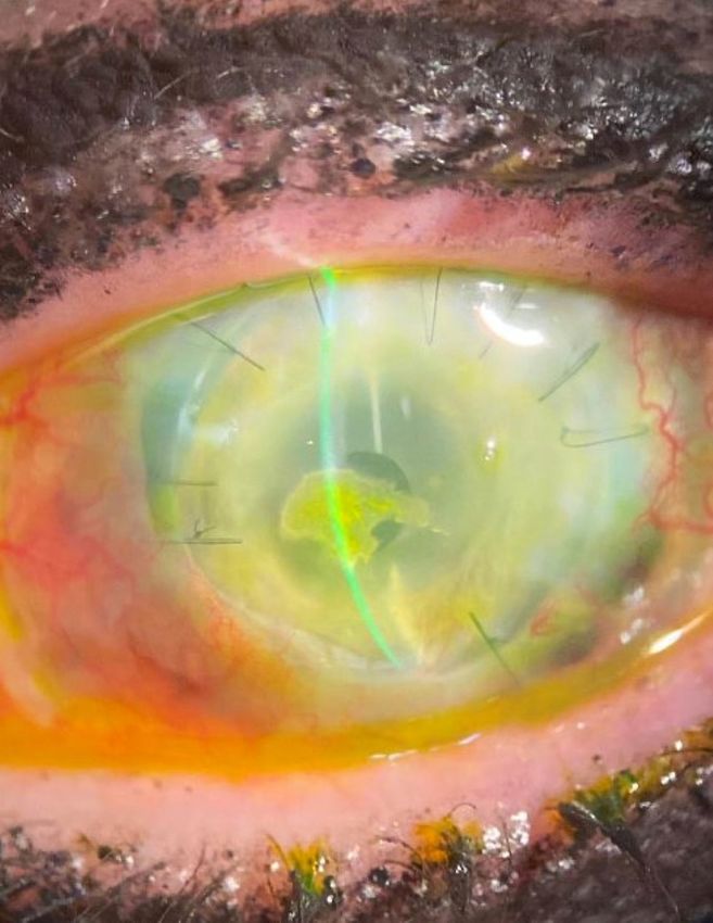
Acanthamoeba keratouveitis secondary glaucoma and neurotrophic keratopathy (Figure 10).
2022 Smith et al. Cureus 14(1): e21112. DOI 10.7759/cureus.21112 10 of 20FIGURE 10: Slit-lamp photograph of left eye after a third corneal
transplantation for Acanthamoeba. There is central calcific band
keratopathy present and a pupillary membrane, both causing a
reduction in vision.
Case 4
A 46-year-old female with a history of eosinophilic esophagitis, positive antinuclear antibody, and
trigeminal neuralgia was referred from an outside ophthalmologist due to approximately five months of
recurrent right eye burning, tearing, vision changes, photophobia, and unilateral headache. Her symptoms
appeared shortly after she began storing her contact lenses in tap water. She was initially treated by an
outside optometrist for presumed herpetic keratitis with minimal relief.
Her examination was notable for the presence of a ring infiltrate and geographic ulcer of the right cornea
(Figure 11). A confocal examination demonstrated cystic amoeba forms in the corneal stroma strongly
suspicious for Acanthamoeba (Figure 12); however, the scrapings sent for culture grew only Pseudomonas
aeruginosa. She was started on topical PHMB, vancomycin, tobramycin, voriconazole, and oral valacyclovir.
Due to incomplete resolution and strong clinical suspicion for Acanthamoeba, oral
trimethoprim/sulfamethoxazole, voriconazole, and miltefosine were added to the therapeutic regimen.
Repeat confocal microscopy demonstrated attenuated organisms near the surface and deep double-walled
cysts (Figure 13). Penetrating keratoplasty was performed, and pathology demonstrated Acanthamoeba
trophozoites, with some “empty” cysts and severe reactive changes of the corneal epithelium.
2022 Smith et al. Cureus 14(1): e21112. DOI 10.7759/cureus.21112 11 of 20FIGURE 11: Slit-lamp examination showing a central geographic ulcer
with ring infiltrate.
2022 Smith et al. Cureus 14(1): e21112. DOI 10.7759/cureus.21112 12 of 20FIGURE 12: Confocal microscopy at the level of Bowman’s layer,
approximately 60 microns deep, showing numerous bright figures with
halos and an occasional signet ring cell, both consistent with
Acanthamoeba cysts.
2022 Smith et al. Cureus 14(1): e21112. DOI 10.7759/cureus.21112 13 of 20FIGURE 13: Confocal microscopy of the affected eye, 387 microns deep
in the corneal stroma, showing bright figures with halos and an
occasional double-walled structure consistent with Acanthamoeba
cysts.
This patient’s postoperative course was complicated by disease persistence with progression involving the
native sclera and uvea (Figure 14). Corneal transplantation via sclerokeratoplasty was performed and was
again complicated by recurrent infection of the grafted tissue. However, failure of the sclerokeratoplasty was
felt to be due to inflammation from Acanthamoeba keratouveitis, a response to dead or attenuated organisms
in the corneal stroma. After the inflammation was controlled with oral mycophenolate mofetil, a third
penetrating keratoplasty with cataract surgery was done to restore vision. Due to intolerances of
trimethoprim/sulfamethoxazole and azole antifungals, she was continued on oral miltefosine with
flucytosine for six months after transplantation with no evidence of persistent or recurrent disease (Figure
15). Her postoperative course was complicated by neurotrophic keratopathy requiring supraorbital nerve
transposition to the right cornea and tarsorrhaphy.
2022 Smith et al. Cureus 14(1): e21112. DOI 10.7759/cureus.21112 14 of 20FIGURE 14: Slit-lamp photograph of the right eye with clear central
corneal transplantation but new 360-degree limbal infiltrate with
extension into the sclera secondary to persistence of Acanthamoeba
infection.
2022 Smith et al. Cureus 14(1): e21112. DOI 10.7759/cureus.21112 15 of 20FIGURE 15: Slit-lamp examination six months after penetrating
keratoplasty inside of sclerokeratoplasty. The best-corrected vision is
20/800 in relation to cystoid macular edema.
Discussion
Amoebic keratitis is an important diagnosis to consider in the presentation of clinical keratitis due to its
ability to cause profound loss of vision and potential to cause life-threatening disease through invasion into
the central nervous system. Its morbidity may be attributed to a variety of factors, some intrinsic to the
pathogen and some ecological. The Acanthamoeba life cycle is inherently virulent due to the ability of the
cystic form to survive inhospitable environments, including treatments that would otherwise be efficacious
[1]. The trophozoite form thrives using the epithelium of the eye as an ideal portal of entry due to the
concentration of mannosylated glycoproteins, which is further upregulated by the microtrauma caused by
contact lens use [1,3,4].
Maintaining contact lenses also enhances the probability of direct inoculation through activities such as
handling, cleaning, and storing the lenses with contaminated hands or storage products. Estimates suggest
that 85% of cases are associated with the use of contact lenses, although studies have been inconclusive as
to whether extended wear lenses or disposable daily lenses have higher risk [3]. Infections are not limited to
contact lens users, and one study suggests that those infections may behave more aggressively [3]. Activities
such as swimming, bathing, and outdoor work can introduce amoeba to the ocular structures in these
2022 Smith et al. Cureus 14(1): e21112. DOI 10.7759/cureus.21112 16 of 20individuals as well.
Another reason for this pathogen’s high morbidity is delay of diagnosis. As demonstrated in the cases above,
this may be due to the overlap of symptoms and co-occurrence with other more common etiologies such as
trauma, viruses (particularly herpes simplex virus), and bacteria [5]. A further complicating factor is that
Acanthamoeba species have a well-documented propensity for symbiosis with fungal and bacterial species;
therefore, multi-organismal infections should not be excluded [4,5].
Diagnosis is complicated by various limitations of diagnostic testing. Although culture remains the gold
standard (with 50%-75% sensitivity and 100% specificity), multiple recent studies have demonstrated its
significantly inferior sensitivity compared to that of in vivo confocal microscopy (>90% sensitivity and
specificity) [6,7]. An added benefit of confocal microscopy is that it offers an immediate diagnosis, allowing
for rapid initiation of treatment. The caveat is that it requires expensive equipment and a trained specialist
and is not readily available in most eye clinics. If a specialist is not readily available, this may result in delay
of diagnosis and treatment [5]. Much like culture, PCR relies heavily on specimen collection technique and
yield, which requires invasive procurement of specimen, although PCR has improved sensitivity (71.4%-
84%) and specificity (100%) [8,9]. Finally, direct cytological evaluation of corneal scrapings has sensitivity
and specificity comparable to PCR and culture but still requires an invasive procurement procedure. Even
minor delays in diagnosis may have associated morbidity because protozoa can invade deeper into
contiguous tissues and transition into more durable and treatment-resistant cystic forms. Therefore, the
goal is to initiate treatment as early and aggressively as reasonable.
Optimizing treatment of AK is an active area of investigation. The most widely accepted first-line
treatments include a combination of two or three of the following topical therapies aimed at disrupting
organism membranes: 0.1% propamidine isethionate, 0.02%-0.04% chlorhexidine, and 0.02%
polyhexamethylene biguanide (PHMB) [10]. More concentrated doses of PHMB have demonstrated superior
efficacy in locally advanced cases [10]. Greatest efficacy has been shown when doses are “pulsed” in a way
that encourages excystment to the more susceptible trophozoite phase [10].
Miltefosine is a recently approved option for treatment-resistant AK with activity against both the
trophozoite and cystic forms, which has shown promise in otherwise refractory cases [11]. Notably, it was
originally developed in the 1980s as an antineoplastic agent; however, levels required to be sufficiently
antineoplastic would be nearly universally lethal [12]. Its mechanism of action is not fully understood;
however, it mimics the structure of membrane phospholipids with notable omission of glycerol. This
provides the ability to disrupt phospholipid-rich membranes and cytochrome c, inducing apoptosis [12]. This
potency was successfully harnessed in early 2017 for leishmaniasis, as the protozoal membranes were found
to be even more susceptible than human cells. Because cellular and organelle membranes are a ubiquitous
target, this medication is not without risk for significant off-target toxicity, including teratogenicity,
nausea, vomiting, fever, headache, and nephrotoxicity. Less frequently, it can have severe outcomes, such as
Stevens-Johnson syndrome and profound thrombocytopenia [13]. Laboratory and animal studies have
yielded promising results; however, large-scale studies of efficacy in humans are lacking due in part to the
rarity of the condition. Numerous case studies have remarked on its anecdotal efficacy by measure of final
visual acuity, avoidance of surgery, and decreased time to cure [14,15].
The role of voriconazole also remains uncertain, although anecdotally it has shown compelling benefit.
When administered topically, it is of very low risk with the main intolerance being discomfort. The
contentious nature of systemic use arises from the relative paucity of in vivo data. One in vitro study
suggested that voriconazole could have an antagonistic effect when coadministered with cysticidal
chlorhexidine and propamidine [16]. However, another in vitro study speculated that voriconazole should be
effective through the inhibition of ergosterol synthesis via 14α-demethylase [17]. Oral voriconazole has also
been shown to be curative as monotherapy in two published cases, and other case studies have suggested
systemic efficacy in combination with topical agents [18].
The role of steroids in the treatment of AK is often disputed. It increases the pathogenicity of the organism
by enhancing excystment whether given intravenously or topically. Although increasing the burden of the
active trophozoite form certainly bears the possibility of worsening the infection, it also increases the
organisms’ overall susceptibility to antiamoebic therapy [3]. Steroids also quell the inflammatory response to
the organism, which is responsible for some of the disease morbidity [1].
Because of well-documented coinfections and known synergism between the protozoa and certain bacteria,
some therapeutic regimens include an antibacterial agent, such as topical moxifloxacin, ciprofloxacin,
trimethoprim-sulfamethoxazole, or erythromycin [6]. These are of unproven efficacy but may be useful
empirically or perioperatively and are generally well tolerated.
Medical management is generally attempted prior to surgery unless there is immediate threat to central
nervous system penetration. Procedures such as penetrating keratoplasty (PKP) and deep anterior lamellar
keratoplasty (DALK), which were originally performed with the intention of debriding diseased tissue and
debulking disease, are now generally reserved for either cases refractory to medical management or for
2022 Smith et al. Cureus 14(1): e21112. DOI 10.7759/cureus.21112 17 of 20restoration of vision due to structural damage [6,19,20].
Conclusions
In conclusion, it is essential to include AK on the differential diagnosis for patients with keratitis, especially,
but not exclusively, in those who wear contact lenses. Should a patient on empiric topical/oral therapies
develop a further decline in vision, worsened eye pain, photophobia, or increased injection, they should
communicate this promptly to their care team and be seen urgently for a repeat examination. Additionally,
AK cannot be excluded in patients proven to have a viral, bacterial, or fungal infection, as coinfection is
relatively common. Similarly, AK should not be excluded if there is high clinical suspicion, even if culture or
PCR is negative due to poor sensitivity.
Rapid referral to an ophthalmologist trained in confocal microscopy can be key to a timely diagnosis and
therefore improved outcome. In cases refractory to standard topical treatments, systemic therapies
including miltefosine and voriconazole may be of benefit. Surgery should generally be reserved for central
nervous system penetration or visual acuity restoration.
Appendices
Table 1 shows the summary of the cases.
Case 1 Case 2 Case 3 Case 4 Literature review
Age 38 59 71 46 Median: 31 ± 13 [20]
Soft contact lens use
(85%) [3], trauma (40% of
Soft contact lens use, non-contact lens cases),
Risk factors Soft contact lens use Soft contact lens use Soft contact lens use
recent AK infection exposure to contaminated
water including pools and
hot tubs and soil sources [3]
Unilateral
Pain (95.3%), photophobia
Unilateral pruritis and Decreased visual photosensitivity,
Symptoms Unilateral photosensitivity (37.2%), foreign body
photosensitivity acuity tearing, decreased
sensation (23%) [20]
visual acuity
Time from
symptom Median: 2 (range: 0–26
2.5–3 months 2 weeks 2 months 4.5 months
onset to weeks) [20]
diagnosis
Stromal infiltrates (68.2%),
Ring infiltrate and advanced AK signs such as
Conjunctival injection with Ring infiltrate and
Examination Geographic corneal ulceration geographic corneal ring infiltrate, stromal
hypopyon hypopyon
ulcer impairment, hypopyon
(68.2%) [20]
IVCM with cysts, but
PCR at OSH, IVCM PCR was negative,
Culture, cytology of
Diagnosis IVCM, confirmed by PCR IVCM, confirmed by PCR and PCR to confirm and culture with
scrapings IVCM, PCR
recurrence only Pseudomonas
aeruginosa
First line: topical
First line: topical chlorhexidine, First line: acyclovir; second First line: topical First line: topical biguanides
chlorhexidine,
voriconazole, moxifloxacin, and line: topical chlorhexidine, PHMB, voriconazole, and diamidines; refractory
ofloxacin, and oral
atropine, as well as oral valacyclovir moxifloxacin, and moxifloxacin, and cases: oral miltefosine;
acyclovir; second line:
Treatment and fluconazole; second line: IV voriconazole; third line: erythromycin; second unproven benefit:
Bactrim DS,
pentamidine; miltefosine, Bactrim, preoperative miltefosine; line: oral Bactrim DS, voriconazole, steroids, and
voriconazole, and
and voriconazole postoperatively voriconazole postoperatively voriconazole, and antibacterial, antifungal,
miltefosine and topical
added added miltefosine antiviral agents
PHMB
Reserved for invasive,
Surgical PKP,
PKP DALK DALK, PKP refractory, or posttreatment
intervention sclerokeratoplasty
sight restoration
Anterior and deep Frequently shows encysted
Pathology Numerous cysts and possible No protozoa corneal stroma Trophozoites, cysts amoebic forms and chronic
2022 Smith et al. Cureus 14(1): e21112. DOI 10.7759/cureus.21112 18 of 20trophozoites demonstrated some “empty” cysts reactive changes, bacteria,
trophozoites and fungal structures [20]
After relapses,
requiring two Recurrence is common;
After multiple relapses,
additional vision improved from
Disease-free, confirmed by disease-free with
Outcome Disease-free, confirmed by IVCM debridement presentation in 63.9%,
IVCM ongoing close follow-
procedures and grafts, unchanged in 16.7%, and
up
the patient is now deteriorated in 19.4% [20]
disease-free
Postinfection
Snellen
20/60 20/150 20/200 Hand movement Variable
acuity in the
affected eye
TABLE 1: Summary of the cases
IVCM: in vivo confocal microscopy
Additional Information
Disclosures
Human subjects: Consent was obtained or waived by all participants in this study. The University of Kansas
Medical Center (KUMC) issued approval STUDY00148068. This project was reviewed and approved under the
KUMC policy for flexible IRB review, and a waiver of Health Insurance Portability and Accountability Act
(HIPAA) authorization was granted. Conflicts of interest: In compliance with the ICMJE uniform disclosure
form, all authors declare the following: Payment/services info: All authors have declared that no financial
support was received from any organization for the submitted work. Financial relationships: All authors
have declared that they have no financial relationships at present or within the previous three years with
any organizations that might have an interest in the submitted work. Other relationships: All authors have
declared that there are no other relationships or activities that could appear to have influenced the
submitted work.
References
1. Austin A, Lietman T, Rose-Nussbaumer J: Update on the management of infectious keratitis .
Ophthalmology. 2017, 124:1678-89. 10.1016/j.ophtha.2017.05.012
2. Alouffi AS, Dawoud TM, Almaary KS, Mubarak AS, JarAllah K, Matin A: First report of detection of IgA anti-
Acanthamoeba antibodies among Saudi population and amoeba isolation from their surroundings. Trop
Biomed. 2021, 38:73-80. 10.47665/tb.38.1.013
3. Carnt N, Stapleton F: Strategies for the prevention of contact lens-related Acanthamoeba keratitis: a review .
Ophthalmic Physiol Opt. 2016, 36:77-92. 10.1111/opo.12271
4. Neelam S, Niederkorn JY: Pathobiology and immunobiology of Acanthamoeba keratitis: insights from
animal models . Yale J Biol Med. 2017, 90:261-8.
5. Fanselow N, Sirajuddin N, Yin XT, Huang AJ, Stuart PM: Acanthamoeba keratitis, pathology, diagnosis and
treatment. Pathogens. 2021, 10:10.3390/pathogens10030323
6. Varacalli G, Di Zazzo A, Mori T, Dohlman TH, Spelta S, Coassin M, Bonini S: Challenges in Acanthamoeba
keratitis: a review. J Clin Med. 2021, 10: 10.3390/jcm10050942
7. Vaddavalli PK, Garg P, Sharma S, Sangwan VS, Rao GN, Thomas R: Role of confocal microscopy in the
diagnosis of fungal and Acanthamoeba keratitis. Ophthalmology. 2011, 118:29-35.
10.1016/j.ophtha.2010.05.018
8. Chew HF, Yildiz EH, Hammersmith KM, et al.: Clinical outcomes and prognostic factors associated with
Acanthamoeba keratitis. Cornea. 2011, 30:435-41. 10.1097/ICO.0b013e3181ec905f
9. Yera H, Ok V, Lee Koy Kuet F, et al.: PCR and culture for diagnosis of Acanthamoeba keratitis . Br J
Ophthalmol. 2021, 105:1302-6. 10.1136/bjophthalmol-2020-316730
10. Awwad ST, Petroll WM, McCulley JP, Cavanagh HD: Updates in Acanthamoeba keratitis. Eye Contact Lens.
2007, 33:1-8. 10.1097/ICL.0b013e31802b64c1
11. Thulasi P, Saeed HN, Rapuano CJ, et al.: Oral miltefosine as salvage therapy for refractory Acanthamoeba
keratitis. Am J Ophthalmol. 2021, 223:75-82. 10.1016/j.ajo.2020.09.048
12. Tavassoli S, Buckle M, Tole D, Chiodini P, Darcy K: The use of miltefosine in the management of refractory
Acanthamoeba keratitis. Cont Lens Anterior Eye. 2018, 41:400-2. 10.1016/j.clae.2018.03.007
13. Hirabayashi KE, Lin CC, Ta CN: Oral miltefosine for refractory Acanthamoeba keratitis. Am J Ophthalmol
Case Rep. 2019, 16:100555. 10.1016/j.ajoc.2019.100555
14. Avdagic E, Chew HF, Veldman P, et al.: Resolution of Acanthamoeba keratitis with adjunctive use of oral
miltefosine. Ocul Immunol Inflamm. 2021, 29:278-81. 10.1080/09273948.2019.1695853
15. Dewan N, Ming W, Holland SP, Yeung SN, Iovieno A: Oral miltefosine as adjunctive treatment for
recalcitrant Acanthamoeba keratitis. Cornea. 2019, 38:914-7. 10.1097/ICO.0000000000001968
2022 Smith et al. Cureus 14(1): e21112. DOI 10.7759/cureus.21112 19 of 2016. Talbott M, Cevallos V, Chen MC, et al.: Synergy testing of antiamoebic agents for Acanthamoeba:
antagonistic effect of voriconazole. Cornea. 2019, 38:1309-13. 10.1097/ICO.0000000000002055
17. Thomson S, Rice CA, Zhang T, Edrada-Ebel R, Henriquez FL, Roberts CW: Characterisation of sterol
biosynthesis and validation of 14α-demethylase as a drug target in Acanthamoeba. Sci Rep. 2017, 7:8247.
10.1038/s41598-017-07495-z
18. Tu EY, Joslin CE, Shoff ME: Successful treatment of chronic stromal Acanthamoeba keratitis with oral
voriconazole monotherapy. Cornea. 2010, 29:1066-8. 10.1097/ICO.0b013e3181cbfa2c
19. Barnett B, McCloskey K: Acanthamoeba scleral abscess reoccurring after penetrating keratoplasty. BMJ Case
Rep. 2021, 14:10.1136/bcr-2021-241864
20. List W, Glatz W, Riedl R, Mossboeck G, Steinwender G, Wedrich A: Evaluation of Acanthamoeba keratitis
cases in a tertiary medical care centre over 21 years. Sci Rep. 2021, 11:1036. 10.1038/s41598-020-80222-3
2022 Smith et al. Cureus 14(1): e21112. DOI 10.7759/cureus.21112 20 of 20You can also read