A Single Point Mutation Controls the Rate of Interconversion Between the g+ and g Rotamers of the Histidine 189 χ2 Angle That Activates Bacterial ...
←
→
Page content transcription
If your browser does not render page correctly, please read the page content below
ORIGINAL RESEARCH
                                                                                                                                               published: 08 July 2021
                                                                                                                                      doi: 10.3389/fmolb.2021.699203

                                           A Single Point Mutation Controls the
                                           Rate of Interconversion Between the
                                           g+ and g− Rotamers of the Histidine
                                           189 χ2 Angle That Activates Bacterial
                                           Enzyme I for Catalysis
                                           Jeffrey A. Purslow 1, Jolene N. Thimmesch 1, Valeria Sivo 2, Trang T. Nguyen 1,
                                           Balabhadra Khatiwada 1, Rochelle R. Dotas 1 and Vincenzo Venditti 1,3*
                                           1
                                             Department of Chemistry, Iowa State University, Ames, IA, United States, 2Department of Environmental, Biological and
                                           Pharmaceutical Sciences and Technologies, Università Degli Studi Della Campania, Caserta, Italy, 3Roy J. Carver Department of
                                           Biochemistry, Biophysics and Molecular Biology, Iowa State University, Ames, IA, United States

                                           Enzyme I (EI) of the bacterial phosphotransferase system (PTS) is a master regulator of
                                           bacterial metabolism and a promising target for development of a new class of broad-
                           Edited by:      spectrum antibiotics. The catalytic activity of EI is mediated by several intradomain,
               Silvina Ponce Dawson,       interdomain, and intersubunit conformational equilibria. Therefore, in addition to its
University of Buenos Aires, Argentina
                                           relevance as a drug target, EI is also a good model for investigating the dynamics/
                        Reviewed by:
                    Felipe Trajtenberg,
                                           function relationship in multidomain, oligomeric proteins. Here, we use solution NMR and
      Institut Pasteur de Montevideo,      protein design to investigate how the conformational dynamics occurring within the
                               Uruguay     N-terminal domain (EIN) affect the activity of EI. We show that the rotameric g+-to-g−
                   Srabani Taraphder,
        Indian Institute of Technology     transition of the active site residue His189 χ2 angle is decoupled from the state A-to-state B
                      Kharagpur, India     transition that describes a ∼90° rigid-body rearrangement of the EIN subdomains upon
                  *Correspondence:         transition of the full-length enzyme to its catalytically competent closed form. In addition,
                    Vincenzo Venditti
                  venditti@iastate.edu
                                           we engineered EIN constructs with modulated conformational dynamics by hybridizing EIN
                                           from mesophilic and thermophilic species, and used these chimeras to assess the effect of
                   Specialty section:      increased or decreased active site flexibility on the enzymatic activity of EI. Our results
        This article was submitted to      indicate that the rate of the autophosphorylation reaction catalyzed by EI is independent
                           Biophysics,
               a section of the journal    from the kinetics of the g+-to-g− rotameric transition that exposes the phosphorylation site
   Frontiers in Molecular Biosciences      on EIN to the incoming phosphoryl group. In addition, our work provides an example of
             Received: 23 April 2021       how engineering of hybrid mesophilic/thermophilic chimeras can assist investigations of
            Accepted: 29 June 2021
            Published: 08 July 2021
                                           the dynamics/function relationship in proteins, therefore opening new possibilities in
                              Citation:
                                           biophysics.
  Purslow JA, Thimmesch JN, Sivo V,
                                           Keywords: NMR, conformational dynamics, thermophile, protein design, enzyme regulation
  Nguyen TT, Khatiwada B, Dotas RR
  and Venditti V (2021) A Single Point
         Mutation Controls the Rate of
Interconversion Between the g+ and g−
                                           INTRODUCTION
Rotamers of the Histidine 189 χ2 Angle
     That Activates Bacterial Enzyme I
                                           Enzyme I (EI) is the first protein in the bacterial phosphotransferase system (PTS), a signal
                          for Catalysis.   transduction pathway that controls multiple cellular functions including sugar uptake, catabolic
         Front. Mol. Biosci. 8:699203.     gene expression, interactions between carbon and nitrogen metabolisms, chemotaxis, biofilm
     doi: 10.3389/fmolb.2021.699203        formation, and virulence, via phosphorylation-dependent protein-protein interactions (Postma

Frontiers in Molecular Biosciences | www.frontiersin.org                           1                                             July 2021 | Volume 8 | Article 699203
Purslow et al. EIN Conformational Dynamics
in controlling bacterial metabolism, EI has been proposed as a
target for antimicrobial design (Kok et al., 2003; Huang et al.,
2013; Nguyen and Venditti, 2020) or for metabolic engineering
efforts aimed at developing more efficient systems for microbial
production of chemicals from biomass feedstocks (Doucette et al.,
2011; Venditti et al., 2013).
In addition to its relevance for pharmaceutical and biotech
applications, EI is an ideal model system for investigating the
interplay between ligand binding, post-translational
modifications, and conformational dynamics that determine
the activity of complex multidomain proteins. Indeed, EI is a
128 kDa dimeric enzyme (Chauvin and Brand, 1996) whose
activity depends on the synergistic action of at least four
conformational equilibria that results in a series of large
intradomain, interdomain, and intersubunit structural
rearrangements modulated by substrate binding and two
subsequent protein phosphorylation steps (from the substrate
to EI and from EI to HPr, the second protein of the PTS)
(Figure 1A). The N-terminal phosphoryl-transfer domain
(EIN, residues 1–249) contains the phosphorylation site
(His189) and the binding site for the phosphocarrier protein,
HPr. The C-terminal domain (EIC, residues 261–575) is
responsible for dimerization and contains the binding site for
the substrate phosphoenolpyruvate (PEP) and the small molecule
regulator α-ketoglutarate (αKG) (Chauvin and Brand, 1996;
Venditti et al., 2013). A long helical linker connects the EIN
and EIC domains. In the absence of substrate, EI adopts an open
conformation in which the EIN domains of the two monomeric
subunits are more than 60 Å apart (Schwieters et al., 2010).
Binding of PEP induces a transition to the catalytically
competent closed form of EI (Venditti et al., 2015a; Venditti
et al., 2015b). In the closed structure, the EIN domains of the two
monomeric subunits are in direct contact and the active site
residue, His189, is inserted in the catalytic pocket on EIC
(Figure 1A) (Teplyakov et al., 2006).
In recent years, we have published several studies revealing
that progressive quenching of the intradomain EIC dynamics is
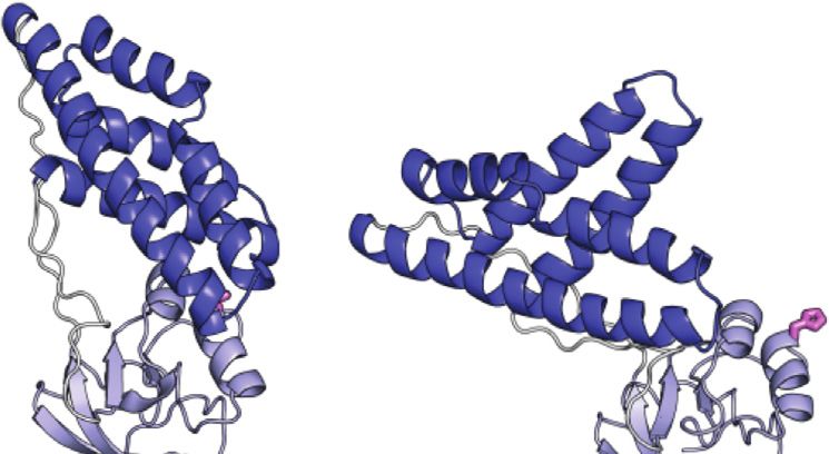
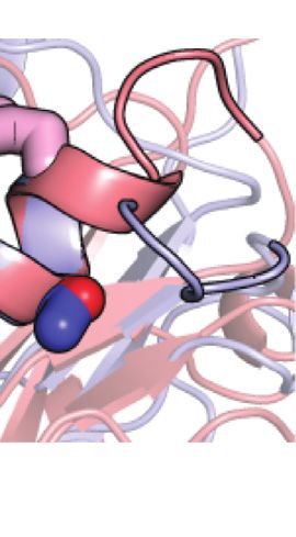
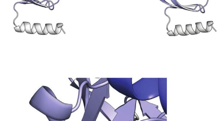
FIGURE 1 | Conformational equilibria of EI during catalysis. (A) an important source of functional regulation that can be exploited
Schematic summary of the EI conformational equilibria during catalysis. The to design allosteric inhibitors of EI. In particular, by using high-
EIN domain is colored blue, the EIC domain is colored red, the PEP molecule is
pressure NMR we have shown that dimerization of EI promotes
colored green (B) The EIN domain adopts the state A and state B
conformation in open (PDB code: 2KX9) and closed (PDB code: 2HWG) EI, substrate binding by providing structural stabilization to the EIC
respectively. The α and α/β subdomains of EIN are colored blue and light blue, catalytic pocket (Nguyen et al., 2021). Coupling NMR relaxation
respectively. The active site His189 is shown as pink sticks. The linkers experiments with Small Angle X-ray Scattering, we showed that
connecting the EIN subdomains and the helical linker connecting EIN to EIC binding of PEP results in further quenching of μs-ms dynamics at
are colored white. (C) The active site His189 adopts the g+ and g− rotameric
states in the structures of unphosphorylated and phosphorylated EI,
the EIC catalytic loops that triggers the open-to-close
respectively. The α and α/β subdomains of EIN are colored blue and light blue, interdomain rearrangement and activates EI for catalysis
respectively. The side chains of Thr168 and of unphosphorylated and (Venditti et al., 2015b). Finally, by combining NMR with
phosphorylated His189 are shown as sticks (carbon is pink, nitrogen is blue, Molecular Dynamics (MD) simulations, we noticed that
oxygen is red, and phosphorus is orange).
residual conformational heterogeneity at the EIC active site in
the activated enzyme-substrate complex determines the
enzymatic turnover (Dotas et al., 2020) and that perturbing
et al., 1993; Clore and Venditti, 2013). The phosphorylation state conformational dynamics at the active site loops is an effective
of EI dictates the phosphorylation state of all other downstream strategy to inhibit the phosphoryl-transfer reaction (Nguyen and
components of the PTS (Deutscher et al., 2014) and malfunction Venditti 2020).
of EI has been linked to reduced growth-rate and attenuated Despite the wealth of knowledge we possess about the coupling
virulence in several bacterial species (Edelstein et al., 1999; Jones between EIC conformational flexibility and enzymatic activity,
et al., 2000; Lau et al., 2001; Kok et al., 2003). Given its central role very little is known about if and how EIN conformational
Frontiers in Molecular Biosciences | www.frontiersin.org 2 July 2021 | Volume 8 | Article 699203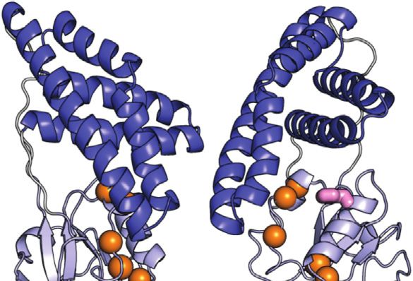
Purslow et al.                                                                                                       EIN Conformational Dynamics

dynamics impact the function of the enzyme. Indeed, while a                 thermophilic EIN, respectively. The melting temperature (Tm)
comparison of the experimental atomic-resolution structures of              was calculated as the maximum value of the derivative of θ222nm
EI indicates that the open-to-close conformational change is                with respect to temperature.
coupled to a rigid body reorientation of the α subdomain
relative to the α/β subdomain of EIN (commonly referred to
as state A-to-state B equilibrium, Figure 1B) and that protein              Nuclear Magnetic Resonance
phosphorylation shifts the χ2 angle of His189 from the g+ to g−             Spectroscopy
rotameric state (Figure 1C), it is not clear if these conformational        NMR samples were prepared in 20 mM Tris-HCl buffer (pH 7.4),
equilibria are active in isolated EIN and if their external                 100 mM NaCl, 4 mM MgCl2, 1 mM ethylenediaminetetraacetic
perturbation can impact turnover. Addressing these questions                acid (EDTA), 2 mM dithiothreitol (DTT), and 90% H2O/10%
will advance our understanding of how synergistic couplings                 D2O (v/v). For protein phosphorylation, samples were incubated
among      intradomain,     interdomain,         and     intersubunit       for 1 h at 37°C with
Purslow et al.                                                                                                        EIN Conformational Dynamics

experiments, with the spin-lock field (ω1) for the R1ρ experiments          (v/v). The reaction volume was 500 μl. All enzymatic assays were
set to 1 kHz. Relaxation delay durations were 0, 120, 280, 440,            run at fixed concentrations of enzyme (∼0.005 μM), HPr
640, 800, 1,040, and 1,200 ms for R1 and 0.2, 4.2, 7.2, 15, 23.4,          (600 μM), and PEP (1 mM). The initial velocities (ν0) for the
32.4, 42, 52.2, and 60 ms for R1ρ, respectively. R1 and R1ρ values         phosphoryl transfer reactions were determined by plotting the
were determined by fitting time-dependent exponential                       concentration of unphosphorylated HPr determined by the 1H-
                                                                           15
restoration of peak intensities at increasing relaxation delays.              N cross-peak intensities, as described by Nguyen et al. (2018) vs.
R2 values were extracted from the measured R1 and R1ρ values.              time (Figure 5D). All assays were performed in triplicates to
Global rotational correlation times (τ c) were estimated from the          estimate the experimental error.
mean R1 and R2 values, excluding residues displaying enhanced
local dynamics on the ps-ns timescale, using the following                 Mass Spectrometry
equation (Kay et al., 1989):                                               A binary ACQUITY UPLC H-Class system coupled with
                                                                   Synapt G2-Si HDMS system (Waters, Milford, MA) and
                              1      R2                                    electrospray ionization (ESI) source was used to determine
                       τc ≈         6 −7                       (2)
                            4π]N     R1                                    the intact masses of phosphorylated and non-
                                                                           phosphorylated tEIN. Starting samples were prepared by
where ]N is the 15N resonance frequency in Hz, and R1 and R2 are           diluting 10 μl of the NMR samples of phosphorylated and
the average determined values of the 15N relaxation rates.                 non-phosphorylated tEIN with HPLC grade H 2 O to final
    15
       N and 13Cmethyl relaxation dispersion (RD) experiments              concentration of 2 µM. 1 µl of each sample was injected in the
were conducted at 5, 10, 15, and 20°C using a well-established             mass spectrometer.
protocol (Singh et al., 2021). In brief, a pulse sequence that                UPLC separations were performed using a Restek Ultra C4
measures the exchange contribution for the TROSY                           column (5 um 50 mm × 1 mm) with a flow rate of 0.4 ml/min.
component of the 15N magnetization (Loria et al., 1999) and a              Solvents used were 0.1% formic acid in HPLC grade H2O
pulse scheme for 13C single-quantum CPMG (Carr-Purcell-                    (solvent A) and 0.1% formic acid in acetonitrile (solvent B,
Meinboom-Gill) RD described by Kay and co-workers                          mobile phase). The gradient used started with an initial
(Lundström et al., 2007) were employed. Off-resonance effects              condition of 5% B for 1 min, followed by a 7 min gradient
and pulse imperfections were minimized using a four-pulse phase            of 5–100% solvent B. This was held for 4 min before dropping
scheme (Yip and Zuiderweg, 2004). CPMG RD experiments were                 back to the initial 5% buffer B in 1 min and held for the
performed at a 1H frequency of 800 and 600 MHz with fixed                   remainder of the run (total 20 min).
relaxation delays (Trelax) of 60 and 30 ms, for 15N and 13Cmethyl             The eluant from the UPLC was introduced to the Waters
experiments, respectively. Different numbers of refocusing pulses          Synapt G2-Si HDMS with TOF mass analyzer using a Waters
were implemented to produce effective CPMG fields (νCPMG)                   Lockspray Source (300–5,000 Da mass range). Finally, the intact
varying from 50 to 1,000 Hz (Mulder et al., 2001). Experimental            mass was determined by deconvolution of mass spectra using the
errors on the transverse relaxation rates (R2) were estimated from         MassLynx 4.2 software.
the noise level estimated with the NMRFAM-SPARKY software.
The resulting RD curves acquired at multiple temperatures and
magnetic fields were globally fit to a two-site exchange model
                                                                           RESULTS
using the Carver-Richard equation (Carver and Richards, 1972),
as described by Dotas et al. (2020).                                       In this contribution, we investigate the structure and
    Backbone amide 1DNH residual dipolar couplings (RDCs)                  conformational dynamics of native and phosphorylated EIN
were measured at 40°C by taking the difference in 1JNH scalar              from two organisms: a mesophilic bacterium (Escherichia coli)
couplings in isotropic and alignment media. Phage pf1 (16 mg/              and     a     thermophilic    organism     (Thermoanaerobacter
ml; ASLA Biotech) was the employed alignment media and the                 tengcongensis). The two proteins are referred to as eEIN and
1
  JNH couplings were measured using the RDCs by TROSY pulse                tEIN in the unphosphorylated state, and eEIN-P and tEIN-P in
scheme (Fitzkee and Bax, 2010). Xplor-NIH (Schwieters et al.,              the phosphorylated state throughout the manuscript,
2003) was used to compute singular value decomposition (SVD)               respectively. Further, we examined the effect of two single-
analysis of the RDC values.                                                point mutations, Ser191→Ala191 within eEIN and Ala191→
                                                                           Ser191 within tEIN, on protein structure and dynamics. These
Enzyme Kinetic Assays                                                      mutants are denoted eEINS191A and tEINA191S, respectively.
eEI, eEIS191A, tEI, and tEIA191S were investigated for their ability       Similar notations are used for HPr and the full-length EI
to catalyze the transfer of the phosphoryl group from PEP to HPr.          (eHPr, tHPr, eEI, tEI, eEIS191A, and tEIA191S). The full-length
Assays were performed on a Bruker 700 MHz spectrometer at                  eEI and tEI share similar sequence (overall identity  54% and
25°C using 1H-15N Selective Optimized Flip Angle Short                     active site identity  100%) (Supplementary Figure 1) and 3D
Transient (SOFAST) NMR experiments (Schanda et al., 2005),                 structure (Oberholzer et al., 2005; Teplyakov et al., 2006;
using a protocol previously described (Nguyen et al., 2018). The           Navdaeva et al., 2011; Evangelidis et al., 2018), but have been
reaction buffer was 20 mM Tris-HCl buffer (pH 7.4), 100 mM                 shown to be optimally active at 37 and 65°C, respectively,
NaCl, 4 mM MgCl2, 1 mM ethylenediaminetetraacetic acid                     (Navdaeva et al., 2011). The sequence identity of isolated eEIN
(EDTA), 2 mM dithiothreitol (DTT), and 90% H2O/10% D2O                     and tEIN is 48% (Supplementary Figure 1).

Frontiers in Molecular Biosciences | www.frontiersin.org               4                                       July 2021 | Volume 8 | Article 699203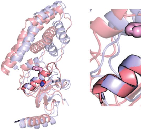
Purslow et al.                                                                                                                                                   EIN Conformational Dynamics

    FIGURE 2 | Phosphorylation induced structural perturbations in tEIN. (A) Deconvoluted masses of tEIN (black) and tEIN-P (red) from LC-MS. (B) Weighted
    combined chemical shift perturbations (ΔH/N) induced by phosphorylation of tEIN on the 1H-15N TROSY spectra of the protein are plotted against residue index. (C) The
    experimental ΔH/N values are plotted on the structure of tEIN (PDB code: 5WOY). Two different tEIN orientations are shown. The relationship between sphere size and
    color and the value of ΔH/N is depicted by the color bar. The His189 side chain is shown as pink sticks. (D) 800 MHz 1H-15N TROSY spectra of tEIN (black) and tEIN-
    P (red). (E) Correlation between the secondary Cα chemical shifts measured for tEIN (y-axis) and tEIN-P (x-axis). (F) SVD fitting of the 1DNH RDC data measured for tEIN
    (black) and tEIN-P (red) against the solution structure of tEIN (PDB code: 5WOY).

TABLE 1 | Kinetic and thermodynamic parameters for the µs-ms dynamics detected by CPMG.

                                kab/kba (s−1)a                      Δ‡Hab/Δ‡Hb,a            Δ‡Sab/Δ‡Sb,a                           pb(%)c                           ΔH                   ΔS
                                                                     (kJ mol−1)b           (J K−1 mol−1)b                                                                −1 d           −1
                  5° C        10°C        15°C          20°C                                                      5°C       10°C        15°C       20°C        (kJ mol )          (J K       mol−1)d

tEIN              171         265          406          613              55   ±   1            −2    ±   1        8.1        8.4         8.7        9.0           6±3                  0 ± 39
                 1946        2,897        4,255        6,168             50   ±   1            −2    ±   1
eEINS191A         361         554          837         1,249             54   ±   1            −2    ±   1        8.9        9.3         9.6       10.0           5±3                 −2 ± 22
                 3,686       5,425        7,879       11,297             48   ±   1            −3    ±   1
a
  The major and minor states of the equilibrium are referred to as a and b, respectively. kab and kba are the rate constants for the transition from a to b and from b to a, respectively, and are
calculated from the values of the optimized parameters kex ( kab + kba) and pb. The upper and lower numbers refer to kab and kba, respectively. Errors on rate constants are < 15% of the
reported values.
b
  Activation enthalpies and entropies for the a to b and b to a transitions were calculated by fitting the temperature dependence of kab and kba to the Eyring equation, respectively. The upper
and lower numbers refer to kab and kba, respectively.
c
  Errors on populations are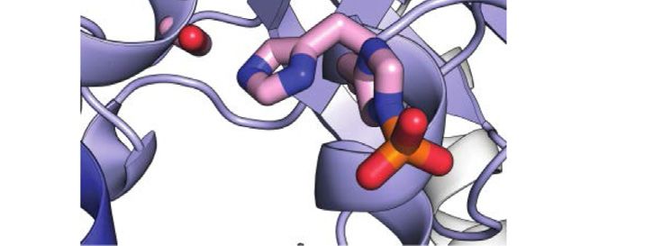
Purslow et al.                                                                                                                                    EIN Conformational Dynamics

  FIGURE 3 | Effect of phosphorylation on the ps-ns dynamics of EIN. (A) 15N-R2/R1 data measured at 800 MHz and 40°C for tEIN (top), eEIN (second from top),
  tEINA191S (second from bottom), and eEINS191A (bottom) are plotted vs. the residue index. The unphosphorylated and phosphorylated states are colored black and red,
  respectively. The localization of linker 1, linker 2, and the C-terminal end are indicated by transparent gray boxes. (B) The 15N-R2/R1 data measured for tEIN are displayed
  as a gradient on the solution structure of the protein (PDB code: 5WOY) according to the color bar. The His189 side chain is shown as pink sticks.

catalytic amounts (0.3 ppm) ΔH/N values are observed exclusively in the vicinity                             the protein.
of the phosphorylation site on the α/β subdomain and are
presumably a result of electronic effects that arise from the
presence of the phosphoryl group as well as ring current                                    Effect of Phosphorylation on the ps-ns
effects from the change in the χ2 angle of His189 to                                        Dynamics
accommodate the phosphoryl group at the Nε2 position. Such                                  ps-ns timescale dynamics were investigated for eEIN, eEIN-P,
hypothesis is supported by the excellent agreement between the                              tEIN, and tEIN-P using NMR relaxation experiments. NMR
secondary Cα chemical shifts measured for phosphorylated and                                samples of eEIN-P were produced as described previously
unphosphorylated tEIN (Figure 2E), which demonstrates that no                               (Suh et al., 2008). Residue-specific 15N-R1 and 15N-R2 values
transition in backbone conformation occurs upon                                             were obtained at 800 MHz and 40°C by acquisition of TROSY-
phosphorylation. Further insight into the effect of                                         detected R1 and R1ρ experiments (Lakomek et al., 2012) on

Frontiers in Molecular Biosciences | www.frontiersin.org                                6                                                 July 2021 | Volume 8 | Article 699203
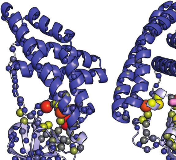
Purslow et al.                                                                                                                             EIN Conformational Dynamics

  FIGURE 4 | Effect of phosphorylation on the μs-ms dynamics for EIN. (A) 15N and 13Cmethyl exchange contributions to the transverse relaxation rate (Rex) measured
  at 10°C and 800 MHz are graphed vs. residue index for tEIN (top), eEIN (second from top), tEINA191S (second from bottom), and eEINS191A (bottom). The
  unphosphorylated and phosphorylated states are colored black and red, respectively. Unassigned cross-peaks were given arbitrary pseudo residue indexes larger than
  300. (B) The NMR correlation showing Rex values larger than 5 s−1 at 10°C and 800 MHz for tEIN are displayed as orange spheres on the solution structure of tEIN
  (PDB code: 5WOY). The His189 side chain is shown as pink sticks. (C) Examples of typical RD profiles measured at 10°C and 800 MHz for unphosphorylated (black) and
  phosphorylated (red) tEIN. Data are shown for the Asp167 NH (top) and the Ile199 δ1-methyl (bottom) groups. The experimental data are shown as circles. The best fit
  curves are shown as lines. The complete set of analyzed data are shown in Supplementary Figure 2. (D) The peak specific ΔωN and ΔωC parameters obtained from
  fitting the RD profiles of tEIN are plotted vs. the change in 15N and 13Cmethyl chemical shift induced by protein phosphorylation. The poor agreement between
  experimental and fitted ΔωN observed for Ile199 might be due to the fact that the phosphoryl group is oriented toward this amide group in the phosphorylated enzyme
  (Figure 1C). Because the RD experiments were acquired on unphosphorylated EIN, the effects on the 15N chemical shift of the electrostatic interactions between the
  phosphoryl group and the Ile199 backbone amide are not observable in the fitted ΔωN.

uniformly 15N-labeled protein. 15N-R2/R1 ratios are graphed as a                       transfer reaction does not affect the ps-ns timescale dynamics of
function of residue index in Figure 3A and depicted as a gradient                      EIN (Figure 3A).
on the solution structure of tEIN in Figure 3B. For globular
diamagnetic proteins, global tumbling is the only significant
contribution to 15N relaxation and the R2/R1 values are                                Effect of Phosphorylation on the μs-ms
expected to be constant throughout the amino acid sequence                             Dynamics
and proportional to the rotational correlation time (τ c) (Kay et al.,                 μs-ms timescale dynamics in eEIN, eEIN-P, tEIN, and tEIN-P were
1989). Therefore, residues that produce lower than average R2/R1                       investigated by 15N and 13Cmethyl Carr-Purcell-Meiboom-Gill
values likely undergo additional local motion on the ps-ns                             (CPMG) relaxation dispersion (RD) spectroscopy (Mittermaier
timescale that decrease the effective correlation time                                 and Kay 2006). Experiments were acquired on U-(2H,15N)/Ile
experienced by the N-H bond. Analysis of the NMR relaxation                            (d1)-13CH3/Val, Leu-(13CH3/12C2H3)-labeled samples at two
data in Figure 3A indicates that eEIN, eEIN-P, tEIN, and tEIN-P                        different static fields (600 and 800 MHz) and four different
tumble with a τ c ∼ 11 ns, which is consistent with the theoretical                    temperatures (5, 10, 15, and 20°C). Simultaneous investigation of
τ c calculated for a globular protein of the EIN size (∼11 ns).                        RD profiles measured at multiple temperatures returns a
Notably, several regions of the protein exhibit lower than average                     comprehensive      characterization   of   the    kinetics    and
R2/R1 values, suggesting the presence of local backbone dynamics                       thermodynamics of conformational exchange processes between
on the ps-ns timescale. These regions cluster into the                                 species with distinct chemical shifts occurring on a timescale
unstructured linkers connecting the α and α/β subdomains                               ranging from ∼0.1 to ∼10 ms, by providing enthalpy (ΔH),
(linker 1: residues 19–32; linker 2: residues 143–156) and at                          entropy (ΔS), activation enthalpy (Δ‡H), and activation entropy
the C-terminal end of EIN (Figure 3B). However,                                        (Δ‡S) for the conformational equilibrium (Purslow et al., 2018).
phosphorylation of His189 elicits no observable change in the                              Exchange contributions toward the transverse relaxation rates
distribution of the R2/R1 values, indicating that the phosphoryl                       (Rex) are plotted against residue index in Figure 4A. Large

Frontiers in Molecular Biosciences | www.frontiersin.org                           7                                              July 2021 | Volume 8 | Article 699203
Purslow et al.                                                                                                                      EIN Conformational Dynamics

                                                                                         between the two conformational states (ΔωN and ΔωC,
                                                                                         respectively) were treated as peak-specific and temperature
                                                                                         independent parameters. The exchange rate (kex) and the
                                                                                         fractional population of the minor conformational state (pb)
                                                                                         were calculated at each temperature from the fitted values of
                                                                                         Δ‡G and ΔG using the general form of the Eyring and reaction
                                                                                         isotherm equations, respectively. This fitting procedure reduces
                                                                                         the number of optimized parameters from 28 (kex, pb, and Δω for
                                                                                         five NMR peaks at four experimental temperatures) to 7 (Δ‡G,
                                                                                         ΔG, and Δω for five NMR peaks), and it is justified if the heat
                                                                                         capacity of activation remains constant over the experimental
                                                                                         temperature range (5–20°C) (Nguyen et al., 2017). For
                                                                                         completeness, it should be noted that the intrinsic 15N and
                                                                                         13
                                                                                            Cmethyl transverse relaxation rates were also optimized as
                                                                                         peak-specific parameters, therefore increasing the overall
                                                                                         number of fitted parameters. Also, in order to improve
                                                                                         convergence of the fitting algorithm, the ΔωN parameters were
                                                                                         restrained to be larger than 1 ppm. Recently we have used a
                                                                                         similar fitting protocol to model the temperature dependence of
                                                                                         the μs-ms dynamics in the EIC domain of the enzyme (Dotas
                                                                                         et al., 2020).
                                                                                             A summary of the optimized parameters is reported in
                                                                                         Table 1. Examples of the global fit are provided in Figure 4C.
                                                                                         Curves for all the analyzed RD profiles are provided in
                                                                                         Supplementary Figure 2. The optimized Δ‡G is 50,228 ±
  FIGURE 5 | Design and characterization of eEIN/tEIN hybrids. (A)                       227J mol−1, which translates into exchange rate constants (sum
  Structure alignment of α-helix 6 in the solution structure of eEIN (PDB code:          of forward and backward rate constants, kab and kba, respectively)
  2EZA) (blue) and tEIN (PDB code: 5WOY) (red). The side chain of His189 is
  shown as pink sticks. The side chain of Ser191 in eEIN is shown as blue
                                                                                         of 2,117 ± 236, 3,162 ± 347, 4,660 ± 503, and 6,780 ± 719 s−1 at 5,
  sticks. The side chain of Ala191 in tEIN is shown as red sticks. (B) Sequence          10, 15, and 20°C, respectively. The optimized ΔG is 5,601 ± 330 J
  alignment of α-helix 6 in eEIN (blue) and tEIN (red). Full sequence alignment of       mol−1, resulting in pb values of 8.0 ± 0.4, 8.4 ± 0.4, 8.7 ± 0.4, and
  the mesophilic and thermophilic protein is provided in Supplementary                   9.0 ± 0.4% at 5, 10, 15, and 20°C, respectively (Table 1). kab and
  Figure 1. (C) Temperature-induced unfolding of eEIN (blue), eEINS191A                  kba were calculated from the values of kex and pb, and their
  (green), tEINA191S (orange), and tEIN (red). (D) Rate of HPr phosphorylation
  catalyzed by 0.005 μM eEI (blue), eEIS191A (green), tEIA191S (orange), and tEI
                                                                                         temperature dependence was modeled using the van’t Hoff and
  (red) in the presence of 1 mM PEP at 25°C. The phosphorylation rate (V0) was           Eyring equations to obtain ΔH, ΔS, Δ‡H, and Δ‡S of the tEIN
  obtained by linear fitting (solid lines) of the decrease in concentration of            conformational equilibrium (Table 1).
  unphosphorylated HPr (open circles) vs. time. A V0 of ∼25 μM/min was                       From the analysis of the kinetic, thermodynamic, and NMR
  obtained for eEI and eEIS191A. A V0 of ∼3 μM/min was obtained for tEI and
                                                                                         parameters obtained by the RD study at multiple temperatures it
  tEIA191S.
                                                                                         is apparent that 1) tEIN is in equilibrium between two
                                                                                         conformational states, 2) the relative thermodynamic stability
                                                                                         of the two states is dictated by enthalpic contributions to the free
(>5 s−1) Rex values were detected for amino acids that cluster in                        energy (Table 1), and 3) the 15N and 13C chemical shift
the vicinity of the active site His189 on the α/β subdomain of tEIN                      differences between the two conformational states correlates
(Figures 4A,B). In particular, Asp167, Ala169, Lys172 localize on                        with the change in 15N and 13C chemical shift (ΔN and ΔC,
the partially structured helix that is in direct contact with His189,                    respectively) induced by phosphorylation of tEIN (Figure 4D).
while Ile199 is located at the C-terminal end of the short helix that                    These findings suggest that the μs-ms dynamics detected in tEIN
comprises the phosphorylation site. Contrarily, eEIN revealed no                         by RD experiments report on the equilibrium between the g+ and
significant Rex values (Figure 4A), suggesting the observed                               g− rotameric states of His189 that breaks the hydrogen bond
dynamics in tEIN may be too fast to be detected by CPMG                                  between the Thr168 and His189 side-chains and makes the Nε2
within its mesophilic analogue. Interestingly, all the 15N and                           atom accessible to the incoming phosphoryl group. Consistent
13
   Cmethyl RD curves measured at multiple temperatures and static                        with this hypothesis, the fitted value for ΔH (6 kJ mol−1) is
magnetic fields for Asp167, Ala169, Lys172, and Ile199 within tEIN                        comparable with the reported energies for the weak
could be fit simultaneously to a model describing the                                     intramolecular hydrogen bonds involving the hydroxyl group
interconversion       between      two      conformational     states                    on Ser or Thr side-chains (∼7 kJ mol−1) (Pace et al., 2014a; Pace
(Figure 4C; Supplementary Figure 2). In this global fitting                               et al., 2014b).
procedure, the activation (Δ‡G) and standard (ΔG) free energy                                To further test this model, the effect of phosphorylation
of the conformational equilibrium were optimized as global                               on the μs-ms dynamics of tEIN was investigated at 10°C by
parameters, whereas the 15N and 13C chemical shift differences                           acquisition of RD experiments on tEIN-P. tEIN-P was

Frontiers in Molecular Biosciences | www.frontiersin.org                             8                                       July 2021 | Volume 8 | Article 699203
Purslow et al. EIN Conformational Dynamics
prepared enzymatically as described above. However, in this case, Although a comparison of the 1H-15N TROSY spectra measured
tEI, tHPr, and excess PEP were purified out of the final NMR sample for the wild type and mutant proteins shows that mutations at the 191
by anion exchange chromatography. This additional purification position provides minimal perturbations to the NMR spectra and,
step is required to remove any possible complex between tEIN and therefore, to the solution fold of EIN (Supplementary Figure 3), the
other molecules in the sample that, even at very dilute concentrations temperature-induced unfolding data acquired by CD reveal that the
( 5 s−1 is
value larger than 5 s−1 was measured for eEIN and, as expected, detected in the NMR spectra of tEINA191S (Figure 4A), which is
phosphorylation of eEIN resulted in no observable change in the Rex consistent with the hypothesis that Ala191Ser mutation in tEIN
distribution (Figure 4A). Being that the experimental structures of speeds up the rotameric equilibrium of His189. In contrast, the
phosphorylated and unphosphorylated eEIN indicate that the Ser191Ala mutation in eEIN introduces Rex > 5 s−1 at three 1H-15N
protein must undergo the His189 rotameric transition to TROSY correlations. Although we were able to confidently assign only
accommodate the phosphoryl group (Figure 1C), our data one of these three NMR signals, we ascribe the appearance of exchange
suggests the g+/g− exchange in eEIN is faster than in tEIN and, induced effects on the spectra of eEINS191A to the rotameric equilibrium
therefore, not detected by RD experiments. of His189 for the following reasons: 1) The assigned NMR correlation
with Rex > 5 s−1 (Thr168) localizes in the same region observed to
experience exchange contributions to R2 in tEIN (Figure 4A), 2) Global
Engineering eEIN/tEIN Hybrids With fitting of the RD profiles acquired for the three NMR signals at multiple
Modulated Conformational Dynamics temperatures and static fields (Supplementary Figure 4) produces
We have recently shown that hybridizing proteins from mesophilic kinetic and thermodynamic parameters that are similar to the ones
and thermophilic bacteria is an effective strategy to produce active obtained for the His189 rotameric equilibrium in tEIN (Table 1), and 3)
enzymes with modulated conformational dynamics and biological Phosphorylation of eEINS191A results in a complete quenching of μs-ms
function (Dotas et al., 2020). Indeed, by merging the scaffold of EIC dynamics (Figure 4A).
from T. tengcongensis with the active site loops of the E. coli enzyme we Overall, the NMR and CD data reported above support the
engineered a hybrid EIC variant that displays the thermal stability of hypothesis that the identity of the residue at position 191 controls
the thermophilic protein and the high active site flexibility and low- helix 6 stability and the dynamics of the g+/g− equilibrium of His189.
temperature activity of the mesophilic enzyme. In contrast, implanting To test the dependency of the EI biological function on the kinetics
the active site loops from T. tengcongensis EIC onto the scaffold of of the His189 rotameric transition, the Ser191Ala and Ala191Ser
E. coli EIC resulted in a construct that is more rigid and less active than mutations were incorporated into the full-length eEI and tEI,
the mesophilic enzyme (Dotas et al., 2020). Here, eEIN/tEIN hybrids respectively. Then, the ability of eEI, eEIS191A, tEI, and tEIA191S,
are engineered to investigate the relationship between the kinetics of to transfer the phosphoryl group from PEP to HPr was investigated
the His189 rotameric equilibrium and turnover number. Comparison at 25°C by 1H-15N SOFAST-TROSY spectra, as recently described
of the experimental atomic-resolution structures shows that the (Nguyen et al., 2018). As expected, the data indicate that at room
N-terminal end of α-helix 6 (which comprises the His189) in tEIN temperature eEI catalyzes the phosphoryl transfer reaction ∼10 times
is two residues longer than in eEIN (Figure 5A). Alignment of the faster than tEI (Figure 5D). Interestingly, the Ser191Ala and
amino acid sequences reveals a single Ser191Ala mutation within Ala191Ser mutations produce no detectable changes in the activity
α-helix 6 moving from the mesophilic to the thermophilic of eEI and tEI, respectively (Figure 5D), indicating that the
construct (Figure 5B; Supplementary Figure 1). As alanine conformational transition from the g+ to g− rotameric state of
residues are known to promote helix formation in proteins (Panja His189 is not rate limiting for catalysis.
et al., 2015), we hypothesize that the Ser191Ala mutation provides
structural stabilization to α-helix six and is responsible for the slower
rotameric equilibrium observed for His189 in tEIN. To test this DISCUSSION
hypothesis, we investigated the structure, dynamics, and thermal
stability of eEINS191A and tEINA191S by solution NMR and circular Protein conformational transitions are fundamental to
dichroism (CD). signaling, enzyme catalysis, and assembly of cellular
Frontiers in Molecular Biosciences | www.frontiersin.org 9 July 2021 | Volume 8 | Article 699203Purslow et al. EIN Conformational Dynamics structures. Yet, our understanding of how the interconversion by tEI (Figure 4, Figure 5). Similarly, increasing the activation among different folded structures affects function continues to energy for the His189 rotameric transition in eEI by lag. One technical challenge limiting our ability to interrogate introducing an Ala residue at position 191 does not affect the dynamics/function relationship is the lack of universal and its enzymatic activity (Figure 4, Figure 5). These results straightforward strategies to selectively perturb indicate that the His189 conformational change is not rate conformational equilibria in complex biomolecular systems. limiting for catalysis and, therefore, regulation of the EI Here, we have shown that it is possible to perturb protein activity cannot be achieved by slight perturbations of the conformational dynamics without dramatically affecting their His189 conformational dynamics. thermal stability by hybridizing the amino acid sequence of a Finally, it is important to highlight that the data reported in this mesophilic and a thermophilic analogue. In particular, we have manuscript show no evidence for an active state A/state B investigated the structure and dynamics of the N-terminal equilibrium in the isolated EIN. This observation implies that domain of EI from a mesophilic (eEIN) and a thermophilic the latter equilibrium is either on a timescale that is not (tEIN) bacterium. We found that the two proteins adopt the compatible with RD experiments (i.e., outside the μs-ms regime) same fold and undergo a rotameric equilibrium at the His189 or completely inactive in the isolated EIN domain. Considering that side chain that exposes the phosphorylation site to react with in the full-length dimeric EI transition to state B is required to avoid the EI substrate, PEP (Figure 1C). Interestingly, CPMG RD steric overlap between the EIN and EIC domains, and that state B is experiments revealed that the rotameric transition in tEIN structurally stabilized by intersubunit EIN-EIN interactions occurs on a slower time scale than in eEIN (Figure 4A). By (Figure 1A), we deduce that state B is inaccessible by the comparing the primary structures of the mesophilic and isolated EIN domain investigated here. In any case, our data thermophilic proteins (Figures 5A,B) we identified a single indicate that the His189 g+-to-g− transition that exposes the EI point mutation in eEIN (Ser191Ala) and tEIN (Ala191Ser) that phosphorylation site to PEP (Figure 1C) is decoupled from the EIN swaps the observed kinetics for this conformational change, state A/state B equilibrium and is not triggered by transition of the with the eEINS191A mutant exchanging between the rotameric full-length enzyme to the catalytically active closed conformation states of His189at a rate similar to the one measured for the wild (Figure 1A). type tEIC, and the tEINA191S mutant undergoing the same rotameric equilibrium on a faster time scale, comparable to wild type eEIN (Figure 4, Table 1). Intriguingly, we have DATA AVAILABILITY STATEMENT recently used the same mesophilic/thermophilic hybridization strategy described here to engineer constructs of the The names of the repository/repositories and accession C-terminal domain of EI (EIC) with modulated active site number(s) can be found below: BioMagResBank. Accession dynamics (Dotas et al., 2020). In contrast to the EIN case that number: 50386 allows for a single-point hybridizing mutation, design of the EIC hybrids required swapping of the entire active site (composed of three catalytic loops) between mesophilic and AUTHOR CONTRIBUTIONS thermophilic species. Nonetheless, as for the EIN case presented here, the engineering effort resulted in production JP, JT, and VV designed the research; JP, JT, VS, TN, RD, and BK, of two enzymatically active hybrids with mixed properties: performed the experiments; JP, JT, VS, BK, and VV analyzed the One hybrid displayed the high thermal stability of the data; JP and VV wrote the article. thermophilic enzyme and the increased active site flexibility and low-temperature activity of the mesophilic analogue; The second hybrid showed the low thermal stability of the FUNDING mesophilic enzyme and the rigid active site and low activity at room temperature of the thermophilic protein (Dotas et al., This work was supported by funds from NIGMS R35GM133488 2020). Therefore, hybridizing homologue proteins from and from the Roy J. Carver Charitable Trust to VV mesophilic and thermophilic bacteria is emerging as a powerful tool in biophysics by providing a straightforward approach to produce functional proteins with modulated ACKNOWLEDGMENTS internal flexibility. In addition of serving as a demonstration of the mesophilic/ We thank Marius Clore and Jeong-Yong Suh for providing the thermophilic hybridization strategy for protein design, the resonance assignments of phosphorylated eEIN. EIN constructs engineered here allowed us to investigate the relationship between enzymatic turnover and the kinetics of the His189 rotameric equilibrium. Indeed, by introducing the SUPPLEMENTARY MATERIAL hybridizing mutations at position 191 into the sequence of the full-length enzyme we have demonstrated that increasing the The Supplementary Material for this article can be found online at: rate of the g+-to-g− transition of the His189 χ2 angle does not https://www.frontiersin.org/articles/10.3389/fmolb.2021.699203/ affect turnover for the phosphoryl transfer reaction catalyzed full#supplementary-material Frontiers in Molecular Biosciences | www.frontiersin.org 10 July 2021 | Volume 8 | Article 699203
Purslow et al.                                                                                                                                           EIN Conformational Dynamics

REFERENCES                                                                                      Kok, M., Bron, G., Erni, B., and Mukhija, S. (2003). Effect of Enzyme I of the
                                                                                                   Bacterial Phosphoenolpyruvate : Sugar Phosphotransferase System (PTS) on
                                                                                                   Virulence in a Murine Model. Microbiology 149 (9), 2645–2652. doi:10.1099/
Carver, J. P., and Richards, R. E. (1972). A General Two-Site Solution for the                     mic.0.26406-0
    Chemical Exchange Produced Dependence of T2 upon the Carr-Purcell Pulse                     Lakomek, N.-A., Ying, J., and Bax, A. (2012). Measurement of 15N Relaxation
    Separation. J. Magn. Reson. (1969) 6, 89–105. doi:10.1016/0022-2364(72)                        Rates in Perdeuterated Proteins by TROSY-Based Methods. J. Biomol. NMR 53
    90090-x                                                                                        (3), 209–221. doi:10.1007/s10858-012-9626-5
Chauvin, F., Brand, L., and Roseman, S. (1996). Enzyme I: the First Protein and                 Lau, G. W., Haataja, S., Lonetto, M., Kensit, S. E., Marra, A., Bryant, A. P., et al.
    Potential Regulator of the Bacterial Phosphoenolpyruvate: Glycose                              (2001). A Functional Genomic Analysis of Type 3 Streptococcus Pneumoniae
    Phosphotransferase System. Res. Microbiol. 147 (6), 471–479. doi:10.1016/                      Virulence. Mol. Microbiol. 40 (3), 555–571. doi:10.1046/j.1365-
    0923-2508(96)84001-0                                                                           2958.2001.02335.x
Clore, G. M., and Garrett, D. S. (1999). R-factor, FreeR, and Complete Cross-                   Lee, W., Tonelli, M., and Markley, J. L. (2015). NMRFAM-SPARKY: Enhanced
    Validation for Dipolar Coupling Refinement of NMR Structures. J. Am. Chem.                      Software for Biomolecular NMR Spectroscopy. Bioinformatics 31 (8),
    Soc. 121, 9008–9012. doi:10.1021/ja991789k                                                     1325–1327. doi:10.1093/bioinformatics/btu830
Clore, G. M., and Gronenborn, A. M. (1998). Determining the Structures of Large                 Loria, J. P., Rance, M., and Palmer, III, A. G. (1999). A TROSY CPMG Sequence for
    Proteins and Protein Complexes by NMR. Trends Biotechnol. 16 (1), 22–34.                       Characterizing Chemical Exchange in Large Proteins. J. Biomol. NMR 15 (2),
    doi:10.1016/s0167-7799(97)01135-9                                                              151–155. doi:10.1023/a:1008355631073
Clore, G. M., and Venditti, V. (2013). Structure, Dynamics and Biophysics of the                Lundström, P., Vallurupalli, P., Religa, T. L., Dahlquist, F. W., and Kay, L. E. (2007).
    Cytoplasmic Protein-Protein Complexes of the Bacterial Phosphoenolpyruvate:                    A Single-Quantum Methyl 13C-Relaxation Dispersion experiment with
    Sugar Phosphotransferase System. Trends Biochemical Sciences 38 (10),                          Improved Sensitivity. J. Biomol. NMR 38 (1), 79–88. doi:10.1007/s10858-
    515–530. doi:10.1016/j.tibs.2013.08.003                                                        007-9149-7
Delaglio, F., Grzesiek, S., Vuister, G. W., Zhu, G., Pfeifer, J., and Bax, A. (1995).           Mittermaier, A., and Kay, L. E. (2006). New Tools Provide New Insights in NMR
    NMRPipe: A Multidimensional Spectral Processing System Based on UNIX                           Studies of Protein Dynamics. Science 312 (5771), 224–228. doi:10.1126/
    Pipes. J. Biomol. NMR 6 (3), 277–293. doi:10.1007/bf00197809                                   science.1124964
Deutscher, J., Aké, F. M. D., Derkaoui, M., Zébré, A. C., Cao, T. N., Bouraoui, H.,             Mulder, F. A. A., Schipper, D., Bott, R., and Boelens, R. (1999). Altered Flexibility in
    et al. (2014). The Bacterial Phosphoenolpyruvate:Carbohydrate                                  the Substrate-Binding Site of Related Native and Engineered High-Alkaline
    Phosphotransferase System: Regulation by Protein Phosphorylation and                           Bacillus Subtilisins 1 1Edited by P. E. Wright. J. Mol. Biol. 292 (1), 111–123.
    Phosphorylation-dependent Protein-Protein Interactions. Microbiol. Mol.                        doi:10.1006/jmbi.1999.3034
    Biol. Rev. 78 (2), 231–256. doi:10.1128/mmbr.00001-14                                       Mulder, F. A. A., Skrynnikov, N. R., Hon, B., Dahlquist, F. W., and Kay, L. E.
Dotas, R. R., Nguyen, T. T., Stewart, C. E., Ghirlando, R., Potoyan, D. A., and                    (2001). Measurement of Slow (μs−ms) Time Scale Dynamics in Protein Side
    Venditti, V. (2020). Hybrid Thermophilic/Mesophilic Enzymes Reveal a Role                      Chains by15N Relaxation Dispersion NMR Spectroscopy: Application to Asn
    for Conformational Disorder in Regulation of Bacterial Enzyme I. J. Mol. Biol.                 and Gln Residues in a Cavity Mutant of T4 Lysozyme. J. Am. Chem. Soc. 123 (5),
    432 (16), 4481–4498. doi:10.1016/j.jmb.2020.05.024                                             967–975. doi:10.1021/ja003447g
Dotas, R. R., and Venditti, V. (2019). Resonance Assignment of the 128 kDa                      Navdaeva, V., Zurbriggen, A., Waltersperger, S., Schneider, P., Oberholzer, A. E.,
    Enzyme I Dimer from Thermoanaerobacter Tengcongensis. Biomol. NMR                              Baḧ ler, P., et al. (2011). Phosphoenolpyruvate: Sugar Phosphotransferase
    Assign 13 (2), 287–293. doi:10.1007/s12104-019-09893-y                                         System from the HyperthermophilicThermoanaerobacter Tengcongensis.
Doucette, C. D., Schwab, D. J., Wingreen, N. S., and Rabinowitz, J. D. (2011).                     Biochemistry 50 (7), 1184–1193. doi:10.1021/bi101721f
    α-Ketoglutarate Coordinates Carbon and Nitrogen Utilization via                             Nguyen, T. T., Ghirlando, R., Roche, J., and Venditti, V. (2021). Structure
    Enzyme I Inhibition. Nat. Chem. Biol. 7 (12), 894–901. doi:10.1038/                            Elucidation of the Elusive Enzyme I Monomer Reveals the Molecular
    nchembio.685                                                                                   Mechanisms Linking Oligomerization and Enzymatic Activity. Proc. Natl.
Edelstein, P. H., Edelstein, M. A. C., Higa, F., and Falkow, S. (1999). Discovery of               Acad. Sci. 118, e2100298118. doi:10.1073/pnas.2100298118
    Virulence Genes of Legionella pneumophila by Using Signature Tagged                         Nguyen, T. T., Ghirlando, R., and Venditti, V. (2018). The Oligomerization State of
    Mutagenesis in a guinea Pig Pneumonia Model. Proc. Natl. Acad. Sci. 96                         Bacterial Enzyme I (EI) Determines EI’s Allosteric Stimulation or Competitive
    (14), 8190–8195. doi:10.1073/pnas.96.14.8190                                                   Inhibition by α-ketoglutarate. J. Biol. Chem. 293 (7), 2631–2639. doi:10.1074/
Evangelidis, T., Nerli, S., Nováček, J., Brereton, A. E., Karplus, P. A., Dotas, R. R.,           jbc.RA117.001466
    et al. (2018). Automated NMR Resonance Assignments and Structure                            Nguyen, T. T., and Venditti, V. (2020). An Allosteric Pocket for Inhibition of
    Determination Using a Minimal Set of 4D Spectra. Nat. Commun. 9 (1),                           Bacterial Enzyme I Identified by NMR-Based Fragment Screening. J. Struct.
    384. doi:10.1038/s41467-017-02592-z                                                            Biol. X 4, 100034. doi:10.1016/j.yjsbx.2020.100034
Fitzkee, N. C., and Bax, A. (2010). Facile Measurement of 1H-15N Residual Dipolar               Nguyen, V., Wilson, C., Hoemberger, M., Stiller, J. B., Agafonov, R. V., Kutter, S.,
    Couplings in Larger Perdeuterated Proteins. J. Biomol. NMR 48 (2), 65–70.                      et al. (2017). Evolutionary Drivers of Thermoadaptation in Enzyme Catalysis.
    doi:10.1007/s10858-010-9441-9                                                                  Science 355 (6322), 289–294. doi:10.1126/science.aah3717
Garrett, D. S., Seok, Y. J., Peterkofsky, A., Gronenborn, A. M., and Clore, G. M.               Oberholzer, A. E., Bumann, M., Schneider, P., Bächler, C., Siebold, C., Baumann,
    (1999). Solution Structure of the 40,000 Mr Phosphoryl Transfer Complex                        U., et al. (2005). Crystal Structure of the Phosphoenolpyruvate-Binding Enzyme
    between the N-Terminal Domain of Enzyme I and HPr. Nat. Struct. Biol. 6 (2),                   I-Domain from the Thermoanaerobacter Tengcongensis PEP: Sugar
    166–173. doi:10.1038/5854                                                                      Phosphotransferase System (PTS). J. Mol. Biol. 346 (2), 521–532.
Huang, K.-J., Lin, S.-H., Lin, M.-R., Ku, H., Szkaradek, N., Marona, H., et al. (2013).            doi:10.1016/j.jmb.2004.11.077
    Xanthone Derivatives Could Be Potential Antibiotics: Virtual Screening for the              Pace, C. N., Scholtz, J. M., and Grimsley, G. R. (2014b). Forces Stabilizing Proteins.
    Inhibitors of Enzyme I of Bacterial Phosphoenolpyruvate-dependent                              FEBS Lett. 588 (14), 2177–2184. doi:10.1016/j.febslet.2014.05.006
    Phosphotransferase System. J. Antibiot. 66 (8), 453–458. doi:10.1038/ja.2013.30             Pace, C. N., Fu, H., Lee Fryar, K., Landua, J., Trevino, S. R., Schell, D., et al. (2014a).
Jones, A. L., Knoll, K. M., and Rubens, C. E. (2000). Identification of Streptococcus               Contribution of Hydrogen Bonds to Protein Stability. Protein Sci. 23 (5),
    Agalactiae Virulence Genes in the Neonatal Rat Sepsis Model Using Signature-                   652–661. doi:10.1002/pro.2449
    Tagged Mutagenesis. Mol. Microbiol. 37 (6), 1444–1455. doi:10.1046/j.1365-                  Panja, A. S., Bandopadhyay, B., and Maiti, s. (2015). Protein Thermostability Is
    2958.2000.02099.x                                                                              Owing to Their Preferences to Non-polar Smaller Volume Amino Acids,
Kay, L. E., Torchia, D. A., and Bax, A. (1989). Backbone Dynamics of Proteins as                   Variations in Residual Physico-Chemical Properties and More Salt-Bridges.
    Studied by Nitrogen-15 Inverse Detected Heteronuclear NMR Spectroscopy:                        PLoS One 10 (7), e0131495. doi:10.1371/journal.pone.0131495
    Application to Staphylococcal Nuclease. Biochemistry 28 (23), 8972–8979.                    Pervushin, K., Riek, R., Wider, G., and Wüthrich, K. (1998). Transverse Relaxation-
    doi:10.1021/bi00449a003                                                                        Optimized Spectroscopy (TROSY) for NMR Studies of Aromatic Spin Systems

Frontiers in Molecular Biosciences | www.frontiersin.org                                   11                                                   July 2021 | Volume 8 | Article 699203
Purslow et al. EIN Conformational Dynamics
in13C-Labeled Proteins. J. Am. Chem. Soc. 120, 6394–6400. doi:10.1021/ Carbonyl Chemical Shifts in Proteins. J. Biomol. NMR 58 (1), 1–8.
ja980742g doi:10.1007/s10858-013-9803-1
Postma, P. W., Lengeler, J. W., and Jacobson, G. R. (1993). Phosphoenolpyruvate: Ulrich, E. L., Akutsu, H., Doreleijers, J. F., Harano, Y., Ioannidis, Y. E., Lin, J., et al.
carbohydrate Phosphotransferase Systems of Bacteria. Microbiol. Rev. 57 (3), (2008). BioMagResBank. Nucleic Acids Res. 36 (Database issue), D402–D408.
543–594. doi:10.1128/mmbr.57.3.543-594.1993 doi:10.1093/nar/gkm957
Purslow, J. A., Nguyen, T. T., Egner, T. K., Dotas, R. R., Khatiwada, B., and Venditti, Venditti, V., Tugarinov, V., Schwieters, C. D., Grishaev, A., and Clore, G. M.
V. (2018). Active Site Breathing of Human Alkbh5 Revealed by Solution NMR (2015b). Large Interdomain Rearrangement Triggered by Suppression of
and Accelerated Molecular Dynamics. Biophysical J. 115 (10), 1895–1905. Micro- to Millisecond Dynamics in Bacterial Enzyme I. Nat. Commun. 6
doi:10.1016/j.bpj.2018.10.004 (1), 5960. doi:10.1038/ncomms6960
and Brutscher, B. (2005). SOFAST-HMQC Experiments for
Schanda, P., Kupče, E., Venditti, V., and Clore, G. M. (2012). Conformational Selection and
Recording Two-Dimensional Deteronuclear Correlation Spectra of Proteins Substrate Binding Regulate the Monomer/Dimer Equilibrium of the
within a Few Seconds. J. Biomol. NMR 33 (4), 199–211. doi:10.1007/s10858- C-Terminal Domain of Escherichia coli Enzyme I. J. Biol. Chem. 287
005-4425-x (32), 26989–26998. doi:10.1074/jbc.m112.382291
Schwieters, C. D., Kuszewski, J. J., Tjandra, N., and Marius Clore, G. (2003). The Venditti, V., Egner, T. K., and Clore, G. M. (2016). Hybrid Approaches to
Xplor-NIH NMR Molecular Structure Determination Package. J. Magn. Reson. Structural Characterization of Conformational Ensembles of Complex
160 (1), 65–73. doi:10.1016/s1090-7807(02)00014-9 Macromolecular Systems Combining NMR Residual Dipolar Couplings and
Schwieters, C. D., Suh, J.-Y., Grishaev, A., Ghirlando, R., Takayama, Y., and Clore, Solution X-ray Scattering. Chem. Rev. 116 (11), 6305–6322. doi:10.1021/
G. M. (2010). Solution Structure of the 128 kDa Enzyme I Dimer acs.chemrev.5b00592
fromEscherichia Coliand its 146 kDa Complex with HPr Using Residual Venditti, V., Ghirlando, R., and Clore, G. M. (2013). Structural Basis for Enzyme I
Dipolar Couplings and Small- and Wide-Angle X-ray Scattering. J. Am. Inhibition by α-Ketoglutarate. ACS Chem. Biol. 8 (6), 1232–1240. doi:10.1021/
Chem. Soc. 132 (37), 13026–13045. doi:10.1021/ja105485b cb400027q
Singh, A., Purslow, J. A., and Venditti, v. (2021). 15N CPMG Relaxation Dispersion Venditti, V., Schwieters, C. D., Grishaev, A., and Clore, G. M. (2015a).
for the Investigation of Protein Conformational Dynamics on the μs-ms Dynamic Equilibrium between Closed and Partially Closed States of the
Timescale. J. Vis. Exp. 170, e62395. doi:10.3791/62395 Bacterial Enzyme I Unveiled by Solution NMR and X-ray Scattering.
Suh, J.-Y., Cai, M., and Clore, G. M. (2008). Impact of Phosphorylation on Proc. Natl. Acad. Sci. USA 112 (37), 11565–11570. doi:10.1073/
Structure and Thermodynamics of the Interaction between the N-Terminal pnas.1515366112
Domain of Enzyme I and the Histidine Phosphocarrier Protein of the Bacterial Yip, G. N. B., and Zuiderweg, E. R. P. (2004). A Phase Cycle Scheme that
Phosphotransferase System. J. Biol. Chem. 283 (27), 18980–18989. doi:10.1074/ Significantly Suppresses Offset-dependent Artifacts in the R2-CPMG 15N
jbc.m802211200 Relaxation experiment. J. Magn. Reson. 171 (1), 25–36. doi:10.1016/
Teplyakov, A., Lim, K., Zhu, P.-P., Kapadia, G., Chen, C. C. H., Schwartz, J., et al. j.jmr.2004.06.021
(2006). Structure of Phosphorylated Enzyme I, the Phosphoenolpyruvate:sugar
Phosphotransferase System Sugar Translocation Signal Protein. Proc. Natl. Conflict of Interest: The authors declare that the research was conducted in the
Acad. Sci. 103 (44), 16218–16223. doi:10.1073/pnas.0607587103 absence of any commercial or financial relationships that could be construed as a
Tjandra, N., and Bax, A. (1997). Direct Measurement of Distances and Angles in potential conflict of interest.
Biomolecules by NMR in a Dilute Liquid Crystalline Medium. Science 278
(5340), 1111–1114. doi:10.1126/science.278.5340.1111 Copyright © 2021 Purslow, Thimmesch, Sivo, Nguyen, Khatiwada, Dotas and
Tugarinov, V., Hwang, P. M., Ollerenshaw, J. E., and Kay, L. E. (2003). Cross- Venditti. This is an open-access article distributed under the terms of the
Correlated Relaxation Enhanced1H−13C NMR Spectroscopy of Methyl Groups Creative Commons Attribution License (CC BY). The use, distribution or
in Very High Molecular Weight Proteins and Protein Complexes. J. Am. Chem. reproduction in other forums is permitted, provided the original author(s) and
Soc. 125 (34), 10420–10428. doi:10.1021/ja030153x the copyright owner(s) are credited and that the original publication in this journal is
Tugarinov, V., Venditti, V., and Marius Clore, G. (2014). A NMR experiment for cited, in accordance with accepted academic practice. No use, distribution or
Simultaneous Correlations of Valine and Leucine/isoleucine Methyls with reproduction is permitted which does not comply with these terms.
Frontiers in Molecular Biosciences | www.frontiersin.org 12 July 2021 | Volume 8 | Article 699203You can also read