A novel murine in vivo model for acute hereditary angioedema attacks
←
→
Page content transcription
If your browser does not render page correctly, please read the page content below
www.nature.com/scientificreports
OPEN A novel murine in vivo model
for acute hereditary angioedema
attacks
Sujata Bupp1, Matthew Whittaker2, Mari Lehtimaki1, JuMe Park1, Jessica Dement‑Brown1,
Zhao‑Hua Zhou1 & Steven Kozlowski1*
Hereditary Angioedema (HAE) is a rare genetic disease generally caused by deficiency or mutations
in the C1-inhibitor gene, SERPING1, a member of the Serpin family. HAE results in acute attacks
of edema, vasodilation, GI pain and hypotension. C1INH is a key inhibitor of enzymes controlling
complement activation, fibrinolysis and the contact system. In HAE patients, contact system
activation leads to uncontrolled production of bradykinin, the vasodilator responsible for the
characteristic symptoms of HAE. In this study, we present the first physiological in vivo model to
mimic acute HAE attacks. We evaluate hypotension, one of the many hallmark symptoms of acute
HAE attacks using Serping1 deficient mice (serping1−/−) and implanted telemetry. Attacks were
induced by IV injection of a silica nanoparticle (SiNP) suspension. Blood pressure was measured in
real time, in conscious and untethered mice using implanted telemetry. SiNP injection induced a
rapid, reversible decrease in blood pressure, in the presence of angiotensin converting enzyme (ACE)
inhibition. We also demonstrate that an HAE therapeutic, ecallantide, can prevent HAE attacks in this
model. The in vivo murine model described here can facilitate the understanding of acute HAE attacks,
support drug development and ultimately contribute to improved patient care.
The intricate coordination between coagulation and inflammation is the basis for a diverse set of disorders in
humans. Hereditary angioedema (HAE[OMIM:106100]) was first described in the early 1800s as a familial form
of angioedema, now known to have an incidence of 1:10,000–1:150,000 in the general population. Reports of
HAE reflect that for more common forms, prevalence of disease is not affected by ethnicity or sex1–3. Patients with
HAE typically have intermittent episodes of angioedema of the extremities, gastrointestinal tract, face, larynx
or external genitalia, and hypotension2,4–9, due to vasodilation and increased vascular permeability. For the vast
majority, the physiologic basis of HAE is low-functioning C1-inhibitor protein. A member of the SERPIN (serine
protease inhibitor) superfamily, C1-inhibitor plays a crucial role in regulating contact system activation10–24.
Unlike histamine-mediated acute allergic reactions, which are usually resolved within 24 h, HAE attacks are
histamine-independent and can last for over 72 h25,26. HAE can be of multiple types including; type I (85% of
cases) and type II HAE (14% of cases), which are generally associated with mutations in the C1INH gene, while
HAE with normal C1INH (~ 1% of cases) is idiopathic and not associated with deficiency in levels or function
of C1INH, however may be associated with mutations in the FXII g ene27–30.
Serping1 deficient mice (serping−/−) have been used as a model to better understand the pathophysiology
of HAE31–33. The readout in these studies was vascular permeability assessed with Evans Blue dye. The role of
bradykinin in this model was verified by the dependence on expression of a bradykinin receptor. This model has
been used to assess potential therapies; however, increased vascular permeability is a chronic change and the
model does not reflect the acute attacks seen in HAE. An HAE animal model that better reflects acute systemic
attacks like hypotension, would be useful in assessing both HAE prophylaxis and treatment.
To develop such a model, we considered stimuli of the contact system.
The contact system involves interaction between coagulation and the kallikrein-kinin cascades. In the physi-
ological setting, activation of factor XII and the subsequent conversion of prekallikrein (PK) to kallikrein, stimu-
lates cleavage of high molecular weight kininogen (HK), leading to generation of bradykinin (BK)34–41. BK has
a short half-life (s) and is catabolized rapidly by carboxypeptidases including angiotensin-converting enzyme
(ACE). As ACE plays a pivotal role in degrading bradykinin, ACE inhibitors (ACEi) have been implicated in
1
Office of Biotechnology Products, Office of Pharmaceutical Quality, Center for Drug Evaluation and Research,
US Food and Drug Administration, 10903 New Hampshire Avenue, Silver Spring, MD 20993, USA. 2Office of New
Drugs, Center for Drug Evaluation and Research, US Food and Drug Administration, Silver Spring, MD 20993,
USA. *email: Steven.Kozlowski@fda.hhs.gov
Scientific Reports | (2021) 11:15924 | https://doi.org/10.1038/s41598-021-95125-0 1
Vol.:(0123456789)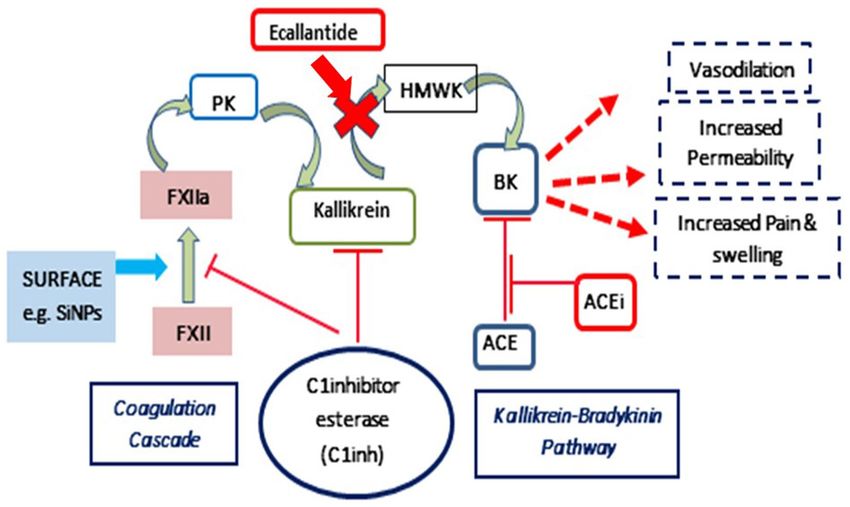
www.nature.com/scientificreports/
angioedema, mainly for causing uncontrollable bradykinin generation42. While natural biological agents, like
NA43, misfolded proteins44, collagen45 and platelet polyphosphate46,47 can cause autoactivation of FXII48,49, a
R
diverse array of biomaterial surfaces like g lass50, dextran s ulphate51 and silica nanoparticles (SiNPs)52 have also
been shown to activate FXII53–58.
In addition to the selection of a driver of acute activation, an objective real time physiologic readout of an
attack, such as hypotension, is also needed for a model of acute HAE attacks. Telemetry devices can be implanted
in mice to allow for real time measurement of blood p ressure59.
In this article, we present the first in vivo murine model to mimic acute HAE attacks. Attacks were induced
by IV injection of a silica nanoparticle (SiNP) suspension60–64. Blood pressure was measured in real time, in
conscious and untethered mice using implanted telemetry. SiNP injection induced a rapid, reversible decrease
in blood pressure, in the presence of angiotensin converting enzyme (ACE) inhibition. The robust and repro-
ducible murine model reported here will facilitate understanding and development of interventions to prevent
and treat acute HAE attacks.
Methods
Animals. Male and female serping1−/− mice as well as wild-type C57BL6J mice (Age 4–6 months) were
used. The serping1-deficient mouse was generated by Deltagen Inc. (San Mateo, CA), applying homologous
recombination using ES cells. A Lac-Z/neo target vector was introduced to disrupt the serping1 (Gene#763,
Serpin 1, Genbank Accession #Y10386 gi:1772997), using mouse ES cells derived from 129/Olahsd mouse sub
strain (line #5961). Southern Blot was used to confirm correct targeting. serping1−/− and serping1+/+ geno-
types were verified by PCR amplification by genomic DNA using primers spanning the LacZ/Neo insertion site
(Supplemental Fig. S1) The resulting serping1−/+ males and females were backcrossed to C57BL6J to generate
congenic strains (Charles River Laboratories). In the FDA vivarium, the serping1−/+ animals were interbred for
more than 20 generations to obtain serping1−/− animals.
We backcrossed the Serping1−/− (129/Olahsd /C57Bl6J background) congenic mouse strain to wild type
C57BL6/J (Stock#000664; Jax Laboratories) for 12 generations to generate the serping1−/− animals on a pure
C57Bl6 background. The genotype and pedigree of the backcrossed mice were tested using the GigaMUGA plat-
form (University of North Carolina at Chapel Hill). A western blot was used to verify the knockout animals lack
expression of an intact C1 inhibitor protein (Supplemental Fig. S2). All experimental animals were maintained
in accordance with the Institutional Animal Care and Use Committee (IACUC) and White Oak Consolidated
Animal Program. The study was carried out in compliance with the ARRIVE guidelines.
Surgical implantation using wireless telemetry. Implantable transmitter HD-X11 (Data Sciences
International, St. Paul, MN) is designed to simultaneously measure mean arterial pressure (MAP), temperature,
locomotor activity and ECG at 10 s intervals allowing for accurate time-resolution of treatment-related effects.
Animals were anesthetized with Isoflurane (by vaporizer—3–5% for induction, 1–3% for maintenance, nose
cone). Buprenorphine SR (sustained release) Lab at 1 mg/kg was used subcutaneously on the day of the surgery.
Because of its sustained release formulation, it can provide analgesic effect for ~ 48–72 h. post-surgery, thereby
reducing handling of post-operative animals. The HD-X11 transmitters were implanted in the intraperitoneal
cavity, with the fluid filled catheter inserted into the left carotid artery, according to manufacturer’s instructions.
After securing the ECG leads on the body muscle, the skin was sutured using non-absorbable 5-O sutures. Post-
operative animals were administered warmed Lactate Ringer solution (LRS). Mice were placed in individual
cages, on heating pads, until the animals regained independent mobility. All procedures conducted on mice were
approved by the IACUC of Division of Veterinary Services (DVS) at the Food and Drug Administration, Silver
Spring, Maryland. Raw data collection and analysis were implemented using Dataquest ART (v. 4.3). A 7-day
period of post-surgery data collection was performed to record baseline data. Later during experimental dosing,
BP data were collected at 10 s intervals for 1 h preceding the intravenous injection and compared to MAP data
collected during a 1-h post injection period (Supplemental data S3–S5).
Efficacy studies in mice. After 1 week of post-surgical recovery serping1−/− mice (3–4 animals/group)
received 100 µl of 0.25 mg of SiNPs (Silica Nanospheres, mean diameter 50 nm, nanoComposix, San Diego,
CA) or saline via bolus intravenous tail injections. The animals were pre-treated with angiotensin converting
enzyme inhibitor (ACEi), Captopril (12.5 mg; Mylan, WV) in their drinking water (0.01 mg/ml), 24 h prior to
injection65. Based on established values for mean daily water c onsumption66 each animal received an approxi-
mate captopril dose of 2.5 mg/kg/day. Individual animals were subjected to up to three separate interventions
under different experimental conditions (e.g. SiNP, Captopril, saline, SiNP+ antagonist). Each intervention was
separated by a 1-week recovery period.
A control group of C57BL6J females (Jax# 00064) was used to compare the effect of SiNPs and ecallantide
(10 mg/ml, Dyax, Burlington,MA) exposing the wild-type (WT) females to same conditions as their serp-
ing1−/− female counterparts.
Effect size calculations. MAP was continuously monitored (every 10 s) via Dataquest ARTv4.3 for each
animal from 30 min prior to experimental intervention (injection of SiNPs) until 60 min after injection. The first
post-injection blood pressure reading was defined as t = 0. Baseline MAP was defined as the mean blood pres-
sure from t = − 15 to t = − 10 min. For each animal, all post-intervention MAP values were expressed relative to
the mean baseline MAP value. The relative MAP at time point n ( RMAPn) was calculated as: MAPn/MAPbaseline.
The effect of SiNP on MAP was calculated at each time point by subtracting the MAP at t = 0 from the meas-
ured MAP. The net RMAP at time point n (Net R MAPn) was expressed as: Net R
MAPn = RMAPn − RMAPt=0. The
Scientific Reports | (2021) 11:15924 | https://doi.org/10.1038/s41598-021-95125-0 2
Vol:.(1234567890)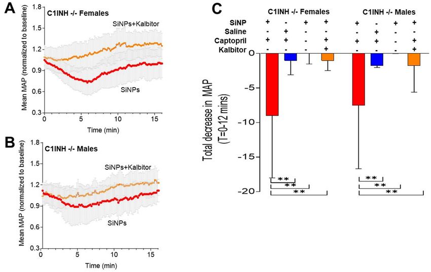
www.nature.com/scientificreports/
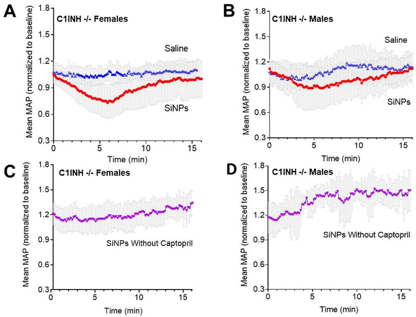
Figure 1. In Vivo stimulation of the contact pathway with SiNPs in serping1−/− mice evokes acute HAE-like
attacks in the presence of Captopril. Bolus IV injection of SiNPs (0.25 mg/100 µl) at t = 0 in captopril treated
serping1−/− females; n = 18 (A) and males; n = 12 (B) evoked a rapid and reversible decrease in MAP. When
serping1−/− females (n = 9) and males (n = 6) were injected with a single bolus IV injection of saline (vehicle), in
the presence of captopril, no change in MAP was reported (A, B). Bolus IV injection of SiNPs (0.25 mg/100 µl)
in the absence of captopril had no such effect on MAP in serping1−/− females; n = 6 (C) or males; n = 3 (D).
Data points represent mean ± SD of normalized MAP.
total effect of SiNP on blood pressure was calculated as the sum of Net RMAP values from t = 0 to 12 min. MAP
values generally returned to baseline values by t = 12 min. Only those time points in which R
MAPn was ≥ 10%
below RMAPt=0 were included in this calculation in order to capture physiologically relevant effects.
Statistical analysis. Statistical analyses were conducted using GraphPad PRISM (8.0 GraphPad Software,
San Diego, CA). All data were expressed as the Mean ± SD. Assessment of the differences between groups of
treatments were performed using 2-way ANOVA (Analysis of Variance) and Bonferroni’s post-hoc tests compar-
ing all group means. A P-value of < 0.05 was considered statistically significant. At least nine animals were evalu-
ated for each experimental group; However, fewer animals were evaluated for some controls. Animal numbers
are included in figure legends.
Results
SiNP injection models acute HAE attack in serping1−/− mice. We chose silica nanoparticles (SiNPs)
to simulate acute HAE attacks in our in vivo murine model as silica nanoparticles (SiNPs)52 have been shown to
activate FXII53–58. A dose of 0.25 mg/100 µl of SiNPs was selected based upon published toxicity data in mice67,68.
With ACEi captopril p retreatment69, intravenous SiNP injection induced a reversible decrease in blood pressure
in serping1−/− females and males (Fig. 1A,B). When the same set of mice with captopril pretreatment were
intravenously injected with saline, they failed to demonstrate any drop-in MAP (Fig. 1A,B). SiNP injections,
in the absence of captopril pretreatment, did not lead to blood pressure drops in serping1−/− males or females
(Fig. 1C,D); in fact, a blood pressure increase post-injection was observed. Examples of individual animal data
are in the “Supplementary Materials S1” (Supplementary Figs. S3 and S4). In addition to decreased blood pres-
sure, observation of captopril and SiNP treated animals revealed markedly decreased locomotor activity relative
to captopril and saline treated controls (Supplementary Fig. S5). These observations reversed on the same time
course as the measured blood pressure changes.
The plasma kallikrein inhibitor ecallantide inhibits SiNP‑induced blood pressure decrease. We
used ecallantide (Kalbitor), an FDA-approved HAE therapy, to evaluate the model’s ability to respond to HAE
Scientific Reports | (2021) 11:15924 | https://doi.org/10.1038/s41598-021-95125-0 3
Vol.:(0123456789)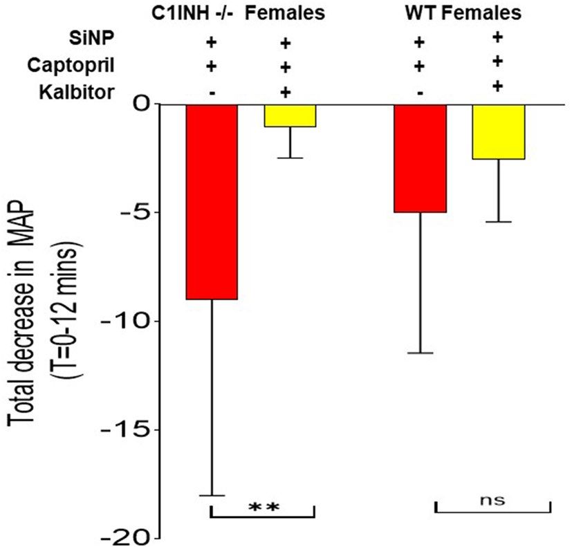
www.nature.com/scientificreports/
Figure 2. Ecallantide (Kalbitor) ameliorates the SiNP-induced MAP decrease in captopril-treated Serping1−/−
mice. (A, B) Kalbitor (100 µg), injected immediately prior to a bolus IV injection of SiNPs (0.25 mg/100 µl)
at t = 0, inhibits the SiNP-induced decrease in mean MAP in both (A) serping1−/− females (n = 9) and
(B) serping1−/− males (n = 9). (C) The histogram compares the total decrease in MAP (t = 0—12 min)
in serping1−/− female and serping1−/− male mice as per the Materials and Methods, with four different
interventions. Data are expressed as mean ± SD. Statistical analysis was performed using 2-way ANOVA with
Bonferroni’s post-hoc tests SiNPs in the presence of captopril vs. saline in the presence of captopril *P = 0.002;
SiNPs in the presence of captopril vs. SiNPs in the absence of Captopril **P = 0.007; SiNPs in the presence of
Captopril vs SiNPs + Ecallantide in the presence of captopril **P = 0.002.
therapeutics. Ecallantide (100 µg/100 μl) was injected immediately prior to SiNP injection in captopril-treated
mice. Ecallantide blocked the SiNP induced MAP decrease in both captopril pretreated male and female serp-
ing1−/− mice (Fig. 2A,B) Silica vs Saline P = 0.002; Silica + Captopril vs Silica Without Captopril P = 0.007, and
Silica vs Kalbitor, P = 0.002. The results of all the interventions were integrated over time as per the “Materials
and methods” in Fig. 2C. Even though the results indicated a potential trend towards sex-based difference in
response to SiNP, post-hoc testing did not indicate significant differences between the sexes (P = 0.87).
Wild‑type females failed to show a significant effect of ecallantide treatment on captopril and
SiNP‑induced in blood pressure drops. Ecallantide inhibited the SiNP-induced MAP decrease in serp-
ing1−/− females (P = 0.01) but not in C57Bl6J females (P = 0.86) as shown in Fig. 3. Assessment of the differences
between groups of treatments was performed using two-way Analysis of Variance (ANOVA) with Bonferroni’s
post-hoc test. SiNP-induced blood pressure decreases were nominally smaller in WT C57Bl6J mice relative to
serping1−/− animals. These data support the conclusion that serping1−/− mice pretreated with captopril and
injected with SiNP are a more sensitive model for HAE-like attacks and treatments compared with WT mice
under the same conditions.
Discussion
Hereditary angioedema is a rare genetic disease with significant mortality that is generally caused by mutations
in the serping1 gene leading to either reduced levels (Type I HAE) or abnormal function (Type II HAE) of the
C1-esterase inhibitor. Although there are murine models for HAE, there has not been an HAE animal model
for acute attacks of angioedema. In this study, we describe a reliable and reproducible in vivo murine model
that mimics acute HAE attacks. In this model, HAE-like attacks are successfully mitigated by the FDA-approved
HAE therapeutic, ecallantide.
The role of bradykinin generation in HAE murine models has been demonstrated in earlier s tudies32. The
importance of captopril, an ACE inhibitor, in this model enhances the role of bradykinin as ACE catabolizes brad-
ykinin rapidly and ACE inhibitors can increase the levels and duration of kallikrein generated b radykinin70–72.
In humans, HAE attacks occur in the absence of ACE i nhibitors69. The need for ACE inhibition in this model
may reflect differences in bradykinin regulation between mice and humans. Human HAE occurs as an autosomal
dominant genetic inheritance pattern and affected patients have residual levels of functional serping1. There
does not seem to be a known syndrome with complete absence of serping1 in humans. Mice with complete loss
of serping1 are viable and do not seem to have spontaneous a ttacks32. These mice may have additional regulation
Scientific Reports | (2021) 11:15924 | https://doi.org/10.1038/s41598-021-95125-0 4
Vol:.(1234567890)www.nature.com/scientificreports/
Figure 3. The Serping1−/− murine model is more sensitive than C57BL6 WT mice for assessing treatment
of acute HAE-like attacks. Comparison of the integrated effect size, as per the Materials and Methods, of
ecallantide treatment of SiNP-induced blood pressure drops in Serping1−/− (n = 18) and WTC57Bl6/J female
mice (n = 9). There is significant amelioration of SiNP-induced MAP decrease with ecallantide treatment in
Serping1−/− females (P = 0.01) as compared to the C57Bl6 females (NS, P = 0.86). Assessment of the differences
between groups of treatments were performed using 2-way Analysis of Variance (ANOVA) with Bonferroni’s
post-hoc test (used to determine the significance of mean MAP change of all groups against each other) with
*P < 0.02; NS = non-significant. Data are expressed as mean ± SD.
Figure 4. Contact system pathways with the kallikrein-bradykinin pathway and Serping1. Serping1 acts as
the primary plasma inhibitor of the contact-kinin system cascades. In the absence of Serping1, FXIIa converts
prekallikrein (PK) to kallikrein, which in turn cleaves high molecular weight kininogen (HK) and releases
bradykinin (BK). Bradykinin is degraded by ACE and other plasma enzymes. The levels and duration of
bradykinin in plasma impacts vascular permeability and vasodilation, leading to decreases in blood pressure
and swelling; a hallmark for HAE. Ecallantide acts as a plasma kallikrein inhibitor and limits the production of
bradykinin from HK.
Scientific Reports | (2021) 11:15924 | https://doi.org/10.1038/s41598-021-95125-0 5
Vol.:(0123456789)www.nature.com/scientificreports/
of kallikrein-mediated bradykinin generation and/or bradykinin degradation (Fig. 4), for example, non-ACE
pathways of bradykinin clearance. There may also be other compensatory mechanisms. Of note, in the absence of
ACE inhibition, the post-SiNP injection trend of increased blood pressure may reflect other regulatory systems
in mice. Such differences may explain the need for ACE inhibition in this model.
A murine model of C1 inhibitor deficiency with a heterozygous genotype and increased vascular perme-
eveloped73. This model used CRISPR‐Cas9 to generate a truncated protein rather than
ability despite has been d
inserting another construct into the Serping1 gene as we and o thers32 have done. Our model targets exon 4 and
allows for antigen expression but deletes an ~ 75 KDa C1 inhibitor reactive band compatible with glycosylated C1
inhibitor (Supplemental Fig. S2). We cannot rule out alternative splice variants and there is some MAP impact
of our approach in WT mice; However, we do observe a difference between the KO mice and WT mice (Fig. 3).
The heterozygote model better mimics heterozygote human disease; However, the model did not exhibit
spontaneous attacks. A rat model of HAE did have some spontaneous intestinal swelling; however, this model
was based on bradykinin overexpression rather than Serping1 mutations. Directly mimicking all features of
human disease is challenging as human disease phenotypes may be dependent on specific m utations74. as well
as species-specific characteristics of the contact system. Our approach of using real-time MAP monitoring and
an inducer of acute attacks, such as SiNP, could be applied across a range of HAE animal models and increase
the utility of such models.
This model can also further explore sex-related effects in HAE with additional studies in larger numbers of
mice, in both serping1−/− and WT animals. The evaluation of sex differences is important because female sex
hormones, mainly estrogen, may impact bradykinin generation75–77. This robust in vivo model may also facili-
tate further study of the mechanisms that can induce acute HAE attacks. In summary, the serping1−/− murine
model is the first model to evoke acute HAE-like attacks and can also be used to study approved HAE drugs
(i.e. danacrine, icatibant, cinryze, lanadelumab)78–82, and support development of future targeted novel drug
therapies for HAE prophylaxis.
Received: 24 July 2020; Accepted: 15 June 2021
References
1. Osler, W. Landmark publication from The American Journal of the Medical Sciences: Hereditary angio-neurotic oedema. 1888.
Am. J. Med. Sci. 339(2), 175–178 (2010).
2. Nzeako, U. C., Frigas, E. & Tremaine, W. J. Hereditary angioedema: A broad review for clinicians. Arch. Intern. Med. 161(20),
2417–2429 (2001).
3. Cicardi, M. & Agostoni, A. Hereditary angioedema. N. Engl. J. Med. 334(25), 1666–1667 (1996).
4. Ebo, D. G., Stevens, W. J. & Bosmans, J. L. An adverse reaction to angiotensin-converting enzyme inhibitors in a patient with
neglected C1 esterase inhibitor deficiency. J. Allergy Clin. Immunol. 99(3), 425–426 (1997).
5. Hoyer, C., Hill, M. R. & Kaminski, E. R. Angio-oedema: An overview of differential diagnosis and clinical management. Continuing
Educ. Anaesthesia Crit. Care Pain. 12(6), 307–311 (2012).
6. Johnston, D. T. Diagnosis and management of hereditary angioedema. J. Am. Osteopathic Assoc. 111(1), 28–36 (2011).
7. Longhurst, H. J. & Bork, K. Hereditary angioedema: An update on causes, manifestations and treatment. Br. J. Hosp. Med. (Lond.).
80(7), 391–398 (2019).
8. Zuraw, B. L. et al. A focused parameter update: Hereditary angioedema, acquired C1 inhibitor deficiency, and angiotensin-
converting enzyme inhibitor-associated angioedema. J. Allergy Clin. Immunol. 131(6), 1491–1493 (2013).
9. Kaplan, A. P. Angioedema. World Allergy Org. J. 1(6), 103–113 (2008).
10. Donaldson, V. H. & Evans, R. R. A biochemical abnormality in hereditary angioneurotic edema. Am. J. Med. 35(1), 37–44 (1963).
11. Kaplan, A. P. & Joseph, K. The bradykinin-forming cascade and its role in hereditary angioedema. Ann. Allergy Asthma Immunol.
Off. Publ. Am. Coll. Allergy Asthma Immunol. 104(3), 193–204 (2010).
12. de Maat, S. et al. Plasmin is a natural trigger for bradykinin production in patients with hereditary angioedema with factor XII
mutations. J. Allergy Clin. Immunol. 138(5), 1414–1423.e9 (2016).
13. Dewald, G. & Bork, K. Missense mutations in the coagulation factor XII (Hageman factor) gene in hereditary angioedema with
normal C1 inhibitor. Biochem. Biophys. Res. Commun. 343(4), 1286–1289 (2006).
14. Lucas, A., Yaron, J. R., Zhang, L. & Ambadapadi, S. Overview of serpins and their roles in biological systems. Methods Mol. Biol.
(Clifton, NJ). 1826, 1–7 (2018).
15. Hofman, Z. L. et al. Angioedema attacks in patients with hereditary angioedema: Local manifestations of a systemic activation
process. J. Allergy Clin. Immunol. 138(2), 359–366 (2016).
16. Bouillet, L., Boccon-Gibod, I. & Massot, C. Bradykinin mediated angioedema. Rev. Med. Interne. 32(4), 225–231 (2011).
17. Agostoni, A. & Cicardi, M. Hereditary and acquired C1-inhibitor deficiency: Biological and clinical characteristics in 235 patients.
Medicine 71(4), 206–215 (1992).
18. Asmis, L. M., Sulzer, I., Furlan, M. & Lammle, B. Prekallikrein deficiency: The characteristic normalization of the severely prolonged
aPTT following increased preincubation time is due to autoactivation of factor XII. Thromb. Res. 105(6), 463–470 (2002).
19. Cugno, M., Nussberger, J., Cicardi, M. & Agostoni, A. Bradykinin and the pathophysiology of angioedema. Int. Immunopharmacol.
3(3), 311–317 (2003).
20. Nussberger, J., Cugno, M. & Cicardi, M. Bradykinin-mediated angioedema. N. Engl. J. Med. 347(8), 621–622 (2002).
21. Schmaier, A. H. Plasma prekallikrein: Its role in hereditary angioedema and health and disease. Front. Med. (Lausanne). 5, 3 (2018).
22. Schmaier, A. H. The hereditary angioedema syndromes. J. Clin. Investig. 129(1), 66–68 (2019).
23. De Maat, S., Hofman, Z. L. M. & Maas, C. Hereditary angioedema: The plasma contact system out of control. J. Thromb. Haemostasis
JTH. 16(9), 1674–1685 (2018).
24. Nussberger, J. et al. Plasma bradykinin in angio-oedema. Lancet (London, England). 351(9117), 1693–1697 (1998).
25. Zuraw, B. L. The pathophysiology of hereditary angioedema. World Allergy Org. J. 3(9 Suppl), S25–S28 (2010).
26. Lara-Marquez, M. L., Christiansen, S. C., Riedl, M. A., Herschbach, J. & Zuraw, B. L. Threshold-stimulated kallikrein activity
distinguishes bradykinin- from histamine-mediated angioedema. Clin. Exp. Allergy J. Br. Soc. Allergy Clin. Immunol. 48(11),
1429–1438 (2018).
27. Zuraw, B. L. Hereditary angioedema with normal C1 inhibitor: Four types and counting. J. Allergy Clin. Immunol. 141(3), 884–885
(2018).
Scientific Reports | (2021) 11:15924 | https://doi.org/10.1038/s41598-021-95125-0 6
Vol:.(1234567890)www.nature.com/scientificreports/
28. Ghannam, A. et al. Contact system activation in patients with HAE and normal C1 inhibitor function. Immunol. Allergy Clin. N.
Am. 33(4), 513–533 (2013).
29. Magerl, M., Germenis, A. E., Maas, C. & Maurer, M. Hereditary angioedema with normal C1 inhibitor: Update on evaluation and
treatment. Immunol. Allergy Clin. N. Am. 37(3), 571–584 (2017).
30. Bjorkqvist, J. et al. Defective glycosylation of coagulation factor XII underlies hereditary angioedema type III. J. Clin. Investig.
125(8), 3132–3146 (2015).
31. Davis, A. E. 3rd. New treatments addressing the pathophysiology of hereditary angioedema. Clin. Mol. Allergy CMA. 6, 2 (2008).
32. Han, E. D., MacFarlane, R. C., Mulligan, A. N., Scafidi, J. & Davis, A. E. 3rd. Increased vascular permeability in C1 inhibitor-
deficient mice mediated by the bradykinin type 2 receptor. J. Clin. Investig. 109(8), 1057–1063 (2002).
33. Shoemaker, L. R., Schurman, S. J., Donaldson, V. H. & Davis, A. E. 3rd. Hereditary angioneurotic oedema: Characterization of
plasma kinin and vascular permeability-enhancing activities. Clin. Exp. Immunol. 95(1), 22–28 (1994).
34. Boulanger, C., Schini, V. B., Moncada, S. & Vanhoutte, P. M. Stimulation of cyclic GMP production in cultured endothelial cells
of the pig by bradykinin, adenosine diphosphate, calcium ionophore A23187 and nitric oxide. Br. J. Pharmacol. 101(1), 152–156
(1990).
35. Colman, R. W. & Schmaier, A. H. Contact system: A vascular biology modulator with anticoagulant, profibrinolytic, antiadhesive,
and proinflammatory attributes. Blood 90(10), 3819–3843 (1997).
36. Dixon, B. S. et al. Effects of kinins on cultured arterial smooth muscle. Am. J. Physiol. 258(2 Pt 1), C299-308 (1990).
37. Parratt, J. R. Cardioprotection by angiotensin converting enzyme inhibitors—The experimental evidence. Cardiovasc. Res. 28(2),
183–189 (1994).
38. Schini, V. B., Boulanger, C., Regoli, D. & Vanhoutte, P. M. Bradykinin stimulates the production of cyclic GMP via activation of
B2 kinin receptors in cultured porcine aortic endothelial cells. J. Pharmacol. Exp. Therapeutics 252(2), 581–585 (1990).
39. Wang, J. et al. Human tissue kallikrein induces hypotension in transgenic mice. Hypertension (Dallas, Tex: 1979). 23(2), 236–243
(1994).
40. Wiemer, G., Scholkens, B. A. & Linz, W. Endothelial protection by converting enzyme inhibitors. Cardiovasc. Res. 28(2), 166–172
(1994).
41. Schapira, M., Scott, C. F. & Colman, R. W. Protection of human plasma kallikrein from inactivation by C1 inhibitor and other
protease inhibitors. The role of high molecular weight kininogen. Biochemistry 20(10), 2738–2743 (1981).
42. Bhoola, K. D., Figueroa, C. D. & Worthy, K. Bioregulation of kinins: Kallikreins, kininogens, and kininases. Pharmacol. Rev. 44(1),
1–80 (1992).
43. Kannemeier, C. et al. Extracellular RNA constitutes a natural procoagulant cofactor in blood coagulation. Proc. Natl. Acad. Sci.
USA 104(15), 6388–6393 (2007).
44. Maas, C. et al. Misfolded proteins activate factor XII in humans, leading to kallikrein formation without initiating coagulation. J.
Clin. Investig. 118(9), 3208–3218 (2008).
45. van der Meijden, P. E. et al. Dual role of collagen in factor XII-dependent thrombus formation. Blood 114(4), 881–890 (2009).
46. Smith, S. A. et al. Polyphosphate exerts differential effects on blood clotting, depending on polymer size. Blood 116(20), 4353–4359
(2010).
47. Smith, S. A. et al. Polyphosphate modulates blood coagulation and fibrinolysis. Proc. Natl. Acad. Sci. USA 103(4), 903–908 (2006).
48. Schmaier, A. H. The elusive physiologic role of Factor XII. J. Clin. Investig. 118(9), 3006–3009 (2008).
49. Kaplan, A. P., Joseph, K. & Silverberg, M. Pathways for bradykinin formation and inflammatory disease. J. Allergy Clin. Immunol.
109(2), 195–209 (2002).
50. Rapaport, S. I., Aas, K. & Owren, P. A. The effect of glass upon the activity of the various plasma clotting factors. J. Clin. Investig.
34(1), 9–19 (1955).
51. Siebeck, M. et al. Dextran sulfate activates contact system and mediates arterial hypotension via B2 kinin receptors. J. Appl. Physiol.
(Bethesda, Md: 1985). 77(6), 2675–2680 (1994).
52. Murugadoss, S. et al. Toxicology of silica nanoparticles: An update. Arch. Toxicol. 91(9), 2967–3010 (2017).
53. Maas, C. & Renne, T. Coagulation factor XII in thrombosis and inflammation. Blood 131(17), 1903–1909 (2018).
54. Wu, Y. Contact pathway of coagulation and inflammation. Thromb. J. 13, 17 (2015).
55. Gryshchuk, V. & Galagan, N. Silica nanoparticles effects on blood coagulation proteins and platelets. J. Biochem. Res. Int. 2016, 6
(2016).
56. Kushida, T., Saha, K., Subramani, C., Nandwana, V. & Rotello, V. M. Effect of nano-scale curvature on the intrinsic blood coagula-
tion system. Nanoscale 6(23), 14484–14487 (2014).
57. Maas, C., Oschatz, C. & Renne, T. The plasma contact system 2.0. Semin. Thromb. Hemost. 37(4), 375–381 (2011).
58. Long, A. T., Kenne, E., Jung, R., Fuchs, T. A. & Renne, T. Contact system revisited: An interface between inflammation, coagula-
tion, and innate immunity. J. Thromb. Haemostasis JTH. 14(3), 427–437 (2016).
59. Zhao, X. et al. Arterial pressure monitoring in mice. Curr. Protocols Mouse Biol. 1, 105–122 (2011).
60. Levy, J. H. & O’Donnell, P. S. The therapeutic potential of a kallikrein inhibitor for treating hereditary angioedema. Expert Opin.
Investig. Drugs 15(9), 1077–1090 (2006).
61. Schneider, L., Lumry, W., Vegh, A., Williams, A. H. & Schmalbach, T. Critical role of kallikrein in hereditary angioedema patho-
genesis: A clinical trial of ecallantide, a novel kallikrein inhibitor. J. Allergy Clin. Immunol. 120(2), 416–422 (2007).
62. Williams, A. & Baird, L. G. DX-88 and HAE: A developmental perspective. Transfusion Apheresis Sci. Off. J. World Apheresis Assoc.
Off. J. Eur. Soc. Haemapheresis. 29(3), 255–258 (2003).
63. Riedl, M. Hereditary angioedema therapy: Kallikrein inhibition and bradykinin receptor antagonism. World Allergy Org. J. 3(9
Suppl), S34–S38 (2010).
64. Lewis, L. M. et al. Ecallantide for the acute treatment of angiotensin-converting enzyme inhibitor-induced angioedema: A multi-
center, randomized, controlled trial. Ann. Emerg. Med. 65(2), 204–213 (2015).
65. Emanueli, C., Angioni, G. R., Anania, V., Spissu, A. & Madeddu, P. Blood pressure responses to acute or chronic captopril in mice
with disruption of bradykinin B2-receptor gene. J. Hypertens. 15(12 Pt 2), 1701–1706 (1997).
66. Sweet DV. Registry of toxic effects of chemical substances (RTECS): Comprehensive guide to the RTECS: US Department of Health
and Human Services, Public Health Service, Centers (1997). https://www.cdc.gov/niosh/docs/97-119/pdfs/97-119.pdf. Accessed
July 26, 2021.
67. Chan, W. T. et al. In vivo toxicologic study of larger silica nanoparticles in mice. Int. J. Nanomed. 12, 3421–3432 (2017).
68. Cho, M. et al. The impact of size on tissue distribution and elimination by single intravenous injection of silica nanoparticles.
Toxicol. Lett. 189(3), 177–183 (2009).
69. Agostoni, A. & Cicardi, M. Contraindications to the use of ace inhibitors in patients with C1 esterase inhibitor deficiency. Am. J.
Med. 90(2), 278 (1991).
70. Gainer, J. V., Morrow, J. D., Loveland, A., King, D. J. & Brown, N. J. Effect of bradykinin-receptor blockade on the response to
angiotensin-converting-enzyme inhibitor in normotensive and hypertensive subjects. N. Engl. J. Med. 339(18), 1285–1292 (1998).
71. Pretorius, M., Rosenbaum, D., Vaughan, D. E. & Brown, N. J. Angiotensin-converting enzyme inhibition increases human vascular
tissue-type plasminogen activator release through endogenous bradykinin. Circulation 107(4), 579–585 (2003).
72. Straka, B. T. et al. Effect of bradykinin receptor antagonism on ACE inhibitor-associated angioedema. J. Allergy Clin. Immunol.
140(1), 242–248.e2 (2017).
Scientific Reports | (2021) 11:15924 | https://doi.org/10.1038/s41598-021-95125-0 7
Vol.:(0123456789)www.nature.com/scientificreports/
73. Qiu, T. et al. Gene therapy for C1 esterase inhibitor deficiency in a Murine Model of Hereditary angioedema. Allergy 74(6),
1081–1089 (2019).
74. Caccia, S. et al. Intermittent C1-inhibitor deficiency associated with recessive inheritance: functional and structural insight. Sci.
Rep. 8(1), 977 (2018).
75. Banerji, A. & Riedl, M. Managing the female patient with hereditary angioedema. Womens Health (Lond. Engl.) 12(3), 351–361
(2016).
76. Bouillet, L. Hereditary angioedema in women. Allergy Asthma Clin. Immunol. 6(1), 17 (2010).
77. Bouillet, L. & Gompel, A. Hereditary angioedema in women: Specific challenges. Immunol. Allergy Clin. N. Am. 33(4), 505–511
(2013).
78. Banerji, A. et al. Effect of lanadelumab compared with placebo on prevention of hereditary angioedema attacks: A randomized
clinical trial. JAMA 320(20), 2108–2121 (2018).
79. Bernstein, J. A. et al. Escalating doses of C1 esterase inhibitor (CINRYZE) for prophylaxis in patients with hereditary angioedema.
J. Allergy Clin. Immunol. Pract. 2(1), 77–84 (2014).
80. Bork, K., Bygum, A. & Hardt, J. Benefits and risks of danazol in hereditary angioedema: A long-term survey of 118 patients. Ann.
Allergy Asthma Immunol. Off. Publ. Am. Coll. Allergy Asthma Immunol. 100(2), 153–161 (2008).
81. Bouillet, L. Icatibant in hereditary angioedema: News and challenges. Expert Rev. Clin. Immunol. 7(3), 267–272 (2011).
82. Sexton, D. J. et al. Comparison of plasma kallikrein inhibition by the endogenous C1-inhibitor versus DX-2930, a monoclonal
antibody inhibitor. Blood 122(21), 1066 (2013).
Acknowledgements
We would like to gratefully acknowledge internal peer reviewers Marjorie Shapiro and Melanie Blank for their
critical reading of the manuscript. We would like to thank Ashwinkumar Bhirde, Mate Tolnay, Christie Jane
Fennell, Odile Engel Tzanko Stantchev and Carole Sourbier for their help and support. The authors thank John
Dennis, Eric Nimako and DVS technical staff (Division of Veterinary Sciences, Silver Spring, FDA) for outstand-
ing support with animal protocol and study.
Author contributions
S.B., M.W., and S.K. conceived the study. S.B., M.W., M.L., S.K., J.P., J.D. and Z.Z. designed experiments. S.B.
and J.P. performed experiments. S.B. performed in vivo surgical manipulation. S.B. and M.W. implemented data
extraction and normalization. M.L. advised and assisted with statistical interpretations. S.B., M.W., M.L. and S.K.
wrote the manuscript. S.B., M.W., M.L., S.K., J.P., J.D. and Z.Z. reviewed the manuscript.
Competing interests
The authors declare no competing interests.
Additional information
Supplementary Information The online version contains supplementary material available at https://doi.org/
10.1038/s41598-021-95125-0.
Correspondence and requests for materials should be addressed to S.K.
Reprints and permissions information is available at www.nature.com/reprints.
Publisher’s note Springer Nature remains neutral with regard to jurisdictional claims in published maps and
institutional affiliations.
Open Access This article is licensed under a Creative Commons Attribution 4.0 International
License, which permits use, sharing, adaptation, distribution and reproduction in any medium or
format, as long as you give appropriate credit to the original author(s) and the source, provide a link to the
Creative Commons licence, and indicate if changes were made. The images or other third party material in this
article are included in the article’s Creative Commons licence, unless indicated otherwise in a credit line to the
material. If material is not included in the article’s Creative Commons licence and your intended use is not
permitted by statutory regulation or exceeds the permitted use, you will need to obtain permission directly from
the copyright holder. To view a copy of this licence, visit http://creativecommons.org/licenses/by/4.0/.
© The Author(s) 2021
Scientific Reports | (2021) 11:15924 | https://doi.org/10.1038/s41598-021-95125-0 8
Vol:.(1234567890)You can also read