A new inflammatory parameter can predict delayed intracranial hemorrhage following ventriculoperitoneal shunt - Nature
←
→
Page content transcription
If your browser does not render page correctly, please read the page content below
www.nature.com/scientificreports
OPEN A new inflammatory
parameter can predict delayed
intracranial hemorrhage
following ventriculoperitoneal
shunt
Shiwei Li1*, Hongcai Wang1, Feng Li2, Maosong Chen1 & Pandi Chen1
Delayed intracerebral hemorrhage (DICH) secondary to ventriculoperitoneal (VP) shunt is considered
to be a potentially severe event. This study aimed to investigate the association between a ratio of
postoperative neutrophil-to-lymphocyte ratio to preoperative neutrophil-to-lymphocyte ratio (NLRR)
and DICH secondary to VP shunt. We performed a retrospective review of patients who underwent
VP shunt between January 2016 and June 2020. Multivariable logistic regression analysis was used to
assess the association of DICH and NLRR. Then patients were divided into two groups according to the
optimal cut-off point of NLRR, propensity score matching (PSM) method was performed to reconfirm
the result. A total of 130 patients were enrolled and DICH occurred in 29 patients. Elevated NLRR and
history of craniotomy were independent risk factors for DICH secondary to VP shunt. The optimal cut
off point of NLRR was 2.05, and the sensitivity was 89.7%, the specificity was 63.4%. Patients with
NLRR > 2.05 had much higher incidence of DICH (40.6% vs 4.5%). Our finding suggested that DICH
following VP shunt was not a rare complication and elevated NLRR could independently predict DICH.
Inflammatory responses might play an important role in the development of DICH following VP shunt.
Abbreviations
DICH Delayed intracerebral hemorrhage
VP Ventriculoperitoneal
NLRR A ratio of postoperative neutrophil-to-lymphocyte ratio to preoperative neutrophil-to-lymphocyte
ratio
PSM Propensity score matching
OR Odds ratio
CI Confidence interval
ROC Receiver operating characteristic
AUC Area under the curve
NLR Neutrophil-to-lymphocyte ratio
GCS Glasgow Coma Scale
GOS Glasgow outcome scale
ICH Spontaneous intracerebral hemorrhage
IQR Interquartile range
INR International normalized ration
PT Prothrombin time
APTT Activated partial thromboplastin time
PLT Thrombocytes
Ventriculoperitoneal (VP) shunt is the most common treatment for h ydrocephalus1, and the placement of VP
shunt is one of the routine neurosurgical procedures w orldwide2. The common complications secondary to VP
shunt are shunt obstruction, infection, seizure, subdural hemorrhage and shunt m alfunction3,4. Mild hemorrhage
1
Neurosurgery Department of Ningbo Medical Center Lihuili Hospital, Zhejiang, China. 2Medical Imaging
Department of Ningbo Medical Center Lihuili Hospital, Zhejiang, China. *email: 516760848@qq.com
Scientific Reports | (2021) 11:13763 | https://doi.org/10.1038/s41598-021-93315-4 1
Vol.:(0123456789)
www.nature.com/scientificreports/
is frequently observed in the ventricle or in the parenchyma soon after operation5, and the rate could be up to
43.1%6. However, the delayed intracerebral hemorrhage (DICH) is considered to be a rare but potentially severe
event7, the mortality is as high as 50%4. The risk factors and underlying mechanisms of DICH are still not fully
elucidated. There are several risk factors proposed by previously published articles8–11: (1) advanced age, age
≥ 60 years; (2) history of craniotomy; (3) delayed partial thromboplastin time (PTT); (4) brain edema around
catheter; (5) postoperative manipulation of valve system; (6) postoperative anticoagulation.
A recent study pointed out that the systemic inflammatory responses might be involved in the pathologic
process of active cerebral h emorrhage12. We hereby hypothesize that inflammatory response is one of the mecha-
nisms associated with DICH following VP shunt. The neutrophil-to-lymphocyte ratio (NLR), as a rapid and
economic biomarker of systemic inflammation13, has been a dependable predictor of clinical outcome in patients
with spontaneous intracerebral h emorrhage14 and traumatic brain i njury15. Considering that the value of NLR
is greatly influenced by the basic systemic inflammatory statuses such as pneumonia or urinary infection, we
proposed a new parameter named NLRR, what is a ratio of postoperative NLR to preoperative NLR. In this
study, we sought to test the hypothesis that elevated NLRR is associated with the DICH secondary to VP shunt.
Methods
Patient selection. We performed a retrospective review of patients who underwent VP shunt between
January 2016 and June 2020 from the Neurosurgery Department of Ningbo Medical Center Lihuili Hospital.
Inclusion criteria were as follows: (1) age ≥ 18 years; (2) the diagnosis of hydrocephalus was confirmed by clin-
ical symptoms and imaging examination, and VP shunt was performed in our hospital; (3) laboratory tests
(blood routine and coagulation function) within 5 days before VP shunt, and blood routine on the first morning
after VP shunt; (4) postoperative brain computed tomography (CT) scan was performed on the first day after
operation (later than the postoperative test of blood routine), and at least one CT scan was performed within
5–10 days after operation. The exclusion criteria were as follows: (1) patients on a regimen of anticoagulant or
antiplatelet therapy; (2) patients with Ommaya reservoir implantation, the Ommaya tube was directly connected
to the shunt pump without ventricular puncture, or the Ommaya tube was removed during the surgery; (3)
cranioplasty and VP shunt were performed simultaneously; (4) a revision of the VP shunt; (5) early intracerebral
hemorrhage after VP shunt, which was defined as bleeding on the first day after operation.
DICH was defined as subsequent hemorrhage in the ventricle or the parenchyma along the catheter path
which was not found in the CT scan on the first day after operation. The patients enrolled in the study were
divided into two groups according to whether or not DICH. The patient flowchart was summarized in Fig. 1.
Data collection. Demographic characteristics and clinical variables were collected such as: sex and age;
history of hypertension and diabetes; history of craniotomy and skull defect; preoperative pneumonia and Glas-
gow Coma Scale (GCS); primary intracranial lesions including normal hydrocephalus, trauma, spontaneous
intracerebral hemorrhage(ICH), tumor and inflammation; hydrocephalus types including low pressure hydro-
cephalus (LPH, cerebrospinal fluid pressure < 80 mm H 2O), normal pressure hydrocephalus (NPH, 80 mm H 2O
≤ cerebrospinal fluid pressure ≤ 180 mm H2O), high pressure hydrocephalus (HPH, cerebrospinal fluid pressure
> 180 mm H2O). For the patients with DICH secondary to VP Shunt, the onset day of hemorrhage, types of
hemorrhage, with or without symptom after hemorrhage and the Glasgow outcome scale (GOS) were also col-
lected. Laboratory variables were retrieved from our hospital’s database. International normalized ration (INR),
prothrombin time (PT), activated partial thromboplastin time (APTT), serum thrombocytes, neutrophils and
lymphocytes were collected within 5 days before operation. Postoperative serum neutrophils and lymphocytes
were collected on the first morning after operation. All of the preoperative and postoperative brain CT scans
were obtained, and respectively reviewed by one neurosurgeon and one radiologist who blinded to the detail
information (demographic and clinical variables, laboratory data) of patients. The different opinions between
them were resolved by consultation. The volume of hematoma was calculated by 3D slicer (version 4.10.2).
Besides, postoperative cerebral edema around catheter on the first postoperative CT scan was also collected.
Procedural technique. All of the patients were implanted with Medtronic Strata Adjustable Pressure Valve
Systems, and the initial pressures were collected. The standard technique for VP shunt was employed, and the
catheter was placed into the left or right anterior frontal horn via a bur hole at Kocher’s point. The postoperative
manipulation of valve system was recorded before the occurrence of DICH, or within 15 days after operation of
patient without DICH.
Statistical analysis. SPSS version 26.0 (IBM Corporation, Armonk, New York, USA) and R Software
(version 4.0.2) were used for data analysis with statistical significance was defined as P < 0.05. The Kolmogo-
rov–Smirnov test was used to determine the distributions of continuous variables. The continuous variables
with non-normally distributions were analyzed by Mann–Whitney test, presented as the median (50th) with
interquartile range (IQR). Categorical variables were presented as number (proportion). Mann–Whitney test
was used for ordered categorical variables, and unordered categorical variables were analyzed by Pearsons chi-
square test, Continuous correction chi-square test or Fishers exact test. The variables with P < 0.1 and variables
proposed by previously published articles, were selected by Least Absolute Shrinkage and Selection Operator
(LASSO) regression, and then included into multivariable logistic regression analysis to assess the association
of DICH and NLRR. The predictive value of NLRR for DICH following VP shunt in patients was evaluated by
receiver operating characteristic (ROC) curve analysis. According to the optimal cut-off point of NLRR (the
point at which the value of “sensitivity + specificity—1” was maximum), patients were divided into two groups
(NLRR ≤ cut-off point group and NLRR > cut-off point group). Propensity score matching (PSM) method was
Scientific Reports | (2021) 11:13763 | https://doi.org/10.1038/s41598-021-93315-4 2
Vol:.(1234567890)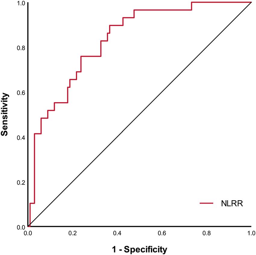
www.nature.com/scientificreports/

                                  Figure 1.  Flowchart of patient selection.

                                  performed to adjust for imbalances of patients’ characteristics between two groups. Covariates such as sex, age,
                                  history of hypertension and diabetes, history of craniotomy and skull defect, preoperative pneumonia and GCS
                                  grade, primary intracranial lesion, hydrocephalus type, preoperative PT and APTT, preoperative INR and PLT,
                                  puncture site, initial pressure of vale system, brain edema around catheter and postoperative manipulation of
                                  valve system were matched at a ratio of 1:1 using a caliper width of 0.2. The estimation algorithm of propensity
                                  score was logistic regression and matching algorithm was nearest neighbor matching. After PSM, 82 patients
                                  (NLRR ≤ 2.05 group: n = 41, NLRR > 2.05 group: n = 41) were selected to analysis.

                                  Ethics approval and consent to participate. This study was performed in accordance with the Declara-
                                  tion of Helsinki of the World Medical Association, and all procedures were carried out in accordance with the
                                  approved guidelines and regulations. This study was approved by our institutional ethics committee (Ningbo
                                  Medical Center Lihuili Hospital, Approval Number: KY2020PJ111). Participant data were retrospectively
                                  reviewed and deidentified. Because of anonymization, consent was waived by the ethics committee of Ningbo
                                  Medical Center Lihuili Hospital.

                                  Results
                                  Characteristics of the patients. A total of 130 patients underwent VP shunt were included in this study.
                                  Because there were 36 patients with early intracerebral hemorrhage after VP shunt who were excluded, the
                                  overall incidence of DICH secondary to VP shunt was 17.5% (29/166). The median age of the 130 patients was
                                  60 years, and 78 (60.0%) patients were male (Table 1). The 130 patients were divided into two groups: non-
                                  DICH group (n = 101) and DICH group (n = 29). Characteristics and clinical data were compared between the

Scientific Reports |   (2021) 11:13763 |               https://doi.org/10.1038/s41598-021-93315-4                                               3

                                                                                                                                           Vol.:(0123456789)
www.nature.com/scientificreports/
Variables n (%) or median [IQR]
Demographics
Male sex 78 (60.0)
Age (years) 60.0 [53.0–66.0]
Clinical history
Hypertension 49 (37.7)
Diabetes mellitus 16 (12.3)
Craniotomy 75 (57.7)
Skull defect 34 (26.2)
Preoperative pneumonia 44 (33.8)
Preoperative GCS 13 [9–15]
Primary intracranial lesion
Normal hydrocephalus 8 (6.2)
Trauma 56 (43.1)
ICH 48 (36.9)
Tumor 15 (11.5)
Inflammation 3 (2.3)
Hydrocephalus type
LPH 11 (8.5)
NPH 101 (77.7)
HPH 18 (13.8)
Laboratory test
Pre-PT (s) 11.9 [11.3–12.6]
Pre-APTT (s) 30.2 [28.0–32.2]
Pre-INR 1.04 [1.00–1.09]
Pre-PLT (*103/μL) 216 [173–264]
Pre-NLR 2.67 [1.60–4.3.8]
Post-NLR 5.45 [3.59–7.35]
NLRR 2.02 [1.38–3.16]
Puncture site
Left precornu 45 (34.6)
Right precornu 85 (65.4)
Initial pressure of vale system
1.0 30 (23.1)
1.5 67 (51.5)
2.0 28 (21.5)
2.5 5 (3.8)
Brain edema around catheter 26 (20.0)
Manipulation of valve system 54 (41.5)
Table 1. Characteristics and clinical data of the patients. GCS Glasgow Coma Scale, ICH spontaneous
intracerebral hemorrhage, LPH low pressure hydrocephalus, NPH normal pressure hydrocephalus, HPH high
pressure hydrocephalus, Pre-PT preoperative prothrombin time, Pre-APTT preoperative activated partial
thromboplastin time, Pre-INR preoperative international normalized ration, Pre-PLT preoperative serum
thrombocyte, Pre-NLR preoperative neutrophil-to-lymphocyte ratio, Post-NLR postoperative neutrophil-to-
lymphocyte ratio, NLRR a ratio of post-NLR to pre-NLR.
two groups and shown in Supplementary Table S1 online. Most of the data were comparable, except history of
craniotomy and preoperative NLR, postoperative NLR, NLRR. 15 patients in the DICH group (51.9%) presented
a history of craniotomy, while the number in the non-DICH group is 34 (33.7%) (P = 0.008). Lower preoperative
NLR (P = 0.037) and higher postoperative NLR (P = 0.001) were observed in patients with DICH secondary to
VP shunt. NLRR in the DICH group was much higher than that in the non-DICH group (P < 0.001).
Association between elevated NLRR and DICH. Hypertension, history of craniotomy, preoperative
APTT, preoperative PTL, preoperative NLR, postoperative NLR, NLRR (P < 0.1) and age, brain edema around
catheter, postoperative manipulation of valve system (proposed by previously published articles), were included.
The linear relationships between the continuous independent variables and the logit conversation of dependent
variable were confirmed by Box–Tidwell test. The indicators of multicollinearity (tolerance, variance inflation
factor, eigenvalue, condition index) were statistically tested, and the results of eigenvalue and condition index
Scientific Reports | (2021) 11:13763 | https://doi.org/10.1038/s41598-021-93315-4 4
Vol:.(1234567890)www.nature.com/scientificreports/
Crude Adjusted
OR (95% CI) P OR (95% CI) P
Hypertension 2.111 (0.914–4.877) 0.080 1.742 (0.612–4.962) 0.298
Craniotomy 3.612 (1.356–9.620) 0.010 3.394 (1.060–10.869) 0.040
Post-NLR 1.142 (1.025–1.274) 0.016 1.110 (0.971–1.267) 0.125
NLRR 2.839 (1.843–4.374) < 0.001 2.792 (1.747–4.460) < 0.001
Manipulation of valve system 0.682 (0.288–1.613) 0.383 0.414 (0.134–1.275) 0.124
Table 2. Association between elevated NLRR and DICH secondary to VP shunt. OR odds ratio, CI confidence
interval, pre-NLR preoperative neutrophil-to-lymphocyte ratio, Post-NLR postoperative neutrophil-to-
lymphocyte ratio, NLRR a ratio of post-NLR to pre-NLR.
Figure 2. Receiver operating characteristic curves of NLRR to predict DICH. Area under the curve was 0.832
(95% CI 0.754–0.910; P < 0.001) for NLRR.
showed that there was multicollinearity among above variables. In order to solve the multicollinearity problem,
Least Absolute Shrinkage and Selection Operator (LASSO) analysis in R software (package glmnet) was used to
select variables (Supplementary Fig. S1 online). Finally, five variables including hypertension, history of crani-
otomy, postoperative NLR, NLRR, postoperative manipulation of valve system were selected into the multivari-
ate logistic regression model. After adjustment of potential confounding variables, NLRR was considered as an
independent risk factors for DICH, as well as history of craniotomy (Table 2).
Receiver operating characteristic curve analysis. ROC analysis of NLRR regarding DICH was shown
in Fig. 2, area under the curve (AUC) was 0.832, with a 95% CI 0.754–0.910 (P < 0.001). The optimal cut off
point of NLRR as a predictor for DICH was determined as 2.05, and the sensitivity was 89.7%, the specificity was
63.4%, the positive predictive value was 40.6%, the negative predictive value was 95.5%.
Propensity score matching (PSM) analysis. Considering that there were five independent variables
included in the multivariate logistic regression analysis, the model might be unstable. PSM analysis method16
was conducted to confirm the results. After PSM, the propensity score distributions were similar between the
two groups (NLRR ≤ 2.05 group and NLRR > 2.05 group) (Fig. 3A), and the standardized mean differences
were much smaller than before (Fig. 3B). The covariates were generally balanced between two groups. Uni-
variable logistic regression analysis showed that the incidence of DICH following VP shunt was much higher in
NLRR > 2.05 group than NLRR ≤ 2.05 group (p = 0.025), and the odds ratio was 11.25 (95% CI 1.35–93.50). It was
similar to the result obtained from the adjusted multivariable logistic regression analysis before PSM, which was
10.01 (95% CI 1.64–61.25; p = 0.013) (Table 3).
Characteristics of hemorrhage in the patients with DICH. The mean onset day of DICH after opera-
tion was 6.10 ± 0.53 day, ranged from 2 to 13 day. Intraventricular hemorrhage was the most common type,
Scientific Reports | (2021) 11:13763 | https://doi.org/10.1038/s41598-021-93315-4 5
Vol.:(0123456789)www.nature.com/scientificreports/
Figure 3. Propensity score distributions (A) and standardized mean differences (B) after PSM. GCS Glasgow
Coma Scale, ICH spontaneous intracerebral hemorrhage, NPH normal pressure hydrocephalus, HPH high
pressure hydrocephalus, Pre-PT preoperative prothrombin time, Pre-APTT preoperative activated partial
thromboplastin time, Pre-INR preoperative international normalized ration, Pre-PLT preoperative serum
thrombocyte.
Original data (n = 130)* After PSM (n = 82)**
OR (95% CI) P OR (95% CI) P
NLRR (> 2.05 vs ≤ 2.5) 10.01 (1.64–61.25) 0.013 11.25 (1.35–93.50) 0.025
Table 3. Association between NLRR(> 2.05 vs ≤ 2.05) and DICH secondary to VP shunt. PSM propensity
score matching, OR odds ratio, CI 95% confidence interval, NLRR a ratio of post-NLR to pre-NLR.
*Multivariable logistic regression analysis adjusted by age, hypertension, preoperative APTT, preoperative PLT,
brain edema around catheter, postoperative valve manipulation, preoperative NLR, postoperative NLR, history
of craniotomy. **Univariable logistic regression analysis.
which was presented in 13 patients (44.8%). 16 patients (55.2%) with hematoma volume less than 1 ml, only
three patients (10.3%) had hematoma volume more than 15 ml, and the maximum hematoma volume was
74.5 ml. 6 patients (20.7%) were found to be symptomatic, such as vomiting, epilepsy and decreased conscious-
ness. 15 patients (51.7%) had a GOS = 3 at the time of discharge, while only one patient (3.4%) had a GOS = 1,
whose hematoma volume was 74.1 ml. The scatter plot of NLRR and hematoma volume was shown in Fig. 4, it
seemed that there was no correlation between the NLRR and hematoma volume.
Discussion
DICH is one of complications of VP shunt surgery and was first reported in 1 98517. It was considered to be a
rare complication with incidence varies from 0.4 to 4%4,5,8,9,18. However, the incidence might be underestimated.
Patient with small hematoma might be missed if CT scan was not performed f requently4. An article published
in 2017 reported that the incidence of postoperative DICH was 7.8% (17/218)10. In our study, the incidence
was 17.5%, which was much higher than previous studies. Another article published in 2018 reported that the
incidence was 23.7% (34/143) without excluding patients with anticoagulant and antiplatelet t herapy11. Just as
our study, they included the patients with hematoma volume less than 1 ml which would be ignored easily and
maybe it is the reason. The incidence of symptomatic DICH was 3.6% (6/166) in our study.
Scientific Reports | (2021) 11:13763 | https://doi.org/10.1038/s41598-021-93315-4 6
Vol:.(1234567890)www.nature.com/scientificreports/
Figure 4. The scatter plot of NLRR value and hematoma volume.
The mechanisms underlying the DICH secondary to VP shunt are still controversial. Several hypotheses
have been proposed: (1) erosion of cerebral vasculature by the insertion of catheter; (2) fragility of cerebral tis-
sue caused by advanced age, craniotomy, trauma or stroke; (3) disseminated intravascular coagulation (DIC)
induced by VP shunt; (4) coagulopathy, anticoagulant or antiplatelet therapy; (5) sudden change of intracranial
pressure after manipulation of the valve system7,19. Savitz and Bobroff5 pointed out that the mechanism of DICH
was more likely erosion of surface or deeper small vessel by catheter. This opinion was supported by most reports
because most hematomas located along the c atheters4. The hypercapnia, hypoxia and venous congestion might
encourage developing of hematoma at the sites of injury just as the mechanism of traumatic delayed ICH20.
A study published in 2017 found that postoperative cerebral edema around the catheter observed on the first
CT scan was an independent risk factor for D ICH9, which might be a sign of vascular erosion. Nevertheless,
this difference was not found in our study. Advanced age, history of craniotomy were considered to be the risk
factors for DICH secondary to VP shunt in several articles8–11, and these factors might increase the fragility of
cerebral tissue. However, one article published in 2017 reported that there was no difference between two groups
with respect to a ge10. In our study, we also found that history of craniotomy was an independent risk factor for
DICH, while advanced age was not. DIC induced by catheter insertion was considered to be another potential
mechanism of DICH. Two cases of DIC associated with VP shunt were r eported21,22. A Korean study found
that prolonged partial thromboplastin time was major risk factor of D ICH11. Some studies showed that dual
antiplatelet therapy and postoperative anticoagulation therapy would increase the risk of D ICH10,23. As for our
study, we excluded the patients with anticoagulant or antiplatelet therapy in order to control the confounding
factors, and we found that preoperative PT, APTT, INR and PLT were not risk factors for DICH. Two articles
presented that postoperative manipulation of valve system might be a risk factor for DICH secondary to VP
shunt4,8, which was not supported in our study either.
As the main purpose of the study, we concluded that elevated NLRR could independently predict DICH
secondary to VP shunt, which suggested that inflammatory responses might play an important role in the
development of DICH. Catheters of VP shunt are made of silicones and may not be immunologically inert24.
A study pointed out that immune response might be elicited by VP shunt in some patients and could lead to
shunt malfunctions25. Neutrophils and giant cells were found on the surface of catheters by scanning electron
microscopy26. In view of the above, we hypothesize that the inflammatory responses may arise from the stimula-
tion of catheter as a foreign body. The acute inflammation phase of foreign body reaction against biomaterials is
characterized by migration, adhesion, activation of neutrophils and mast cells, and lasts for hours to few d ays27.
28
Just as the process of brain i njury , neutrophils’ number increase greatly in the peripheral blood and they can
Scientific Reports | (2021) 11:13763 | https://doi.org/10.1038/s41598-021-93315-4 7
Vol.:(0123456789)www.nature.com/scientificreports/
enter central nervous system through the damaged blood brain barrier e arly15. Recruitment and infiltration of
neutrophils around catheter could induce neurotoxicity by following pathways: production of cytotoxic mediators
and proinflammatory cytokines, activation of matrix metalloproteinases and increase of oxidate s tress29–31. The
ensuing further destruction of blood brain barrier, cellular swelling and increased permeability of capillary might
trigger cerebral edema and active bleeding32–36. Lymphocytes play an important part in the cellular and humoral
immune. It was found that autoreactive T cells could promote vascular reconstruction and healing after cerebral
trauma37. Studies showed that depletion of blood neutrophils would reduce Blood–Brain Barrier b reakdown38,39,
while increase of blood regulatory T lymphocytes could alleviate the degradation of Blood–Brain Barrier40.
Therefore, Elevated NLRR (an increase of blood neutrophils or decrease of lymphocytes) might aggravate the
degradation of Blood–Brain Barrier and induce DICH secondary to VP shunt. We suggest that the patients with
NLRR > 2.05 should be more carefully observed after VP shunt. However, there was no correlation between
the NLRR and hematoma volume. The volume of hematoma might be affected by many factors, such as blood
pressure, coagulation function and so on. Since limited understanding of the mechanisms of DICH, our finding
would also contribute to identify potential preventive and curative strategies.
NLRR, as a new inflammatory parameter, has smaller variation range than NLR, and can roughly represent
the change of inflammatory status due to surgery (including anesthesia) and perioperative treatment. Maybe it
could be used as predictors of other diseases requiring surgery, such as postoperative rebleeding of ICH follow-
ing minimally invasive surgery.
There are several limitations in our study. The first, it is a retrospective study with small sample size, and a
quarter of patients were excluded due to incomplete laboratory or radiological data, which may induce potential
selection of bias. The second, there were five variables included in the multivariate logistic regression analysis,
the model might be unstable, even though the result was reconfirmed by PSM analysis. The third, the time of
ventricular puncture attempt and postoperative treatments such as hemostatic therapy were not included in our
study, which might be confounding factors. Finally, preoperative neutrophils and lymphocytes were collected
within 5 days before surgery. In general, patient’s condition was stable before operation, fever or other unusual
situation would lead to cancellation of operation, and the laboratory indexes would not change greatly in these
days. However, there were still small deviations in these data and they could not represent the preoperative
inflammatory status accurately. NLRR’s predictive value should be verified by further larger prospective studies.
Conclusions
In this study, we proposed a new parameter named NLRR, and suggested that DICH following VP shunt was not
a rare complication. History of craniotomy and elevated NLRR were independent risk factors for DICH second-
ary to VP shunt. According to the results, we proposed that inflammatory responses might play an important
role in the developing of DICH. More attention should be paid to the patients with NLRR > 2.05 after VP shunt.
Data availability
All data are available within the text of the article. Further anonymized data could be made available to qualified
investigators upon reasonable request.
Received: 26 November 2020; Accepted: 23 June 2021
References
1. Mallucci, C. L. et al. Antibiotic or silver versus standard ventriculoperitoneal shunts (BASICS): A multicentre, single-blinded,
randomised trial and economic evaluation. Lancet 394, 1530–1539 (2019).
2. Musali, S. R. et al. Delayed intracerebral hemorrhage after placement of a ventriculoperitoneal shunt in a case of hydrocephalus:
A rare case report and review of literature. J. Neurosci. Rural Pract. 10, 533–536 (2019).
3. Wu, Y., Green, N., Wrensch, M., Zhao, S. & Gupta, N. Ventriculoperitoneal shunt complications in California: 1990 to 2000.
Neurosurgery 61, 557–562 (2007).
4. Ma, L., Chen, Y. L., Yang, S. X. & Wang, Y. R. Delayed intracerebral hemorrhage secondary to ventriculoperitoneal shunt: A case
report and literature review. Medicine (Baltimore) 94, e2029 (2015).
5. Savitz, M. & Bobroff, L. Low incidence of delayed intracerebral hemorrhage secondary to ventriculoperitoneal shunt insertion. J.
Neurosurg. 91, 32–34 (1999).
6. Ko, J. et al. Hemorrhage rates associated with two methods of ventriculostomy: External ventricular drainage vs ventriculoperi-
toneal shunt procedure. Neurol. Medico-chirurgica 54, 545–551 (2014).
7. Mavridis, I. N., Mitropoulos, A., Mantas, C., Karagianni, A. & Vlachos, K. Delayed intraventricular hemorrhage following a ven-
triculoperitoneal shunt placement: exploring the surgical anatomy of a rare complication. Case Rep. Med. 2017, 3953248 (2017).
8. Gong, W. et al. Characteristics of delayed intracerebral hemorrhage after ventriculoperitoneal shunt insertion. Oncotarget 8,
42693–42699 (2017).
9. Guo, L., Chen, X., Yu, B., Shen, L. & Zhang, X. Delayed intracerebral hemorrhage secondary to ventriculoperitoneal shunt: A
retrospective study. World Neurosurg. 107, 160–167 (2017).
10. Qian, Z., Gao, L., Wang, K. & Pandey, S. Delayed catheter-related intracranial hemorrhage after a ventriculoperitoneal or ven-
triculoatrial shunt in hydrocephalus. World Neurosurg. 107, 846–851 (2017).
11. Jang, S. Y., Kim, C. H., Cheong, J. H. & Kim, J. M. Risk factors of delayed intracranial hemorrhage following ventriculoperitoneal
shunt. Korean J. Neurotrauma 14, 112–117 (2018).
12. Zhang, F. et al. Neutrophil to lymphocyte ratio predicts island sign in patients with intracranial hemorrhage. Medicine (Baltimore)
97, e13057 (2018).
13. Zahorec, R. Ratio of neutrophil to lymphocyte counts-rapid and simple parameter of systemic inflammation and stress in critically
ill. Bratisl. Lek. Listy 102, 5–14 (2001).
14. Lattanzi, S. et al. Neutrophil-to-lymphocyte ratio in acute cerebral hemorrhage: A system review. Transl. Stroke Res. 10, 137–145
(2019).
15. Siwicka-Gieroba, D. et al. The neutrophil/lymphocyte count ratio predicts mortality in severe traumatic brain injury patients. J.
Clin. Med. 8, 1453 (2019).
Scientific Reports | (2021) 11:13763 | https://doi.org/10.1038/s41598-021-93315-4 8
Vol:.(1234567890)www.nature.com/scientificreports/
16. Lee, J. & Little, T. D. A practical guide to propensity score analysis for applied clinical research. Behav. Res. Ther. 98, 76–90 (2017).
17. Matsumura, A., Shinohara, A., Munekata, K. & Maki, Y. Delayed intracerebral hemorrhage after ventriculoperitoneal shunt. Surg.
Neurol. 24, 503–506 (1985).
18. Zhou, F., Liu, Q., Ying, G. & Zhu, X. Delayed intracerebral hemorrhage secondary to ventriculoperitoneal shunt: Two case reports
and a literature review. Int. J. Med. Sci. 9, 65–67 (2012).
19. Snow, R., Zimmerman, R. & Devinsky, O. Delayed intracerebral hemorrhage after ventriculoperitoneal shunting. Neurosurgery
19, 305–307 (1986).
20. Fujioka, S., Matsukado, Y., Kaku, M., Yano, T. & Yoshioka, S. Delayed apoplexy following ventricular puncture—A case report. No
shinkei geka. Neurol. Surg. 10, 955–958 (1982).
21. Frazier, J. L. et al. Disseminated intravascular coagulation associated with ventriculoperitoneal shunt surgery. J. Neurosurg. Pediatr.
5, 306–309 (2010).
22. Shurin, S. & Rekate, H. Disseminated intravascular coagulation as a complication of ventricular catheter placement. Case report.
J. Neurosurg. 54, 264–267 (1981).
23. Hudson, J. S. et al. Hemorrhage associated with ventriculoperitoneal shunt placement in aneurysmal subarachnoid hemorrhage
patients on a regimen of dual antiplatelet therapy: A retrospective analysis. J. Neurosurg. 129, 916–921 (2018).
24. Goldblum, R., Pelley, R., O’Donell, A., Pyron, D. & Heggers, J. Antibodies to silicone elastomers and reactions to ventriculoperi-
toneal shunts. Lancet (London, England) 340, 510–513 (1992).
25. VandeVord, P. et al. Immune reactions associated with silicone-based ventriculo-peritoneal shunt malfunctions in children. Bio-
materials 25, 3853–3860 (2004).
26. Gower, D., Lewis, J. & Kelly, D. Sterile shunt malfunction. A scanning electron microscopic perspective. J. Neurosurg. 61, 1079–1084
(1984).
27. Klopfleisch, R. & Jung, F. The pathology of the foreign body reaction against biomaterials. J. Biomed. Mater. Res. Part A 105,
927–940 (2017).
28. Nejat, F. et al. Effect of shunt catheter on the systemic immune response: Evaluation of neutrophil count, function, and rate of
chemotaxis. J. Neurosurg. 106, 288–291 (2007).
29. Chen, S., Yang, Q., Chen, G. & Zhang, J. An update on inflammation in the acute phase of intracerebral hemorrhage. Transl. Stroke
Res. 6, 4–8 (2015).
30. Liao, Y., Liu, P., Guo, F., Zhang, Z. & Zhang, Z. Oxidative burst of circulating neutrophils following traumatic brain injury in human.
PLoS ONE 8, e68963 (2013).
31. Sharma, R. et al. Infections after a traumatic brain injury: The complex interplay between the immune and neurological systems.
Brain Behav. Immun. 79, 63–74 (2019).
32. Silva, Y. et al. Molecular signatures of vascular injury are associated with early growth of intracerebral hemorrhage. Stroke 36,
86–91 (2005).
33. Aronowski, J. & Zhao, X. Molecular pathophysiology of cerebral hemorrhage: Secondary brain injury. Stroke 42, 1781–1786 (2011).
34. Wang, J. Preclinical and clinical research on inflammation after intracerebral hemorrhage. Prog. Neurobiol. 92, 463–477 (2010).
35. Zheng, H., Chen, C., Zhang, J. & Hu, Z. Mechanism and therapy of brain edema after intracerebral hemorrhage. Cerebrovasc.
Diseases (Basel, Switzerland) 42, 155–169 (2016).
36. Wang, J. Y. et al. Admission neutrophil-lymphocyte ratio predicts rebleeding following aneurismal subarachnoid hemorrhage.
World Neurosurg. 138, e317–e322 (2020).
37. Hofstetter, H. et al. Autoreactive T cells promote post-traumatic healing in the central nervous system. J. Neuroimmunol. 134,
25–34 (2003).
38. Moxon-Emre, I. & Schlichter, L. Neutrophil depletion reduces blood–brain barrier breakdown, axon injury, and inflammation
after intracerebral hemorrhage. J. Neuropathol. Exp. Neurol. 70, 218–235 (2011).
39. Kang, L. et al. Neutrophil extracellular traps released by neutrophils impair revascularization and vascular remodeling after stroke.
Nat. Commun. 11, 2488 (2020).
40. Gao, W. et al. IL-2/Anti-IL-2 complex attenuates inflammation and bbb disruption in mice subjected to traumatic brain injury.
Front. Neurol. 8, 281 (2017).
Author contributions
S.L. designed and conceptualized study, data analysis, drafted manuscript and figures. P.C. collected the data.
H.W. and F.L. reviewed CT scans. M.C. critically revised the manuscript. All authors read and approved the
final manuscript.
Funding
None.
Competing interests
The authors declare no competing interests.
Additional information
Supplementary Information The online version contains supplementary material available at https://doi.org/
10.1038/s41598-021-93315-4.
Correspondence and requests for materials should be addressed to S.L.
Reprints and permissions information is available at www.nature.com/reprints.
Publisher’s note Springer Nature remains neutral with regard to jurisdictional claims in published maps and
institutional affiliations.
Scientific Reports | (2021) 11:13763 | https://doi.org/10.1038/s41598-021-93315-4 9
Vol.:(0123456789)www.nature.com/scientificreports/
Open Access This article is licensed under a Creative Commons Attribution 4.0 International
License, which permits use, sharing, adaptation, distribution and reproduction in any medium or
format, as long as you give appropriate credit to the original author(s) and the source, provide a link to the
Creative Commons licence, and indicate if changes were made. The images or other third party material in this
article are included in the article’s Creative Commons licence, unless indicated otherwise in a credit line to the
material. If material is not included in the article’s Creative Commons licence and your intended use is not
permitted by statutory regulation or exceeds the permitted use, you will need to obtain permission directly from
the copyright holder. To view a copy of this licence, visit http://creativecommons.org/licenses/by/4.0/.
© The Author(s) 2021
Scientific Reports | (2021) 11:13763 | https://doi.org/10.1038/s41598-021-93315-4 10
Vol:.(1234567890)You can also read