A case report of non-arteritic anterior ischaemic optic neuropathy
←
→
Page content transcription
If your browser does not render page correctly, please read the page content below
African Vision and Eye Health
ISSN: (Online) 2410-1516, (Print) 2413-3183
Page 1 of 8 Case Report
A case report of non-arteritic anterior
ischaemic optic neuropathy
Author: Non-arteritic anterior ischaemic optic neuropathy (NA-AION) is a common cause of
Raymond G. Mabaso1 sudden, unilateral, painless vision loss in people over the age of 50 years. The purpose of
Affiliation: this article was to report a case of NA-AION in a 56-year-old black man with reduced vision
1
Department of Optometry, in the left eye. The patient had relative afferent pupillary defect and diffuse papilledema
Faculty of Health Sciences, in the left eye, but no visual symptoms and/or associated risk factors. Correct diagnosis
University of Limpopo,
is essential.
Polokwane, South Africa
Keywords: non-arteritic anterior ischaemic optic neuropathy; ischaemic optic neuropathy;
Corresponding author:
Raymond Mabaso,
optic nerve atrophy; optic disc oedema; ocular disease diagnosis and treatment.
mabaso@mweb.co.za
Dates:
Received: 16 June 2020
Introduction
Accepted: 14 Jan. 2021 Non-arteritic anterior ischaemic optic neuropathy (NA-AION) is a vision-threatening
Published: 08 July 2021 disease that is caused by infarction of the short posterior ciliary arteries that supply the optic
How to cite this article:
nerve head (ONH).1,2 Non-arteritic anterior ischaemic optic neuropathy is a common cause of
Mabaso RG. A case report acute or subacute optic neuropathy that results in sudden unilateral vision loss in people
of non-arteritic anterior usually over the age of 50 years.3,4 It is the second most common cause of permanent optic
ischaemic optic neuropathy.
nerve-related vision loss in adults after glaucoma.2,5
Afr Vision Eye Health. 2021;
80(1), a586. https://doi.
org/10.4102/aveh.v80i1.586 The annual incidence of NA-AION amongst people of European descent in the United States
aged above 50 years was estimated at 2.3–10.2 cases per 100 000 population.6,7 However, the
Copyright:
© 2021. The Author(s).
annual incidence in a large Medicare database study7,8 was as high as 82 cases per 100 000
Licensee: AOSIS. This work population, with an annual prevalence of 0.3%.7,9 The Beijing Eye Study estimated the prevalence
is licensed under the of NA-AION at 1 in 45 000 in a population aged above 40 years.7,9 At the time of writing this
Creative Commons report, no previous reports on NA-AION in African countries, including South Africa, were found
Attribution License.
in the literature.
A patient with NA-AION usually presents with acute unilateral vision loss, which some
patients describe as a sudden or transient dimming or blurring of vision especially around the
central area of the visual field (VF).2,5 Peripheral VF defects can also occur as a result of factors
such as optic disc oedema. The early clinical findings of NA-AION include sector or diffuse
hyperaemic optic disc oedema with or without retinal and/or optic disc haemorrhages.1,2,10
Correct diagnosis and proper management of this condition are important to minimise the risk
of recurrence and involvement also in the unaffected eye.
Case presentation and special investigations
A 56-year-old man initially visited the author’s practice in 2012 for a routine eye examination to
get new spectacles. He reported that his general health was good and that he was not using any
medication. Slit-lamp examination did not reveal any abnormalities of the external structures of
the eyes. The pupillary response to light was normal on the right eye but slow and reduced in
the left eye. The author performed a swinging flashlight test to rule out relative afferent
pupillary defect (RAPD), which is the hallmark of unilateral optic neuropathy or retinal
disease.11 When the light beam was shined on the patient’s right eye, both pupils constricted
normally (at the same rate and amount). However, when the beam was shined onto the left eye,
Read online: there was a reduced direct response in that eye and reduced consensual response in the right
Scan this QR eye, suggesting RAPD in the left eye.
code with your
smart phone or
mobile device Non-contact tonometry showed 13 mmHg and 16 mmHg, respectively, for the right and left eyes.
to read online.
Direct ophthalmoscopy revealed a small optic disc with a cup-to-disc (C/D) ratio of 0.3 in the
http://www.avehjournal.org Open Access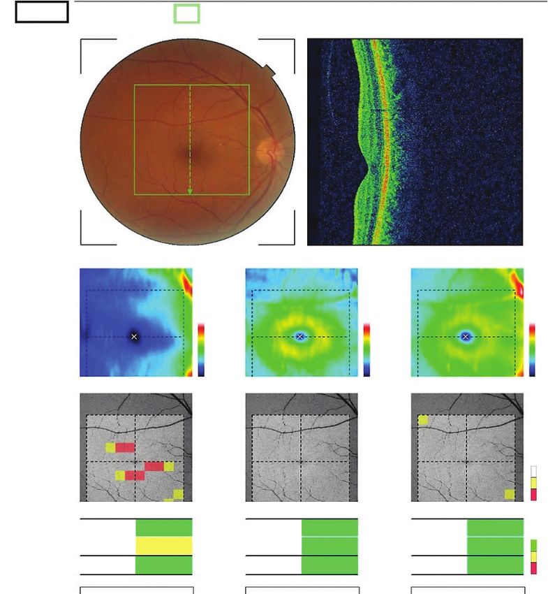
Page 2 of 8   Case Report

right eye, and because of diffuse disc oedema, the author           coagulopathies.18,19 In addition, bilateral optic disc drusen
could not estimate the C/D ratio in the left eye. Uncorrected       and moderate-to-severe obstructive sleep apnoea in patients
visual acuity (VA) measurements for the right and left eyes         non-compliant with continuous positive airway pressure
were 6/7.5 and 6/18, respectively. Optical correction               treatment were found to be significant risk factors for the
improved VA to 6/6 in the right eye, but no improvement             fellow eye involvement in NA-AION.20
was noted in the left eye.
                                                                    Treatment, outcomes and follow-up
Differential diagnosis and risk                                     The patient was referred to an ophthalmologist for
factors                                                             further evaluation; however, the patient failed to
                                                                    comply with the recommendation to see an ophthalmologist
Non-arteritic anterior ischaemic optic neuropathy must
                                                                    (see below).
be differentiated from other anterior optic neuropathies,
including anterior optic neuritis (idiopathic, demyelinating,
sarcoid-related, etc.), anterior compressive optic neuropathy       In 2013, the patient revisited the author’s practice for vision
(from anterior orbital lesions) and infiltrative optic              screening to renew his driver’s licence in compliance with
neuropathy.5 Optic neuritis may resemble NA-AION in terms           Regulation 102 of the National Road Traffic Act 93 of 1996.
of the rate of onset, pattern of VF loss and the appearance of      This regulation requires that any person in South Africa
the optic disc. However, the presence of ocular pain especially     seeking a new driver’s licence or renewing their driver’s
with eye movements, hyperaemic optic disc oedema (without           licence needs to undergo a vision screening test.
haemorrhages), early recovery and the fact that optic neuritis      The patient’s vision screening results showed the corrected
mostly affects younger patients, distinguishes this condition       VA of 6/6 for the right eye and 6/20 for the left eye. The
from NA-AION.5,12 Contrary to NA-AION, which presents               author became concerned about the possible further
with acute unilateral vision loss and an optic disc oedema          deterioration in VA in the patient’s left eye since his
that tends to result in sectoral or diffuse optic nerve atrophy     previous visit in 2012. The author thus requested the
usually within 6–11 weeks after the onset of vision loss,           ophthalmologist report from the patient but discovered
orbital lesion and infiltrative optic neuropathy present with       that the patient had not actually visited the ophthalmologist
gradual progressive vision loss and optic disc oedema that          as advised in 2012.
persist beyond 6–11 weeks. In addition, optic neuropathy
from orbital lesion is associated with mild ptosis and              The author referred the patient again and emphasised
abnormal lid and eye movements.                                     the need for him to see an ophthalmologist. Three days after
                                                                    the consultation, he came back with the medical report,
                                                                    which showed a VA of 6/6 in the right eye and 6/20 in the
It is of the utmost importance to differentiate NA-AION
                                                                    left eye, the presence of RAPD, an incipient cataract and
from arteritic anterior ischaemic optic neuropathy
                                                                    optic nerve atrophy in the left eye. The Humphrey Visual
(A-AION), as the two can be easily confused. The primary
                                                                    Field Test for the Central 30-2 thresholds showed that the
differentiating factors are prodromal symptoms and the
                                                                    VF in the right eye was within normal limits (Figure 1), and
appearance of optic disc oedema.13 Arteritic anterior
                                                                    in the left eye there was an altitudinal defect of inferior
ischaemic optic neuropathy is a disease of the elderly and
                                                                    hemianopia caused mainly by NA-AION (Figure 2). The
late middle-aged persons14 and typically presents with
                                                                    computerised tomography (CT) scan report did not show
pain, jaw claudication, scalp tenderness, fever and
                                                                    the presence of any masses/tumours. The ophthalmologist
malaise.2,13 Arteritic anterior ischaemic optic neuropathy is
                                                                    did not prescribe any treatment for the patient but advised
characterised by chalky white papilledema and in the                him to return to his surgery in 6 months for review.
majority of cases, when the oedema has resolved the optic
disc shows cupping like that of glaucoma (except that there         In 2015, the patient revisited the author’s practice for a
is pallor of the neuroretinal rim in A-AION and no pallor of        routine examination to get new glasses. His best corrected
the neuroretinal rim in glaucoma).15,16 On the contrary,            visual acuities (BCVA) in the right and left eyes were 6/6
NA-AION affects slightly younger patients over 50 years of          and 6/18+1, respectively. The patient visited the
age2,8,14 and is typically painless and characterised by severe     ophthalmologist again in 2017 and brought an optical
optic disc oedema with haemorrhages.17 The glaucoma-like            coherence tomography (OCT) scan, which corroborated
cupping is not seen in NA-AION.14 Furthermore, the                  the presence of left eye optic atrophy (Figure 3) and no
prognosis for untreated A-AION is quite poor and could              atrophy in the right eye (Figure 4). The BCVA in 2017
lead to rapid blindness, making the differentiation of              were the same as they were in 2015, showing that the VA
A-AION and NA-AION paramount.13                                     had stabilised.

Non-arteritic anterior ischaemic optic neuropathy is
associated with several risk factors, including advanced age,       Discussion
‘disc at risk’ (a small C/D ratio or an absent physiologic cup)     The pathogenesis of NA-AION remains unclear, although
and common systemic vascular risk factors, such as                  it is thought to result from transient non-perfusion or
hypertension, diabetes mellitus, hyperlipidaemia, obstructive       hypoperfusion of the ONH circulation, leading to
sleep apnoea, smoking, migraine, drugs and various                  ischaemia.2,14 The exact aetiology of the ischaemia is also

                                       http://www.avehjournal.org   Open Access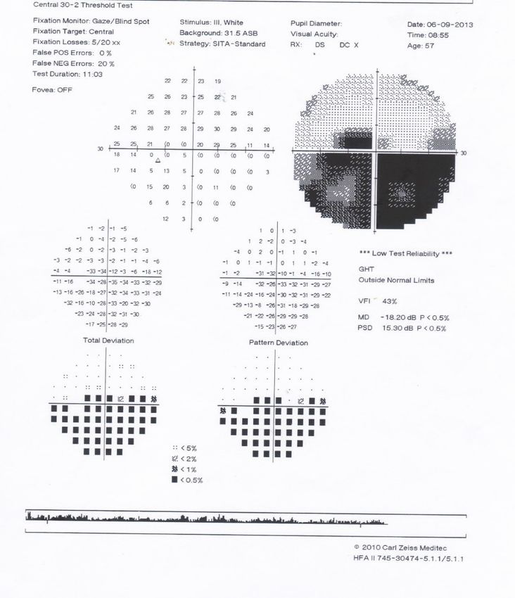
Page 3 of 8 Case Report
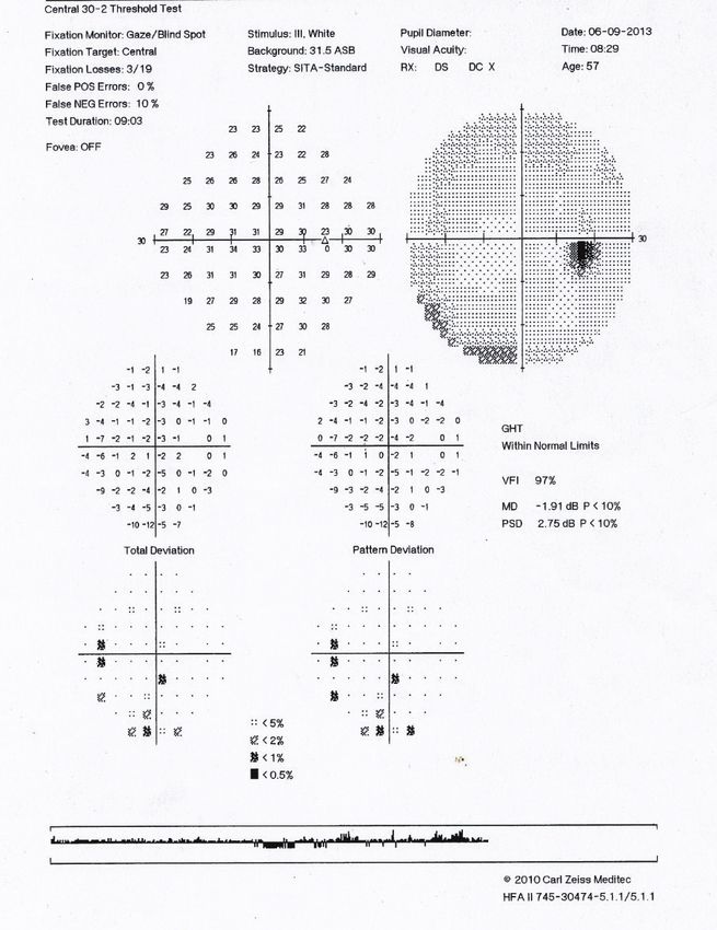
Source: Visual field courtesy of S.M. Thompson
OD, oculus dextrus or right eye; POS, positive; NEG, negative; RX, prescription; DS, dioptre sphere; DC, dioptre cylinder; GHT, glaucoma hemifield test; VFI, visual field index; MD, mean deviation;
PSD, pattern standard deviation.
FIGURE 1: Humphrey Visual Field Test (Central 30-2 threshold) showing a normal visual field for the right eye (or OD).
http://www.avehjournal.org Open Access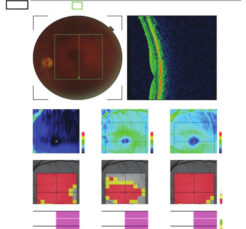
Page 4 of 8             Case Report

Source: Visual field courtesy of S.M. Thompson
VF, visual field; OD, oculus dextrus or right eye; POS, positive; NEG, negative; RX, prescription; DS, dioptre sphere; DC, dioptre cylinder; GHT, glaucoma hemifield test; VFI, visual field index; MD,
mean deviation; PSD, pattern standard deviation.
FIGURE 2: Humphrey Visual Field Test (Central 30-2 threshold) for the left eye suggests a mainly inferior altitudinal or hemi-field visual field defect that might extend even
further outwards. Note the result for the fixation losses (5/20), which indicates possible intratest unreliability or difficulties with central fixation or unstable eye movement,
possibly because of the visual field loss affecting the foveal or central region of the visual field. The patient might be seeking the vision stimuli by moving the eye about
rather than maintaining steady fixation (see also the gaze tracking record below the visual field and compare with OD).

not clear, but several risk factors have been implicated,                                                occlusion and generalised hypoperfusion.2,14,21 In addition,
including arterial hypertension, diabetes mellitus,                                                      an absent or a small C/D ratio, referred to as a ‘disc at
nocturnal hypotension, ischaemic heart disease, venous                                                   risk’, has been found to be significantly associated with

                                                          http://www.avehjournal.org                    Open Access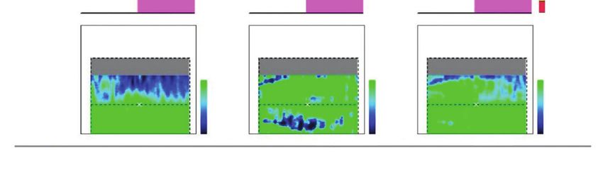
Page 5 of 8 Case Report
OS(L) 45
TopQ image quality : mode : Fine(1.1.0)
Capture date : 2017-12-06
RNFL
GCL+ GCL++
thickness
150 µm 150 µm 200 µm
100 100
100
50 50
0 0 0
Superpixel-600
N T
(%)
5
1
Average (6 mm × 6 mm)
Superior 8 µm Superior 52 µm Superior 60 µm
16 µm 52 µm 68 µm (%)
Inferior Inferior Inferior
5
Total 11µm Total 52 µm Total 63 µm 1
Asymmetry (relave thining)
-6 -7 -11
-20 µm -15 µm -30 µm
Comments : Signature : Date :
Source: Optical coherence tomography courtesy of S.M. Thompson
OCT, optical coherence tomography; RNFL, retinal nerve fibre layer; GCL, ganglion cell layer.
FIGURE 3: Topcon optical coherence tomography scan for the left eye showing the presence of left eye optic atrophy. The retinal nerve fibre layer and ganglion cell layer are
both thinner than expected, suggesting possible retinal nerve fibre layer atrophy.
NA-AION.22 Hence, there is a higher prevalence of who generally have smaller optic discs compared with
NA-AION amongst people of European and Asian descent, people of African descent.7,9
http://www.avehjournal.org Open AccessPage 6 of 8 Case Report
OD(R) TopQ image quality : 47 mode : Fine(1.1.0)
Capture date : 2017-12-06
RNFL
GCL+ GCL++
thickness
150 µm 150 µm 200 µm
100 100
100
50 50
0 0 0
Superpixel-600
T N
(%)
5
1
Average (6 mm × 6 mm)
Superior 31 µm Superior 68 µm Superior 99 µm
(%)
Inferior 31 µm Inferior 71 µm Inferior 102 µm
5
Total 31 µm Total 69 µm Total 101 µm 1
Asymmetry (relave thining)
-6 -7 -11
-20 µm -15 µm -30 µm
Comments : Signature : Date :
Source: Optical coherence tomography courtesy of S.M. Thompson
OCT, optical coherence tomography; RNFL, retinal nerve fibre layer; GCL, ganglion cell layer.
FIGURE 4: Optical coherence tomography scan for the right eye showing a more normal fundus with a healthier ganglion cell layer.
http://www.avehjournal.org Open AccessPage 7 of 8 Case Report
The patient in this report did not present with any symptoms advising NA-AION patients on better control and
of vision loss and supposedly only became aware of the management of the vasculopathic risk factors, including
vision loss for the left eye during VA measurements. This smoking cessation, exercise and weight loss to minimise
was contrary to other reports where patients experienced the risk of recurrence in the affected eye and involvement
symptoms of vision loss.2,5 Some patients reported in the fellow eye.14
experiencing vision loss on awakening.14 In a study involving
544 episodes of NA-AION, 51.8% were symptomatic on
awakening, 21.5% early in the morning and 26.7% later
Conclusion
during the day.1 However, this feature was not confirmed in This report has shown that NA-AION sometimes can occur
the Ischemic Optic Neuropathy Decompression Trial Study.1,5 in patients with apparently no visual symptoms and/or
The vision loss in NA-AION typically progresses over the associated risk factors. The clinical signs and symptoms
first 2 weeks1,10 and stabilises within 2–3 months,2 and there as well as risk factors associated with this condition
may be an improvement of three or more lines of VA in could help optometrists to take relevant case histories
13%–43% of the cases after vision loss has stabilised.2,5,10 and to perform careful eye examinations to differentially
diagnose this condition from other causes of unilateral
As mentioned above, the early clinical findings of vision loss such as central retinal artery occlusions, acute
NA-AION include sector or diffuse optic disc oedema with
angle closure glaucoma, unilateral retinal detachment at or
hyperaemia1 with or without haemorrhages.1,2,10 The oedema
involving the fovea or traumatic ocular injury. Early
tends to result in generalised or sectoral pallor of the optic
detection and timely referral potentially could reduce the
disc within 4–11 weeks.1,23 In 2012, the patient in this report
risk of vision loss in the unaffected eye and recurrence or
presented with diffuse optic disc oedema in the left eye, and
later in 2013 the ophthalmologist’s report showed that the progression in the affected eye. Further research is required
left eye had optic nerve atrophy and an incipient cataract. on the epidemiology of NA-AION, especially in the African
This suggests that when the patient presented to the continent, including South Africa, where data on this
author’s practice in 2012, the NA-AION was still at an early condition are lacking.
stage (the presence of optic disc oedema vs. optic nerve
atrophy), and the cataract in the left eye had not yet Acknowledgements
developed. The presence of inferior altitudinal VF defects in
The author is grateful to S.M. Thompson for his assistance
the patient’s left eye is consistent with reports from other
with diagnosis and the optical coherence tomography scans
authors,2,8,23 who reported that the inferior altitudinal and
and visual field images.
arcuate defects are the most common in NA-AION.
In this case, the ophthalmologist did not prescribe any Competing interests
treatment for the patient but advised him to return to his The author declares that he has no financial or personal
surgery in 6 months for review, probably because the patient relationships that may have inappropriately influenced him
did not present with any symptoms and because the CT scan in writing this research article.
was normal. Despite the improvement in the understanding
of the risk factors and clinical findings, there is no definitive
proven treatment for NA-AION.24 Several treatments that Author’s contribution
were advocated in the past, such as surgical decompression R.G.M. is the sole author of this article.
of the optic nerve, steroid therapy and aspirin to prevent
NA-AION in the fellow eye, were found to be ineffective.14
Ethical considerations
Despite the lack of definitive evidence, many practitioners A consent letter was taken from the patient to publish the
use aspirin as a treatment for NA-AION and for its report and anonymity was maintained.
role in preventing stroke and coronary artery disease.4,10
Patients with NA-AION are usually advised by some Funding information
ophthalmologists and neurologists that nothing can be done
with their condition.14 However, Hayreh14 believes that such This research received no specific grant from any funding
agency in the public, commercial or not-for-profit sectors.
advice is inadequate because NA-AION is a multifactorial
disease, with many risk factors contributing to its
development. He argues that the correct strategy is to manage Data availability
the risk factors, especially nocturnal arterial hypotension, to Data sharing is not applicable to this article.
minimise the risk of any further episodes in the affected eye
or involvement in the unaffected eye.14 Reports have shown
that there is aPage 8 of 8 Case Report
References 12. Arnold AC, Costa RMS, Dumitrascu OM. The spectrum of optic disc ischemia in
patients younger than 50 years (an American Ophthalmological Society thesis).
Trans Am Ophthalmol Soc [serial online]. 2013 [cited 2019 Dec 27];111:93–118.
1. Desai N, Pate MR, Prisant LM, Thomas DA. Nonarteritic anterior ischemic Available from: https://pubmed.ncbi.nlm.nih.gov/24167327
optic neuropathy. J Clin Hypertens. 2007;7(2):130–133. https://doi.org/10.1111/ 13. Luber S, Alweis R. Keeping NAION visual loss: Discriminating urgent versus
j.1524-6175.2005.04095.x emergent visual loss in an elderly female. BMJ Case Rep. 2014;2014:bcr2013202262.
2. Berry S, Lin WV, Sadaka A, Lee AG. Nonarteritic anterior ischemic optic neuropathy: https://doi.org/10.1136/bcr-2013-202262
Cause, effect, and management. Eye Brain. 2017;9:23–28. https://doi. 14. Hayreh SS. Management of ischemic optic neuropathies. Indian J Ophthalmol.
org/10.2147/EB.S125311 2011;59(2):123–136. https://doi.org/10.4103/0301-4738.77024
3. Chen J, Zhu J, Chen L, Hu C, Du Y. Steroids in the treatment of nonarteritic 15. Punjabi OS, Tanna AP, Rosenberg MA. Optic disk excavation in nonarteritic
anterior ischemic optic neuropathy: A PRISMA-compliant meta-analysis. Medicine anterior ischemic optic neuropathy. J Glaucoma. 2011;20(2):71–73. https://doi.
(Baltimore). 2019;98(46):e17861. https://doi.org/10.1097/MD.00000000000 org/10.1097/IJG.0b013e3181dddf5c
17861
16. Hayreh SS, Jonas JB. Optic disc morphology after arteritic anterior ischemic optic
4. Atkins EJ. Nonarteritic anterior ischemic optic neuropathy. Curr Treat Options neuropathy. Ophthalmology. 2001;108(9):1586–1594. https://doi.org/10.1016/
Neurol. 2011;13(1):92–100. https://doi.org/10.1007/s11940-010-0099-0 S0161-6420(01)00649-2
5. Miller NR, Arnold AC. Current concepts in the diagnosis, pathogenesis and 17. Voss E, Raab P, Trebst C, Stangel M. Clinical approach to optic neuritis: Pitfalls, red
management of nonarteritic anterior ischaemic optic neuropathy. Eye (Lond). flags and differential diagnosis. Ther Adv Neurol Disord. 2011;4(2):123–134.
2015;29(1):65–79. https://doi.org/10.1038/eye.2014.144 https://doi.org/10.1177/1756285611398702
6. Johnson LN, Arnold AC. Incidence of nonarteritic and arteritic anterior ischemic 18. Hayreh SS, Zimmerman MB. Nonarteritic anterior ischemic optic neuropathy: Clinical
optic neuropathy: Population-based study in the state of Missouri and Los characteristics in diabetic patients versus nondiabetic patients. Ophthalmology.
Angeles county, California. J Neuro-Ophthalmology. 1994;14(1). https://journals. 2008;115(10):1818–1825. https://doi.org/10.1016/j.ophtha.2008.03.032
lww.com/jneuro-ophthalmology/Fulltext/1994/03000/Incidence_of_ 19. Patil A, Mahesh KV, Prabhat N, et al. Non-arteritic anterior ischaemic optic
Nonarteritic_and_Arteritic_Anterior.11.aspx neuropathy – Myths and misconceptions: Experience from a Tertiary Care Centre
7. Patil M, Ganger A, Saxena R. Non-arteritic anterior ischemic optic neuropathy in North India. Neuroophthalmology. 2019;44(4):246–254. https://doi.org/10.10
(NAION) – A brief review. 2016;6(3):158–163. https://doi.org/10.4236/ojoph. 80/01658107.2019.1688356
2016.63022 20. Chang MY, Keltner JL. Risk factors for fellow eye involvement in nonarteritic
8. Lee MS, Grossman D, Arnold AC, Sloan FA. Incidence of nonarteritic anterior anterior ischemic optic neuropathy. J Neuro-Ophthalmology. 2019;39(2):147–152.
ischemic optic neuropathy: Increased risk among diabetic patients. Ophthalmology. https://doi.org/10.1097/WNO.0000000000000715
2011;118(5):959–963. https://doi.org/10.1016/j.ophtha.2011.01.054 21. Hayreh SS, Joos KM, Podhajsky PA, Long CR. Systemic diseases associated with
9. Wang Y, Xu L, Jonas JB. Frequency of non-arteritic anterior ischaemic optic nonarteritic anterior ischemic optic neuropathy. Am J Ophthalmol.
neuropathy in adult Chinese: The Beijing eye study. Br J Ophthalmol. 1994;118(6):766–780. https://doi.org/10.1016/S0002-9394(14)72557-7
2007;91(3):401. https://doi.org/10.1136/bjo.2006.103788 22. Beck RW, Servais GE, Hayreh SS. Anterior ischemic optic neuropathy: IX. Cup-to-
10. Atkins EJ, Bruce BB, Newman NJ, Biousse V. Treatment of nonarteritic anterior disc ratio and its role in pathogenesis. Ophthalmology. 1987;94(11):1503–1508.
ischemic optic neuropathy. Surv Ophthalmol. 2010;55(1):47–63. https://doi. https://doi.org/10.1016/S0161-6420(87)33263-4
org/10.1016/j.survophthal.2009.06.008 23. Espino Barros A, Amram AL, Derham AM, Smith SV, Lee AG. Management of
ischemic optic neuropathies. Expert Rev Ophthalmol. 2017;12(2):99–109. https://
11. Broadway DC. How to test for a relative afferent pupillary defect doi.org/10.1080/17469899.2017.1291341
(RAPD). Community Eye Health [serial online]. 2012 [cited 2019 Dec 20];
25(79–80):58–59. Available from: http://www.ncbi.nlm.nih.gov/pmc/articles/ 24. Foroozan R. New treatments for nonarteritic anterior ischemic optic neuropathy.
PMC3588138/ Neurol Clin. 2017;35(1):1–15. https://doi.org/10.1016/j.ncl.2016.08.003
http://www.avehjournal.org Open AccessYou can also read


