2020 Compendium A collection of NA PRRS Symposium extended abstracts and NC-229 station reports - University of Illinois College of Veterinary ...
←
→
Page content transcription
If your browser does not render page correctly, please read the page content below
Compendium                                       2020
A collection of NA PRRS Symposium extended abstracts and NC-229 station reports.
Contents
      Special Thank You                                                                                   Page 2-89
                                                                                                          Page 2
                                                                                                                        North American PRRS Symposium Extended Abstracts
                                                                                                                        Association of the Blood Transcriptome of Healthy Piglets with Response to Natural Polymicrobial

            to the
                                                                                                                        Disease, Include PRRS
                                                                                                          Page 4        Attenuation of a PCV2B Vaccine Candidate by Directed Suicidal Replication
                                                                                                          Page 10       Cytokine Response in Cells Co-Infected with Type I Interferon Suppression-Negative and NF-

 2020 Organizing Committees
                                                                                                                        KBActivation-Negative PRRS Virus and Bacterial Pathogen
                                                                                                          Page 14       Development and Characterization of Monoclonal Antibodies Against African Swine Fever Virus
                                                                                                          Page 20       Development of an Infectious Clone for Porcine Deltacoronavirus Strain USA/IL/2014/026
                                                                                                          Page 28       Diagnostic Performance vs Surveillance Performance – The Case of PRRSV Oral Fluid ELISA
                                                                                                          Page 34       Effects of Immunodominance on Porcine Circovirus Type 2 (PCV2) Vaccine Design
                                                                                                          Page 40       An Emerging Porcine Sapelovirus in the United States: Genetic Characterization and Diagnostic Tool
                                                                                                                        Development
                                                                                                          Page 44       Evaluation of Antibody Response Directed Against Porcine Reproductive and Respiratory Syndrome
         Advisory Committee                               Joint Scientific Committee                                    Virus Structural Proteins
                                                                                                          Page 52       Evaluation for Genetic Stability of a US Commercially Licensed Modified Live Porcine Reproductive and
Dr. Fernando Osorio, University of Nebraska-Lincoln   Co-Chairs, Joint Scientific Committee:                            Respiratory Syndrome Virus Vaccine
Dr. Raymond R.R. Rowland, University of Illinois             Dr. Ying Fang, University of Illinois
                                                                                                          Page 58       Host Transcriptional Response to Persistent Infection with a Live-Attenuated Porcine Reproductive and
                                                             Dr. Sheela Ramamoorthy, North Dakota State
                                                                                                                        Respiratory Syndrome Virus Strain
                                                      		University
                                                                                                          Page 66       How to Best Protect Pigs Against PRRSV Based on Our Understanding of the Adaptive Immune
                                                                                                                        Response to PRRSV
         Planning Committee                           Committe Members:
                                                            Dr. Diego G. Diel, Cornell University                       A Neutralizing Monoclonal Antibody-Based Competitive ELISA with the Emphasis on the Replacement
                                                                                                          Page 72
Executive Director:                                         Dr. Scott Kenney, The Ohio State University                 of Virus Neutralization Test for Classical Swine Fever C-Strain Post-Vaccination Monitoring
       Dr. Ying Fang, University of Illinois                Dr. Pablo Pineyro, Iowa State University      Page 74       A Risk-Free In Situ Non-Animal (RISNA) Surrogate Assay for ASFV
                                                            Dr. Roman Pogranichniy, Kansas State
Industry Liaison:                                     		University                                        Page 78       Role of Host Restriction Factors in Porcine Reproductive and Respiratory Syndrome Virus (PRRSV)
                                                                                                                        Replication
        Dr. Lisa Becton, National Pork Board                Dr. Hiep Vu, University of Nebraska
                                                            Dr. Zhengguo Xiao, University of Maryland     Page 82       Shedding and Transmission of a Lineage One Modified Live Porcine Reproductive and Respiratory
NA PRRS Internal Coordinators:                                                                                          Syndrome Virus Vaccine
      Kristen Eighner, University of Illinois                                                             Page 90-175   NC-229 Station Reports
      Crystal Zulauf, University of Illinois
                                                                                                          Page 90       Iowa State University
NA PRRSS-CRWAD Virtual Conference Coordinator:                                                            Page 100      Kansas State University
      Jennifer Stalley, Midwest Solutions                                                                               North Dakota State University
                                                                                                          Page 110
                                                                                                          Page 114      Purdue University
                                                                                                          Page 118      South Dakota State University
                                                                                                          Page 124      University of Florida
                                                                                                          Page 128      University of Illinois
                                                                                                          Page 140      University of Maryland
                                                                                                          Page 142      University of Minnesota
                                                                                                          Page 164      University of Nebraska-Lincoln
                                                                                                          Page 168      USDA-ARS-National Animal Disease Center

                                                                                                                                                                                                                             1
Association of the Blood Transcriptome of Healthy Piglets
         with Response to Natural Polymicrobial Disease,
                           Include PRRS

       Authors     K.S. Lim¹, J. Cheng¹, A. Putz¹ 6, Q. Dong¹,7, X. Bai², C.K. Tuggle¹, M.K. Dyck²,                       protein localization and viral gene expression tended to be associated with
                   PigGen Canada³, F. Fortin4, J.C.S. Harding5, G.S. Plastow², and J.C.M.                                 reduced performance and health traits after but not before challenge.
                   Dekkers¹
                                                                                                           Conclusion     In conclusion, gene expression profiles in blood from young healthy piglets
    Affiliations   1. Department of Animal Science, Iowa State University                                                 provide insight into their performance when exposed to disease. This in-
                   2. Department of Agriculture, Food and Nutritional Science, University of                              cludes the level of expression of genes that appear to respond to the various
                      Alberta                                                                                             stressors that piglets are exposed to even without major disease, of genes
                   3. PigGen Canada Industry Consortium                                                                   related to heme metabolism, as well as the baseline expression of host genes
                   4. Centre de Développement du Porc du Québec Inc. (CDPQ)                                               related to virus propagation.
                   5. Department of Large Animal Clinical Sciences, University of
                      Saskatchewan                                                                    Financial Support   Funding from USDA-NIFA, Genome Canada, Genome Alberta, and PigGen
                   6. Hypor Inc.                                                                                          Canada.
                   7. Department of Epidemiology and Cancer Control, St. Jude Children’s
                      Research Hospital

    Key Words      Pigs, Disease Resilience, Disease Challenge, Blood, Transcriptomics

    Objectives     Our objective was to investigate population-level gene expression profiles in
                   the blood of 912 healthy nursery pigs for associations with performance and
                   health before and after exposure to the natural disease challenge.

      Methods      We applied a natural polymicrobial disease challenge model, including PRRS,
                   to collect detailed data on disease resilience on F1 barrows. Blood samples
                   were collected at ~27 days of age prior to exposure to disease, and gene
                   expression profiles were quantified using 3’ mRNA-sequencing.

       Results     The most significant (q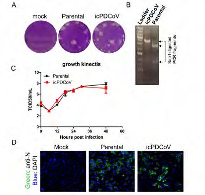
Attenuation of a PCV2B Vaccine Candidate by Directed
                          Suicidal Replication

        Authors     AGM Rakibuzzaman¹, Pablo Piñeyro², Angela Pillatzki³, and Sheela                                   predominant one. [4-6]. Although a DNA virus, PCV2 has a high mutation rate.
                    Ramamoorthy¹*                                                                                      which is close to a single stranded RNA viruses [7, 8]. The frequent evolution
                                                                                                                       of PCV2 despite the availability of standard vaccines indicates that there is
     Affiliations   1. Department of Microbiological Sciences, North Dakota State University,                          room for the improvement of current vaccines.
                       Fargo, ND, USA
                    2. College of Veterinary Medicine, Iowa State University, Ames, Iowa, USA                          In this study, the high mutation rates of PCV2 were used to redesign the
                    3. Animal Disease Research and Diagnostic Laboratory, South Dakota State                           serine and leucine codons of the capsid gene in a way that the chances of
                       University, Brookings, SD                                                                       accumulation of stop codons will increase during viral replication to eventu-
                                                                                                                       ally attenuate and eliminate the virus from vaccinated hosts. For example, if
                    *Correspondence: sheela.ramamoorthy@ndsu.edu; Tel.: +01-701-231-8504                               the serine codon UCU is changed to UCA in the vaccine virus, a mutation of
                                                                                                                       C to A during vaccine viral replication in the host would result in a sequence
     Key Words      Vaccine, Porcine Circovirus, PCV2, Attenuated PCV2, Antibody, Virus                                change to UAA, a stop codon. Under selection pressure in vitro or the im-
                    Neutralization                                                                                     mune system in vivo, the likelihood that rapidly mutating viruses with mod-
                                                                                                                       ified serine and leucine codons will acquire mutations which result in stop
       Abstract     Postweaning multisystemic wasting syndrome (PMWS), is caused by porcine
                                                                                                                       codons is high for viruses with high mutation rates. Although this strategy
                    circovirus type 2 (PCV2), an economically important swine virus which affects
                                                                                                                       was successfully applied for the RNA viruses [9] it has not been for DNA
                    production worldwide. Although a DNA virus, PCV2 has a high rate of mutation
                                                                                                                       viruses yet. Further, with the increasing number of newly emerging viruses,
                    and continues to evolve into new subtypes. The aim of this study is to take
                                                                                                                       necessitates the availability of tools for rapid response vaccine development.
                    the advantage of the high mutation rate of PCV2 to target the premature
                                                                                                                       The strategy developed in this study is both elegant and highly effective for
                    termination of the gene expression as a strategy for rapid attenuation and
                                                                                                                       the rapid attenuation of viruses for application as vaccine candidates.
                    vaccine development (suicidal PCV2 vaccine or sPCV2-Vac) by altering
                    serine and leucine codons of the capsid gene to increase the probability                           Cloning and attenuation of the virus: An infectious clone of PCV2b subtype
                                                                                                        Methods and
                    of accumulating stop mutations during viral replication. The sPCV2-Vac                             41513 (accession number KR816332) [10] was used as backbone for the vac-
                                                                                                           Materials
                    candidate developed was successfully rescued by transfection and showed                            cine virus. The serine and leucine codons of the ORF2 were redesigned as de-
                    a reduction in fluorescence over serial passages when compared to wild type                        scribed above and commercially synthesized (Eurofin genomics). The ORF2
                    virus, indicating attenuation of the virus in vitro. Vaccination of pigs with the                  of PCV2b 41513 was replaced with the redesigned ORF2 gene (sPCV2-Vac).
                    sPCV2-Vac showed that the binding antibody response in test group was higher                       sPCV2-Vac was rescued by transfection of PK-15 cells and viral replication in
                    than the unvaccinated control in pre-challenged sera. As hypothesized, the                         infected cells visualized with an immunofluorescence assay (IFA) [11].
                    sPCV2-Vac virus was cleared in vaccinated pigs within 2 weeks of exposure but
                    elicited strong binding antibody and virus neutralization responses. Vaccinated                    Immunization of pigs: Approximately 3-week-old piglets from a PCV2 PCR
                    pigs were protected against heterologous challenge with a PCV2d virus, as                          negative herd were administered treatments as follows: Group I- unvacci-
                    vaccinated pigs were protected against viremia and the development of lesions                      nated control (N=9), Group II – one 2.0 ml-i/m dose of an inactivated com-
                    due to challenge. Thus, the described strategy has potential application as a                      mercial PCV2 vaccine (N=9), Group III - sPCV2-Vac (N=9) ,104 TCID50 , 2ml
                    rapid attenuation method for newly emerging viruses, including DNA viruses.                        i/n, 2ml i/m. Serum was collected on day 0,14 and 28 to assesses antibody
                                                                                                                       responses. The animal study was conducted at South Dakota State Universi-
    Introduction    Post-Weaning Multi-Systemic Wasting Syndrome (PMWS) is an important                                ty, Animal Resource Wing, following the guidelines of the Institutional Animal
                    manifestation of porcine circovirus type2 (PCV2) infection in weanling pigs.                       Care and Use Committee.
                    [1,2] Porcine circovirus associated diseases (PCVAD) continue to cause a huge
                    economic losses to the swine industry [3] each year, even though commercial                        Read-outs: Serum antibody responses against PCV2 in vaccinated pigs
                    vaccines are able to reduce the clinical signs of PCV2. Over time, new subtypes                    was achieved with a commercial PCV2 ELISA kit (Ingezim Circovirus IgG kit,
                    of PCV2 have been emerged periodically, and currently PCV2d is the most

4                                                                                                                                                                                                       5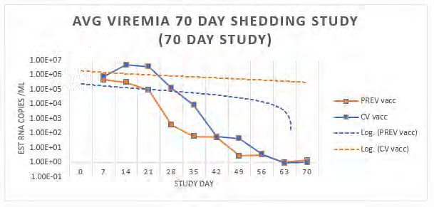
Attenuation of a PCV2B Vaccine Candidate by Directed
                  Suicidal Replication (Continued)

                  Ingenasa, Madrid, Spain). The titer of neutralizing antibody (NA) against PCV2a,
                  2b, and 2d were assessed by the fluorescence focus neutralization (FFN)                                                                                                          Figure 3. Microscopic lesions scores of pig
                  assay essentially as described before [12] . The challenge virus replication                                                                                                     tissues resulting from PCV2d challenge.
                  was measured by a PCV2d specific qPCR from post challenged sera. The                                                                                                             Bar shows the average lesions score of
                  pathological scores were measured by a board-certified veterinarian.                                                                                                             respective tissues. A, B, C, and D represents
                                                                                                                                                                                                   lesions scores of Lymph node, Tonsils, Lung
    Results and   Rescue of the sPCV2-Vac construct: The vaccine virus was successfully                                                                                                            and Ileum respectively.
    Discussions   rescued by transfection of PK-15 cells, at titers comparable to the wild type
                  virus (fig. 1, A and B). However, the titer of sPCV2-Vac decreased over serial
                  passages when detected with an immune fluorescence assay (fig. 1, C and D),
                  indicating possible attenuation in vitro.
                                                                                                                               Conclusion     Thus, the strategy described for rapid attenuation of viruses has significant
                  sPCV2-Vac induces heterologous virus neutralization (V/N) responses: Unlike                                                 application to animal health, considering the increasing emergence of new
                  the commercial vaccine, the test vaccine is not dose optimized or adjuvanted.                                               viruses and the long lag time with current strategies for producing attenuated
                  However, testing of serum from day post vaccine 28 (DPV 28) for homologous                                                  vaccine candidates.
                  and heterologous V/N titers showed that robust responses against all three
                  circulating PCV2 subtypes.                                                                           Acknowledgement        We thank Dr. Michele Mucciante and Ms. Amanda Zubke from SDSU for help
                                                                                                                                              with the animal experimentation.
                  sPCV2-Vac protects against heterologous PCV2d challenge: As hypothesized,
                  the sPCV2-Vac was cleared in vaccinated pigs within 2 weeks of vaccination,                          Conflict of Interest   The authors declare no financial conflict of interest.
                  indicating that the rapid-attenuation strategy was successful. In addition,                                 References      1. Tischer, I., et al., A very small porcine virus with circular single-stranded DNA. 1982.
                  the PCV2d challenge virus was completely cleared in vaccinated pigs, which                                                  2. Clark, E. Pathology of the post-weaning multisystemic wasting syndrome of pigs. in Proc
                  remained PCR negative until DPC 21 (data not shown). Pigs vaccinated with the                                                   Western Can Assoc Swine Pract. 1996.
                  sPCV2-had significantly lower lesions scores in lymph nodes, tonsils, ileum and                                             3. Hu, Y., et al., Evidence of natural co-infection with PCV2b subtypes in vivo. Archives of
                  lungs (fig. 2), compared to unvaccinated control and commercial vaccine.                                                        Virology, 2017: p. 1-6.
                                                                                                                                              4. Afghah, Z., et al., Ten years of PCV2 vaccines and vaccination: Is eradication a possibility?
                                                                                                                                                  Vet Microbiol, 2017. 206: p. 21-28.
                                                                                                                                              5. Karuppannan, A.K. and T. Opriessnig, Porcine Circovirus Type 2 (PCV2) Vaccines in the
                                                                                                                                                  Context of Current Molecular Epidemiology. Viruses, 2017. 9(5).
                                                                                                                                              6. Ssemadaali, M.A., M. Ilha, and S. Ramamoorthy, Genetic diversity of porcine circovirus type
                                                                                                                                                  2 and implications for detection and control. Res Vet Sci, 2015. 103: p. 179-86.
                                                                                                                                              7. Kwon, T., et al., Genotypic diversity of porcine circovirus type 2 (PCV2) and genotype shift
                                                                                                                                                  to PCV2d in Korean pig population. Virus research, 2017. 228: p. 24-29.
                                                                                                                                              8. Karuppannan, A.K. and T. Opriessnig, Porcine circovirus type 2 (PCV2) vaccines in the
                                                                                                                                                  context of current molecular epidemiology. Viruses, 2017. 9(5): p. 99.
                                                                                                                                              9. Moratorio, G., et al., Attenuation of RNA viruses by redirecting their evolution in sequence
                  Figure 1. Immune fluorescence assay of                                                                                          space. Nature microbiology, 2017. 2(8): p. 17088.
                  transfection and infection with the wild type PCV2b   Figure 2. Virus neutralization assay: Virus                           10. Constans, M., et al., Antigenic Determinants of Possible Vaccine Escape by Porcine
                  41513 and sPCV2-Vac construct and rescued virus       neutralizing antibodies measured by a                                     Circovirus Subtype 2b Viruses. Bioinform Biol Insights, 2015. 9(Suppl 2): p. 1-12.
                  on PK-15 cells. Viable viruses have stained with a    fluorescent focus neutralization assay using                          11. Opriessnig, T., et al., Differences in virulence among porcine circovirus type 2 isolates are
                  PCV2 specific polyclonal antibody. A. transfected     days post vaccination 28 pre-challenge sera.                              unrelated to cluster type 2a or 2b and prior infection provides heterologous protection.
                  wildtype PCV2b, B. Transfected sPCV2-Vac, C.          X-axis— PCV2 subtypes used in the assay.                                  Journal of General Virology, 2008. 89(10): p. 2482-2491.
                  Passage 5 of wildtype PCV2b, D. Passage 5 of          Y axis mean % reduction in fluorescent foci                           12. Kolyvushko, O., et al., Efficacy of a Commercial PCV2a Vaccine with a Two-Dose Regimen
                  sPCV2-Vac.                                            compared to the untreated virus culture.                                  Against PCV2d. Veterinary sciences, 2019. 6(3): p. 61.

6                                                                                                                                                                                                                                                 7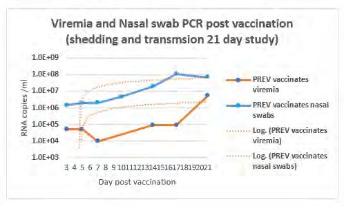
Co-vaccination and Schedule of Immunization with
    Attenuated Porcine Reproductive and Respiratory Syndrome
    (PRRS) Vaccine Could Potentially Affect Efficacy of Subunit
                   Classical Swine Fever Vaccine
        Authors     Rachel Madera¹, Lihua Wang¹, Ada Giselle Cino-Ozuna², and Jishu Shi¹*                            various multiple vaccination combinations in swine would be an interesting
                                                                                                                     aspect for future investigations.
     Affiliations   1. Department of Anatomy and Physiology, College of Veterinary Medicine,
                       Kansas State University, Manhattan, KS, USA                                Acknowledgements   This research is supported by awards from the National Bio and Agro-
                    2. Department of Diagnostic Medicine and Pathobiology, College of                                Defense Facility Transition Fund, the USDA NIFA Hatch-Multistate project
                       Veterinary Medicine, Kansas State University, Manhattan, KS, USA                              1021491, the USDA ARS Non-Assistance Cooperative Agreements (58-8064-
                                                                                                                     8-011, 58-8064-9-007, 58-3020-9-020, and 59-0208-9-222), and National Pork
                    *Correspondence: jshi@ksu.edu                                                                    Board Grant #18-059. Authors declare no financial conflict of interest.

     Key Words      Classical Swine Fever (CSF), E2, Vaccine, PRRS, Subunit

    Introduction    Commercial pigs have been routinely injected with multiple vaccines that
                    are either administered separately or co-administered at the same time
                    for convenience, and to minimize pig stress. However, viruses, including
                    attenuated and modified live virus (MLV) vaccines, can modulate host
                    immune responses that could potentially impact the efficacy of co-
                    administered vaccines.

       Methods      Here we report the effects of pre- and co-administered Chinese highly
                    pathogenic porcine reproductive and respiratory syndrome (PRRS) virus MLV,
                    JXA1-R, on the efficacy of an emulsion-based classical swine fever virus
                    (CSFV) subunit vaccine, KNB-E2. Immune responses to the CSFV and JXA-
                    1R vaccines were evaluated by testing CSFV-specific and PRRSV-specific
                    sera antibodies and then challenged with CSFV at 4 weeks post KNB-E2
                    vaccination.

        Results     Pigs co-administered with JXA1-R vaccine exhibited slightly lower levels
                    of PRRSV-specific antibodies than pigs vaccinated with JXA1-R two weeks
                    before KNB-E2 vaccination. On the other hand, both JXA1-R/KNB-E2
                    vaccinated pig groups had slightly lower CSFV-specific antibodies than pigs
                    vaccinated with KNB-E2 alone at 3 weeks post KNB-E2 vaccination.

     Discussion     These observed differences imply an effect of live MLV vaccination on
                    other vaccines, and that should be considered in multiple swine vaccination
                    schedules. In this study, both groups of JXA-1R/KNB-E2 vaccinated pigs
                    were amply protected from CSF clinical symptoms upon challenge. However,
                    the observed differences in the pre- and co-administered JXA1-R/KNB-E2
                    vaccinated pig groups compared with single KNB-E2 vaccination pig group
                    indicate an unintended effect of PRRS MLV on the elicited immune response
                    to the CSF KNB-E2 vaccine. The immunological responses affected by

8                                                                                                                                                                                                 9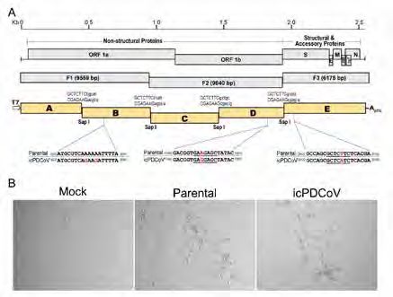
Cytokine Response in Cells Co-Infected with Type I Inter-
      feron Suppression-Negative and NF-KBActivation-Negative
                 PRRS Virus and Bacterial Pathogen

         Authors     Chia-Ming Su¹, Jineui Kim¹, Dongwan Yoo¹*                                                              IL‐1β, IL‐6, IL‐8, TNF‐α, CCL4, IL-10, and INF-β in pulmonary alveolar macrophages
                                                                                                                            (PAMs) (3). Several pieces of evidence indicate that the co-infection of PRRSV
      Affiliations   1. Department of Pathobiology, College of Veterinary Medicine, University of                           and other pathogens would enhance proinflammatory cytokines response and
                        Illinois at Urbana-Champaign, Urbana, IL, USA                                                       further cause a severe outcome.
                     *Correspondence: dyoo@illinois.edu                                                                     PRRSV non-structure protein 1β (nsp1β) has been demonstrated as an IFN
                                                                                                                            antagonist, and a highly conserved SAP (SAF-A/B, Acinus, and PIAS) motif
      Key Words      PRRSV, nsp1-beta, Nucleocapsid, Co-Infection, NF-kB, Cytokine.                                         inhibits cytoplasmic translation of host mRNAs, further suppresses type I
                                                                                                                            IFNs (4, 5). Previously, we show that the mutation at position 135 in the SAP
        Abstract     Porcine reproductive and respiratory syndrome virus (PRRSV) suppresses innate
                                                                                                                            motif was attenuated and released a higher concentration of IFN in pigs during
                     immunity, namely, type I interferon responses, and activates NF-κB signaling
                                                                                                                            infection (4). Furthermore, the nuclear localization signal (NLS) of the PRRSV
                     during infection. The NF-kB activation by PRRSV may predispose to the secondary
                                                                                                                            nucleocapsid (N) protein was identified as the NF-κB activation domain (6). We
                     bacterial infections and increase the clinical severity. Among the PRRSV proteins,
                                                                                                                            hypothesize that PRRSV mutant in which both the SAP motif in nsp1β protein
                     non-structure protein 1β (nsp1β) has been identified as the potent interferon
                                                                                                                            and the NLS motif in N protein may exhibit both type I IFN suppression-negative
                     antagonist, and leucine at position 135 in the SAP motif is determined as the active
                                                                                                                            and NF-κB activation-negative phenotypes and may lead to relieving the severity
                     residue for nsp1β-mediated IFN suppression. The viral nucleocapsid (N) protein
                                                                                                                            of PRDC causing by co-infection. In the present study, a double-mutant PRRSV
                     has been found as the effector protein for the NF-κB activation, and the nuclear
                                                                                                                            was generated by reverse genetics to eliminate both IFN suppression and NF-κB
                     localization signal (NLS) overlaps the NF-κB activation domain. In the present
                                                                                                                            activation functions. The immunological phenotype was examined in cells during
                     study, SAP motif-deleted and NLS motif-deleted double mutant virus was generated
                                                                                                                            co-infection of PRRSV and a bacteria pathogen.
                     by reverse genetics. The double mutant PRRSV was characterized in cells and
                     examined for its immunological phenotypes. The results demonstrated that the           Materials and   Baby hamster kidney-21 (BHK-21) cells were cultivated in modified Eagle’s
                     SAP motif-deleted and NLS motif-deleted double mutant PRRSV attenuated the                 Methods     medium supplemented with 10% heat-inactivated fetal bovine serum (FBS).
                     expression of proinflammatory cytokines in macrophages compared to wild-type                           MARC-145 cells were grown in Dulbecco’s modified Eagle’s medium containing
                     PRRSV. While TNF-α and IL-1β were significantly upregulated in cells co-infected                       10% heat-inactivated FBS. Extralong inverse PCR was conducted using full-
                     with wild-type PRRSV and Streptococcus suis (S. suis), the double mutant PRRSV                         length infectious clones to delete L135 in nsp1β and NLS in N from PRRSV.
                     and S. suis co-infection did not enhance the TNF-α and IL-1β productions. This                         A series of mutant PRRSVs were then rescued by reverse genetics in BHK-21
                     study suggests that the intervention of nsp1β-mediated IFN suppression and N                           cells and MARC-145 cells. Mutations were confirmed by sequencing ‘passage
                     protein-mediated NF-κB activation relieves the clinical severity that may be caused                    4’ virus. RT-qPCR was used to determine mRNA expressions for cytokines and
                     by co-infection of PRRSV and other swine pathogens. Our findings pave a way to                         chemokines in infection with individual mutant PRRSV at different times post-
                     developing a novel vaccine candidate that may reduce a risk of developing clinical                     infection. For co-infection, MARC-145 cells were inoculated with P129 wild
                     severity which may be induced by current vaccines in swine farms.                                      type PRRSV or mutant PRRSV. After 48 h of infection, the cells were infected
                                                                                                                            with 1 multiplicity of infection (MOI) of Streptococcus suis for four hours. After
     Introduction    Porcine reproductive and respiratory syndrome virus (PRRSV) suppresses type I
                                                                                                                            incubation, supernatants were removed, and penicillin G (5 μg/ml) and gentamicin
                     interferon (IFNs-α/β) response and also activates NF-κB signaling during infection
                                                                                                                            (100 μg/ml) were added to each well for two hours to kill extracellular bacteria.
                     and predisposes infected hosts to secondary pathogen infection. In swine farms,
                                                                                                                            NF-κB response was examined by luciferase reporter assay, and pro-inflammatory
                     pigs are commonly co-infected with PRRSV and other pathogens, which will
                                                                                                                            cytokines were determined by RT-qPCR. Statistical analyses were performed
                     trigger enhanced expression of proinflammatory cytokines. PRRSV predisposes
                                                                                                                            using Student t-tests, and statistical significance was expressed as P < 0.05.
                     a secondary infection caused by other infectious agents, including Mycoplasma
                     hyopneumoniae (M. hyo) and Streptococcus suis (S. suis), leading to severe porcine                     Generation of L135-deleted and NLS motif-deleted double mutant virus:
                                                                                                                 Results
                     respiratory disease complex (PRDC) and increased morbidity and mortality in pigs                       Deletion of L135 in the SAP motif of nsp1β and deletion of NLS motif in N were
                     (1, 2). An in vitro study showed that PRRSV and S. suis co-infection upregulated

 1
10                                                                                                                                                                                                           11
Cytokine Response in Cells Co-Infected with Type I Inter-
     feron Suppression-Negative and NF-KBActivation-Negative
          PRRS Virus and Bacterial Pathogen (Continued)

                hypothesized to confer the mutant PRRSV to lose the type I IFN suppression and                             mutant PRRSV has attenuated activation of NF-κB and thus reduced cytokine
                NF-kB activation functions. To determine this possibility, three mutant viruses were                       productions during co-infection with other pathogens.
                generated by the reverse genetics approach using infectious clones. PN-ΔNLS
                was first generated to delete NSL from the N gene as described previously (7, 8),             Discussion   The SAP motif-deleted and NLS motif-deleted double mutant PRRSV was
                and P1β-Δ135 was generated by introducing a specific deletion in the SAP motif                             generated in this study. This virus represents type I IFN suppression-negative and
                of nsp1β. (7, 8). Then using PN-ΔNLS, the double mutant virus was generated by                             NF-κB activation-negative in its phenotype. This double-deletion mutant virus
                introducing a deletion in the SAP motif. The double mutant was named PD-Δ135-                              attenuates proinflammatory cytokine productions in cells. During co-infection,
                NLS. After transfection with wild-type P129 infectious clone, P1β-Δ135, PN-ΔNLS,                           wild-type PRRSV can activate NF-κB and induces higher proinflammatory cytokine
                and PD-Δ135-NLS full-length genomic mutant clones in BHK-21 cells, infectious                              responses compared to those of single infection, but our double-deletion mutant
                PRRSVs were rescued. The viruses were subsequently amplified in MARC-145                                   PRRSV shows noticeable attenuation in the NF-κB activation and cytokines
                cells. The viral infectivity was determined by CPE and IFA for nsp1β and N protein                         responses. This double mutant PRRSV may lessen the clinical severity and lead to
                expressions. The multi-step growth kinetics revealed that P1β-Δ135 and PD-Δ135-                            attenuation of the clinical outcome of secondary pathogen infections compared
                NLS exhibited decreased growth rates compared to that of wild-type P129 PRRSV.                             to those of wild-type PRRSV. Our study provides a viable platform for development
                These results showed that mutant PRRSVs were successfully generated.                                       of a better vaccine candidate to reduce the clinical severity of PRDC.

                Cytokine responses by nsp1β-mediated IFN-suppression negative and                      Acknowledgements    This project was supported by Agriculture and Food Research Initiative
                N-mediated NF-kB-activation negative mutant viruses: To determine the NF-                                  Competitive Grants no.2013-67015-21243 and 2018-67015-28287 from the US
                κB directed proinflammatory cytokine expressions regulated by mutant PRRSV,                                Department of Agriculture National Institute of Food and Agriculture (USDA NIFA).
                MARC-145 cells were infected with wild-type P129 PRRSV or PD-Δ135-NLS, and                   References    1. Thanawongnuwech R, Young TF, Thacker BJ, Thacker EL. 2001. Differential production of
                RT-qPCR were conducted. The expressions of IL-6, IL-8, and TNF-α were lower at                                proinflammatory cytokines: In vitro PRRSV and Mycoplasma hyopneumoniae co-infection
                12 h postinfection in MARC-145 by the double mutant PRRSV. The expression of                                  model. Vet Immunol Immunopathol 79:115-127.
                IL-1β was also significantly lower at 24 h postinfection by double mutant PRRSV                            2. Chae C. 2016. Porcine respiratory disease complex: Interaction of vaccination and porcine
                compared to wild-type p129 PRRSV. These results indicated that the double                                     circovirus type 2, porcine reproductive and respiratory syndrome virus, and Mycoplasma
                deletion of both NLS and SAP motif attenuate NF-κB directed proinflammatory                                   hyopneumoniae. Vet J 212:1-6.
                cytokine productions.                                                                                      3. Li J, Wang J, Liu Y, Yang J, Guo L, Ren S, Chen Z, Liu Z, Zhang Y, Qiu W, Li Y, Zhang S, Yu
                                                                                                                              J, Wu J. 2019. Porcine reproductive and respiratory syndrome virus NADC30-like strain
                Cytokine responses to co-infection of double mutant PRRSV and S. suis: A                                      accelerates Streptococcus suis serotype 2 infection in vivo and in vitro. Transbound Emerg
                previous study showed that co-infection of PRRSV and S. suis activated NF-kB                                  Dis 66:729-742.
                                                                                                                           4. Ke H, Han M, Zhang Q, Rowland R, Kerrigan M, Yoo D. 2018. Type I interferon suppression-
                and enhanced the expression of proinflammatory cytokines (3). Since the double
                                                                                                                              negative and host mRNA nuclear retention-negative mutation in nsp1β confers attenuation
                mutant PRRS virus decreased the expression of NF-κB directed proinflammatory
                                                                                                                              of porcine reproductive and respiratory syndrome virus in pigs. Virology 517:177–187.
                cytokines, this mutant was expected to lose the NF-κB activation during co-                                5. Ke H, Han M, Kim J, Gustin KE, Yoo D. 2019. Porcine Reproductive and Respiratory
                infection. To test this hypothesis, MARC-145 cells or macrophages were coinfected                             Syndrome Virus Nonstructural Protein 1 Beta Interacts with Nucleoporin 62 To Promote Viral
                with mutant PRRSVs and S. suis, and NF-κB luciferase assay and RT-qPCR were                                   Replication and Immune Evasion. J Virol 93:e00469-19.
                conducted. Co-infection with wild-type PRRSV and S. suis significantly activated                           6. Ke H, Lee S, Kim J, Liu H-C, Yoo D. 2019. Interaction of PIAS1 with PRRS virus nucleocapsid
                NF-κB reporter expression compared to that of wild-type PRRSV alone. However,                                 protein mediates NF-κB activation and triggers proinflammatory mediators during viral
                co-infection with mutant PRRSV and S. suis showed no significant differences in                               infection. Sci Rep 9:11042.
                NF-κB reporter expression compared to mutant PRRSV infection alone. Compared                               7. Lee C, Hodgins D, Calvert JG, Welch SKW, Jolie R, Yoo D. 2006. Mutations within the nuclear
                to the co-infection of wild type PRRSV and S. suis, expressions of IL-1b, IL-6, and                           localization signal of the porcine reproductive and respiratory syndrome virus nucleocapsid
                TNF-α were decreased in co-infection with the double mutant virus and S. suis.                                protein attenuate virus replication. Virology 346:238-250.
                                                                                                                           8. Lee C, Hodgins DC, Calvert JG, Welch SKW, Jolie R, Yoo D. 2006. The nuclear localization
                These results indicate that the SAP motif-deleted and NLS motif-deleted double-
                                                                                                                              signal of the PRRS virus nucleocapsid protein modulates viral replication in vitro and
                                                                                                                              antibody response in vivo. Adv Exp Med Biol 581:145-148.

12                                                                                                                                                                                                                     13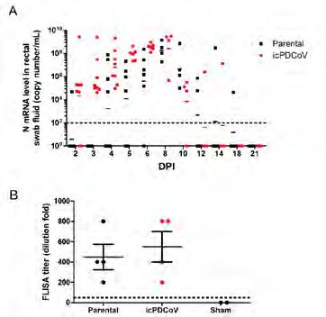
Development and Characterization of Monoclonal Antibodies
                Against African Swine Fever Virus

         Authors     Fangfeng Yuan¹,²#, Xingyu Yan¹,²#, Ana Stoian², Han Gao¹, Vlad Petrovan²,                       responses, which could be implicated in vaccine development as well as in
                     Lihua Wang³, Jishu Shi³, Raymond R. Rowland¹,², Ying Fang¹,²*                                   diagnostic assays for detection of ASFV infection.

      Affiliations   1. Department of Pathobiology, College of Veterinary Medicine, University of                    ASFV causes high morbidity and mortality in domestic pigs (5). Currently,
                        Illinois at Urbana-Champaign, Urbana, IL, USA                                                the only strategy to control the disease is to quarantine and eliminate the
                     2. Department of Diagnostic Medicine and Pathobiology, College of                               infected animals; therefore, highly sensitive and specific diagnostic reagents
                        Veterinary Medicine, Kansas State University, Manhattan, KS, USA                             are urgently needed for rapid detection and isolation of the ASFV-infected
                     3. Department of physiology, College of Veterinary Medicine, Kansas State                       pigs. Monoclonal antibody (mAb) is a key reagent for the detection of viral
                        University, Manhattan, KS, USA                                                               infection. In our previous study, we generated and characterized specific
                                                                                                                     mAbs against p30 (6). Here, we applied our technology to generate mAbs
                     #These authors contributed equally.                                                             against additional viral proteins based on their immunogenic nature and
                     *Corresponding author: yingf@illinois.edu                                                       functional importance in viral replication as described above (Table 1). This
                                                                                                                     panel of mAbs were further characterized in various assays for future use in
      Key Words      ASFV, Monoclonal Antibody, Diagnostic Reagent                                                   ASFV diagnostics and research.

        Abstract     This study generated a panel of specific monoclonal antibodies (mAbs)                Methods    ASFV genomic region encoding antigenic domain of each viral protein was
                     against selected immunogenic ASFV proteins, including p10, p14.5,                               amplified by PCR and then cloned into the protein expression vector pET 28a
                     p22, p49, p54, p72, and CD2v. These mAbs were initially screened by                             (Novagen) as a fusion protein containing an amino-terminal 6-His tag for
                     immunofluorescent assay using in vitro expression system. The antibody                          facilitating protein purification. Recombinant proteins were expressed in E.
                     reactivity was confirmed in virus-infected cells. Their application in the                      coli BL21 (DE3) cells and purified by nickel-affinity chromatography using the
                     detection of ASFV infection was further tested using the methods of Western                     method described in our previous publications (7). The purified antigens were
                     blotting, immunoprecipitation and ELISA. The availability of these mAbs                         used for mice immunization, and spleenocytes from immunized mouse were
                     provides an important tool in aid of ASFV diagnostics and research.                             fused with mouse myeloma cells to generate hybridomas as we described
                                                                                                                     previously (8). ASFV specific mAbs from hybridoma cell culture were
     Introduction    African swine fever virus (ASFV) is a large double-stranded DNA virus that                      initially screened by immunofluorescent assay (IFA) using MARC145 cells
                     belongs to family Asfarviridae, genus Asfivirus (1). The virus is enveloped                     transfected by a plasmid DNA expressing each individual ASFV protein. The
                     with two membranes at its inner and outer sides, wrapped around an                              antibody reactivity was confirmed by IFA using Vero cells infected by ASFV
                     icosahedral capsid. The viral genome varies in size between 170 and 190                         strain BA71 V. Specificity of each mAb was further determined by Western
                     kb, which encodes over 170 proteins. Among all the ASFV proteins that have                      blot and immunoprecipitation using the methods as we described preciously
                     been analyzed as far, p30 protein is determined to be a highly immunogenic                      (6). In addition, indirect ELISA was performed using the previous described
                     protein, which is capable to stimulate the highest level of antibody response                   method (9).
                     during the virus infection (2). Besides p72 and p54 were also reported to
                     be highly immunogenic (3). In a recent study (4),a total of 47 ASFV proteins      Results and   Initial hybridoma screening by IFA using MARC-145 cells that express each
                     were screened in order to identify the immunogenic and protective antigens        Discussions   individual ASFV protein resulted in a total of 62 mAbs against ASFV p10, p22,
                     for vaccine development. Among these proteins, 8 antigens including p10,                        CD2v, p49, p14.5, p72, and p54 proteins (Figure 1A). Two to three hybridoma
                     p14.5, p22, p30, p49, p54, p72, and CD2v, were tested to be able to induce                      clones from each antigen were expanded for further characterization (Table
                     certain levels of antibody responses in immunized pigs. These antigens were                     1).
                     selected based on their (known or predicted) properties of being present on
                     the surface of the intracellular mature virion or extracellular viral particles
                     (Table 1). They are potentially important for induction of protective antibody

14                                                                                                                                                                                                    15
Development and Characterization of Monoclonal Antibodies
           Against African Swine Fever Virus (Continued)

                                                                                                   A                                                      B

                                                                                                   C                                                      D
                Table 1. Characterization of ASFV specific monoclonal antibodies*
                *Refer to Petrovan et al (2) for the information about mAbs against p30.

                Western blot (WB) and immunoprecipitation (IP) were performed using the
                lysate of transfected 293T cells expressing a specific viral protein. In western
                blot analysis (Fig. 1B, top panel), all mAbs specifically detected protein bands
                for p10, p49, p54, CD2v, p14.5, p72, and p22. As expected, these bands were
                not detected in mock-transfected cells. MAbs against p54, p22, p10, p49,               Figure 1. Characterization of mAbs against ASFV. (A) IFA detection of specific ASFV antigens
                CD2v, and p72 also detected corresponding proteins in immunoprecipitated               expressed in transfected MARC145 cells. Fixed cells were stained by the corresponding mAb
                proteins from transfected cells (Fig. 1B, bottom panel). This panel of mAbs            and FITC-conjugated goat anti-mouse IgG was used as the secondary antibody. Nuclei were
                                                                                                       counterstained with DAPI (blue). (B) MAb reactivity in Western blot and immunoprecipitation
                were further tested on indirect ELISA using recombinant proteins as the
                                                                                                       analysis. Lysates from transfected 293T cells expressing a specific viral protein were harvested
                coating antigens. As is shown in Figure 1C, mAbs against p72, p54 (#114-69,            and utilized for WB and IP. Red arrows point to specific viral proteins detected by WB or IP. (C)
                #22-22), p10 (#53-23, #62-59), and p22 (#71-52) showed high reactivity with            MAb reactivity tested on indirect ELISA. Immunlon 2HB plates were coated with a recombinant
                OD405 value above 1.0, while low reactivity was observed for mAbs against              viral protein and further incubated with a corresponding mAb. HRP-conjugated goat anti-mouse
                p49 (#39-68, #88-41, and #7-90). No reactivity was detected for mAbs                   IgG was used as the secondary antibody. (D) IFA detection of ASFV antigens using Vero cells
                against CD2v and p14.5. Optimization of the ELISA conditions for these two             infected with ASFV BA71 V strain (top panel) or Vietnam virus (bottom panel).
                antigens are in the process.

                                                                                                       Specificity of these mAbs were further confirmed by IFA test using ASFV-
                                                                                                       infected Vero cells. As shown in Figure 1D, anti-p10, anti-p54, anti-p14.5,
                                                                                                       anti-p49, and anti-p72 mAbs showed strongly reactivity with the viral proteins,
                                                                                                       while anti-p22 and anti-CD2v showed weak reactivity. The weak binding
                                                                                                       activity could be due to the lower expression levels of the specific viral
                                                                                                       protein in infected cells. Another reason is that original protein structure on
                                                                                                       the virion may be different from that in the in vitro expression system.

16                                                                                                                                                                                                     17
Development and Characterization of Monoclonal Antibodies
Against African Swine Fever Virus (Continued)
Taken together, we have generated a total of 18 mAbs against 8 ASFV
antigens. This panel of mAbs provides a valuable tool for ASFV diagnostics.
They are also important reagent for basic mechanism studies toward
developing vaccines and antiviral agents.
Acknowledgements We thank Rachel Madera, Yuzhen Li and Tori Matta from Kansas State
University for providing the technical support. This project was supported
by National Pork Board grant (11-117) and Kansas National Bio and Agro-
Defense Facility Transition Fund. Fangfeng Yuan was partially supported by
Illinois Distinguished Fellowship for graduate student, University of Illinois at
Urbana-Champaign, IL.
References 1. Dixon LK, Escribano JM, Martins C, Rock DL, Salas ML, Wilkinson PJ. 2005. Asfarviridae In
Virus taxonomy. VIIIth Report of the ICTV. London, UK: Elsevier/Academic Press.
2. Giménez-Lirola LG, Mur L, Rivera B, Mogler M, Sun Y, Lizano S, Goodell C, Harris DLH,
Rowland RRR, Gallardo C, Sánchez-Vizcaíno JM, Zimmerman J. 2016. Detection of African
Swine Fever Virus Antibodies in Serum and Oral Fluid Specimens Using a Recombinant
Protein 30 (p30) Dual Matrix Indirect ELISA. PLOS ONE 11:e0161230.
3. Jia N, Ou Y, Pejsak Z, Zhang Y, Zhang J. 2017. Roles of African Swine Fever Virus Structural
Proteins in Viral Infection. J Vet Res 61:135-143.
4. Netherton CL, Goatley LC, Reis AL, Portugal R, Nash RH, Morgan SB, Gault L, Nieto R, Norlin
V, Gallardo C, Ho CS, Sanchez-Cordon PJ, Taylor G, Dixon LK. 2019. Identification and
Immunogenicity of African Swine Fever Virus Antigens. Front Immunol 10:1318.
5. Gallardo MC, Reoyo AdlT, Fernández-Pinero J, Iglesias I, Muñoz MJ, Arias ML. 2015.
African swine fever: a global view of the current challenge. Porcine Health Management
1:21.
6. Petrovan V, Yuan F, Li Y, Shang P, Murgia MV, Misra S, Rowland RRR, Fang Y. 2019.
Development and characterization of monoclonal antibodies against p30 protein of African
swine fever virus. Virus Res 269:197632.
7. Brown E, Lawson S, Welbon C, Gnanandarajah J, Li J, Murtaugh MP, Nelson EA, Molina RM,
Zimmerman JJ, Rowland RR, Fang Y. 2009. Antibody response to porcine reproductive and
respiratory syndrome virus (PRRSV) nonstructural proteins and implications for diagnostic
detection and differentiation of PRRSV types I and II. Clin Vaccine Immunol 16:628-35.
8. Li Y, Tas A, Snijder EJ, Fang Y. 2012. Identification of porcine reproductive and respiratory
syndrome virus ORF1a-encoded non-structural proteins in virus-infected cells. J Gen Virol
93:829-839.
9. Brown E, Lawson S, Welbon C, Gnanandarajah J, Li J, Murtaugh MP, Nelson EA, Molina RM,
Zimmerman JJ, Rowland RRR, Fang Y. 2009. Antibody Response to Porcine Reproductive
and Respiratory Syndrome Virus (PRRSV) Nonstructural Proteins and Implications for
Diagnostic Detection and Differentiation of PRRSV Types I and II. Clinical and Vaccine
Immunology 16:628.
18 19Development of an Infectious Clone for Porcine
Deltacoronavirus Strain USA/IL/2014/026
Authors Xufang Deng¹*, Alexandra C. Buckley², Angela Pillatzki³, Kelly M. Lager², Susan the family Coronaviridae of the order Nidovirales and have been genetically
C. Baker¹, Kay S. Faaberg²* grouped into four genera: alpha-, beta-, gamma-, and deltacoronavirus.
To date, six porcine CoVs have been identified: four alphacoronaviruses
Affiliations 1. Department of Microbiology and Immunology, Loyola University Chicago, [Transmissible gastroenteritis virus (TGEV), porcine respiratory coronavirus
Stritch School of Medicine, Maywood, IL 60153, USA (PRCoV), porcine epidemic diarrhea virus (PEDV), and swine acute
2. Virus and Prion Research Unit, USDA-ARS-National Animal Disease Center, diarrhea syndrome coronavirus (SADS-CoV, also known as swine enteric
Ames, IA 50010, USA alphacoronavirus)], one betacoronavirus [porcine haemagglutinating
3. Animal Disease Research & Diagnostic Laboratory, South Dakota State encephalomyelitis virus (PHEV)], and one deltacoronavirus [porcine
University, Brookings, SD 57007, USA deltacoronavirus (PDCoV)] [reviewed in (6)].
*Corresponding authors: Xufang Deng (xudeng@luc.edu) and Kay S. Faaberg PDCoV has been circulating in the US swine population since 2013 and there
(kay.faaberg@usda.gov) is no vaccine commercially available (7, 8). Generating a reverse genetic
tool for PDCoV is pivotal for understanding the molecular signatures of its
Key Words Porcine Deltacoronavirus, Infectious Clone, Enteric, Pathogenesis, Piglets pathogenesis and developing genetically modified live-attenuated vaccines.
Abstract We report the generation of a full-length infectious cDNA clone for porcine Results Designing an infectious PDCoV clone and rescuing recombinant virus. We
deltacoronavirus strain USA/IL/2014/026. The rescued virus, designated used a previously published strategy to develop an infectious clone of a US
icPDCoV, replicates as efficiently as the parental strain in cell culture. Both strain of PDCoV (9). Briefly, three DNA fragments (F1 to F3) composing the
parental and icPDCoV had comparable growth kinetics and formed similar complete genomic sequence of PDCoV strain USA/IL/2014/026 (GenBank
sizes of plaques in PK1 cells. To evaluate the replication and pathogenesis accession number KP981395) were synthesized. These synthetic DNAs
of the infectious clone, 7-day-old conventional piglets were inoculated were used as the template for PCR amplification of five segments that were
orally with either the parental strain or icPDCoV. Although only mild clinical then cloned into plasmid vector backbones, designated PDCoV subclones
symptoms were observed, we detected similar amounts of viral RNA in A to E (Figure 1A). All subclones were joined by a unique Sap I restriction
rectal swabs and comparable virus-specific IgG titers in the serum of both endonuclease cleavage site that allowed for directional assembly into a
groups of pigs. Immunohistochemistry analysis and histological examination full-length cDNA without alteration of the viral amino acid sequence. A T7
indicate that both viruses infect the jejunum and ileum epithelial cells RNA polymerase promoter sequence and a poly(A) tail (23 As) were added
and disrupt the integrity of the epithelium. Taken together, these results to the 5’ and 3’ ends of subclones A and E, respectively, allowing for the
collectively indicate that the infectious clone PDCoV replicates as efficiently generation of capped and polyadenylated full-length transcripts using in
as the parental strain in cell culture and in piglets. vitro RNA transcription. In subclones D and E, two naturally occurring Sap
I sites were removed by introducing silent mutations at positions 18001
Introduction Porcine deltacoronavirus (PDCoV) was first detected in 2012 and later and 20143, respectively. In subclone B, two silent mutations (A6545G and
identified as an enteric pathogen of swine (1). PDCoV infection causes A6548G) were introduced to disrupt a stretch of six A nucleotides that might
an age-dependent gastroenteritis with symptoms of acute diarrhea and interfere with in vitro RNA transcription. In addition, the coding sequence of
dehydration seen in neonatal pigs (reviewed in (2)). It primarily spreads in the N gene was cloned into a pcDNA3.1 vector that carries a T7 promoter
the swine population but can also experimentally infect calves and chickens, sequence. To recover infectious virus from the full-length cDNA clone, the
possibly due to its broad receptor usage (3–5). This highlights the potential five PDCoV DNA subclones were ligated and in vitro transcribed into viral
of diverse cross-species transmission. RNA. A linearized N gene plasmid was used to generate N gene transcripts.
These RNA transcripts were electroporated into LLC-PK1 cells. 24 hours post
Coronaviruses (CoVs) are infamous for their ability to transmit across electroporation, PDCoV-specific cytopathic effect (CPE) was observed (Figure
species barriers and can cause significant economic impact. CoVs belong to 1B), indicating an infectious PDCoV was rescued. This recombinant PDCoV
was designated icPDCoV.
20 21Development of an Infectious Clone for Porcine
Deltacoronavirus Strain USA/IL/2014/026 (Continued)
Characterization of recombinant PDCoV. To characterize the recombinant
virus, we first conducted a plaque assay and found that both the parental and
icPDCoV formed plaques with a similar size in LLC-PK1 cells (Figure 2A). To
verify the sequence of icPDCoV, a genomic region that contains the removal
of naturally occurring Sap I site (T20143A) was amplified and the PCR
fragment was subjected to Sap I digestion. As expected, the PCR fragment
amplified from the parental virus was digested by the Sap I enzyme, while the
PCR product derived from icPDCoV was resistant to Sap I-digestion (Figure
2B). We further performed whole-genome sequencing on a plaque-purified
icPDCoV clone and confirmed the fidelity of the genomic sequence. To
characterize viral replication in cell culture, both the parental and recombinant
PDCoVs were used to infect LLC-PK1 cells and the extracellular titer in the
cell culture supernatant was determined. As shown in Figure 2C, the growth
kinetics of icPDCoV was similar to that of the parental virus and both viruses
replicated to peak titers at 48 hours post-infection. In addition, we detected
the expression of the nucleocapsid (N) protein in PK1 cells that were infected
with either parental or recombinant PDCoV using a specific N protein
monoclonal antibody (Figure 2D) (10). These results together demonstrate
that the rescued icPDCoV replicates as efficiently as the parental strain in
LLC-PK1 cells.
Figure 2. Characterization of the rescued PDCoV from infectious clone. (A) Plaque assay results
Figure 1. Schematic diagram of show that both the parental and rescued PDCoV form plaques in PK1 cells with similar size. (B)
a PDCoV infectious clone and The PCR product derived from icPDCoV was resistant to Sap I-digestion, while the PCR fragment
CPE induced by PDCoV infection. amplified from the parental virus was digested by the Sap I enzyme. (C) Growth kinetics of the
(A) The gene order of PDCoV is parental strain (Parental) and infectious clone strain (icPDCoV) in PK1 cells. (D) Detection of
5’-UTR-ORF1a/1b-S-E-M-NS6-N- N protein expression in parental- or icPDCoV-infected PK1 cells using an immunofluorescence
NS7-3’-UTR. Two thirds of the assay with a mouse anti-N monoclonal antibody.
genome encodes a non-structural
polyprotein while the rest of Evaluating the replication and pathogenesis of the parental strain and
genome encodes structural and recombinant PDCoV in conventional piglets. Eight 7-day-old piglets were
accessory proteins. Three DNA
orally inoculated with either parental strain or icPDCoV at a dose of 105
fragments (F1-F3) composing the
entire genome were synthesized
TCID50 per animal. Both groups of infected piglets exhibited negligible or
and used as template for mild clinical symptoms, while control animals were healthy and showed no
the amplification of five DNA sign of disease through the entire 21 day infection course. Although only mild
segments (A-E). Restriction clinical symptoms were observed, high and comparable levels of viral RNA in
enzyme sites used for directional the rectal swabs taken from both infected groups were detected (Figure 3A).
ligation and the intentional The peak viral RNA level (~108 copy number) of fecal shedding was detected
nucleotide substitutions between 6-8 days post infection (dpi). To assess the antibody response to
described in this study are infection, sera were collected at 21 dpi and subject to a fluorescence-linked
depicted. (B) Cytopathic effect immunosorbent assay (FLISA) to detect virus-specific IgG titer. As shown
induced by parental PDCoV and
recombinant icPDCoV infection.
22 23Development of an Infectious Clone for Porcine
Deltacoronavirus Strain USA/IL/2014/026 (Continued)
in Figure 3B, both virus-infected groups had high titers of virus-specific
IgG with a similar mean of 400~500-fold dilution. We further performed
histopathological examination on the sections of the small intestines from
two pigs from each group that were euthanized on both 4 and 7 dpi. At 4 dpi,
all examined tissue sections exhibited healthy histology and no lesion was
identified. At 7 dpi, lesions consisted of villus atrophy and fusion, contraction
of superficial villus lamina with necrotic cells, vacuolar degeneration of
villus tip, and necrotic, attenuation and sloughing of enterocytes were
observed in the ileum and jejunum sections of both infected groups but not
of the control animals. We report that lesion severity was not significantly
different in affected pigs between the two virus-infected groups; however,
individual variation was observed in both groups. Representative images
of ileum sections from control piglet and from one piglet per virus-infected
group are shown in Figure 4 (upper panel). To determine the sites of virus
replication, immunohistochemistry analysis was performed to detect the
nucleocapsid (N) protein. Similar to previous studies (11–13), N protein was
mainly detected in epithelial cells, and no apparent difference in the sites of
replication between challenge groups was observed (Figure 4, middle and
lower panels). These results collectively indicate that the infectious clone
PDCoV replicates efficiently as the parental strain in piglets.
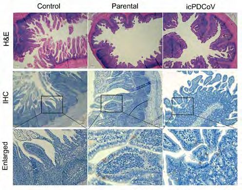
Figure 4. Histology and immunohistochemistry staining of uninfected control, parental PDCoV-,
and icPDCoV-infected piglet ileum. (Upper panel) Representative images of H&E stained
Figure 3. Evaluating viral
histological sections of ileum specimens collected at day 7 post-infection (magnification, 4×).
replication and virus-specific IgG
(Middle and lower panels) Immunohistochemistry (IHC) staining of ileum specimens collected
response to PDCoV infection.
at day 4 post-infection (middle panel, 4×; lower panel, enlarged) using a mouse anti-PDCoV-N
5~7-day-old piglets were orally
antibody.
inoculated with either the
parental strain or infectious
clone PDCoV at a dose of 105 In this study, we describe the construction of a full-length cDNA infectious
TCID50 per pig. (A) RT-qPCR Conclusion
clone for PDCoV strain USA/IL/2014/026. With this clone, a recombinant
was performed on fecal swab
samples that were taken at
PDCoV was rescued and assessed to have similar replication in cell culture
the indicated time points in a and animals as the parental strain. This infectious clone will be useful tool
21-day infection course. The for understanding the function of viral components of PDCoV and developing
bars represent the geometric genetically modified live-attenuated vaccines.
means of N gene copy numbers
of the animal group on a specific
day. (B) The titer of PDCoV-
specific IgG in serum samples
was measured using FLISA
(see details in Materials and
Methods). Error bars represent
mean ± SEM.
24 25Development of an Infectious Clone for Porcine
Deltacoronavirus Strain USA/IL/2014/026 (Continued)
References 1. Woo PCY, Lau SKP, Lam CSF, Lau CCY, Tsang AKL, Lau JHN, Bai R, Teng JLL, Tsang CCC,
Wang M, Zheng B-J, Chan K-H, Yuen K-Y. 2012. Discovery of Seven Novel Mammalian and
Avian Coronaviruses in the Genus Deltacoronavirus Supports Bat Coronaviruses as the Gene
Source of Alphacoronavirus and Betacoronavirus and Avian Coronaviruses as the Gene
Source of Gammacoronavirus and Deltacoronavirus. J Virol 86:3995–4008.
2. Jung K, Hu H, Saif LJ. 2016. Porcine deltacoronavirus infection: Etiology, cell culture for virus
isolation and propagation, molecular epidemiology and pathogenesis. Virus Res 226:50–59.
3. Jung K, Hu H, Saif LJ. 2017. Calves are susceptible to infection with the newly emerged
porcine deltacoronavirus, but not with the swine enteric alphacoronavirus, porcine epidemic
diarrhea virus. Arch Virol 162:2357–2362.
4. PA B, MA A, G L, KK Y, M V-L, LJ S, SP K. 2020. Porcine Deltacoronavirus Infection and
Transmission in Poultry, United States 1. Emerg Infect Dis 26:255–265.
5. Li W, Hulswit RJG, Kenney SP, Widjaja I, Jung K, Alhamo MA, van Dieren B, van Kuppeveld
FJM, Saif LJ, Bosch B-J. 2018. Broad receptor engagement of an emerging global
coronavirus may potentiate its diverse cross-species transmissibility. Proc Natl Acad Sci U S
A 115:E5135–E5143.
6. Wang Q, Vlasova AN, Kenney SP, Saif LJ. 2019. Emerging and re-emerging coronaviruses in
pigs. Curr Opin Virol 34:39–49.
7. Wang L, Byrum B, Zhang Y. 2014. Detection and genetic characterization of deltacoronavirus
in pigs, Ohio, USA, 2014. Emerg Infect Dis 20:1227–1230.
8. Sinha A, Gauger P, Zhang J, Yoon KJ, Harmon K. 2015. PCR-based retrospective evaluation
of diagnostic samples for emergence of porcine deltacoronavirus in US swine. Vet Microbiol
179:296–298.
9. Yount B, Denison MR, Weiss SR, Ralph S, Baric RS. 2002. Systematic assembly of a full-length
infectious cDNA of mouse hepatitis virus strain A59. J Virol 76:11065–11078.
10. Okda F, Lawson S, Liu X, Singrey A, Clement T, Hain K, Nelson J, Christopher-Hennings J,
Nelson EA. 2016. Development of monoclonal antibodies and serological assays including
indirect ELISA and fluorescent microsphere immunoassays for diagnosis of porcine
deltacoronavirus. BMC Vet Res 12:95.
11. Chen Q, Gauger P, Stafne M, Thomas J, Arruda P, Burrough E, Madson D, Brodie J, Magstadt
D, Derscheid R, Welch M, Zhang J. 2015. Pathogenicity and pathogenesis of a United States
porcine deltacoronavirus cell culture isolate in 5-day-old neonatal piglets. Virology 482:51–
59.
12. Hu H, Jung K, Vlasova AN, Saif LJ. 2016. Experimental infection of gnotobiotic pigs with the
cell-culture-adapted porcine deltacoronavirus strain OH-FD22. Arch Virol 161:3421–3434.
13. Ma Y, Zhang Y, Liang X, Lou F, Oglesbee M, Krakowka S, Li J. 2015. Origin, Evolution, and
Virulence of Porcine Deltacoronaviruses in the United States. MBio 6:e00064-15.
26 27Diagnostic Performance vs Surveillance Performance – The
Case of PRRSV Oral Fluid ELISA
Authors Alexandra Henao-Diaz¹*, Min Zhang², Luis Giménez-Lirola¹, Phil Gauger¹, Introduction Oral fluids are a diagnostic specimen easy to collect that provide for the
David Baum¹, Maria J. Clavijo¹,³, Marisa Rotolo³, Esteban Ramirez4, Rodger sufficient and accurate epidemiological data necessary for surveillance and
Main¹, Jeffrey Zimmerman¹ control of PRRSV(1, 2). For this reason, this specimen has been adapted
to the main assays routinely used in veterinary diagnostic laboratories
Affiliations 1. Department of Veterinary Diagnostic and Production Animal Medicine (VDL), i.e., RT-PCR and ELISA. PRRSV oral fluids RT-PCR detects PRRSV-
2. Department of Statistics, Iowa State University, USA RNA during the short viremia period and PRRSV oral fluids ELISA detects
3. PIC North America, USA the specific antibody response against the virus for at least 6 months
4. Grupo Porcícola Mexicano Kekén, México post-infection(3). One of the best assays to test PRRSV-antibody is the
commercial PRRS OF Ab ELISA Test (IDEXX Laboratories, Inc., Westbrook,
* Corresponding Author: ME). This test has demonstrated an excellent diagnostic performance(4) and
Veterinary Medicine Research Institute, College of Veterinary Medicine, the interpretation of the ELISA results using the manufacturer cut-off (S/P
Iowa State University, ≥ 0.4) offers a balance between diagnostic sensitivity (dxSe) and specificity
1907 ISU C Drive, Ames, IA, USA. 50011. (dxSp). However, rather than a balance, the major consideration for routine
Email address: yulyh@iastate.edu surveillance is an almost perfect diagnostic specificity because false positive
results disrupt the work flow in the farms.
Key Words Diagnostic performance, ELISA, Oral fluids, PRRSV, Surveillance performance
Herein, we evaluated the effect of using an alternative cut-off on the PRRS OF
Abstract The PRRS OF Ab ELISA Test has demonstrated a good diagnostic Ab ELISA test diagnostic performance and surveillance performance using
performance, with balance between diagnostic sensitivity (dxSe) and oral fluids of known and unknown PRRSV status.
specificity (dxSp). However, rather than a balanced diagnostic performance,
a higher diagnostic specificity and, thus the absence of false positives, is the Materials and Two sets of oral fluids (OFs) were used:
major consideration for routine surveillance. Herein, we evaluated the use of Methods Set 1: 596 experimental OFs of known PRRSV positive and negative status.
an alternative cut-off S/P ≥ 1.0 instead of the manufacturer cut-off S/P ≥ 0.4 Set 2: 1574 field OFs of known PRRSV-negative status.
and it’s potential to provide for both, diagnostic performance and surveillance
performance. Set 1 was collected from 12 experimental vaccinated pigs (PRRSV MLV) at
-7 to 42 days post vaccination(4). Set 2 consisted of samples submitted for
Two set of samples were used in this study, Set 1 included 596 oral fluids routine testing at the Iowa State University VDL from known PRRSV-negative
of known positive and negative PRRSV status and Set 2 included 1574 oral status farms. All samples were tested on the PRRS OF Ab ELISA (IDEXX
fluids from PRRSV-negative sites. Using the alternative cut-off S/P ≥ 1.0 Laboratories Inc.) and the data analyzed using non-parametrical statistical
on Set 1 resulted in a dxSe - dxSp of 96.2% - 99.4%. In set 2 the dxSp was procedures:
improved from 95.2% to 99.2% and the number of false positives reduced
from 76 to 12. Interestingly, from 76 samples classified as false positives • The test diagnostic performance (dxSe, dxSp) was analyzed by
by the manufacturer cut-off (S/P ≥ 0.4), 46 were identified as “Extreme S/P receiver operating characteristic curve (ROC) (Set 1);
outliers” and most of them associated with specific pig age (9, 24, 25 week) • The effect of an alternative cut-off on proportion of samples
or gestation. classified as positives analyzed using Cochran’s Q (Sets 1, 2);
• The relationship between ELISA results and pig age was described
The manufacturer cut-off offers a good diagnostic performance, but using using linear regression and Tukey’s box plot (Set 2);
the alternative cut-off S/P ≥ 1.0 to interpret the test results provides also for
an enhanced surveillance performance by reducing the occurrence of false
positives.
28 29You can also read