Migratory and anti-fibrotic programmes define the regenerative potential of human cardiac progenitors
←
→
Page content transcription
If your browser does not render page correctly, please read the page content below
Articles
                                                                                                         https://doi.org/10.1038/s41556-022-00899-8

Migratory and anti-fibrotic programmes define
the regenerative potential of human cardiac
progenitors
Christine M. Poch 1,16, Kylie S. Foo2,3,16, Maria Teresa De Angelis1,4,16, Karin Jennbacken 5,16,
Gianluca Santamaria1,4,16, Andrea Bähr1,16, Qing-Dong Wang 5, Franziska Reiter1,
Nadja Hornaschewitz1, Dorota Zawada1,4, Tarik Bozoglu1, Ilaria My1, Anna Meier 1,4, Tatjana Dorn1,4,
Simon Hege1, Miia L. Lehtinen3, Yat Long Tsoi 2, Daniel Hovdal 5, Johan Hyllner5,6, Sascha Schwarz7,
Stefanie Sudhop7, Victoria Jurisch1, Marcella Sini8, Mick D. Fellows8, Matthew Cummings9,
Jonathan Clarke10, Ricardo Baptista10, Elif Eroglu 2, Eckhard Wolf 11, Nikolai Klymiuk1,12,
Kun Lu13, Roland Tomasi13, Andreas Dendorfer 12,13, Marco Gaspari 14, Elvira Parrotta14,
Giovanni Cuda 14, Markus Krane12,15, Daniel Sinnecker 1,12, Petra Hoppmann1, Christian Kupatt 1,12 ✉,
Regina Fritsche-Danielson 5 ✉, Alessandra Moretti 1,4,12 ✉, Kenneth R. Chien 2,3 ✉ and
Karl-Ludwig Laugwitz 1,12 ✉

Heart regeneration is an unmet clinical need, hampered by limited renewal of adult cardiomyocytes and fibrotic scarring.
Pluripotent stem cell-based strategies are emerging, but unravelling cellular dynamics of host–graft crosstalk remains elusive.
Here, by combining lineage tracing and single-cell transcriptomics in injured non-human primate heart biomimics, we uncover
the coordinated action modes of human progenitor-mediated muscle repair. Chemoattraction via CXCL12/CXCR4 directs cellu-
lar migration to injury sites. Activated fibroblast repulsion targets fibrosis by SLIT2/ROBO1 guidance in organizing cytoskeletal
dynamics. Ultimately, differentiation and electromechanical integration lead to functional restoration of damaged heart mus-
cle. In vivo transplantation into acutely and chronically injured porcine hearts illustrated CXCR4-dependent homing, de novo
formation of heart muscle, scar-volume reduction and prevention of heart failure progression. Concurrent endothelial differ-
entiation contributed to graft neovascularization. Our study demonstrates that inherent developmental programmes within
cardiac progenitors are sequentially activated in disease, enabling the cells to sense and counteract acute and chronic injury.

W
            hereas mammals undergo endogenous cardiac regen-                       to a cardiac fate15 and exogenous transplantation of human plu-
            eration during development and shortly after birth1,2,                 ripotent stem cell (hPSC)-derived CMs16–18 or cardiac progenitors19
            the regenerative capacity of the human heart in adult-                 have been recently explored as potential approaches to generate
hood is markedly low3. The inability to replace lost myocardium is                 de novo myocardium.
accompanied by extensive tissue remodelling and fibrosis4, leaving                    Studies in lower vertebrates, where robust cardiac regeneration
patients with cardiac disease vulnerable to heart failure. Although                occurs throughout life, have demonstrated that endogenous heart
several drugs and mechanical devices can moderately improve                        repair is a highly coordinated process involving inter-lineage com-
cardiac function, they do not replace lost cardiomyocytes (CMs)                    munication, cellular de-/re-differentiation, migration and extra-
or abolish fibrotic scar formation5,6. Biotherapies have emerged as                cellular matrix (ECM) remodelling without fibrotic scarring20–23.
innovative strategies for heart repair7–10. Induction of endogenous                Similar programmes are the foundation of organ morphogenesis
CM proliferation11–14, in vivo direct reprogramming of non-CMs                     and are inherent of embryonic cardiac progenitors. During heart

1
 Medical Department I, Cardiology, Angiology, Pneumology, Klinikum rechts der Isar, Technical University of Munich, Munich, Germany. 2Department of
Cell and Molecular Biology, Karolinska Institutet, Stockholm, Sweden. 3Department of Medicine, Karolinska Institutet, Huddinge, Sweden. 4Institute of
Regenerative Medicine in Cardiology, Technical University of Munich, Munich, Germany. 5Research and Early Development, Cardiovascular, Renal and
Metabolism (CVRM), BioPharmaceuticals R&D, AstraZeneca, Gothenburg, Sweden. 6Division of Biotechnology, IFM, Linköping University, Linköping,
Sweden. 7Center for Applied Tissue Engineering and Regenerative Medicine (CANTER), Munich University of Applied Sciences, Munich, Germany. 8Clinical
Pharmacology and Safety Sciences, BioPharmaceuticals R&D, AstraZeneca, Cambridge, UK. 9Western Michigan School of Medicine, Kalamazoo, MI, USA.
10
   Procella Therapeutics, Stockholm, Sweden. 11Chair for Molecular Animal Breeding and Biotechnology, Gene Center and Department of Veterinary Sciences,
LMU Munich, Munich, Germany. 12DZHK (German Centre of Cardiovascular Research), Munich Heart Alliance, Munich, Germany. 13Walter-Brendel-Centre
of Experimental Medicine, University Hospital, LMU Munich, Munich, Germany. 14Department of Experimental and Clinical Medicine, University of Magna
Grecia, Catanzaro, Italy. 15Department of Cardiovascular Surgery, INSURE, German Heart Center Munich, Technical University of Munich, Munich, Germany.
16
  These authors contributed equally: Christine M. Poch, Kylie S. Foo, Maria Teresa De Angelis, Karin Jennbacken, Gianluca Santamaria, Andrea Bähr.
✉e-mail: Christian.Kupatt@tum.de; Regina.Fritsche-Danielson@astrazenca.com; amoretti@mytum.de; kenneth.chien@ki.se; KL.Laugwitz@mri.tum.de

Nature Cell Biology | VOL 24 | May 2022 | 659–671 | www.nature.com/naturecellbiology                                                                659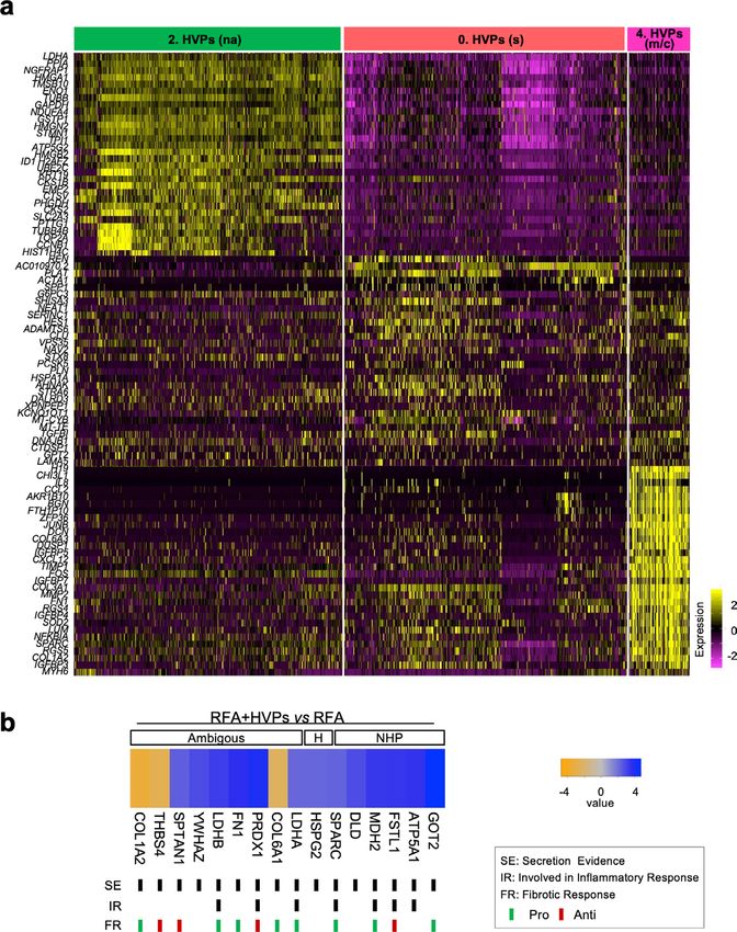
Articles                                                                                                         NATURE CELL BIOLOgy

development, defined embryonic precursors, including first heart         lineages: endothelial-committed progenitors and HVPs with their
field (FHF) and second heart field (SHF), give rise to distinct car-     CMs (Fig. 2c and Extended Data Fig. 2b).
diac compartments and cell types24,25. While FHF cells differentiate         Gene Ontology (GO) enrichment analysis of differentially
early into CMs of the primitive heart tube, ISL1+ SHF has                expressed genes (DEGs) in cells from D0 to D21 revealed progres-
broader lineage potential and its differentiation is preceded by an      sive activation of terms related to cardiac ventricular morphogen-
extensive proliferation and directed migration into the form-            esis or maturation, while pathways relevant to cardiac progenitor
ing myocardium26–28. We recently reported the generation of an           state, such as ECM organization, cell cycle and canonical BMP sig-
enriched pool of hPSC-derived ISL1+ ventricular progenitors              nalling, were gradually suppressed (Fig. 2d). On D3, a significant
(HVPs), which can expand and differentiate into functional ven-          enrichment of pathways important for progenitor proliferation and
tricular CMs in vitro and in vivo29.                                     cardiac growth was detected, including canonical Wnt, ERK1/2 and
    In this Article, we sought to determine whether HVPs could           TOR signalling (Fig. 2d). Interestingly, genes upregulated in HVPs
effectively promote heart regeneration by orchestrating sequential       at the early time of co-culture also associated with cell migration, cell
programmes of cardiac development, ultimately leading to de novo         projection organization, cytokine production and response to TGFβ
myocardium formation and positively influencing fibrotic scar            (Fig. 2d), suggesting a specific sensing-reacting response of HVPs
remodelling.                                                             to the tissue environment. Notably, enriched vasculature develop-
                                                                         ment confirmed the potential of some early precursors to differen-
Results                                                                  tiate into vessels. To define the maturation of HVP-derived CMs,
HVPs functionally repopulate a tissue model of chronic heart             we integrated our data with published scRNA-seq of in vivo human
failure. To molecularly dissect HVP-mediated cardiac repair at           adult ventricular muscle35 in pseudotime (Fig. 2e and Extended
the single-cell level, we utilize an ex vivo non-human primate           Data Fig. 2c). D21 eGFP+ cells partially allocated together with
(NHP) adult heart tissue model imitating key steps of heart failure.     adult ventricular CMs at the end of the differentiation trajectory
NHP left ventricle (LV) slices were cultured in biomimetic cham-         and expressed high levels of structural, functional and metabolic
bers30, allowing structural and functional preservation for 14 days      genes characteristic of the adult state (Fig. 2e and Extended Data
(Fig. 1a,b and Extended Data Fig. 1a). Thereafter, progressive loss of   Fig. 2c). Quantitative PCR with reverse transcription (qRT–PCR)
contractile force coincided with increased CM apoptosis (Fig. 1b,c       confirmed progressive myofibril maturation (sarcomeric isoform
and Extended Data Fig. 1a,b). NKX2.5eGFP/wt human embryonic              switching) and electrophysiological/Ca2+-handling maturation of
stem cells (hESC) were coaxed towards ISL1+/NKX2.5+ heart pro-           eGFP+-CMs from D14 to D21 (Extended Data Fig. 2d).
genitors using our protocol enriching for HVPs29, with small num-            Collectively, our single-cell transcriptomic analyses facilitated
bers of multipotent ISL1+ precursors31 (Fig. 1a and Extended Data        the construction of a differentiation route through which early
Fig. 1c). After magnetic-activated cell sorting (MACS)-based deple-      mesodermal cardiac progenitors generate mature CMs in response
tion of undifferentiated hESCs, cells were seeded onto NHP-LV slices     to signalling cues of a dying myocardium.
by bioprinting (Extended Data Fig. 1c,d). Expression of enhanced
green fluorescent protein (eGFP) enabled live tracing of HVPs            HVPs migrate and remuscularize acutely damaged myocar-
and their derivative CMs (Extended Data Fig. 1e). Labelling with         dium. Next, we designed an acute injury model in NHP-LV slices
5-ethynyl-2-deoxyuridine (EdU) and activated caspase-3 (ClCasp3)         to simulate tissue death and elucidate HVP properties in response
indicated that eGFP+ cells were highly proliferative until day 14        to injury (Fig. 3). We used radiofrequency ablation (RFA), clini-
(D14), but stopped by D21 when NHP-CMs underwent substantial             cally employed to terminate arrhythmogenic foci, to consistently
apoptosis (Fig. 1c and Extended Data Fig. 1b,f). This corresponded       destroy a defined area of cellular compartment, leaving the ECM
to extensive differentiation towards CMs and ISL1 downregulation         scaffold intact (Extended Data Fig. 3a). Gradually, progressive
(Extended Data Fig. 1g). Remarkably, heart slices gradually regained     invasion of activated cardiac fibroblasts (CFs) expressing the dis-
contraction in the third week of co-culture (Extended Data Fig. 1e),     coidin domain receptor 2 (DDR2) and increased collagen type I
reaching 2 mN force, and maintained to D50 (Fig. 1b and Extended         deposition were visible in the RFA-injured area, with tissue scar-
Data Fig. 1e). Atrial and ventricular markers (MLC2a/MLC2v)              ring by D21 (Extended Data Fig. 3b). We seeded equal amounts of
revealed that ∼81% of eGFP+ cells acquired ventricular identity by       NKX2.5eGFP/wt HVPs or CMs onto bioprinted pluronic frames on
D50 (Fig. 1d). By then, most eGFP+/MLC2v+ CMs were rod shaped            one side of the NHP-LV slices, generated RFA injury on the oppo-
with well-aligned myofibrils, structural characteristics of matura-      site and evaluated the cellular response to the damage by eGFP live
tion (Fig. 1d). A small proportion of cells expressing endothelial       imaging (Fig. 3a). Fluorescence-activated cell sorting (FACS) analy-
marker CD31 were detected (Fig. 1e), probably from multipotent           sis of the cells before seeding indicated their purity (Extended Data
precursors within the HVP pool.                                          Fig. 3d). Contrary to CMs, HVPs departed from their deposition site
    To establish a molecular roadmap for HVP specification and           and directionally migrated towards the injured region, colonizing it
maturation, we profiled cells on D0 and eGFP+ cells from D3 and          within 4 days (Fig. 3a and Extended Data Fig. 3e). By D15, HVPs
D21 ex vivo co-culture by single-cell RNA sequencing (scRNA-seq).        differentiated into CMs and the RFA area appeared remuscular-
We integrated data with our published scRNA-seq from D − 3 of            ized, with new eGFP+-CMs properly organized on D21 (Fig. 3b,c).
in vitro differentiation32. Unsupervised clustering identified seven     Proliferation rate of eGFP+ cells at the RFA injury declined pro-
stage-dependent subpopulations (Fig. 2a). On D − 3, corresponding        gressively from D7 to D21 (Fig. 3d), confirming CM maturation.
to cardiac lineage commitment33, cells expressed high levels of early    Significant reduction of scar volume was measured in HVP-treated
cardiac mesodermal genes (EOMES, MESP1 and LGR5). On D0,                 heart slices, and tissue contractile function improved (Fig. 3e,f and
cells distributed into four distinct clusters: transcriptomes of early   Extended Data Fig. 3f). Real-time intracellular Ca2+ analysis dem-
(KRT18 and ID1), intermediate (KRT8 and PRDX1) and prolifer-             onstrated that, unlike CM-treated heart slices, Ca2+ waves propa-
ating (TOP2A and CCNB1-2) progenitor states including cardiac            gated through the RFA injury when HVPs had been applied; here,
mesenchymal cells (PLCE1 and PPA1). Transcripts related to ECM           HVP-derived CMs displayed intracellular Ca2+ concentration oscil-
organization (DCN, TIMP1, LUM, FN1 and COL3A1) and ven-                  lations similar to and synchronized with the adjacent native NHP
tricular structure/maturation (MYL3, TTN, TNNC1, ACTC1 and               myocardium (Fig. 3g), indicative of electromechanical integration.
PLN) defined late eGFP+ cells and ventricular CMs on D3 and D21              To dissect the mechanisms underlying directed HVP migration
(Fig. 2a,b, Extended Data Fig. 2a and Source Data Fig. 2). Once          towards RFA and the subsequent positive remodelling during the
aligned in a pseudotime trajectory34, D3 cells bifurcated into two       scarring process, we evaluated the cellular composition of the tissue

660                                                                  Nature Cell Biology | VOL 24 | May 2022 | 659–671 | www.nature.com/naturecellbiology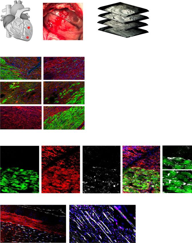
NATURE CELL BIOLOgy Articles
a In vitro Ex vivo
NHP LV Biomimetic
NKX2.5 eGFP/wt hESCs chamber
CHIR Wnt
98014 -C59
D0 D3 D21 D50
D–6 D–3 D0
hESCs HVPs
D2
b – HVPs + HVPs c
EdU+/GFP+ CICasp3+/GFP–
*** *** 100 *
4
*** 80
Contractile force (mN)
***
Cells (%)
60
2
40
**
20 **
0 **
0
*
D7 D14 D21 D50 D0 D3 D7 D14 D21
d a-GFP MLC2v MLC2a DNA
MLC2a+ MLC2v+ MLC2a+/2v+
***
100 ***
** *** ***
80 *** ***
GFP+ cells (%)
60
40
20
0
D21 D50
e a-GFP CD31 DNA
GFP+/HuNu+ GFP–/HuNu+
20
**
CD31+ cells (%)
15
*
10
5
0
D21 D50
Fig. 1 | HVPs expand, repopulate and functionally mature in an ex vivo 3D NHP heart model. a, Schematic of the experimental setup for in vitro
differentiation of HVPs from NKX2-5eGFP/wt hESCs (left) and their ex vivo co-culture with native NHP-LV slices in biomimetic chambers (right).
b, Contractile force of ex vivo cultured NHP heart slices with and without HVPs on indicated days of co-culture. Box plot shows all data points as well
as minimum, maximum, median and quartiles; n = 11 biological replicates per group; ***P < 0.001 (two-way ANOVA). c, Percentage of EdU+/eGFP+ and
ClCasp3+/eGFP− cells during co-culture. Data are mean ± s.e.m.; n = 3 biological replicates per timepoint for EdU analysis; n = 4 biological replicates
per timepoint for ClCasp3 analysis; *P < 0.05, **P < 0.005 versus D0 (one-way ANOVA). d,e, Left: representative immunofluorescence images of D50
chimeric human–NHP heart constructs using an antibody against GFP (a-GFP) together with antibodies for MLC2a and MLC2v (d) or CD31 (e). Scale
bars, 100 µm (d), 50 µm (e) and 10 µm (insets). Right: percentage of eGFP+ cells expressing MLC2v, MLC2a or both (d) and human cells expressing CD31
(e) on D21 and D50. HuNu, human nuclear antigen. Data are mean ± s.e.m. and individual data points; n = 5 biological replicates per timepoint in d, n = 3
biological replicates per timepoint in e; *P < 0.05, **P < 0.005, ***P < 0.001 (two-way ANOVA for d and t-test for e). For b–e, exact P values and numerical
data are provided in Source Data Fig. 1.
around and at the injury site. One day after RFA, activated DDR2+ and surrounding regions after 1 week (Fig. 3h). Subsequently, the
NHP CFs heavily populated the border zone and reached the dam- RFA site was predominantly colonized by eGFP+ cells and the border
aged area before eGFP+-HVPs; both cells co-existed in the injured zone by NHP DDR2+ CFs (Fig. 3h). These observations suggested
Nature Cell Biology | VOL 24 | May 2022 | 659–671 | www.nature.com/naturecellbiology 661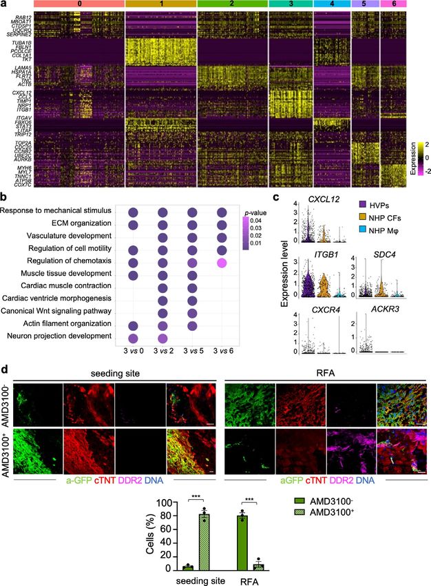
Articles NATURE CELL BIOLOgy
a c
IM HPs EC fate D–3
D0
Early HPs
D3
cMeso D21
CMCs
Component 2
Prolif
UMAP 2
HPs
Late
HPs cMeso IM HPs
D–3 D0
CMCs Prolif HPs
vCMs
Early HPs Late HPs
D3 D21 CM fate
vCMs
UMAP 1 Component 1
b d
Oxidative phosphorylation
ID1 PLCE1
Cardiac muscle contraction
4 Response to hypoxia
Expression level
Expression level
2 Cardiac ventricle morphogenesis
Cardiac muscle development
2
1 Muscle cell proliferation
Muscle cell migration
0 0 Neuron projection development Gene count
10
Endothelial cell migration
PLN PRDX1 20
Endothelial cell proliferation
Vasculature development
Expression level
4
Expression level
4 Angiogenesis
Metallopeptidase activity
2 P value
2 ECM disassembly
Extracellular structure organization 0.04
0.03
0 0 Cytokine production 0.02
Response to TGFβ 0.01
MESP1 TIMP1 TOR signalling
Wnt signalling pathway
2
Expression level
Expression level
4 ERK1 and ERK2 cascade
BMP signalling pathway
1
2 D0 D3 D21
0 0 e D–3 D21
D0 Adult vCMs
TOP2A D3
Early HPs
Component 2
4 CMCs
Expression level
vCMs
Maturation
IM HPs
2 cMeso
Late HPs
Prolif HPs
0
Component 1
Fig. 2 | scRNA-seq reveals dynamic transcriptional changes of HPs in the ex vivo 3D NHP heart model. a, UMAP clustering of single cells captured on
D − 3 and D0 of in vitro differentiation together with D3 and D21 of ex vivo co-culture. cMeso, cardiac mesoderm; CMCs, cardiac mesenchymal cells; early
HPs, early heart progenitors; IM HPs, intermediate heart progenitors; late HPs, late heart progenitors; prolif HPs, proliferating heart progenitors; vCMs,
ventricular CMs. b, Violin plots of cluster-specific marker genes; P < 0.05. c, Developmental trajectory analysis of captured cells coloured by population
identity and time of collection (inset). EC, endothelial cell. d, Representative GO terms upregulated during ex vivo co-culture. e, Pseudotime trajectory of
captured cells combined with adult vCMs from Wang et al.35. Colour gradient (from dark to light) according to maturation. For a and c–e, single cells have
been dissociated from three biological replicates. Numerical data are provided in Source Data Fig. 2.
that cell–cell communication through chemokines or physical inter- CXCL12/CXCR4 signalling mediates HVP chemotaxis to injury
action between host CFs and human progenitors might instruct sites. Developmentally, ISL1+ HVPs are highly migratory during
HVP migration, HVP differentiation and scar remodelling. heart tube extension24. To elucidate the mode of HVP migration, we
662 Nature Cell Biology | VOL 24 | May 2022 | 659–671 | www.nature.com/naturecellbiology
NATURE CELL BIOLOgy Articles
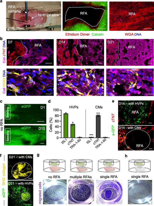
performed a trans-well migration assay, where HVPs were placed injury site as a chemoattracting signal to repopulate the damaged
on a permeable membrane and RFA-injured or uninjured NHP-LV myocardium. Similarly, chemokine-controlled deployment of SHF
slices at the bottom (Extended Data Fig. 3g). RFA significantly cells has been identified as intra-organ crosstalk between progeni-
boosted migration. Interestingly, while multiple, homogeneously tors and FHF CMs during mouse cardiogenesis37, suggesting that
distributed RFAs prompted HVPs to migrate evenly, a directional migration programmes that are functional during development are
migration towards the injured area was observed with a single RFA. re-activated in HVPs during regeneration.
No migration after RFA in decellularized NHP-LV slices confirmed
that HVP migration is dependent on a chemoattractant gradient Dynamical cellular states underlie HVP regenerative potential.
specifically arising from NHP cells at the damaged area (Extended To capture transition cell types and analyse the stepwise process
Data Fig. 3g,h). of HVP-mediated cardiac repair, we integrated scRNA-seq data
To molecularly examine the directed HVP chemotaxis and from HVPs (D0, 24 h, 48 h after RFA injury, and HVP-derived
response, we profiled eGFP+ cells migrating (24 h, 485 cells) and CMs on D21 co-culture; 2,114 cells) and generated a diffusion
arriving at the RFA injury (48 h, 269 cells) together with eGFP− map of tissue-damage-induced cardiac differentiation (Fig. 4d,e).
tissue-resident host cells (315 cells) by scRNA-seq (Fig. 4a). Seven Heat mapping of gene expression with cells ordered in the trajec-
clusters (0-6) were recovered, grouped into three populations tory revealed a temporal sequence of events and identified cells at
(Fig. 4a, Extended Data Fig. 4a and Source Data Fig. 4). Clusters 1 intermediate stages of injury sensing and injury response (Extended
and 4 belonged to the NHP group and mapped to CFs and mono- Data Fig. 5a). Dot plotting illustrated gene signature shifts among
cytes/macrophages. Human cells formed the other two groups. One different stages (Fig. 4f). In the first 24 h after injury, HVPs ‘sense’
contained four clusters, which were classified as: early HVPs (rich the tissue damage and activate gene programmes for ECM remod-
in metabolic genes such as MBOAT1, UQCRQ and MT-ND1,2,4,5,6, elling (COL6A1, ADAMTS9 and FLRT2), secretion and response
but lacking CM transcripts; cluster 0), activated HVPs (LAMA5, to cytokine (SPP1, STX8, TGFBI and IL6ST), as well as initiation
FLRT2 and TNC; cluster 2), proliferating HVPs (TOP2A, CDC20 of migration (PLAT). Subsequently (48 h), they upregulate genes
and CCNB2; cluster 5), and early ventricular CMs (MYH6, MYL3 typical of migratory cells, including transcripts for chemoattrac-
and TNNC1; cluster 6). The second group encompassed a homo- tion (PLXNA2, CMTM3 and CXCL12), cell motility (SNAI1, SNAI2,
geneous population of HVPs (cluster 3) characterized by high FAT1 and TIMP1), cytoskeleton organization (ARPC2), axon guid-
expression of genes involved in chemotaxis (NRP1, CCL2-19-21, ance (SLIT2, NFIB and UNC5B) and cell projection (RGS2, THY1
CXCL2-6-8-12, ITGB1, WASF1, RPS4X and INPPL1), a unique gene and ITGA1). During the migratory state, gene signatures of secre-
signature not captured before. GO analysis of DEGs between cluster tion (COPB2, VPS35 and SPTBN1) and cardiac muscle differentia-
3 and the other HVP clusters identified enrichment related to cell tion (VCAM1, MHY6, PALLD and TMOD1) become increasingly
motility, chemotaxis, actin filament organization, axon-guidance important as a counteracting response to injury (Fig. 4f). Mass
cues and ECM organization (Extended Data Fig. 4b), supporting spectrometry analysis of supernatants from NHP-LV slices 48 h
the migratory feature of this population. We also characterized after RFA injury revealed a significant upregulation of secreted pro-
the intercellular communication signals between HVPs and NHP teins in the presence of HVPs (Extended Data Fig. 5b). The major-
cardiac cells by performing an in silico single-cell receptor–ligand ity are involved in ECM organization (HSPG2, SPARC and FN1)
pairing screen. We found over-representation pairing of CXCL12 and fibrotic/inflammation response (FSTL1, PRDX1 and SPTAN1),
as ligand with several membrane receptors: CXCR4, SDC4, ITGB1 reinforcing the concept of HVP-influenced scar remodelling.
and ACKR3 (Fig. 4b). While CXCL12, SDC4 and ITGB1 were
expressed in HVPs and NHP fibroblasts, CXCR4 and ACKR3 recep- SLIT2/ROBO1 mediates HVP-guided fibroblast repulsion. CFs
tors were highly enriched in the HVPs (Extended Data Fig. 4c). are essential in cardiac development and repair2,38. To investigate
Trans-well migration assays under gain- and loss-of-function the temporal and spatial crosstalk between CFs and HVPs in our
conditions demonstrated that HVPs exhibited enhanced migra- ex vivo cardiac injury model, we isolated CFs from NHP hearts,
tory behaviour under CXCL12 as chemoattractant, which was stably expressed dsRed by lentiviral transduction and performed
reduced by blocking antibodies of CXCR4 or SDC4 and pharma- live imaging of co-culture with NKX2.5eGFP/wt HVPs. RFA injury
cological inhibition of CXCR4 via ADM3100 (Fig. 4c). Notably, was applied on one site of the dsRed+-CF monolayer, while seeding
binding of CXCL12 to SDC4 facilitates its presentation to CXCR4 of NKX2.5eGFP/wt HVPs on the other (Fig. 5a). Like the native tis-
(ref. 36). ADM3100 treatment was sufficient to inhibit HVP migra- sue, dsRed+-CFs were the first to invade the injured area, followed
tion towards the RFA-injured area in NHP-LV slices (Extended by eGFP+-HVPs within 5 days (Fig. 5a). Remarkably, while HVPs
Data Fig. 4d). Collectively, our data support the hypothesis that were directly chemoattracted to the injury, CFs appeared dynami-
HVPs expressing CXCR4 sense CXCL12 secreted by CFs at the cally repelled at the contact sites with migrating HVPs (Fig. 5b and
Fig. 3 | HVPs show directed migration towards acute cardiac RFA injury and remuscularize the scar. a, Left: schematic of experimental design for
selective seeding of NKX2-5eGFP/wt hESC-derived HVPs or CMs onto bioprinted frame on NHP heart slices and RFA injury on the opposite tissue site.
Right: live imaging of eGFP signal on indicated days. Scale bars, 200 µm. b, Representative immunostaining of a-GFP and cTNT in NHP constructs on D15
and D21 after RFA. Magnifications of framed areas are shown in adjacent panels. Scale bars, 200 µm (D15), 100 µm (D21) and 10 µm (magnifications).
c, Statistical analysis of GFP+ HVPs expressing cTNT on D15 and D21. Data are mean ± s.e.m. and individual data points; n = 6 biological replicates per
timepoint; ***P < 0.001 (t-test). d, Left: immunofluorescence images of proliferating (PH3+) cells on D7 and D21. Right: statistical analysis of PH3+/GFP+
cells on D7, D15 and D21. Data are mean ± s.e.m. and individual data points; n = 3 biological replicates per timepoint; **P < 0.005 (one-way ANOVA).
Scale bar, 100 µm. e, Statistical analysis of relative reduction of scar volume with HVPs compared with CMs on D21. Data are shown as mean ± s.e.m.
and individual data points; n = 3 biological replicates per group; **P < 0.005 (t-test). f, Left: representative recordings of contractile force before and after
RFA, separated by a blanking period of 2 days for re-adjustment of preload. Right: corresponding statistical analysis. Data are shown as mean ± s.e.m.;
n = 3 biological replicates per condition; *P < 0.05 versus D7 of the same group (two-way ANOVA). g, Representative images of Fluo-4-loaded NHP-HVP
and NHP-CM constructs (left) and corresponding Ca2+ transients at indicated regions of interest (ROI) (right). Scale bar, 100 μm. Red box indicates
stimulation point (1 Hz). h, Left: representative immunostaining of a-GFP and DDR2 in NHP constructs at indicated days after RFA. Scale bars, 200 µm.
Right: percentage of a-GFP+ and DDR2+ cells at RFA injury or border zone. Data are mean ± s.e.m.; n = 3 biological replicates per timepoint; *P < 0.05,
**P < 0.005 versus D1 of corresponding group (two-way ANOVA). For c–f and h, exact P values and numerical data are provided in Source Data Fig. 3.
Nature Cell Biology | VOL 24 | May 2022 | 659–671 | www.nature.com/naturecellbiology 663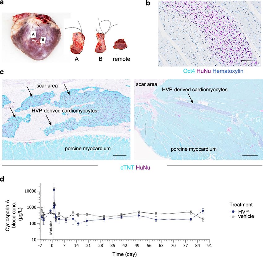
Articles NATURE CELL BIOLOgy
a
D1 D4
CPs/CMs-
eGFP+ HVPs
Seeding
NKX2.5 eGFP/wt
Seeding
RFA RFA
RFA
D0 injury
D1 D4
eGFP+ CMs
Seeding
Seeding
D1–D4
RFA RFA
Cellular migration
b
D15 D21
RFA RF1
a-GFP cTNT DNA
c d e
*** D7 D21 ** **
100 40 100
+CMs
GFP+/cTNT+ (%)
Scar volume (%)
GFP+/PH3+ (%)
75 30 75
+HVPs
50 20 50
25 10 25
0 0 0
D15 D21 a-GFP PH3 DNA D7 D15 D21
f g ROI1
RFA ROI3
RFA RFA + CMs
RFA + HVPs ROI2 RFA ROI2
Fluo-4 fluorescence (a.u.)
+HVPs
3
ROI1
ROI3
Force (2 mN)
2
Force (mN)
ROI1 1s
ROI3
ROI2
1 RFA
+CMs
ROI2
*
ROI1
0 * ROI3
3–7 9–21 days D7 D14 D21
days 1s
h
D1 D7 D21 GFP+ DDR2+
100 * 100
80 * 80
Cells (%)
Cells (%)
RFA RFA RFA 60 60
**
40 40
20 20 *
0 0
D1 D7 D21 D1 D7 D21
a-GFP DDR2 DNA
Injury Border zone
664 Nature Cell Biology | VOL 24 | May 2022 | 659–671 | www.nature.com/naturecellbiology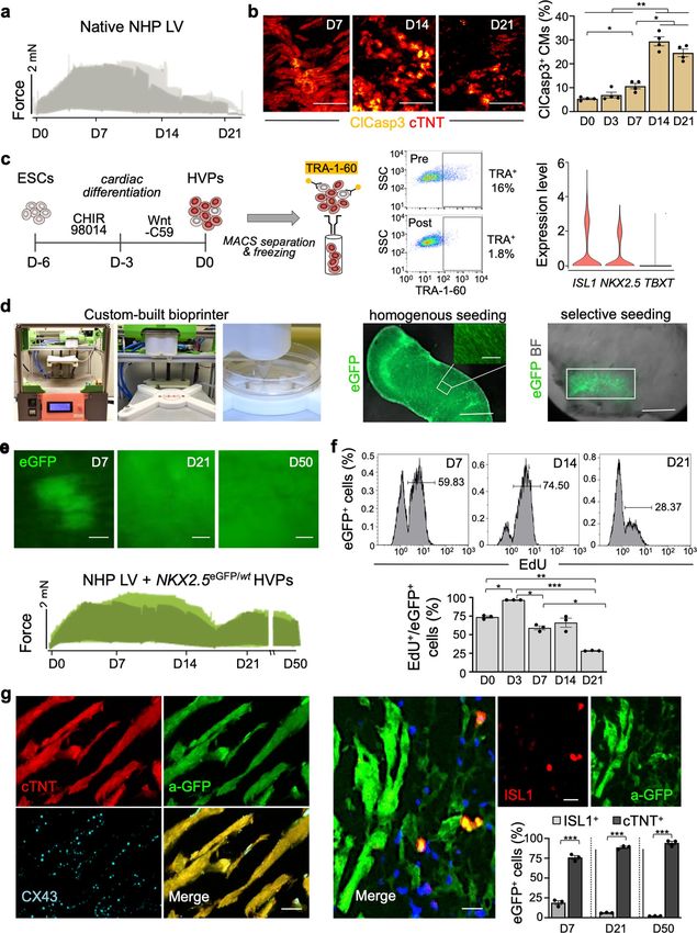
NATURE CELL BIOLOgy                                                                                                                 Articles
Supplementary Video 1). Live-cell tracking of over 100 cells for                   (Fig. 6f,g). Remarkably, their highest concentration was at the epi-
3 days demonstrated that most CFs, after interacting with HVPs,                    cardial layers with the largest damage (Fig. 6g). Most of eGFP+ cells
indeed deviate from the HVP-migratory path and were repelled                       engrafted in the injured tissue were elongated cardiac troponin T
from the injured area when the HVPs started to densely populate                    (cTNT)+ CMs with aligned myofibrils (Fig. 6h). Gap-junction pro-
it on D8 (Fig. 5b). Immunocytochemistry of filamentous (F)-actin                   tein connexin-43 was detected at the eGFP+-CMs’ intercalated discs
revealed a specific retraction of cell protrusions precisely at cellu-             and at graft and host CMs’ contact zone (Fig. 6h). CD31 immu-
lar contact sites with the HVPs (Extended Data Fig. 6a), suggesting                nostaining documented enhanced neo-angiogenesis at the RFA site
that the latter possibly control actin dynamics of CFs at the inter-               after HVP transplantation, with ∼6% of CD31+ cells of human ori-
action sites. Given the upregulation of axon-guidance genes in the                 gin (Fig. 6i and Extended Data Fig. 7b). No acute graft rejection was
migratory HVP state, including SLIT2, we postulated that SLIT2/                    detected on D14 post-transplantation, as assessed by CD68 immu-
ROBO1, a known repulsive guidance cue for axons39, might control                   nodetection. Interestingly, we even observed a reduction of CD68+
HVP-mediated CF repulsion by regulating cytoskeletal organization                  cells in HVP-treated RFA areas (Extended Data Fig. 7c), suggesting
and cell motion. Co-immunofluorescence demonstrated expression                     that HVPs might mitigate post-injury inflammation.
of SLIT2 ligand and ROBO1 receptor in migrating HVPs on D3,
while the signal was absent in the surrounding CFs (Fig. 5c). On                   HVPs remuscularize chronic scars and preserve cardiac function
D8, however, co-localization of SLIT2 and ROBO1 was observed                       in vivo. With a translational aim, we investigated HVPs’ ability to
mainly at the repulsed CFs membrane, with enriched SLIT2 signal                    engraft host myocardium in a porcine model of chronic ischaemic
at the contact sites with HVPs (Fig. 5c). qRT–PCR confirmed SLIT2                  injury. Myocardial infarction (MI) was created by occluding the left
production by HVPs and ROBO1 expression in both cell types at                      anterior descending (LAD) coronary artery for 90 min, followed
the stage of CF repulsion (Extended Data Fig. 6b). Loss-of-function                by reperfusion (Fig. 7a). Twenty-one days later, ~1 × 10⁹ HVPs
experiments using an antibody blocking ROBO1 substantiated that,                   (from WA09 hESCs) or vehicle were injected into the border zone
under ROBO1 inhibition, HVPs failed to induce actin polymeriza-                    and necrotic tissue of the MI region (Fig. 7a). Immunosuppressant
tion and lamellipodia formation in the interacting CFs, leading to                 started 6 days before cell delivery (Methods). Seventeen pigs
reduced CF motility and lack of repulsion (Fig. 5d,e and Extended                  underwent cardiac magnetic resonance imaging (cMRI) to study
Data Fig. 6c). No effects were observed in distant CFs (Extended                   LV function and infarct volume 7 days before and 12 weeks after
Data Fig. 6c). Conversely, treatment with recombinant human                        transplantation. No signs of teratoma or human DNA were detected
SLIT2 enhanced F-actin content and membrane protrusions in CFs                     in heart or other organs (lung, liver, kidney, spleen, brain, thy-
communicating with HVPs (Extended Data Fig. 6d), resulting in                      roid, adrenal glands, pituitary, prostate and lymph nodes) over the
enhanced repulsion (Fig. 5e). FACS analysis indicated that most                    3-month follow-up (Extended Data Fig. 8a,b).
ROBO1+ CFs expressed periostin, a TGFβ superfamily-responsive                         Histological examination at 12 weeks indicated large cTNT+
protein defining a specialized reparative subpopulation of CFs                     human grafts in the fibrotic scar within the MI area and near the nor-
required for healing and scar formation after injury40 (Fig. 5f).                  mal host tissue (Fig. 7b, Extended Data Fig. 8c and Supplementary
                                                                                   Video 3). Graft size ranged from 3.0% to 9.4% of the scar area (mean
HVPs migrate and regenerate injured porcine myocardium                             4.2 ± 1.3%). Expression of MLC2v confirmed that most human
in vivo. To investigate HVPs’ ability to migrate and remuscu-                      CMs in the graft had ventricular identity and well-organized sarco-
larize injured myocardium in vivo, we performed transplanta-                       meres (Fig. 7c). Strong signals of N-cadherin, which anchor myo-
tion in pigs ubiquitously expressing LEA29Y, a human CTLA4-Ig                      fibrils with connexin-43, were observed at the intercalated discs of
derivative blunting systemic T-cell response41. Two epicardial RFA                 the human CMs within the transplants and at the graft–host tissue
injuries were induced afar in the anterior heart wall and 6 × 10⁷                  interconnection, suggesting functional maturation and graft inte-
NKX2.5eGFP/wt HVPs were injected ∼1 cm apart from one damaged                      gration (Fig. 7d). CD31 immunohistochemistry indicated enhanced
site, while the other served as control (Fig. 6a and Supplementary                 neo-angiogenesis at the MI site following treatment (Fig. 7e).
Video 2). Assessment of RFA-induced tissue damage demonstrated                        At 3 months, cMRI (Fig. 8a) documented a significant reduction
consistent size of myocardial injury (Fig. 6b,c). Animals were                     in infarct volume in the HVP group (7.0 ± 1.3% versus 2.5 ± 1.6%)
treated daily with methylprednisolone and killed on D3 (n = 1),                    (Fig. 8b). After induction of ischaemia before treatment, both groups
D5 (n = 4) and D14 (n = 2) post-transplantation. None showed any                   exhibited equally depressed LV functions, with left-ventricular ejec-
signs of tumour formation (Extended Data Fig. 7a). D3 and D5                       tion fraction (LVEF) averaging 38% (vehicle 39.4 ± 1.3%, HVP
immunohistology documented a directed, CXCR4-guided migra-                         37.3 ± 2.8%). Over 12 weeks, LVEF further deteriorated signifi-
tion of eGFP+-HVPs towards the RFA-injured area (Fig. 6d,e).                       cantly by ∼10% in controls (29.4 ± 3.9%) and only by half (∼5%) in
On D5, eGFP+ cells reached the RFA site in clusters, and repop-                    HVP-treated animals (31.9 ± 3.0%), though differences between the
ulated 6.3 ± 0.6% of the scar (Fig. 6d,g). By D14, they constituted                groups did not reach statistical significance (Fig. 8c). However, the
21.0 ± 2.9% of the injured area, reducing control scar volume by half              global longitudinal strain (GLS), a sensitive measure of LV function,

Fig. 4 | HVPs are chemoattracted to sites of cardiac injury via CXCL12/CXCR4 signalling and undergo dynamic functional states in the process of
injury repair. a, Left: representative images of HVPs seeded on an injured NHP heart slice at the timepoints used for cell collection (24 h and 48 h) (top)
and UMAP plot of all captured cells (bottom). Right: relative UMAP clustering of captured cells. Mφ, macrophages. b, Circos plot for ligand–receptor
pairing showing top ten interactions identified in scRNA-seq of NHP-HVP constructs at 24 and 48 h after RFA injury and HVP application. Fraction of
expressing cells and link direction (chemokine to receptor) are indicated. c, Percentage of chemoattracted HVPs in trans-well migration assays in absence
and presence of low dose (LD) or high dose (HD) of CXCL12 (left), after addition of the indicated receptor blockers (middle) or after application of the
pharmacologic CXCR4 blockage AMD3100 in LD or HD (right). Data are indicated as mean ± s.e.m. with individual data points; n = 3 biological replicates
per condition; *P < 0.05, **P < 0.005, ***P < 0.001 versus CXCL12 HD (one-way ANOVA). d, Human scRNA-seq 24 h and 48 h datasets are integrated with
D0 and D21 CM dataset and projected onto UMAP plots, coloured by cluster assignment and annotated post hoc. Both the aligned (left) and split (right)
views are shown. HVPs (na), non-activated; HVPs (s), sensing; HVPs (m/c), migrating and counteracting. e, PCA plot of different cell clusters, with the
principal curve indicating the pathway of injury response. f, Dot plot showing gene signature shifts among different dynamic cellular states. The shadings
denote average expression and the size of dots the fractional expression. For d–f, single cells have been isolated from three biological replicates. Exact
P values and numerical data are provided in Source Data Fig. 4.

Nature Cell Biology | VOL 24 | May 2022 | 659–671 | www.nature.com/naturecellbiology                                                                   665
Articles NATURE CELL BIOLOgy
significantly worsened in the vehicle-treated (−3.1 ± 1.0) compared Discussion
with the HVP group (−0.2 ± 0.6) (Fig. 8d), demonstrating that HVP Human CMs have poor proliferative potential, resulting in virtu-
treatment attenuated the progressive decline of cardiac function in ally non-existent de novo CM renewal after injury. The inability
this model. to replace lost contractile units after acute MI is paralleled by
a
Cell seeding Early HVPs
RFA
0 NHP Mφ
Sample 24 h Sample 48 h
6 Early vCMs 4
Activated 2
HVPs
5
Proliferating HVPs
1
NHP CFs
Human cells 24 h
3
Human cells 48 h Migrating HVPs
NHP cells
b c ITGB1-RB No AMD3100
No chemokine No RB
HVPs
CXCL12 LD CXCR4-RB ACKR3-RB AMD3100 LD
CX
B1
CXCL12 HD SDC4-RB AMD3100 HD
CL
ITG
C4
12
R4
SD
C *
CX 3 *** ** **
KR
SDC4 AC ***
** 100
100 100
ITGB1 NHP
Attracted HVPs (%)
CXCL12 attracted
CXCL12 attracted
1 Mφ 75 75
ITGB 75
HVPs (%)
HVPs (%)
50 50 50
CXCL12
NHP
CFs 25 25 25
0 0 0
d e
HVPs (m/c) vCMs
4
HVPs (s) HVPs (s)
vCMs Split
UMAP 2
PC2
view 0
CMCs HVPs (m/c)
D21
CMCs
–4
D0 HVPs (na) HVPs (na)
UMAP 1 24 h CPs 48 h CPs 0 10 20 30 40
PC1
f
ADAMTS9
ADAMTS1
CCDC80
MBTPS1
SPTBN1
COL6A1
PLXNA2
HSPA1A
CXCL12
CMTM3
TMOD1
VCAM1
HAND1
UNC5B
COPB2
ARPC2
TNNC1
LAMA5
ANXA1
ACTC1
ACTN2
NR4A1
TNNT2
VPS35
PALLD
FLRT2
MEIS1
TGFBI
TIMP1
SNAI2
SNAI1
SORD
ITGA1
MEST
MYH6
RGS2
BMP7
UBR4
IL6ST
SLIT2
HES1
BTG2
SSR1
MYL3
NNAT
SPP1
THY1
GLB1
DLK1
TBX5
STX8
PLD3
PLAT
NFIB
FAT1
ISL1
CLU
FN1
JUN
TTN
PLN
ID3
CMCs
Cell ECM Cell motility Cell Secretion
HVPs (na) activation projection
HVPs (s) CM genes
HVPs (m/c)
vCMs CP genes
25 100 0 1.5
Percent expressed Average expression
666 Nature Cell Biology | VOL 24 | May 2022 | 659–671 | www.nature.com/naturecellbiology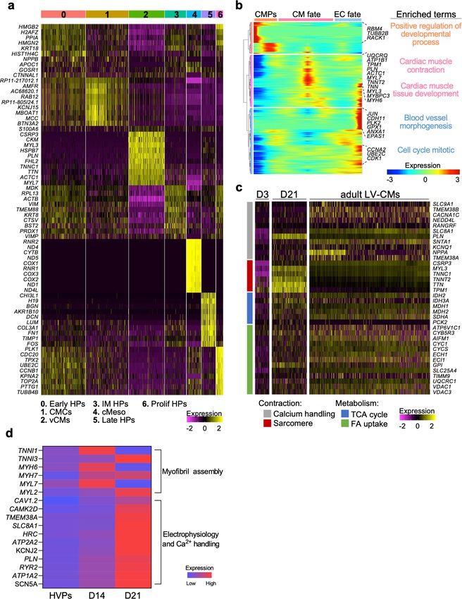
NATURE CELL BIOLOgy Articles
a c D3 D8
NHP-CFsdsRed NKX2.5 eGFP/wt HVPs
RFA
D3 D5
HVP seeding
RFA
dsRed eGFP
b
6 D6 a-GFP dsRed SLIT2 ROBO1 DNA
5 0
4
3 7
100
d e
y position (µm)
2 8 a-ROBO1 (10 min) a-ROBO1 (40 min)
200 Untreated
1
300 a-ROBO1
9 11 rhSLIT2
400
10 12
500
D7 HVP tracking – CF tracking
*
4 6 600
3 ! D7.5
*
7 8 0 100 200 300 400 500 600
x position (µm)
2
1
9 8
11
10 150
Average movement
12 HVPseGFP
**
D8 100 D8
(pixel)
4 6
3 8
7
50 CFsdsRed
2
12 0 30 60
10
0 Repulsed CFs
9 11 D6 D7 D8 a-GFP dsRed F-actin DNA (% relative to D7)
f CFs-dsRed
103 103 ***
100 ***
78.7 % ***
102 102 80
7.23%
Goat IgG
60
Cells (%)
ROBO1
101 101
10.54% 3.53%
40 *
10 0
10 0
20
*
0
+
–
+
–
10–1 100 101 102 10–1 100 101 102
1
1
1
1
BO
BO
BO
BO
O
O
O
O
Rabbit IgG POSTN
R
R
R
R
POSTN+ POSTN–
Fig. 5 | SLIT2/ROBO1 signalling mediates activated CF repulsion and prevents myocardial scarring. a, Top: schematic of 2D model for RFA injury of NHP
CFs expressing dsRed followed by NKX2-5eGFP/wt HVP seeding and monitoring of co-culture. Bottom: sequential live imaging of dsRed+ and eGFP+ cells
during migration. Scale bars, 200 µm. b, Left: representative time-lapse images of dsRed+ and eGFP+ cells at the RFA injury site during CF repulsion on
indicated days. Dotted line delineates HVP migration front. Scale bar, 100 µm. The numbers indicate individual cells followed and tracked during the time
lapse imaging. Right: cell tracking over time (top) and average movement (bottom) analysis of HVPs and CFs. c, Representative immunostaining for eGFP,
SLIT2 and ROBO1 on D3 and D8. Scale bars, 25 µm. d, F-actin and eGFP immunofluorescence an D8 after ROBO1 antibody exposure for 10 and 40 min.
Change of CF shape (arrow head) and F-actin localized on protrusion side of CFs (arrow). Scale bars, 75 µm. e, Percentage of repulsed CFs at the injured
site analysed on D7.5 and D8 in standard condition (untreated) or after ROBO1 antibody and rhSLIT2 treatment on D7. Data are normalized to D7 and
presented as mean ± s.e.m. and individual data points; n = 3 biological replicates per condition; *P < 0.05, **P < 0.005 versus untreated (t-test). f, Flow
cytometry analysis for ROBO1 and POSTN in CFsdsRed after 8 days of co-culture with HVPs. Data are shown as mean ± s.e.m. and individual data points;
n = 4 biological replicates per condition; *P < 0.05, ***P < 0.001 (t-test). For e and f, exact P values and numerical data are provided in Source Data Fig. 5.
Nature Cell Biology | VOL 24 | May 2022 | 659–671 | www.nature.com/naturecellbiology 667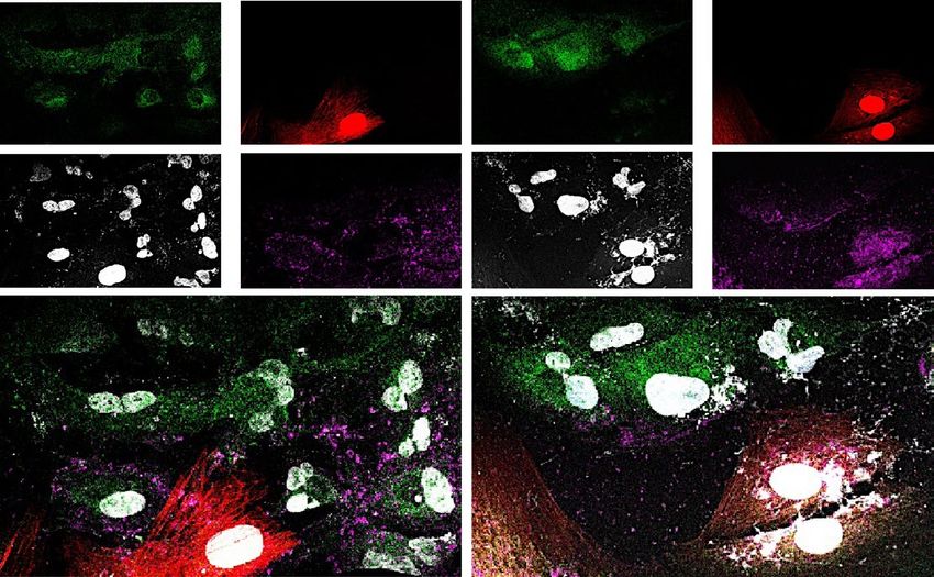
Articles NATURE CELL BIOLOgy
a b c 75
RFA HVP injection Medium injection Bright-field RFA
Volume
Depth
RFA volume (mm3)
10
RFA depth (mm)
70
Depth
5
65 0
In vivo RFA
d e RFA Application site
100 100
Adjacent Injury
Cells (%)
Cells (%)
–CXCR4 blockage
50 50
+CXCR4 blockage
D3
0 0
f RFA +HVPs g
Scar depth (mm) 10 D5
40
D14
40
D5
5
eGFP+ area (%)
30
eGFP+ area (%)
20 20
0
D5 D14
80 0
0
D14
Scar volume
m)
(mm3)
40 (m 1
th
Dep 2
3
4
a-GFP WGA DNA 0 5
D5 D14
h
Border zone
RFA
a-GFP cTNT CX43 DNA
i
HuNu–
RFA *
**
HuNu+
1,000
CD31+ per mm2
750
500
250
RFA 0
WGA CD31 DNA HuNu CD31 DNA –HVPs +HVPs
scar formation and fibrosis in the injury zone2,21. To unleash the cells to target areas, modulate electrical integration and govern
full regenerative potential of cardiac cell therapy, it is essential the cellular/molecular host–graft crosstalk. Our ex vivo model of
to identify the cues that guide the recruitment of transplanted HVPs and NHP heart tissue provides an unprecedented system to
668 Nature Cell Biology | VOL 24 | May 2022 | 659–671 | www.nature.com/naturecellbiologyNATURE CELL BIOLOgy Articles
Fig. 6 | HVPs regenerate RFA-injured porcine myocardium in vivo. a, Schematic of in vivo experimental design with two left ventricular RFA injuries
and adjacent injection of HVPs or cell-free medium. b, Representative 3D reconstruction of non-transmural RFA injury. Scale bar, 2 mm. c, Statistical
analysis of scar volume and depth of RFA injuries in freshly explanted wild-type pig hearts indicating standardized injury size. Box plot shows minimum,
maximum, median and quartiles; n = 3 biological replicates. d, Representative fluorescence images of injury and adjacent sites after wheat germ agglutinin
(WGA) and a-GFP co-staining on days D3, D5 and D14. Scale bars, 100 µm. e, Analysis of cells at application site and RFA in the presence or absence of
pharmacological CXCR4 blockage (AMD3100) on day 5. Data are mean with individual data points; n = 2 biological replicates per condition. f, Analysis of
in vivo scar depth and volume on D5 and D14 with or without HVP treatment. Data are mean ± s.e.m. with individual data points; n = 3 biological replicates
per group on day 5, n = 2 biological replicates per group on day 14. g, Percentage of GFP+ area within the RFA injury (left) and according to depth of the
cutting plane (right). Data are mean with individual data points; n = 2 biological replicates per group. h, Representative immunofluorescence images of RFA
and border zone on D14 for anti-GFP, cTNT and CX43. Magnifications on the right correspond to the boxed area in the merged image. Scale bars, 50 µm
and 10 µm (magnifications). i, Representative fluorescence images of HVP-treated RFA injury site after immunostaining for CD31 in combination with
WGA (left) or with anti-human nuclei (HuNu, right). Scale bars, 50 µm (left), 25 µm (right). Bar graph shows the average number of CD31+ cells per mm2
cells from host (HuNu−) and human HVPs (HuNu+) in HVP-treated and untreated RFAs. Data are presented as mean ± s.e.m. with individual data points;
n = 6 biological replicates per group; *P < 0.05, **P < 0.005 (two-way ANOVA). For c, e–g and i, exact P values and numerical data are provided in Source
Data Fig. 6.
a
90 min
1 × 10⁹
cMRI IMS HVPs cMRI
D – 21 D–7 D–6 D0 12 weeks
LAD occlusion Vehicle or cell injection Histology
b
Porcine
myocardium
Infarcted zone
Grafts
cTNT+ graft
Infarct zone
Porcine myocardium
c d e
MLC2V HuNu N-cadherin HuNu CD31 haematoxylin
Fig. 7 | HVPs remuscularize chronic scars in a porcine model of chronic ischaemia in vivo. a, Schematic of in vivo experimental design of acute MI by
balloon occlusion of the LAD coronary artery (ischaemia) and reperfusion after 90 min. Triple- immunosuppressive regimen (IMS) with cyclosporine
(D − 6 to D84), methylprednisolone (D − 1 to D84) and abatacept (D − 1 to D84). Analysis of baseline infarct volume by cMRI on day −6 followed by
epicardial cell injection (15 injection sites, total 1 × 10⁹ HVPs) into myocardial injury. Follow-up period of 12 weeks with cMRI scans at 12 weeks before
termination and histological work-up. b, Overview of infarct zone and human grafts with labelling of porcine myocardium (HuNu− cTNT+), infarct zone and
cTNT+ graft (HuNu+ cTNT+). Scale bar, 2 mm. c–e, Immunohistochemistry of graft for cardiac ventricular muscle marker (MLC2v) (c), electrical coupling
(N-cadherin) (d), and vessel formation (CD31) (e) at 12 weeks. Scale bar, 50 µm. Lower panels show magnifications of boxed areas. Scale bar, 15 µm.
refine molecular pathways implicated in cardiac regeneration at programmes for directed migration, fibroblast repulsion and ulti-
a single-cell resolution, thus offering an innovative approach. We mate muscle differentiation within an injured heart (Fig. 8e). Future
demonstrated that HVPs harbour the unique potential to sense studies should investigate whether ex vivo human heart slices
and counteract injury by re-activating sequential developmental could predict outcome of cell-based regeneration in patients with
Nature Cell Biology | VOL 24 | May 2022 | 659–671 | www.nature.com/naturecellbiology 669Articles NATURE CELL BIOLOgy
a b Vehicle group HVP group
*
50 NS *** 5
Diastole (LV) Systole (LV)
∆infarct volume (%)
40 0
Infarct volume
(% of LV)
30
–5
20
–10
10
0 –15
0 12 0 12 12 weeks
Time (weeks)
c d
0 NS *
60
* NS
20 NS *** 10
10
∆ LVEF (%)
–5
∆ GLS (%)
40 0
LVEF (%)
GLS (%)
5
–10
20 –20 –10
–30 0
0 –40 –15
0 12 0 12 12 weeks 0 12 0 12 12 weeks
Time (weeks) Time (weeks)
e Sensing Migrating Counteracting/differentiating
ECM genes CXCR4 CXCL12 SLIT2 ROBO1
HVPs
CFs
Injury
Fig. 8 | HVPs preserve cardiac function in vivo. a, Representative LV cMRI images of diastole and systole used for calculation of infarct volume, LVEF and
GLS. b–d, Statistical analysis of infarct volume (b), LVEF (c) and GLS (d). Infarct volume, LVEF and GLS (%) are shown as minimum-to-maximum range
with mean and individual data points; NS, not significant; *P < 0.05, ***P ≤ 0.001 (two-way ANOVA). Delta values (Δ) are shown as mean ± s.e.m. and
individual data points; *P < 0.05 (t-test); n = 10 pigs in the vehicle group and n = 7 pigs in the HVP-treated group. For b–d, exact P values and numerical
data are provided in Source Data Fig. 8. e, Schematic summary of HVPs undergoing dynamic cellular states during cardiac tissue repair. HVPs sense tissue
damage by activating programmes of ECM remodelling and migration and are chemoattracted to sites of cardiac injury via CXCR4/CXCL12 signalling.
Counteraction to injury occurs via CF repulsion in a SLIT2/ROBO1-dependent manner and subsequent CM differentiation to remuscularize scar tissue.
different aetiologies of heart failure (for example, ischaemic, genetic response is sufficient to restore physiological blood flow, since robust
and inflammatory). arterial input is crucial for permanent functional improvement.
Cell homing constitutes an indispensable step in repair pro- Moreover, before HVP transplantation can be translated clinically,
cesses of many organs42,43. We uncovered that the CXCL12/CXCR4 one should determine whether HVP-based therapies could achieve
pathway mediates the inherent homing potential of HVPs, an abil- higher remuscularization compared with CM transplantation with
ity lost once they entered the fully differentiated myocytic lineage. reduced ventricular arrhythmia and the use of hypo-immunogenic
CXCL12 is implicated in migration of haematopoietic progenitors44. PSC lines can circumvent long-term rejection. Recently, the ESCORT
We demonstrate that HVPs utilize similar molecular pathways to trial performed first transplants of hPSC-derived cardiac progenitor
facilitate homing and repopulation like the haematopoietic system. patches in patients with ischaemic cardiomyopathy and reported no
Upon injury, activated CFs produce ECM components for tis- adverse effects including tumour formation19. Concordantly, after
sue reconstructions while sending signals to neighbouring CMs cardiac HVP injection, we did not detect tumourigenesis over the
and others for initiating reparative processes45. Our scRNA-seq 3-month follow-up, demonstrating that MACS depletion of undif-
unravelled that the SLIT2/ROBO1 axis mediates the HVPs’ ability ferentiated hESCs is safe for clinical translation.
to repel CFs, thus reducing scarring. It will be of particular interest In conclusion, our data indicate that HVPs harbour the unique
to evaluate whether such signalling pathway acts similarly in vivo capability to target both loss of myocardium and fibrotic scar-
and whether its pharmacological manipulation could circumvent ring of the injured heart, supporting their therapeutic potential.
cell application. Developing innovative therapeutic strategies rooted in fundamental
The study demonstrates rapid engraftment of HVPs with exten- biology of cardiac development could pave the way for successful
sive de novo myocardium generation in porcine models of acute cell-based cures of heart disease.
injury and chronic ischaemic heart failure. In both settings, scar
volume was reduced and, in the latter, deterioration of cardiac func- Online content
tion was prevented. This repairment cannot occur without sufficient Any methods, additional references, Nature Research report-
blood supply. Increased neovascularization was documented in HVP ing summaries, source data, extended data, supplementary infor-
grafts. Additional analysis is needed to demonstrate whether such mation, acknowledgements, peer review information; details of
670 Nature Cell Biology | VOL 24 | May 2022 | 659–671 | www.nature.com/naturecellbiologyNATURE CELL BIOLOgy Articles
author contributions and competing interests; and statements of 27. Spater, D. et al. A HCN4+ cardiomyogenic progenitor derived from
data and code availability are available at https://doi.org/10.1038/ the first heart field and human pluripotent stem cells. Nat. Cell Biol. 15,
1098–1106 (2013).
s41556-022-00899-8. 28. Liang, X. et al. HCN4 dynamically marks the first heart field and conduction
system precursors. Circ. Res. 113, 399–407 (2013).
Received: 12 August 2021; Accepted: 11 March 2022; 29. Foo, K. S. et al. Human ISL1+ ventricular progenitors self-assemble into an
Published online: 12 May 2022 in vivo functional heart patch and preserve cardiac function post infarction.
Mol. Ther. 26, 1644–1659 (2018).
30. Fischer, C. et al. Long-term functional and structural preservation of
References precision-cut human myocardium under continuous electromechanical
1. Porrello, E. R. et al. Transient regenerative potential of the neonatal mouse
stimulation in vitro. Nat. Commun. 10, 117 (2019).
heart. Science 331, 1078–1080 (2011).
31. Bu, L. et al. Human ISL1 heart progenitors generate diverse multipotent
2. Tzahor, E. & Poss, K. D. Cardiac regeneration strategies: staying young at
cardiovascular cell lineages. Nature 460, 113–117 (2009).
heart. Science 356, 1035–1039 (2017).
32. Sahara, M. et al. Population and single-cell analysis of human cardiogenesis
3. Bergmann, O. et al. Dynamics of cell generation and turnover in the human
reveals unique LGR5 ventricular progenitors in embryonic outflow tract.
heart. Cell 161, 1566–1575 (2015).
Dev. Cell 48, 475–490 (2019).
4. Travers, J. G., Kamal, F. A., Robbins, J., Yutzey, K. E. & Blaxall, B. C. Cardiac
33. Lescroart, F. et al. Defining the earliest step of cardiovascular lineage
fibrosis: the fibroblast awakens. Circ. Res. 118, 1021–1040 (2016).
segregation by single-cell RNA-seq. Science 359, 1177–1181 (2018).
5. Lozano, R. et al. Global and regional mortality from 235 causes of death for
34. Trapnell, C. et al. The dynamics and regulators of cell fate decisions are
20 age groups in 1990 and 2010: a systematic analysis for the Global Burden
revealed by pseudotemporal ordering of single cells. Nat. Biotechnol. 32,
of Disease Study 2010. Lancet 380, 2095–2128 (2012).
381–386 (2014).
6. Fang, L., Murphy, A. J. & Dart, A. M. A clinical perspective of anti-fibrotic
35. Wang, L. et al. Single-cell reconstruction of the adult human heart during
therapies for cardiovascular disease. Front. Pharm. 8, 186 (2017).
heart failure and recovery reveals the cellular landscape underlying cardiac
7. Hnatiuk, A. & Mercola, M. Stars in the night sky: iPSC-cardiomyocytes return
function. Nat. Cell Biol. 22, 108–119 (2020).
the patient context to drug screening. Cell Stem Cell 24, 506–507 (2019).
36. Brule, S. et al. The shedding of syndecan-4 and syndecan-1 from HeLa cells
8. Desgres, M. & Menasche, P. Clinical translation of pluripotent stem cell
and human primary macrophages is accelerated by SDF-1/CXCL12 and
therapies: challenges and considerations. Cell Stem Cell 25, 594–606 (2019).
mediated by the matrix metalloproteinase-9. Glycobiology 16, 488–501 (2006).
9. Shi, Y., Inoue, H., Wu, J. C. & Yamanaka, S. Induced pluripotent stem cell
37. Xiong, H. et al. Single-cell transcriptomics reveals chemotaxis-mediated
technology: a decade of progress. Nat. Rev. Drug Discov. 16, 115–130 (2017).
intraorgan crosstalk during cardiogenesis. Circ. Res. 125, 398–410 (2019).
10. Smart, N. et al. De novo cardiomyocytes from within the activated adult
38. Gonzalez-Rosa, J. M., Martin, V., Peralta, M., Torres, M. & Mercader, N.
heart after injury. Nature 474, 640–644 (2011).
Extensive scar formation and regression during heart regeneration after
11. Gabisonia, K. et al. MicroRNA therapy stimulates uncontrolled cardiac repair
cryoinjury in zebrafish. Development 138, 1663–1674 (2011).
after myocardial infarction in pigs. Nature 569, 418–422 (2019).
39. Nguyen Ba-Charvet, K. T. et al. Diversity and specificity of actions of Slit2
12. Wei, K. et al. Epicardial FSTL1 reconstitution regenerates the adult
proteolytic fragments in axon guidance. J. Neurosci. 21, 4281–4289 (2001).
mammalian heart. Nature 525, 479–485 (2015).
40. Tallquist, M. D. & Molkentin, J. D. Redefining the identity of cardiac
13. Bassat, E. et al. The extracellular matrix protein agrin promotes heart
fibroblasts. Nat. Rev. Cardiol. 14, 484–491 (2017).
regeneration in mice. Nature 547, 179–184 (2017).
41. Bahr, A. et al. Ubiquitous LEA29Y expression blocks T cell co-stimulation
14. Monroe, T. O. et al. YAP partially reprograms chromatin accessibility to
but permits sexual reproduction in genetically modified pigs. PLoS ONE 11,
directly induce adult cardiogenesis in vivo. Dev. Cell 48, 765–779 (2019).
e0155676 (2016).
15. Srivastava, D. & DeWitt, N. In vivo cellular reprogramming: the next
42. Chavakis, E., Urbich, C. & Dimmeler, S. Homing and engraftment of
generation. Cell 166, 1386–1396 (2016).
progenitor cells: a prerequisite for cell therapy. J. Mol. Cell. Cardiol. 45,
16. Chong, J. J. et al. Human embryonic-stem-cell-derived cardiomyocytes
514–522 (2008).
regenerate non-human primate hearts. Nature 510, 273–277 (2014).
43. Ganju, R. K. et al. The α-chemokine, stromal cell-derived factor-1α,
17. Liu, Y. W. et al. Human embryonic stem cell-derived cardiomyocytes restore
binds to the transmembrane G-protein-coupled CXCR-4 receptor and
function in infarcted hearts of non-human primates. Nat. Biotechnol. 36,
activates multiple signal transduction pathways. J. Biol. Chem. 273,
597–605 (2018).
23169–23175 (1998).
18. Romagnuolo, R. et al. Human embryonic stem cell-derived cardiomyocytes
44. Ceradini, D. J. et al. Progenitor cell trafficking is regulated by hypoxic
regenerate the infarcted pig heart but induce ventricular tachyarrhythmias.
gradients through HIF-1 induction of SDF-1. Nat. Med. 10, 858–864 (2004).
Stem Cell Rep. 12, 967–981 (2019).
45. Pellman, J., Zhang, J. & Sheikh, F. Myocyte–fibroblast communication in
19. Menasche, P. et al. Transplantation of human embryonic stem cell-derived
cardiac fibrosis and arrhythmias: mechanisms and model systems. J. Mol.
cardiovascular progenitors for severe ischemic left ventricular dysfunction.
Cell. Cardiol. 94, 22–31 (2016).
J. Am. Coll. Cardiol. 71, 429–438 (2018).
20. Chien, K. R. et al. Regenerating the field of cardiovascular cell therapy.
Nat. Biotechnol. 37, 232–237 (2019). Publisher’s note Springer Nature remains neutral with regard to jurisdictional claims in
21. Sadek, H. & Olson, E. N. Toward the goal of human heart regeneration. published maps and institutional affiliations.
Cell Stem Cell 26, 7–16 (2020). Open Access This article is licensed under a Creative Commons
22. Vagnozzi, R. J. et al. An acute immune response underlies the benefit of Attribution 4.0 International License, which permits use, sharing, adap-
cardiac stem cell therapy. Nature 577, 405–409 (2020). tation, distribution and reproduction in any medium or format, as long
23. Eschenhagen, T. et al. Cardiomyocyte regeneration: a consensus statement. as you give appropriate credit to the original author(s) and the source, provide a link to
Circulation 136, 680–686 (2017). the Creative Commons license, and indicate if changes were made. The images or other
24. Brade, T., Pane, L. S., Moretti, A., Chien, K. R. & Laugwitz, K. L. Embryonic third party material in this article are included in the article’s Creative Commons license,
heart progenitors and cardiogenesis. Cold Spring Harb. Perspect. Med. 3, unless indicated otherwise in a credit line to the material. If material is not included in
a013847 (2013). the article’s Creative Commons license and your intended use is not permitted by statu-
25. Sahara, M., Santoro, F. & Chien, K. R. Programming and reprogramming a tory regulation or exceeds the permitted use, you will need to obtain permission directly
human heart cell. EMBO J. 34, 710–738 (2015). from the copyright holder. To view a copy of this license, visit http://creativecommons.
26. Moretti, A. et al. Multipotent embryonic isl1+ progenitor cells lead to cardiac, org/licenses/by/4.0/.
smooth muscle, and endothelial cell diversification. Cell 127, 1151–1165 (2006). © The Author(s) 2022
Nature Cell Biology | VOL 24 | May 2022 | 659–671 | www.nature.com/naturecellbiology 671You can also read


