Maternal Nutrition and Developmental Programming of Male Progeny
←
→
Page content transcription
If your browser does not render page correctly, please read the page content below

animals
Review
Maternal Nutrition and Developmental Programming of
Male Progeny
Sarah McCoski *, Amanda Bradbery                     , Rodrigo da Silva Marques, Christian Posbergh               and Carla Sanford

                                          Department of Animal and Range Sciences, Montana State University, Bozeman, MT 59718, USA;
                                          amanda.bradbery@montana.edu (A.B.); rodrigo.marques@montana.edu (R.d.S.M.);
                                          christian.posbergh@montana.edu (C.P.); carla.sanford@montana.edu (C.S.)
                                          * Correspondence: sarah.mccoski@montana.edu; Tel.: +1-406-994-5574

                                          Simple Summary: The objective of the following review is to describe available literature on the
                                          interaction between maternal nutrition and developmental programming in male offspring. The
                                          majority of current research focuses on female offspring or fails to take offspring sex into account,
                                          though sexual dimorphisms in response to maternal diet are well-recognized. This leaves a large
                                          gap in the understanding of male developmental programming. This review will specifically discuss
                                          the impacts of maternal dietary energy and protein on bull and ram growth, development, and
                                          reproductive capacity in later life.

                                          Abstract: Poor maternal nutrition can cause several maladaptive phenotypes in exposed offspring.
                                          While non-sex-specific and female-specific adaptations are well-documented, male-specific outcomes
                                          are still poorly understood. Of particular interest are the outcomes in bulls and rams, as develop-

                                          mental programming directly impacts long-term productivity of the animal as well as human food
                                   security. The following review discusses the impact of poor maternal dietary energy and protein on
Citation: McCoski, S.; Bradbery, A.;      bull and ram developmental programming as it relates to growth, development, and reproductive
Marques, R.d.S.; Posbergh, C.;            capacity. The review also highlights the importance of the timing of maternal dietary insult, as early-,
Sanford, C. Maternal Nutrition and        mid-, and late-gestational insults can all have varying effects on offspring.
Developmental Programming of Male
Progeny. Animals 2021, 11, 2216.          Keywords: bull; developmental programming; growth; ram; reproduction
https://doi.org/10.3390/ani11082216

Academic Editor: Rebecca
R. Cockrum                                1. Introduction
                                                Developmental programming is the response to a specific maternal challenge, par-
Received: 30 June 2021
Accepted: 22 July 2021
                                          ticularly nutritional, during a critical developmental window that persistently alters the
Published: 27 July 2021
                                          trajectory of growth, physiology, and/or metabolism of the progeny [1,2]. Maternal nutri-
                                          tion during embryonic and fetal development can have short- and long-term consequences
Publisher’s Note: MDPI stays neutral
                                          for the progeny [3–5]. The following review will discuss available literature examining the
with regard to jurisdictional claims in
                                          impacts of maternal energy and protein availability during gestation on subsequent bull
published maps and institutional affil-   and ram growth, development, and reproductive capacity. The review will also highlight
iations.                                  the impact timing of the dietary insult has on fetal outcomes.
                                                Worldwide, beef cattle and sheep production systems rely largely on forage-based di-
                                          ets as the source of the majority of nutrients. However, seasonal variation in forage quality
                                          and quantity frequently affect nutrient utilization and animal performance by inadequate
Copyright: © 2021 by the authors.
                                          dietary intake, including energy and protein [6]. Regardless of operation location and
Licensee MDPI, Basel, Switzerland.
                                          environment, cattle herds and sheep flocks have nutritional challenges which require sup-
This article is an open access article
                                          plementation. However, management practices can vary, especially considering intensive
distributed under the terms and           versus extensive operations and differences in parturition timing to meet market needs.
conditions of the Creative Commons        Furthermore, changes in nutritional resource availablity, as in drought years, or projected
Attribution (CC BY) license (https://     market values for a calf or lamb crop may cause producers to inadequately supplement
creativecommons.org/licenses/by/          their gestating animals due to financial restraints or inability to source feedstuffs. Thus,
4.0/).                                    beef cattle and sheep are often exposed to suboptimal nutrition during critical periods of

Animals 2021, 11, 2216. https://doi.org/10.3390/ani11082216                                              https://www.mdpi.com/journal/animals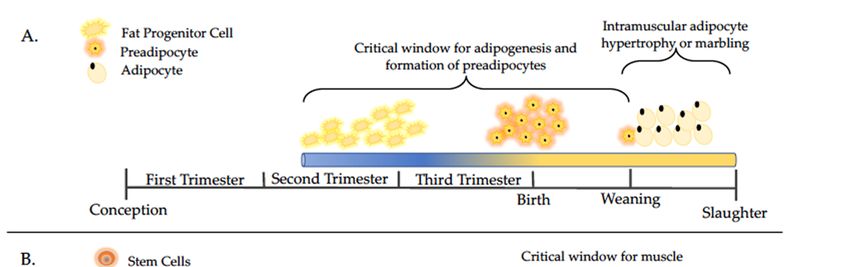
Animals 2021, 11, 2216                                                                                            2 of 13

                         fetal development, which consequently alter metabolic, energetic, and body composition
                         response of the offspring [2,5,7]. To maintain maximum long-term productivity, producers
                         must design and integrate supplementation programs, such as those focused on protein
                         and energy, according to animal requirements and forage characteristics. The effects of
                         dietary manipulation during gestation on offspring outcomes might vary depending on
                         the timing, severity, and duration of nutrient restriction or overnutrition, as well as the sex
                         of the fetus [8–10]. A limited number of studies have investigated the combined influence
                         of maternal diet and fetal sex on offspring performance, metabolism, and health. Those
                         that have included fetal sex in analyses, a majority are focused on female offspring because
                         of their importance as replacement animals. The work discussed herein is focused on the
                         interaction between maternal energy and protein availability during gestation on male
                         offspring growth, development, and reproductive parameters in cattle and sheep. The
                         purpose of this review is to highlight available information on this topic, but also to bring
                         attention to the lack of understanding on how male offspring are impacted by maternal
                         diet.
                               The loss of quality and quantity of available forage during gestation often results in a
                         state of nutrient restriction and may alter the intrauterine environment of the developing
                         offspring. While undernutrition is more common in livestock production systems, overnu-
                         trition presents a similar concern, though the effects are not as well defined. Regardless,
                         when exposed to changes in maternal plane of nutrition in utero, offspring are at greater
                         risk of developing metabolic dysregulation. While little work has been performed with the
                         intent to compare the effects of maternal plane of nutrition and fetal sex, it is an important
                         consideration given the evidence of sexual dimorphism in fetal development [11,12]. This
                         may be especially true with regards to skeletal muscle and adipose tissue development, as
                         well as postpubertal reproductive efficiency. Hence, the focus of this review is to summa-
                         rize recent advancements in the understanding of interactions between maternal dietary
                         management and growth, performance, and reproductive capacity of male offspring.

                         2. Maternal Under- and Over-nutrition Impacts on Skeletal Muscle and Adipose
                         Tissue Development
                              Maternal nutrient status during gestation is critical for fetal skeletal muscle and
                         intramuscular adipocyte development, which can be a determinant factor of the future
                         performance and carcass characteristics of the progeny [13–15]. The impacts of maternal
                         diet on offspring growth and performance are variable and dependent on the timing of
                         the dietary intervention, as muscle and adipose development occur at specific times in
                         gestation (see Figure 1A,B). During the prenatal stage, the formation of muscle fibers can
                         be altered by maternal nutrient restriction, leading to a reduction in the total number of
                         secondary muscle fibers, which permanently reduces animal performance [13]. Conversely,
                         maternal overnutrition promotes the preeminent expression of adipogenic genes in fetal
                         skeletal muscle [16], which likely program a greater number and size of adipocytes of
                         progeny skeletal muscle postnatally [15]. Therefore, maternal nutritional status during fetal
                         development is a major factor affecting the lifetime productivity of the offspring [2,4,5].
Animals
Animals 2021,2021, 11, x FOR PEER REVIEW
11, 2216 3 of 13
3 of 13
Figure 1. Timelines of adipocyte (A) and muscle (B) development with a focus on major events discussed in this review.
Figure 1. Timelines of adipocyte (A) and muscle (B) development with a focus on major events discussed in this review.
Adapted fromfrom
Adapted [14,17,18].
[14,17,18].
Undernutrition often results in offspring that are small for gestational age, culminating
Undernutrition often results in offspring that are small for gestational age, culminat-
in smaller birth weights followed by a compensatory response during postpartum growth
ing in smaller birth weights followed by a compensatory response during postpartum
in sheep and cattle [19–21]. The observed reduction in birth weights may be due to a priori-
growth in sheep and cattle [19–21]. The observed reduction in birth weights may be due
tization of vital organs over the development of muscle mass in animals that are exposed
to a prioritization of vital organs over the development of muscle mass in animals that are
to nutrient restriction in utero. However, weight and mass of some fundamental organs
exposed
involved to nutrient restriction
in metabolism and growthinare utero.
alsoHowever,
known to weight and mass
be sensitive of some
to maternal fundamental
plane of nu-
organs involved in metabolism and growth are also known to be
trition including the pancreas, liver, intestines, skeletal muscle, adrenals, etc. [22–25]. Much sensitive to maternal
plane
of the of nutrition
resulting including
postpartum the pancreas,
compensatory liver,isintestines,
growth skeletal
due to altered muscle,
offspring adrenals, etc.
metabolism
[22–25]. Much of the resulting postpartum compensatory growth
from multifactorial changes at the molecular level of several organ systems [19,20,24,25]. is due to altered off-
spring
Despite the “catch-up” growth often observed following fetal exposure to organ
metabolism from multifactorial changes at the molecular level of several mater-sys-
tems [19,20,24,25].
nal nutrient restriction, offspring tend to have higher levels of adiposity and less lean
Despite the
mass compared “catch-up”whose
to offspring growthdamsoftenwere
observed
fed tofollowing fetal exposure
meet nutrient to maternal
requirements dur- nu-
ing trient restriction,
gestation offspring
[14,26–28]. tend toand
Adipose have highermuscle
skeletal levels oftissues
adiposityareand
bothless lean mass
derived fromcompared
the
to offspring whose dams were fed to meet nutrient requirements
mesenchymal stem cell populations (Figure 1) thus, the effects of maternal nutrition on during gestation [14,26–28].
the Adipose and skeletal
differentiation of one muscle
tissuetissues are influence
will also both derived thefrom the mesenchymal
differentiation stem cell popu-
and development
lations (Figure 1) thus, the effects of maternal nutrition on the
of the other [15]. The mechanism by which this occurs is still under investigation; differentiation of one tissue
how-will
ever,also
aninfluence
interplaythe ofdifferentiation and development
molecular alterations appears to of the other [15]. The
be implicated. mechanism
Results by which
describing
this occurs is still
molecular-specific undervary
effects investigation; however,
based on timing an interplay
of dietary of molecular
manipulation, butalterations
the long-term appears
to be implicated.
implications Results describing molecular-specific effects vary based on timing of dietary
are similar.
manipulation, but the long-term
Increased adiposity and decreased implications
lean mass are similar.
in response to maternal overnutrition
Increased adiposity and decreased
are a product of changes in gene expression in both adipose lean mass in response
tissue toand
maternal
skeletalovernutrition
muscle.
are a skeletal
Because productmuscle
of changesis the inprimary
gene expression in both adipose tissue
site of insulin-stimulated glucose anduptake,
skeletalit muscle.
is a
primeBecause
targetskeletal muscle
for altered is the primary
metabolism resulting site
inof insulin-stimulated
disturbances glucoseto
that contribute uptake,
the devi- it is a
prime
ations target forgrowth
in offspring altered andmetabolism resulting
development. in disturbances
Reduced insulin-likethatgrowth
contribute
factor to 1the
and devi-
ations indue
2 (IGF1/2), offspring growth
to maternal and development.
nutrient Reduced
restriction, with insulin-like
upregulation growth
of their factor 1 re-
respective and 2
ceptors on skeletal
(IGF1/2), due toand cardiacnutrient
maternal musclerestriction,
has been reported, which is associated
with upregulation with insulin
of their respective recep-
dysregulation [24,29]. Other studies do not report the same effects,
tors on skeletal and cardiac muscle has been reported, which is associated with insulin but instead observed
differences in gene[24,29].
dysregulation expressionOther andstudies
signaling molecules
do not report intheother
samemetabolic
effects, butpathways
insteadinclud-
observed
ing adipogenic, lipogenic, and myogenic factors [30–32]. Additionally, Sandoval et al. [24]
Animals 2021, 11, 2216                                                                                           4 of 13

                         found alterations in metabolic pathways in skeletal muscle and a lower proportion of type
                         I skeletal muscle fibers, which are the most sensitive of the myofiber types to insulin and
                         oxidative metabolism, further contributing to altered energy metabolism, growth, and
                         development [24,25]. None of these data, however, infer a difference between fetal sex in
                         response to maternal plane of nutrition in developing lambs.
                               Differences between fetal sex response to maternal undernutrition is variable, and
                         a critical gap remains in the scientific literature. It is suggested that male offspring may
                         be more susceptible to maternal nutrient restriction because their energy demands for
                         development may be higher [12,33]. Ford et al. [26] found glucose intolerance in male
                         lambs following a glucose tolerance test at 12 months of age following exposure to maternal
                         undernutrition; however, the study did not compare male and female lambs. Similarly,
                         beef steers exposed to different levels of nutrient restriction in utero had increased average
                         daily gain consistent with compensatory growth, but heifer offspring were not included in
                         the study [21].
                               Although undernutrition often results in reduced birth weights, the same is not al-
                         ways the case with maternal overnutrition where birth weights tend to be similar between
                         offspring exposed to maternal overnutrition or control diets meeting nutrient require-
                         ments [22,23,34]. The influence of maternal overnutrition on organ weights and mass is not
                         as predictable as that observed with nutrient restriction, although many similar molecular
                         changes to organ systems have been described [23,34–37]. The influence of maternal over-
                         nutrition specific to the development of male offspring is not well investigated; however,
                         sex-specific responses are becoming more frequently considered.
                               Tissue-specific responses to maternal overnutrition include the pancreas, skeletal
                         muscle, adipose tissues, and intestines, resulting in reduced insulin sensitivity, increased
                         adipogenesis, and altered growth and performance [23,26,28]. Several genes and biochem-
                         ical pathways have been implicated in these responses; however, they may differ based
                         on fetal sex and timing and duration of the nutritional insult. A comparison of male and
                         female offspring revealed that male offspring prioritize skeletal muscle development over
                         intestinal development compared to female offspring when exposed to maternal overnu-
                         trition in utero [34,35]. Furthermore, male offspring tend to develop skeletal muscle faster
                         than female offspring, which could alter the response to maternal overnutrition and the
                         resulting performance phenotypes [34,35]. Future work should focus on elucidating sex ×
                         tissue-specific responses to maternal nutritional status during gestation.

                         3. Maternal Dietary Energy
                         3.1. Maternal Dietary Energy and Developmental Programming
                              Most energy models available consider the energy required for the first months of
                         gestation to be negligible due to minimal energy retention in the pregnant uterus, whereas
                         with the progression of the gestation energy requirements and retention in the gravid uterus
                         is maximized to support fetal growth and development [6,38]. Net energy for maintenance
                         increases by 30–50% during the last trimester of gestation, of which half of the increase
                         contributes to gravid uterine tissues and one-quarter to the fetus to support rapid fetal
                         growth and one-quarter for increased maternal metabolic function [6,38]. Nevertheless,
                         the early stage of embryo differentiation, fetal organogenesis, and utero-placental tissues
                         formation are critical events that impact progeny later in life [4,5,38,39]. Accordingly, ewes
                         subjected to a 50% nutrient restriction during early gestation and then realigned to meet
                         energy requirements produced lambs with normal birth weights, whereas lambs from these
                         restriction-realigned dietary regimen ewes exhibited altered growth rates, dysfunction in
                         glucose metabolism, increased carcass fat, and reduced carcass muscle compared with
                         lambs from ewes offered a control diet throughout gestation [26,27,39]. Therefore, dams that
                         undergo nutritional stress during early period of development, rather than late gestation,
                         are expected to calve a normal weight offspring that will still experience undesired growth
                         and metabolic problems later in life [26,27,38,39]. These findings contradict the common
                         misconception that maternal nutrition during early gestation is less critical than that during
Animals 2021, 11, 2216 5 of 13
mid- and late gestation. Additionally, it should be noted that these studies did not take
offspring sex into account. Similar studies should be performed to determine if these
results vary depending on offspring sex.
Maternal nutrient status affects nutrient partitioning of pregnant livestock and fe-
tal development [38,40]. This may disturb the glucose and amino acid metabolism of
the gravid uterus, and these nutrients are the primary source of energy for the normal
development of the fetus [41,42]. In sheep, Gardner et al. [43] demonstrated that a 50%
restriction in energy during late gestation did not impact lamb birth weight, but decreased
glucose tolerance, caused insulin resistance, and increased adiposity in lamb progeny. Long
et al. [28] reported that 30% of energy restriction during early gestation in beef cows did not
impact birth, weaning, or carcass characteristics, but adipocyte diameter was increased, and
muscle weight was reduced in the male progeny born to restricted cows. Corah et al. [44]
demonstrated that calves from beef cows offered an energy-restricted diet during the last
100 days of gestation were lighter at birth and weaning compared with calves from cows
fed 100% of energy requirements. Furthermore, energy deficiency during the last trimester
of gestation increased rates of morbidity and mortality of the progeny [44], suggesting that
maternal energy status during gestation can also impact the health of the offspring. In
an effort to explain the mechanism by which these phenotypic changes occur, Sanglard
et al. [43] evaluated the skeletal muscle and blood transcriptomes of beef calves that were
exposed to energy restriction in utero. Authors describe effects of diet, sex, and diet ×
sex interactions on expression of genes involved in energy metabolism, skeletal muscle
development and contraction, immune response, and responses to stress. These effects
emphasize the sex-specific response of offspring to maternal energy status [43].
Source of maternal dietary energy may also promote adaptations that permanently
alter the trajectory of growth, physiology, and metabolism of the progeny. Loerch et al. [45]
indicated that calves from cows limit-fed a starch-based diet have greater birth weights
compared with calves from cows fed a forage-based diet. Radunz et al. [46] offered hay-,
corn-, or distillers grains-based diets to late-gestating beef cows and observed a reduced
birth weight in calves from hay-fed cows compared with cohorts born to cows receiving
corn or distillers grains, whereas calves from hay-fed cows were lighter at weaning and
required 10 more days on feed to reach a similar fat thickness compared with calves
from corn-fed cows. These outcomes suggest that high-starch diets offered to gestating
cows alter rumen fermentation towards propionate production, which may increase the
circulating blood glucose and insulin secretion [47] resulting in greater maternal nutrient
supply to the fetus growth [48,49]. Collectively, nutritional energy manipulation during
periods of developmental plasticity such as the embryonic, fetal, and neonatal periods exert
lasting effects on muscle and adipose tissue development, health, and overall performance
of offspring. It should be noted that these specific studies did not take offspring sex
into account. Further studies are necessary to determine these impacts on bull and ram
offspring.
3.2. Dietary Energy In Utero and Male Reproductive Capacity
Testes size has long been used as an indicator of fertility, as scrotal circumference
is directly related to semen production capacity [50–52]. Scrotal size can change based
on nutritional status, breeding season, and age of the animal thus, it seems logical that
maternal nutrition during testes development (beginning on day 41 and 31 post-fertilization
in bull and ram embryos, respectively [53,54]) can also impact testes size and weight
(Figure 2). Studies show varying results, likely an effect of the differing times of dietary
intervention. Ram lambs from ewes fed to 70% of energy requirements from week 10
of gestation to birth had lower paired testes weight compared to those exposed to 110%
energy requirements [55]. Similar results were reported when ewes were restricted to
50% energy requirements from day 100 until parturition, with ram lambs experiencing
reduced testicular weight at birth [56]. Conversely, ram lambs from ewes fed 50% energy
requirements from breeding to day 95 of gestation showed no effect on testicular weightvention. Ram lambs from ewes fed to 70% of energy requirements from week 10 of gesta-
tion to birth had lower paired testes weight compared to those exposed to 110% energy
requirements [55]. Similar results were reported when ewes were restricted to 50% energy
Animals 2021, 11, 2216 requirements from day 100 until parturition, with ram lambs experiencing reduced testic- 6 of 13
ular weight at birth [56]. Conversely, ram lambs from ewes fed 50% energy requirements
from breeding to day 95 of gestation showed no effect on testicular weight compared to
controls [57]. These differences in results may be explained by the timing of the nutritional
compared to controls [57]. These differences in results may be explained by the timing of
insult. Nutrient restriction encompassing the entirety of testes development may result in
the nutritional insult. Nutrient restriction encompassing the entirety of testes development
more profound impacts on testes formation than restrictions ending during early/mid de-
may result in more profound impacts on testes formation than restrictions ending during
velopment. Scenarios such as the later, may give the testes enough time during develop-
early/mid development. Scenarios such as the later, may give the testes enough time during
ment to “catch up” to normal growth. Additionally, the timing of nutritional insult may
development to “catch up” to normal growth. Additionally, the timing of nutritional insult
differentially impact
may differentially the development
impact the development of various testicular
of various structures
testicular structures(i.e.
(i.e.seminiferous
seminiferous
tubules,
tubules,Sertoli
Sertoli cells,
cells, vasculature, etc.) which
vasculature, etc.) which may
mayresult
resultinindiffering
differingphenotypic
phenotypicchanges
changes to
to testes structure.
testes structure.
Figure 2. A timeline depicting the development of the bull and ram reproductive tracts. Events included are discussed
Figure 2. A timeline depicting the development of the bull and ram reproductive tracts. Events included are discussed
within the review. Adapted from [58–60].
within the review. Adapted from [58–60].
Sertoli cell number and function may be used to assess male reproductive capacity, as
Sertoli
a single cell number
Sertoli andtofunction
cell is able supportmay be used to assess
the development of a male
finite reproductive
number of sperm capacity,
cells
as a single Sertoli cell is able to support the development of a finite
within the seminiferous tubule [61], play a role in male sex determination [62], and produce number of sperm cells
within the seminiferous
secretions necessary fortubule [61], play a role
spermatogenesis. Early in in
male sex determination
embryogenesis, Sertoli[62], and
cells pro-
secrete
duce secretions necessary for spermatogenesis. Early in embryogenesis,
Mullerian inhibiting factor, which prevents the differentiation of the female reproductive Sertoli cells se-
crete Mullerian inhibiting factor, which prevents the differentiation
tract [63]. Additionally, the number of Sertoli cells dictates the total number of germ cells of the female repro-
ductive tract [63].
in adulthood, Additionally,
as Sertoli the number
cells secretions are of Sertoli cells in
indispensable dictates
promotingthe total number of
spermatogene-
germ cells in adulthood,
sis [61,64,65]. as Sertoli
Studies highlight the cells
impacts secretions
of poor are indispensable
maternal nutritionin onpromoting sper-
offspring Sertoli
matogenesis
cell number.[61,64,65]. Studies highlight
A study utilizing the impacts
sheep to examine the of poor maternal
impacts of reduced nutrition on off-
metabolizable
spring
energySertoli
and crudecell protein
number.(50% A study utilizing
vs 100% sheep tofound
requirement) examine the impacts
that males exposed of to
reduced
energy
metabolizable energy and crude protein (50% vs 100% requirement)
restriction from days 31–100 of gestation had lower Sertoli cell numbers than controls, found that males ex-
posed to energy restriction from days 31–100 of gestation had lower
though testes weight did not differ between groups [66]. Additional work reported similar Sertoli cell numbers
than controls,
findings though
in which testes energy
maternal weightrestriction
did not differ reducedbetween
Sertoligroups
cell and[66]. Additional tubule
seminiferous work
reported
numberssimilar
in ram findings in which
lambs [55,56]. maternal
However, energy
these restriction
studies reduced Sertoli
are contradicted by work cellwhich
and
seminiferous
found ram fetuses tubuleexposed
numbers in ram
to low lambs
energy (50%[55,56]. However,
requirement) these studies
maternal diets had areunaltered
contra-
dicted
Sertolibycellwork
numberwhich
[67].found ram fetuses
Collectively, theseexposed
findings to low energy
indicate that the (50%
timingrequirement)
of the maternal ma-
ternal diets had unaltered Sertoli cell number [67]. Collectively,
dietary insult and when the effects of maternal dietary insults are assessed are important tothese findings indicate
that the timing
consider. Sertoliofcell
thenumbers
maternal dietarydrastically
increase insult and when
from the effects to
mid-gestation of birth
maternal
in sheepdietary
and
insults are assessed
cattle (reviewed are important
in [59]). Considering to this,
consider. Sertoli cell
implementing numbers dietary
a maternal increaseinsult
drastically
during
from mid-gestation
this window to birthdifferent
may produce in sheepresults
and cattle (reviewed
compared in [59]).
to those Considering
following maternal this, im-
dietary
interventiona during
plementing maternal early gestation.
dietary insultAdditionally,
during this window Sertoli cellmay numbers
produce continue
differentto increase
results
from birthtothrough
compared the peripubertal
those following maternal period.
dietary Sampling
interventionpriorduring
to this early
window of Sertoli
gestation. cell
Addi-
proliferation [55,67] may yield different results than studies in which
tionally, Sertoli cell numbers continue to increase from birth through the peripubertal pe- samples are collected
during
riod. or after prior
Sampling this period. The effectofofSertoli
to this window timingcellshould be investigated
proliferation [55,67] further.
may yield Thedifferent
impacts
of maternal
results undernutrition
than studies in which onsamples
Sertoli cell
aredevelopment
collected during is notorfully
afterunderstood.
this period.Sertoli
The effectcells
are an undisputed necessity in male reproduction, and disruption in their development
and function has damaging effects on male fertility. However, it is important to recognize
that Sertoli cells continue to proliferate after birth, therefore, a sufficient postnatal diet
may recover the damaging effects of gestational dietary insults. The biological impacts of
altered Sertoli cells during early life on reproductive performance in adulthood remains
unclear.Animals 2021, 11, 2216 7 of 13
The mechanisms resulting in the structural differences in the gonads of males exposed
to maternal high or low dietary energy are still not fully understood. There are two possible
causes for the connection between maternal diet and later reproductive capacity in male
offspring. First, the effects discussed above may be the direct consequence of nutrition on
the gonads, as made evident by gene expression data. Bull fetuses exposed to maternal
overnutrition experienced a decrease in expression of steroidogenic acute regulatory protein
(StAR), hydroxysteroid 17-Beta dehydrogenase 3 (HSD17B3), IGF1, IGF2, and IGF1 receptor
(IGF1R), genes involved in testes development and steroidogenesis [68]. In particular, IGF1
is produced by Leydig and Sertoli cells, and IGF1 receptor (IGF1R) is found on germ and
somatic cells of the testis [69]. When inactivated in mouse testes, decreased IGF1R resulted
in a decrease in Sertoli cell proliferation [70]. As previously discussed, Sertoli cells play an
indispensable role in testes development and later reproductive capacity, and alterations
in their proliferation may have lasting impacts on male reproductive performance. A
second possibility is that the observed gonadal effects are secondary to disruptions in
hypotholamo-pituitary function. Studies show that exposure to maternal energy restriction
alters pituitary sensitivity to gonadotropin releasing hormone (GnRH), which may disrupt
gonadotropin release [71,72]. Seminiferous tubule function and Sertoli cell development
are dependent on gonadotropin production during fetal development [73] thus, a reduction
in GnRH responsiveness may result in reproductive insufficiency in adult life. Further
work is needed to identify the mechanisms responsible for the link between maternal
dietary energy availability and male offspring reproduction in later life.
Limited information is available on the impacts of in utero growth-restriction on adult
reproductive parameters in bull and ram offspring. A study utilizing ewes fed either a
normal or a growth restricted diet found that male offspring experienced delayed puberty
and had lower plasma testosterone and testicular volume until 28–35 weeks of age [74].
Furthermore, though peak testosterone production occurred at a similar age, the growth-
restricted males saw a much lower peak value, 5.8 ± 1.1 versus 9.4 ± 0.5. Puberty was not
attained in these rams until live weights similar to those of the control rams at puberty was
achieved. The delay in the onset of puberty in the growth-restricted male lambs indicates a
significant impact in maternal diet and the onset of sexual maturation in sheep [74]. Rams
and bulls that attain puberty at an earlier age compared to their counterparts allow for
earlier progeny testing, increase the rate of genetic selection, and can reduce production
costs for breeders. These findings suggest that male reproductive capacity is susceptible to
developmental programming; however, studies examining testicular structure and semen
quality during adult life are necessary.
4. Maternal Dietary Protein
4.1. Dietary Protein and Developmental Programming
As previously mentioned, supplementation is often required in livestock production
systems based typically on forage diets, and protein is often considered the limiting nutrient
in livestock operations [75]. Research has suggested protein supply during gestation affects
fetal, growth, and development, and is critical to offspring performance and health [76–79].
The crucial role of specific amino acids on cell metabolism and function of the fetus might
exert lasting effects on muscle and adiposity development, health, and overall performance
of the progeny [77,80,81]. For example, arginine is a conditionally-essential amino acid
that contributes to nitric oxide and polyamine production, both of which play important
roles in placental development and function [82–84]. Therefore, it seems reasonable that
maternal dietary protein supply during gestation is indispensable for adequate progeny
trajectory prenatally and postnatally.
A growing body of evidence suggests that maternal protein nutrition might alter
offspring body composition and growth, hormonal balance, metabolic function, neonatal
health, organ development, and function [2,85,86]. Maternal undernutrition during early
gestation, which is a critical window for establishing normal fetal development of all organs
and tissues [87,88], leads to reduced fetal muscle mass and increased adipocyte size thatAnimals 2021, 11, 2216 8 of 13
could modify progeny metabolism later in life [13,85]. Accordingly, Copping et al. reported
that pregnant heifers exposed to low dietary protein during the first trimester of gestation
affected the development of the fetal heart, liver, lung, brain, and pancreas with sex-specific
responses to the gestational dietary protein restriction [10]. Specifically, maternal protein
restriction during first trimester gestation resulted in differential gene expression in the
liver between male and female offspring [62]. Male offspring had increased expression of
glucose transporter protein type 1 (GLUT1) over female offspring, whereas female offspring
more greatly expressed peroxisome proliferator-activated receptor γ (PPARγ) with reduced
expression of IGF2 and glucocorticoid receptor. This suggests altered metabolism between
the sexes, which would both result in dyslipidemia and increased adipogenesis postnatally,
but programmed through different molecular mechanisms based on fetal sex [62]. Sex-
specific cardiovascular and central nervous system effects have also been observed in
cattle and sheep which are believed to further drive metabolic differences through the
preferential allocation of nutrients during periods of protein restriction [10,89,90].
Maternal nutrient intake affects fetal growth trajectory from early stages of fetal de-
velopment to birth. A large proportion of available data is focused on late gestation when
nearly 75% of the fetal growth occurs, and nutrient requirements for fetal development
are maximal [6,28]. Furthermore, variations in maternal dietary protein might alter the
performance and carcass characteristics of male progeny reared for slaughter [7,78,91,92].
Funston et al. [93] reported that protein supplementation during gestation has later life
effects on the male offspring, including weaning weight and carcass characteristics com-
pared with non-supplemented cohorts. Bohnert et al. [76] also observed that calves from
cows supplemented with dried distillers grains with solubles during the last trimester
of gestation were heavier at weaning compared with un-supplemented cohorts. Stalker
et al. [94] and Larson et al. [78] reported increased weaning and carcass weights in steers
from protein-supplemented dams compared with un-supplemented cohorts. Supplying
protein to late-gestating cows has also been shown to enhance the proportion of carcasses
graded USDA choice and marbling score of the male progeny [78]. Taken together, these
findings indicate that dietary protein during gestation impacts fetal growth and devel-
opment [2], leading to short- and long-term consequences on performance and carcass
characteristics of male progeny [76,78,94]. Furthermore, maternal protein status during
the gestation affects metabolic and endocrine function of the fetus, which might be pro-
gramming the offspring to exhibit undesirable productivity, as well as diseases, later in
life [2,14,84,85]. Nonetheless, maternal nutrient status for other macronutrients may exert
additional impacts on male progeny performance trajectory, though this requires further
investigation.
4.2. Dietary Protein In Utero and Male Reproductive Capacity
Seminiferous tubules are the site of spermatogenesis within the testicles, and they
contain both spermatogenic cells and somatic Sertoli cells. As they are the sole site of sper-
matogenesis in the male, changes in their length or diameter may alter spermatozoa output
by altering the area available for spermatogenesis to occur. Interestingly, seminiferous
tubule development is shown to be sensitive to nutritional insults in utero. At 98 days of
gestation, bull fetuses of dams exposed to a low protein peri-conception diet showed an
increase in the proportion of seminiferous tubules and a decrease in blood vessel area,
but did not exhibit an effect on the tubule area in the testis. [95]. This indicates protein
insufficiency may impact testicular development in a cell-specific manner.
The plasticity of seminiferous tubule development is highlighted in scenarios of pro-
tein over-supplementation. Bull calves exposed to over-supplementation during gestation
had a tendency to have a smaller tubule diameter than those exposed to reduced pro-
tein levels [96]. These findings compliment a later report of reduced seminiferous tubule
length and diameter, and a reduced percentage of testicular tissue made up of seminiferous
tubules in calves exposed to overnutrition, though no difference in testicular weight was
detected [68]. It is possible that this reduction in seminiferous tubule diameter affectsAnimals 2021, 11, 2216 9 of 13
sperm output in these males, however, postnatal assessments are needed to confirm this
idea.
Few reports exist on the impacts of low protein exposure in utero on reproductive
parameters in mature rams and bulls. One study examining the impact of a low protein
diet fed to nulliparous crossbred (Bos taurus x Bos indicus) heifers for 60 days prior to
insemination, found male offspring had an increased proportion of seminiferous tubules,
decreased blood vessel area in the testis [95]. Additionally, bulls from dams fed a low
protein peri-conception diet experienced delayed attainment of puberty compared to their
counterparts and had lower sperm quality at day 598 of age [95]. Seasonal variations
and occurrence of drought-stricken range results in decreased forage quantity and in low-
quality forages. Therefore, protein restriction during peri-conception and first trimester
may be more prevalent in both sheep and cattle than once thought. Further research is
needed to determine the mechanisms by which protein restriction impacts male offspring
reproductive parameters. Future research may assist producers identify the most critical
timepoints of developmental programming and allow for more strategic supplementation
to ewes and cows to maximize their offspring’s potential.
5. Conclusions
Despite many advancements in developmental programming, sex-specific impacts
on male offspring growth and reproductive performance remain largely undetermined.
This is partially due to the variable results between studies by different types and timing of
dietary insults during critical periods of embryonic and fetal development. Maternal un-
dernutrition during early gestation may result in offspring with differing phenotypes than
those exposed to the same nutritional insult during mid-late gestation. Tissue and organ
system development is coordinated by a highly regulated interplay of multiple biological
mechanisms. Accordingly, the effects of inadequate maternal nutrition span a multitude of
systems influencing offspring health and production in postnatal life. Both maternal under-
and overnutrition have resulted in a shift in favor of adipose development over skeletal
muscle, impacting carcass characteristics and nutrient metabolism. Additionally, the same
nutritional insults in utero have resulted in altered testicular size, Sertoli cell numbers, and
seminiferous tubule morphology which may have long-term implications on reproductive
efficiency. These findings expose the effects of a single nutritional insult on multiple organ
systems which persist into postnatal life. Understanding the effects of maternal nutrition
on male offspring development will benefit the development of management practices to
optimize growth and postpubertal reproductive efficiency. Furthermore, new knowledge
on paternal effects of developmental programming is being revealed, supporting the need
to focus on male offspring development like the emphasis already placed on female de-
velopment. Providing optimal maternal nutrition during all stages of embryonic and fetal
development is imperative for normal growth, health, and reproductive performance in
postnatal life.
Author Contributions: S.M., A.B., R.d.S.M., C.P., and C.S. participated in the conceptualization, draft
preparation, review and editing of this review. All authors have read and agreed to the published
version of the manuscript.
Funding: This research received no external funding.
Conflicts of Interest: The authors declare no conflict of interest.
References
1. Barker, D.J.; Clark, P.M. Fetal undernutrition and disease in later life. Rev. Reprod. 1997, 2, 105–112. [CrossRef] [PubMed]
2. Wu, G.; Bazer, F.W.; Wallace, J.M.; Spencer, T.E. Board-invited review: Intrauterine growth retardation: Implications for the
animal sciences. J. Anim. Sci. 2006, 84, 2316–2337. [CrossRef]
3. Bell, A.W. Prenatal programming of postnatal productivity and health of livestock: A brief review. Aust. J. Exp. Agric. 2006, 46,
725–732. [CrossRef]Animals 2021, 11, 2216 10 of 13
4. Funston, R.N.; Larson, D.M.; Vonnahme, K.A. Effects of maternal nutrition on conceptus growth and offspring performance:
Implications for beef cattle production. J. Anim. Sci. 2010, 88, E205–E215. [CrossRef]
5. Caton, J.S.; Crouse, M.S.; McLean, K.J.; Dahlen, C.R.; Ward, A.K.; Cushman, R.A.; Grazul-Bilska, A.T.; Neville, B.W.; Borowicz,
P.P.; Reynolds, L.P. Maternal periconceptual nutrition, early pregnancy, and developmental outcomes in beef cattle. J. Anim. Sci.
2020, 98. [CrossRef] [PubMed]
6. National Academies of Sciences, Engineering, and Medicine. Nutrient Requirements of Beef Cattle: Eighth Revised Edition; The
National Academies Press: Washington, DC, USA, 2016; p. 494.
7. Greenwood, P.; Clayton, E.; Bell, A. Developmental programming and beef production. Anim. Front. 2017, 7, 38–47. [CrossRef]
8. Micke, G.C.; Sullivan, T.M.; McMillen, I.C.; Gentili, S.; Perry, V.E. Heifer nutrient intake during early- and mid-gestation programs
adult offspring adiposity and mRNA expression of growth-related genes in adipose depots. Reproduction 2011, 141, 697–706.
[CrossRef] [PubMed]
9. Micke, G.C.; Sullivan, T.M.; Kennaway, D.J.; Hernandez-Medrano, J.; Perry, V.E. Maternal endocrine adaptation throughout
pregnancy to nutrient manipulation: Consequences for sexually dimorphic programming of thyroid hormones and development
of their progeny. Theriogenology 2015, 83, 604–615. [CrossRef]
10. Copping, K.J.; Hoare, A.; Callaghan, M.; McMillen, I.C.; Rodgers, R.J.; Perry, V.E.A. Fetal programming in 2-year-old calving
heifers: Peri-conception and first trimester protein restriction alters fetal growth in a gender-specific manner. Anim. Prod. Sci.
2014, 54, 1333–1337. [CrossRef]
11. Hinde, K.; Carpenter, A.J.; Clay, J.S.; Bradford, B.J. Holsteins favor heifers, not bulls: Biased milk production programmed during
pregnancy as a function of fetal sex. PLoS ONE 2014, 9, e86169. [CrossRef]
12. Trivers, R.L.; Willard, D.E. Natural selection of parental ability to vary the sex ratio of offspring. Science 1973, 179, 90–92.
[CrossRef]
13. Zhu, M.J.; Ford, S.P.; Nathanielsz, P.W.; Du, M. Effect of maternal nutrient restriction in sheep on the development of fetal skeletal
muscle. Biol. Reprod. 2004, 71, 1968–1973. [CrossRef] [PubMed]
14. Du, M.; Tong, J.; Zhao, J.; Underwood, K.R.; Zhu, M.; Ford, S.P.; Nathanielsz, P.W. Fetal programming of skeletal muscle
development in ruminant animals. J. Anim. Sci. 2010, 88, E51–E60. [CrossRef] [PubMed]
15. Yan, X.; Zhu, M.J.; Dodson, M.V.; Du, M. Developmental programming of fetal skeletal muscle and adipose tissue development. J.
Genom. 2013, 1, 29–38. [CrossRef] [PubMed]
16. Tong, J.; Zhu, M.J.; Underwood, K.R.; Hess, B.W.; Ford, S.P.; Du, M. AMP-activated protein kinase and adipogenesis in sheep fetal
skeletal muscle and 3T3-L1 cells1. J. Anim. Sci. 2008, 86, 1296–1305. [CrossRef]
17. Wang, Q.A.; Tao, C.; Gupta, R.K.; Scherer, P.E. Tracking adipogenesis during white adipose tissue development, expansion and
regeneration. Nat. Med. 2013, 19, 1338–1344. [CrossRef]
18. Du, M.; Zhao, J.X.; Yan, X.; Huang, Y.; Nicodemus, L.V.; Yue, W.; McCormick, R.J.; Zhu, M.J. Fetal muscle development,
mesenchymal multipotent cell differentiation, and associated signaling pathways. J. Anim. Sci. 2011, 89, 583–590. [CrossRef]
19. Blasio, M.J.D.; Gatford, K.L.; Robinson, J.S.; Owens, J.A. Placental restriction of fetal growth reduces size at birth and alters
postnatal growth, feeding activity, and adiposity in the young lamb. Am. J. Physiol.-Regul. Integr. Comp. Physiol. 2007, 292,
R875–R886. [CrossRef]
20. Desai, M.; Crowther, N.J.; Lucas, A.; Hales, C.N. Organ-selective growth in the offspring of protein-restricted mothers. Br. J. Nutr.
1996, 76, 591–603. [CrossRef]
21. Ramirez, M.; Testa, L.M.; Lopez Valiente, S.; Latorre, M.E.; Long, N.M.; Rodriguez, A.M.; Pavan, E.; Maresca, S. Maternal energy
status during late gestation: Effects on growth performance, carcass characteristics and meat quality of steers progeny. Meat. Sci.
2020, 164, 108095. [CrossRef]
22. Ford, S.P.; Zhang, L.; Zhu, M.; Miller, M.M.; Smith, D.T.; Hess, B.W.; Moss, G.E.; Nathanielsz, P.W.; Nijland, M.J. Maternal obesity
accelerates fetal pancreatic beta-cell but not alpha-cell development in sheep: Prenatal consequences. Am. J. Physiol. Regul. Integr.
Comp. Physiol. 2009, 297, R835–R843. [CrossRef] [PubMed]
23. George, L.A.; Uthlaut, A.B.; Long, N.M.; Zhang, L.; Ma, Y.; Smith, D.T.; Nathanielsz, P.W.; Ford, S.P. Different levels of
overnutrition and weight gain during pregnancy have differential effects on fetal growth and organ development. Reprod. Biol.
Endocrinol. 2010, 8, 75. [CrossRef]
24. Sandoval, C.; Askelson, K.; Lambo, C.A.; Dunlap, K.A.; Satterfield, M.C. Effect of maternal nutrient restriction on expression of
glucose transporters (SLC2A4 and SLC2A1) and insulin signaling in skeletal muscle of SGA and Non-SGA sheep fetuses. Domest.
Anim. Endocrinol. 2021, 74, 106556. [CrossRef]
25. Sandoval, C.; Lambo, C.A.; Beason, K.; Dunlap, K.A.; Satterfield, M.C. Effect of maternal nutrient restriction on skeletal muscle
mass and associated molecular pathways in SGA and Non-SGA sheep fetuses. Domest. Anim. Endocrinol. 2020, 72, 106443.
[CrossRef]
26. Ford, S.P.; Hess, B.W.; Schwope, M.M.; Nijland, M.J.; Gilbert, J.S.; Vonnahme, K.A.; Means, W.J.; Han, H.; Nathanielsz, P.W.
Maternal undernutrition during early to mid-gestation in the ewe results in altered growth, adiposity, and glucose tolerance in
male offspring. J. Anim. Sci. 2007, 85, 1285–1294. [CrossRef] [PubMed]
27. Gilbert, J.S.; Lang, A.L.; Grant, A.R.; Nijland, M.J. Maternal nutrient restriction in sheep: Hypertension and decreased nephron
number in offspring at 9 months of age. J. Physiol. 2005, 565, 137–147. [CrossRef] [PubMed]Animals 2021, 11, 2216 11 of 13
28. Long, N.M.; Rule, D.C.; Zhu, M.J.; Nathanielsz, P.W.; Ford, S.P. Maternal obesity upregulates fatty acid and glucose transporters
and increases expression of enzymes mediating fatty acid biosynthesis in fetal adipose tissue depots. J. Anim. Sci. 2012, 90,
2201–2210. [CrossRef] [PubMed]
29. Gonzalez, J.M.; Camacho, L.E.; Ebarb, S.M.; Swanson, K.C.; Vonnahme, K.A.; Stelzleni, A.M.; Johnson, S.E. Realimentation of
nutrient restricted pregnant beef cows supports compensatory fetal muscle growth. J. Anim. Sci. 2013, 91, 4797–4806. [CrossRef]
30. Duarte, M.S.; Gionbelli, M.P.; Paulino, P.V.; Serao, N.V.; Nascimento, C.S.; Botelho, M.E.; Martins, T.S.; Filho, S.C.; Dodson, M.V.;
Guimaraes, S.E.; et al. Maternal overnutrition enhances mRNA expression of adipogenic markers and collagen deposition in
skeletal muscle of beef cattle fetuses. J. Anim. Sci. 2014, 92, 3846–3854. [CrossRef]
31. Moisa, S.J.; Shike, D.W.; Shoup, L.; Rodriguez-Zas, S.L.; Loor, J.J. Maternal Plane of Nutrition during Late Gestation and Weaning
Age Alter Angus x Simmental Offspring Longissimus Muscle Transcriptome and Intramuscular Fat. PLoS ONE 2015, 10, e0131478.
[CrossRef] [PubMed]
32. Paradis, F.; Wood, K.M.; Swanson, K.C.; Miller, S.P.; McBride, B.W.; Fitzsimmons, C. Maternal nutrient restriction in mid-to-late
gestation influences fetal mRNA expression in muscle tissues in beef cattle. BMC Genom. 2017, 18, 632. [CrossRef] [PubMed]
33. Hewison, A.J.; Gaillard, J.M. Successful sons or advantaged daughters? The Trivers-Willard model and sex-biased maternal
investment in ungulates. Trends Ecol. Evol. 1999, 14, 229–234. [CrossRef]
34. Gionbelli, T.R.S.; Veloso, C.M.; Rotta, P.P.; Valadares Filho, S.C.; Carvalho, B.C.; Marcondes, M.I.; Cunha, S.C.; Novaes, M.A.S.;
Prezotto, L.D.; Duarte, M.S.; et al. Foetal development of skeletal muscle in bovines as a function of maternal nutrition, foetal sex
and gestational age. J. Anim. Physiol. Anim. Nutr. 2018, 102, 545–556. [CrossRef]
35. Gionbelli, T.R.S.; Rotta, P.P.; Veloso, C.M.; Valadares Filho, S.C.; Carvalho, B.C.; Marcondes, M.I.; Ferreira, M.F.L.; Souza, J.V.F.;
Santos, J.; Lacerda, L.C.; et al. Intestinal development of bovine foetuses during gestation is affected by foetal sex and maternal
nutrition. J. Anim. Physiol. Anim. Nutr. 2017, 101, 493–501. [CrossRef]
36. Long, N.M.; Rule, D.C.; Tuersunjiang, N.; Nathanielsz, P.W.; Ford, S.P. Maternal obesity in sheep increases fatty acid synthesis,
upregulates nutrient transporters, and increases adiposity in adult male offspring after a feeding challenge. PLoS ONE 2015, 10,
e0122152. [CrossRef]
37. Nicholas, L.M.; Rattanatray, L.; MacLaughlin, S.M.; Ozanne, S.E.; Kleemann, D.O.; Walker, S.K.; Morrison, J.L.; Zhang, S.;
Muhlhausler, B.S.; Martin-Gronert, M.S.; et al. Differential effects of maternal obesity and weight loss in the periconceptional
period on the epigenetic regulation of hepatic insulin-signaling pathways in the offspring. FASEB J 2013, 27, 3786–3796. [CrossRef]
38. Caton, J.S.; Crouse, M.S.; Reynolds, L.P.; Neville, T.L.; Dahlen, C.R.; Ward, A.K.; Swanson, K.C. Maternal nutrition and
programming of offspring energy requirements. Transl. Anim. Sci. 2019, 3, 976–990. [CrossRef]
39. Vonnahme, K.A.; Hess, B.W.; Hansen, T.R.; McCormick, R.J.; Rule, D.C.; Moss, G.E.; Murdoch, W.J.; Nijland, M.J.; Skinner, D.C.;
Nathanielsz, P.W.; et al. Maternal undernutrition from early- to mid-gestation leads to growth retardation, cardiac ventricular
hypertrophy, and increased liver weight in the fetal sheep. Biol. Reprod. 2003, 69, 133–140. [CrossRef]
40. Wallace, L.R. The growth of lambs before and after birth in relation to the level of nutrition. J. Agric. Sci. 1948, 38, 243–302.
[CrossRef]
41. Baumann, M.U.; Deborde, S.; Illsley, N.P. Placental glucose transfer and fetal growth. Endocrine 2002, 19, 13–22. [CrossRef]
42. Bell, A.; Greenwood, P.; Ehrhardt, R. Chapter 1 Regulation of metabolism and growth during prenatal life. Biol. Grow. Anim. 2005,
3, 3–34.
43. Sanglard, L.P.; Nascimento, M.; Moriel, P.; Sommer, J.; Ashwell, M.; Poore, M.H.; Duarte, M.S.; Serao, N.V.L. Impact of energy
restriction during late gestation on the muscle and blood transcriptome of beef calves after preconditioning. BMC Genom. 2018,
19, 702. [CrossRef] [PubMed]
44. Corah, L.R.; Dunn, T.G.; Kaltenbach, C.C. Influence of prepartum nutrition on the reproductive performance of beef females and
the performance of their progeny. J. Anim. Sci. 1975, 41, 819–824. [CrossRef]
45. Loerch, S.C. Limit-feeding corn as an alternative to hay for gestating beef cows. J. Anim. Sci. 1996, 74, 1211–1216. [CrossRef]
46. Radunz, A.E.; Fluharty, F.L.; Relling, A.E.; Felix, T.L.; Shoup, L.M.; Zerby, H.N.; Loerch, S.C. Prepartum dietary energy source
fed to beef cows: II. Effects on progeny postnatal growth, glucose tolerance, and carcass composition. J. Anim. Sci. 2012, 90,
4962–4974. [CrossRef]
47. Harmon, D.L. Impact of nutrition on pancreatic exocrine and endocrine secretion in ruminants: A review. J. Anim. Sci. 1992, 70,
1290–1301. [CrossRef]
48. Gardner, D.S.; Van Bon, B.W.; Dandrea, J.; Goddard, P.J.; May, S.F.; Wilson, V.; Stephenson, T.; Symonds, M.E. Effect of
periconceptional undernutrition and gender on hypothalamic-pituitary-adrenal axis function in young adult sheep. J. Endocrinol.
2006, 190, 203–212. [CrossRef]
49. Radunz, A.E.; Fluharty, F.L.; Day, M.L.; Zerby, H.N.; Loerch, S.C. Prepartum dietary energy source fed to beef cows: I. Effects on
pre- and postpartum cow performance. J. Anim. Sci. 2010, 88, 2717–2728. [CrossRef] [PubMed]
50. Willett, E.L.; Ohms, J.I. Measurement of Testicular Size and Its Relation to Production of Spermatozoa by Bulls. J. Dairy Sci. 1957,
40, 1559–1567. [CrossRef]
51. Hahn, J.; Foote, R.H.; Seidel, G.E., Jr. Testicular growth and related sperm output in dairy bulls. J. Anim. Sci. 1969, 29, 41–47.
[CrossRef] [PubMed]Animals 2021, 11, 2216 12 of 13
52. Fernandez-Abella, D.; Becu-Villalobos, D.; Lacau-Mengido, I.M.; Villegas, N.; Bentancur, O. Sperm production, testicular size,
serum gonadotropins and testosterone levels in Merino and Corriedale breeds. Reprod. Nutr. Dev. 1999, 39, 617–624. [CrossRef]
[PubMed]
53. Zamboni, L.; Bezard, J.; Mauleon, P. Role of the Mesonephros in the Development of the Sheep Fetal Ovary. Ann. Biol. Anim.
Bioch. 1979, 19, 1153–1178. [CrossRef]
54. Vigier, B.; Prepin, J.; Jost, A. Chronology of development of the genital tract of the calf fetus. Arch. Anat. Microsc. Morphol. Exp.
1976, 65, 77–101. [PubMed]
55. Alejandro, B.; Perez, R.; Pedrana, G.; Milton, J.T.; Lopez, A.; Blackberry, M.A.; Duncombe, G.; Rodriguez-Martinez, H.; Martin,
G.B. Low maternal nutrition during pregnancy reduces the number of Sertoli cells in the newborn lamb. Reprod. Fertil. Dev. 2002,
14, 333–337. [CrossRef]
56. Hoffman, F.; Boretto, E.; Vitale, S.; Gonzalez, V.; Vidal, G.; Pardo, M.F.; Flores, M.F.; Garcia, F.; Bagnis, G.; Queiroz, O.C.M.; et al.
Maternal nutritional restriction during late gestation impairs development of the reproductive organs in both male and female
lambs. Theriogenology 2018, 108, 331–338. [CrossRef]
57. Rae, M.T.; Kyle, C.E.; Miller, D.W.; Hammond, A.J.; Brooks, A.N.; Rhind, S.M. The effects of undernutrition, in utero, on
reproductive function in adult male and female sheep. Anim. Reprod. Sci. 2002, 72, 63–71. [CrossRef]
58. Brooks, A.N.; Hagan, D.M.; Sheng, C.; McNeilly, A.S.; Sweeney, T. Prenatal gonadotrophins in the sheep. Anim. Reprod. Sci. 1996,
42, 471–481. [CrossRef]
59. Hochereau-de Reviers, M.T.; Monet-Kuntz, C.; Courot, M. Spermatogenesis and Sertoli cell numbers and function in rams and
bulls. J. Reprod. Fertil. Suppl. 1987, 34, 101–114. [CrossRef]
60. Senger, P.L. Pathways to Pregnancy & Parturition, 3rd ed.; Current Conceptions: Redmond, OR, USA, 2012; 381p.
61. Orth, J.M.; Gunsalus, G.L.; Lamperti, A.A. Evidence from Sertoli cell-depleted rats indicates that spermatid number in adults
depends on numbers of Sertoli cells produced during perinatal development. Endocrinology 1988, 122, 787–794. [CrossRef]
62. Copping, K.J.; Hernandez-Medrano, J.; Hoare, A.; Hummitzsch, K.; McMillen, I.C.; Morrison, J.L.; Rodgers, R.J.; Perry, V.E.A.
Maternal periconceptional and first trimester protein restriction in beef heifers: Effects on placental parameters and fetal and
neonatal calf development. Reprod. Fertil. Dev. 2020, 32, 495–507. [CrossRef] [PubMed]
63. Tran, D.; Muesy-Dessole, N.; Josso, N. Anti-Mullerian hormone is a functional marker of foetal Sertoli cells. Nature 1977, 269,
411–412. [CrossRef] [PubMed]
64. Rebourcet, D.; Darbey, A.; Monteiro, A.; Soffientini, U.; Tsai, Y.T.; Handel, I.; Pitetti, J.L.; Nef, S.; Smith, L.B.; O’Shaughnessy, P.J.
Sertoli Cell Number Defines and Predicts Germ and Leydig Cell Population Sizes in the Adult Mouse Testis. Endocrinology 2017,
158, 2955–2969. [CrossRef] [PubMed]
65. Meachem, S.J.; McLachlan, R.I.; de Kretser, D.M.; Robertson, D.M.; Wreford, N.G. Neonatal exposure of rats to recombinant
follicle stimulating hormone increases adult Sertoli and spermatogenic cell numbers. Biol. Reprod. 1996, 54, 36–44. [CrossRef]
66. Kotsampasi, B.; Balaskas, C.; Papadomichelakis, G.; Chadio, S.E. Reduced Sertoli cell number and altered pituitary responsiveness
in male lambs undernourished in utero. Anim. Reprod. Sci. 2009, 114, 135–147. [CrossRef]
67. Andrade, L.P.; Rhind, S.M.; Rae, M.T.; Kyle, C.E.; Jowett, J.; Lea, R.G. Maternal undernutrition does not alter Sertoli cell numbers
or the expression of key developmental markers in the mid-gestation ovine fetal testis. J. Negat. Results Biomed. 2013, 12, 2.
[CrossRef]
68. Weller, M.; Fortes, M.R.S.; Marcondes, M.I.; Rotta, P.P.; Gionbeli, T.R.S.; Valadares Filho, S.C.; Campos, M.M.; Silva, F.F.; Silva, W.;
Moore, S.; et al. Effect of maternal nutrition and days of gestation on pituitary gland and gonadal gene expression in cattle. J.
Dairy Sci. 2016, 99, 3056–3071. [CrossRef]
69. Cailleau, J.; Vermeire, S.; Verhoeven, G. Independent control of the production of insulin-like growth factor I and its binding
protein by cultured testicular cells. Mol. Cell. Endocrinol. 1990, 69, 79–89. [CrossRef]
70. Pitetti, J.L.; Calvel, P.; Zimmermann, C.; Conne, B.; Papaioannou, M.D.; Aubry, F.; Cederroth, C.R.; Urner, F.; Fumel, B.; Crausaz,
M.; et al. An essential role for insulin and IGF1 receptors in regulating sertoli cell proliferation, testis size, and FSH action in mice.
Mol. Endocrinol. 2013, 27, 814–827. [CrossRef] [PubMed]
71. Rae, M.T.; Rhind, S.M.; Kyle, C.E.; Miller, D.W.; Brooks, A.N. Maternal undernutrition alters triiodothyronine concentrations and
pituitary response to GnRH in fetal sheep. J. Endocrinol. 2002, 173, 449–455. [CrossRef]
72. Deligeorgis, S.G.; Chadio, S.; Menegatos, J. Pituitary responsiveness to GnRH in lambs undernourished during fetal life. Anim.
Reprod. Sci. 1996, 43, 113–121. [CrossRef]
73. Thomas, G.B.; McNeilly, A.S.; Gibson, F.; Brooks, A.N. Effects of pituitary-gonadal suppression with a gonadotrophin-releasing
hormone agonist on fetal gonadotrophin secretion, fetal gonadal development and maternal steroid secretion in the sheep. J.
Endocrinol. 1994, 141, 317–324. [CrossRef] [PubMed]
74. Da Silva, P.; Aitken, R.P.; Rhind, S.M.; Racey, P.A.; Wallace, J.M. Influence of placentally mediated fetal growth restriction on the
onset of puberty in male and female lambs. Reproduction 2001, 122, 375–383. [CrossRef]
75. DelCurto, T.; Hess, B.W.; Huston, J.E.; Olson, K.C. Optimum supplementation strategies for beef cattle consuming low-quality
roughages in the western United States. J. Anim. Sci. 2000, 77, 1–16. [CrossRef]
76. Bohnert, D.W.; Stalker, L.A.; Mills, R.R.; Nyman, A.; Falck, S.J.; Cooke, R.F. Late gestation supplementation of beef cows differing
in body condition score: Effects on cow and calf performance. J. Anim. Sci. 2013, 91, 5485–5491. [CrossRef]You can also read