Inappropriate sinus tachycardia in post COVID 19 syndrome
←
→
Page content transcription
If your browser does not render page correctly, please read the page content below
www.nature.com/scientificreports
OPEN Inappropriate sinus tachycardia
in post‑COVID‑19 syndrome
Júlia Aranyó1, Victor Bazan1, Gemma Lladós2, Maria Jesús Dominguez3, Felipe Bisbal1,
Marta Massanella4, Axel Sarrias1, Raquel Adeliño1, Ariadna Riverola1, Roger Paredes4,
Bonaventura Clotet2,4, Antoni Bayés‑Genís1,5,6, Lourdes Mateu2,6,7,8 & Roger Villuendas1,5,6,8*
Inappropriate sinus tachycardia (IST) is a common observation in patients with post-COVID-19
syndrome (PCS) but has not yet been fully described to date. To investigate the prevalence and
the mechanisms underlying IST in a prospective population of PCS patients. Consecutive patients
admitted to the PCS Unit between June and December 2020 with a resting sinus rhythm rate
≥ 100 bpm were prospectively enrolled in this study and further examined by an orthostatic test,
2D echocardiography, 24-h ECG monitoring (heart rate variability was a surrogate for cardiac
autonomic activity), quality-of-life and exercise capacity testing, and blood sampling. To assess
cardiac autonomic function, a 2:1:1 comparative sub-analysis was conducted against both fully
recovered patients with previous SARS-CoV-2 infection and individuals without prior SARS-CoV-2
infection. Among 200 PCS patients, 40 (20%) fulfilled the diagnostic criteria for IST (average age
of 40.1 ± 10 years, 85% women, 83% mild COVID-19). No underlying structural heart disease, pro-
inflammatory state, myocyte injury, or hypoxia were identified. IST was accompanied by a decrease
in most heart rate variability parameters, especially those related to cardiovagal tone: pNN50 (cases
3.2 ± 3 vs. recovered 10.5 ± 8 vs. non-infected 17.3 ± 10; p < 0.001) and HF band (246 ± 179 vs. 463 ± 295
vs. 1048 ± 570, respectively; p < 0.001). IST is prevalent condition among PCS patients. Cardiac
autonomic nervous system imbalance with decreased parasympathetic activity may explain this
phenomenon.
More than 100 million people have been infected with SARS-CoV-2 worldwide. Most of these patients experience
mild symptoms that do not warrant hospital admission. However, approximately 20–40% of patients remain
symptomatic weeks, or even months, after overcoming the acute infection p hase1. This phenomenon is regarded
as ongoing symptomatic COVID-19 or post-COVID-19 syndrome (PCS) when remnant symptoms persist from
4 to 12 weeks and for more than 12 weeks, respectively2. Clinical manifestations of PCS usually include fatigue,
chest pain, joint/muscle pain, dizziness, fever, shortness of breath, gastrointestinal symptoms, headache, sore
throat, neurocognitive disorder, and altered sleep structure. Limited understanding of the pathological mecha-
nisms underlying PCS represents a critical challenge to effectively testing and treating this syndrome. Injury to the
autonomic nervous system (ANS) has recently been suggested to be responsible for many of the aforementioned
manifestations and may be key in the pathogenesis of PCS3.
At the cardiovascular level, ANS dysfunction produces orthostatic syndromes, such as orthostatic hypoten-
sion and postural orthostatic tachycardia syndrome (POTS), chest pain, and cardiac arrhythmias, including
inappropriate sinus tachycardia (IST)4. Postural orthostatic tachycardia has already been described in the setting
of PCS3,5. In our initial experience with PCS patients, IST, which often overlaps with POTS, is also a common
observation that has not been fully described to date. Similar to POTS, decreased parasympathetic activity has
been postulated in the etio-pathogenesis of IST6,7. The aim of this study was to investigate the prevalence and
underlying pathophysiological mechanisms of IST in a consecutive and prospective population of PCS patients.
1
Cardiology Department, Heart institute, Hospital Universitari Germans Trias I Pujol, Carretera de Canyet s/n,
08916 Badalona, Spain. 2Department of Infectious Disease, Hospital Universitari Germans Trias I Pujol, Badalona,
Spain. 3Emergency Department, Hospital Universitari Germans Trias I Pujol, Badalona, Spain. 4AIDS Research
Institute (IrsiCaixa), Badalona, Spain. 5Centro de Investigación Biomédica en Red Enfermedades Cardiovascualres
(CIBERCV), Madrid, Spain. 6Autonomous University of Barcelona, Barcelona, Spain. 7Centro de Investigación
Biomédica en Red Enfermedades Respiratorias (CIBERES), Madrid, Spain. 8These authors contributed equally:
Lourdes Mateu and Roger Villuendas. *email: rogervilluendas@hotmail.com
Scientific Reports | (2022) 12:298 | https://doi.org/10.1038/s41598-021-03831-6 1
Vol.:(0123456789)
www.nature.com/scientificreports/
Methods
Study population. At our institution, patients with persistent symptoms, such as tiredness, shortness of
breath, dizziness, brain fog, chest pain, or headache, 3 months after an acute SARS-CoV-2 infection are referred
to a multi-disciplinary PCS unit supported by infectologists, cardiologists, neurologists, rheumatologists, nutri-
tionists, rehabilitators, and psychologists. All consecutive patients seen at this unit from June to December 2020
underwent a resting 12-lead ECG. Patients with sinus rhythm rates ≥ 100 bpm were prospectively enrolled in
the study database and underwent further cardiovascular assessment. The assessment included an orthostatic
test during a 10-min period of standing (to detect concomitant POTS), 2-D echocardiography, 24-h Holter
monitoring, a quality-of-life test (EQ-5D-5L), 6-min walking test (6MWT), and blood sample collection to the
search for biological markers of inflammation and myocardial damage. The EQ-5D-5L has five response levels:
no problems (level 1), slight, moderate, severe, and extreme problems (level 5).
Secondary causes of tachycardia, such as anemia, thyroid pathology, pregnancy, infection, or pulmonary
embolism, were investigated, and patients with a systemic condition justifying tachycardia were excluded from
the study analysis. Patients using sympathomimetic drugs were also excluded.
Study approval. The study was approved by the institutional ethics committee (Hospital Universitari Ger-
mans Trias i Pujol, Badalona, Barcelona, Spain; PI 20-288). The participants signed a written informed consent
form before enrolling in the study. All research activities were carried out in accordance with the Declaration of
Helsinki.
Definition of inappropriate sinus tachycardia. Following conventional criteria, IST was defined as a
symptomatic sinus rhythm rate ≥ 100 bpm at rest with a mean 24-h heart rate above 90 beats/min in the absence
of any acute physiological demand or conditions known to commonly produce sinus t achycardia8.
Assessment of cardiac autonomic function. In this study, we based our assessment of ANS imbal-
ance on the time-and-frequency-domain heart rate variability (HRV) parameters obtained during 24-h ECG
monitoring. Time-domain measurements included the average RR interval (in ms), the standard deviation of
the inter-beat interval (SDNN, in ms), and the percentage of adjacent NN intervals that differed from each
other by more than 50 ms (PNN50, %). Frequency-domain parameters included the very low frequency (VLF;
0.003–0.04 Hz), low frequency (LF; 0.04–0.15 Hz), and high frequency (HF; 0.15–0.40 Hz) bands. The ratio
between the LF and HF bands was also calculated. Briefly, the HF and PNN50 are regarded as specific indicators
of the parasympathetic influence on the heart rate, whereas the LF and VLF components have a complex physiol-
ogy that integrates both the sympathetic and parasympathetic c omponents9. The remaining parameters are less
specific to a determined sympathetic versus parasympathetic influence on the heart rate and, thus, become less
useful in characterizing a specific ANS disturbance.
In the absence of reliable reference values for the HRV parameters in the literature, we conducted a 2:1:1 com-
parative sub-study using two healthy populations. The sub-study included the following groups: group 1, all IST
patients (cases); group 2, age- and gender-matched PCR-confirmed SARS-COV-2 patients without IST criteria;
and group 3, age- and gender-matched patients who had no history of SARS-COV-2 disease, as confirmed by
negative serology. Patients in group 2 were also matched by disease chronology, and their acute infection had
to have the same severity and be within the same 1-month period as the corresponding cases. The severity of
the infection was determined by the following criteria. Mild disease was defined as the presence of symptoms
without evidence of viral pneumonia or hypoxia; moderate disease as hospitalization due to abnormal chest
X-ray, hypoxia, or sepsis; and critical disease as requiring intensive care management. This 2:1:1 comparative
design allowed us to establish study reference values for the assessment of HRV and to characterize presumable
damage to the sympathetic versus parasympathetic input to the heart rate in the setting of PCS.
All of the Holter recordings were analyzed using an AFT 1000 + B recorder (Holter Supplies SAS, Paris,
France).
Statistical analysis. Demographic data were summarized by basic descriptive statistics in the three groups.
All analyses treated the three groups independently, whereas the matching process for every two “cases” was
individual. For qualitative variables, numbers and percentages within specified groups were calculated, and p
values were obtained using χ2 tests. For quantitative variables, the arithmetic mean and standard deviation (SD)
or median and interquartile range were reported as appropriate. Continuous variables were tested for normal
distribution using Q–Q plots. The quantitative variables were compared between the three groups using a one-
way ANOVA model and p-values for post-hoc comparisons were adjusted using the Scheffe method. All statisti-
cal analyses were performed using SPSS version 25.0 (IBM, Armonk, NY, USA). Significance was set at p < 0.05.
Results
During the study period, 200 patients visited the PCS unit due to persistent symptoms beyond the third month
of acute infection. Forty-four patients with a resting heart rate ≥ 100 bpm were initially screened, 4 of whom were
excluded due to mean 24-h heart rate < 90 bpm (n = 2), hyperthyroidism (n = 1), or severe mitral regurgitation
(n = 1). Therefore, 40 patients fulfilled the strict diagnostic criteria for IST, yielding an estimated prevalence of
the disorder of 20%. No patient had complained of palpitations prior to the SARS-CoV-2 infection, endorsing
the principle of post-infective IST. No patient was under any cardiovascular treatment at the time of the evalu-
ation. The IST subjects had a mean heart rate of 105 ± 2 bpm supine and 125 ± 11 bpm in the upright position.
The baseline characteristics of the 40 IST cases and their matched controls are presented in Table 1. All patients
Scientific Reports | (2022) 12:298 | https://doi.org/10.1038/s41598-021-03831-6 2
Vol:.(1234567890)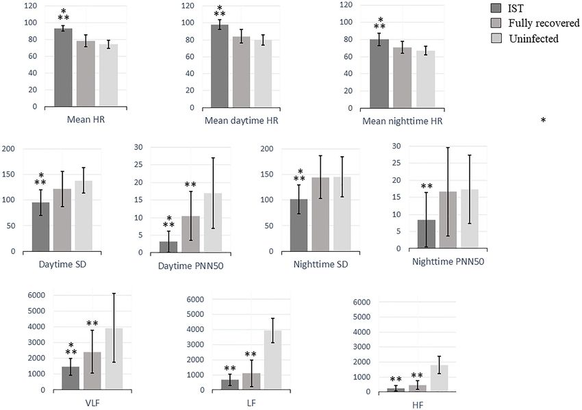
www.nature.com/scientificreports/
Inappropriate sinus tachycardia
(Group 1) Fully recovered (Group 2) Uninfected (Group 3)
N = 40 N = 19 N = 17 P value
Demographic and clinical characteristics
Age (years) 40.1 ± 10 42.2 ± 11 39.5 ± 13 0.243
Males, n (%) 6 (15) 2 (11) 2 (12) 0.655
Body mass index, mean ± SD 25.2 ± 6.1 24.5 ± 3.6 22.5 ± 2.3 0.374
Smoking, n (%) 1 (3) 1(5) 0 0.623
Hypertension, n (%) 3(8) 0 0 0.589
Hyperlipidemia, n (%) 3 (8) 0 0 0.589
Diabetes mellitus, n (%) 0 0 0 NA
Asthma, n (%) 4 (10) 1 (5) 0 0.734
Environmental allergy, n (%) 10 (25) 0 0 0.01
Symptoms of the acute infection
Palpitations, n (%) 36 (90) 1 (5) – < 0.001
Dyspnea, n (%) 33 (83) 3 (16) – < 0.001
Myalgia and joint pain, n (%) 32 (80) 17 (89) – 0.545
Chest pain, n (%) 31 (78) 4 (21) – < 0.001
Fever, n (%) 29 (73) 16 (84) – 0.462
Headache, n (%) 29 (73) 7 (37) – 0.007
Dizziness, n (%) 21 (53) 1 (5) – 0.002
Diarrhea, n (%) 21 (53) 3 (16) – 0.003
Anosmia, n (%) 19 (48) 15 (79) – 0.03
Ageusia, n (%) 19 (48) 8 (42) – 0.454
Dermatologic alterations, n (%) 14 (35) 1 (5) – 0.009
Severity of clinical presentation
Mild 33 (83) 16 (84) – 0.387
Moderate 6 (15) 3 (16) – 0.550
Intensive care management 1 (3) 0 – 0.254
Table 1. Demographic and clinical characteristics of the cases and their matched controls. Values are
expressed as mean ± standard deviation unless otherwise stated. A P value of < 0.05 is considered statistically
significant. Significant values are in [bold].
were Caucasian. Thirty-four (85%) were women, with a mean age of 40.1 ± 10 years. All patients had normal
2-D echocardiography results, and no remnant respiratory disease was identified in any patient. All patients had
O2 saturation > 97%. Notably, IST patients had a higher prevalence of environmental allergy compared to the
control group (25% vs. 0%; p = 0.01).
During the acute phase of SARS-CoV-2 infection, 33 patients (83%) had experienced mild symptoms not
requiring hospital admission; 6 patients (15%) had moderate disease with pulmonary infiltrates and required
hospitalization; and only 1 patient (3%) required intensive care management. Compared to fully recovered
patients, patients with PCS and IST more frequently complained of palpitations (90% vs. 5%; p < 0.001), dyspnea
(82% vs. 16%; p < 0.001), chest pain (78% vs. 21%; p < 0.001), headache (73% vs. 37%; p = 0.007), dizziness (53%
vs. 5%; p = 0.002), diarrhea (53% vs. 16%; p = 0.003), and dermatological alterations (35% vs. 5%; p = 0.009).
Interestingly, IST patients had a lower incidence of anosmia (48% vs. 79%; p = 0.03).
The results of the exercise capacity and quality of life assessment are presented in Table 2, along with the
results of the laboratory tests. Echocardiography yielded normal results in all patients. Overall, biochemistry data
were consistent with a lack of inflammation or myocardial damage at this stage after the acute phase of SARS-
CoV-2 infection. The 6MWT showed that IST patients had a significantly diminished exercise capacity, with a
median walking distance of 392 ± 83 m, which is only 60% of the estimated reference distance after adjusting
for age, sex, and body mass index. Impaired quality of life was also identified, as suggested by a mean score in
the health-state scale of 39 out of 100 points. The most affected domains were mobility (mean score 3.6), usual
activities (mean score 3.5), and pain/discomfort (mean score 3).
Results of the 24-h ECG monitoring are summarized in Table 3 and Fig. 1. Patients with IST had a higher
mean heart rate, predominantly during the daytime, compared to recovered asymptomatic and uninfected sub-
jects (98 ± 6 vs. 84 ± 8 vs. 81 ± 6 bpm, respectively; p < 0.001). The burden of supraventricular premature beats was
lower in IST-PCS patients. No differences were observed in the maximum and minimum heart rates. All HRV
variables were significantly diminished among patients with IST compared to both the recovered subjects and the
uninfected group, with a significant decrease in the following time-domain parameters: daytime pNN50 (3.2 ± 3
vs. 10.5 ± 8 vs. 17.3 ± 10.0, respectively; p < 0.001) and daytime SDNN (95.0 ± 25 vs. 121.5 ± 34 vs. 138.1 ± 25,
respectively; p < 0.001). A significant decrease in frequency-domain parameters was also observed in PCS
patients with IST: VLF (1463.1 ± 538 vs. 2415.7 ± 1361 vs. 3931 ± 2194, respectively; p < 0.001), LF (670.2 ± 380
vs. 1093.2 ± 878 vs. 1801.5 ± 800, respectively; p < 0.001), and HF (246.0 ± 179 vs. 463.7 ± 295 vs. 1048.5 ± 570,
Scientific Reports | (2022) 12:298 | https://doi.org/10.1038/s41598-021-03831-6 3
Vol.:(0123456789)www.nature.com/scientificreports/
Reference values Mean values
Laboratory tests
NT-proBNP (pg/mL) < 125 67.6 ± 59.6
Hs-Troponin I (pg/mL) < 14 3.09 ± 4.2
Leucocytes (× 109/L) 4.00–11.00 6.83 ± 1.7
Hemoglobin (g/dL) 12.0–16.0 13.4 ± 0.7
TSH (µm/U) 0.350–4.940 1.4 ± 0.5
IL-6 (pg/mL) 0–6.40 2.2 ± 1.1
Ferritin (ng/mL) 15–160 50.4 ± 37.4
C-reactive protein (mg/L) 0.00–5.00 1.1 ± 1.3
D-Dimer (ng/mL) 0–500 247.7 ± 133.2
Fibrinogen 150–450 374.9 ± 54.1
24-h urine 3-Metoxiadrenaline (mg/L) 0.02–0.350 0.123 ± 0.120
24-h urine 3-Metoxinoradrenaline (mg/L) 0.03–0.440 0.178 ± 0.189
Exercise capacity test
6MWT (m) 392.7 ± 83.2
6MWT theoretical (%) 60.1 ± 12.1
Quality of life test (EQ-5D-5L) Score reference valuesa Mean values
Mobility 1–5 3.6 ± 1.1
Self-care 1–5 2.4 ± 1.2
Usual activities 1–5 3.5 ± 0.9
Pain/discomfort 1–5 3.0 ± 1.0
Anxiety/depression 1–5 2.9 ± 1.2
Health state scale (Visual Analogue Scale) 0–100 39.6 ± 20.7
Table 2. Characterization of patients with IST. Values are expressed as mean ± standard deviation unless
otherwise stated. NT-proBNP N-terminal pro B-type natriuretic peptide, TSH thyroid-stimulating hormone,
IL-6 interleukin-6, 6MWT 6-min walking test, EQ-5D 5-L EuroQol 5-Dimension 5-Level. a Score values for
each health dimension: 1: No problems, 2: Slight problems, 3: Moderate problems, 4: Severe problems, 5:
Unable/Extreme. For Health state scale, 0 means the best health you can imagine, 100 means the worst health
you can imagine.
respectively; p < 0.001). Numerical but non-significant differences were also observed between both control
groups, with the fully recovered patients presenting with higher heart rates and lower HRV than the uninfected
subjects.
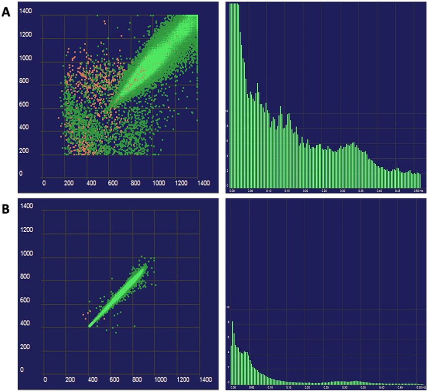
An illustrative example of 24-h ECG monitoring showing altered versus normal HRV in a PCS patient vs.
control is shown in Fig. 2.
Discussion
To the best of our knowledge, this is the first prospective series of consecutive PCS patients in whom a compre-
hensive cardiovascular evaluation has been performed for the investigation of IST. We found that IST is prevalent
among PCS patients (affecting 20% in our series), and this disorder was more common in young women without
previous comorbidities and with mild SARS-CoV-2 infection. IST provides a plausible explanation for some of
the prevalent symptoms of fatigue, impaired exercise capacity, and palpitations that characterize PCS and limit
the affected individuals’ ability to carry out a normal life (Fig. 3). Finally, our results suggest a major role of the
ANS in the pathophysiology of IST. This is supported by the 24-h ECG monitoring, as IST was accompanied
by a decrease in most HRV parameters, predominantly during the daytime, and the most reduced components
were those related to the cardiovagal tone (pNN50 and HF band). Accordingly, the loss of HRV is suggestive of
a cardiac ANS imbalance with decreased parasympathetic activity and compensatory sympathetic activation.
Hypoactivity of the parasympathetic tone could explain not only our findings of PCS-related IST, but also other
prevalent symptoms in this setting, such as fatigue, gastrointestinal discomfort, headache, sore throat, neuro-
cognitive disorder, and altered sleep structure (Central Illustration). Our findings are consistent with previous
investigations suggesting that PCS could be a form of post-infectious dysautonomia.
Anxiety disorders, an acknowledged cause of sinus tachycardia, were not systematically evaluated in our
patient population. However, the observed low HRV in our cohort and manifest physical limitations during the
6MWT makes anxiety-driven IST rather unlikely.
The mechanisms of IST, with or without previous viral infection, are poorly understood and investigated,
but many of the postulated mechanisms include alterations in the nervous system: sympathovagal imbalance,
beta-adrenergic receptor hypersensitivity, and brain stem dysregulation, among others. Considering this, it seems
reasonable that the mechanisms leading to IST after SARS-CoV-2 infection are mixed, with injury of the ANS,
which constantly regulates heart rate and vascular tone, playing an important role.
Scientific Reports | (2022) 12:298 | https://doi.org/10.1038/s41598-021-03831-6 4
Vol:.(1234567890)www.nature.com/scientificreports/
P value
Inappropriate
sinus tachycardia Fully recovered Uninfected Recovered vs
N = 40 N = 19 N = 17 IST vs recovered IST vs uninfected uninfected
Time-domain parameters
Mean HR (bpm) 93.6 ± 3 78.7 ± 7 74.3 ± 5 < 0.001 < 0.001 0.309
Maximum HR
154.6 ± 16 148.9 ± 24 140.8 ± 18 0.653 0.167 0.657
(bpm)
Minimum HR
59 ± 10 53.8 ± 9 51.9 ± 4 0.373 0.177 0.845
(bpm)
Supraventricular
191.6 ± 313 546.6 ± 665 955.8 ± 1180 0.172 0.003 0.234
PB
Ventricular PB 85.6 ± 120 354.1 ± 542 186.9 ± 287 0.018 0.628 0.362
Mean daytime HR
97.5 ± 6 84.1 ± 8 80.6 ± 6 < 0.001 < 0.001 0.656
(bpm)
Daytime PNN50
3.2 ± 3 10.5 ± 8 17.3 ± 10 0.001 < 0.001 0.019
(%)
Daytime SD (ms) 95.0 ± 25 121.5 ± 34 138.1 ± 25 0.011 < 0.001 0.270
Mean nighttime
80.0 ± 7 71.2 ± 7 67.4 ± 5 0.001 < 0.001 0.405
HR (bpm)
Nighttime PNN50
8.4 ± 8 16.6 ± 15 21.4 ± 11 0.051 0.004 0.498
(%)
Nighttime SD (ms) 101.3 ± 28 144.5 ± 42 145.4 ± 39 < 0.001 0.003 0.997
Spectral-domain parameters
VLF (Hz) 1463.1 ± 538 2415.7 ± 1361 3931.1 ± 2194 0.044 < 0.001 0.007
LF (Hz) 670.2 ± 380 1093.2 ± 878 1801.5 ± 800 0.092 < 0.001 0.015
HF (Hz) 246.0 ± 179 463.7 ± 295 1048.5 ± 570 0.060 < 0.001 < 0.001
LF/HF ratio (Hz) 3.6 ± 1 2.7 ± 1.3 2.0 ± 1 0.259 0.040 0.612
Table 3. 24-h ECG monitoring and HRV parameters. Values are presented in mean ± standard deviation. HR
heart rate, SD standard deviation of the interbeat interval, PNN50 percentage of adjacent NN intervals that
differ from each other by more than 50 ms, VLF very low frequency, LF low frequency, HF high frequency.
Significant values are in [bold].
Some studies have shown that COVID-19 has significant cardiovascular involvement, but no previous
research has focused on IST after SARS-CoV-2 infection. However, this is not the first time that IST has been
described after coronavirus infection. In 2006, Yu et al. found that IST was the most common cardiovascular
complication in a cohort of 121 patients with SARS. IST occurred in the absence of fever and was persistent in
nearly 40% of patients during 3 weeks of follow-up. The authors observed that cardiovascular outcomes did not
correlate with the occurrence of hypoxemia, admission to the intensive care unit, or analytical abnormalities9.
Madjid et al. previously described a weak association between acute Severe Acute Respiratory Syndrome Corona-
virus-1 (SARS-CoV-1) and acute Middle Respiratory Syndrome (MERS) and cardiovascular complications, such
as arrhythmia and transient diastolic dysfunction. In addition, a review of 28 studies evaluating the long-term
manifestations of SARS-CoV-1 and MERS observed that the most common symptoms were fatigue, dyspnea,
and weakness, similar to PCS10.
On the one hand, post-infectious dysautonomia has previously been described in relation to other patho-
gens, including Chagas disease, human immunodeficiency virus (HIV), Epstein-Barr virus, and rabies v irus11,12.
Autonomic dysfunction is relatively common among HIV-infected patients, as inferred from a decrease in the
HRV in the early stages of infection in many of these patients13. On the other hand, that patients with IST or
POTS often report experiencing a previous trigger, such as a viral infection14,15. In previous observational stud-
ies, previous infectious illness was the precipitating event for IST in 5–10% of cases, and the reported pathogens
were the influenza virus, Epstein-Barr virus, and herpes zoster, among o thers16. All of these studies mentioned
ANS disruption. A number of mechanisms have been proposed to explain the occurrence of ANS dysfunction
after a viral infection: denervation of the ANS, virus-dependent tissue damage due to persistent infection, and
immune-mediated injury, among others. However, the prevalence and the mechanisms underlying the cardio-
vascular consequences of post-infectious dysautonomia are not clear and need to be investigated further.
As with other pathogens, there is convincing evidence that SARS-CoV-2 can damage the ANS. Previous
studies have suggested a number of concurrent mechanisms, including direct brain invasion across the ethmoid
bone or via the olfactory bulb during acute infection or blood dissemination of the virus and use of the ACE2
receptor for intracellular penetration. This receptor is also present on the glial cells and neurons. Several lines
of evidence also support “indirect” mechanisms as the most important mechanisms involved in neurological
injury, including vasculitis, thrombosis, and endothelial damage, along with exaggerated inflammation and
immune responses17–22.
Furthermore, the evidence not only supports that SARS-CoV-2 can affect the nervous system during the acute
phase, there is growing evidence in patients with orthostatic syndromes and syncope following SARS-CoV-2
infection that endorses a patho-physiological link between PCS and ANS dysfunction. A clear example of the
Scientific Reports | (2022) 12:298 | https://doi.org/10.1038/s41598-021-03831-6 5
Vol.:(0123456789)www.nature.com/scientificreports/
Figure 1. 24-h ECG monitoring and HRV parameters. HRV parameters in the three studied groups: IST,
fully recovered and uninfected subjects. HR indicates heart rate; PNN50, percentage of adjacent NN intervals
that differ from each other by more than 50 ms; SD, standard deviation of the interbeat interval; VLF, very low
frequency; LF, low frequency; HF, high frequency. A P value of < 0.05 is considered statistically significant.
*Significant differences compared with fully recovered patients. **Significant differences compared with
uninfected patients.
capacity of the virus to alter the ANS is the so-called “silent hypoxia”, a characteristic sign of COVID-19. This
condition has been associated with endothelial damage affecting the central and peripheral nervous receptors,
altering respiratory control and dyspnea perception. Specifically, the injury has been postulated to occur in
the vagal fibers, the glossopharyngeal afferents, and in the nucleus of the tractus solitarius, which are all key in
respiratory and autonomic h omeostasis23,24. In our study, most of the patients could not be evaluated for silent
hypoxemia because arterial blood gases were not performed during the acute phase. Nevertheless, nearly all
patients with silent hypoxemia are hospitalized at some point, as this condition leads to a critical diagnostic
delay; in contrast to our study population of patients with mild disease who did not require hospital admission
(therefore, assuming the absence of silent hypoxemia).
Finally, interference of angiotensin II synthesis by COVID-19 can be postulated as the last possible patho-
physiological mechanism leading to dysautonomia. As discussed above, SARS-CoV-2 penetrates cells by attach-
ing to the ACE2 receptor, influencing the synthesis of endogenous angiotensin II, a hormone that directly
activates the SNS. Dysfunction of the renin–angiotensin–aldosterone system with compensatory activation of
the SNS may also contribute to IST. However, our study was unable to demonstrate SNS participation in IST, and
further investigations are needed to elucidate and characterize this patho-physiological aspect.
The reasons for the absolute predominance of this pathological phenomenon in young females, the concomi-
tant high prevalence of environmental allergies, and the lack of correspondence with the severity of the index
SARS-CoV-2 acute infection remain uncertain. Although IST and POTS are complex, heterogeneous syndromes
with overlapping clinical manifestations and potential common mechanisms, it remains important to distinguish
between these entities in order to provide the most appropriate treatment. The results of our study suggest that
patients with PCS and IST may likely benefit from pharmacological treatment, such as beta-blockers, which
blunt the sympathetic nervous system response. However, the pharmacological agent of choice, the timing of its
administration, and the clinical response will warrant a separate investigation.
Scientific Reports | (2022) 12:298 | https://doi.org/10.1038/s41598-021-03831-6 6
Vol:.(1234567890)www.nature.com/scientificreports/
Figure 2. (A) Uninfected subject. Poincaré plot of 24-hour ECG monitoring showing the beat-to-beat
variability from an uninfected subject and histogram of the frequencydomain parameters. (B) IST patient.
Poincaré plot of 24-h ECG monitoring and histogram of the frequency-domain parameters from a patient with
IST. A lower heart rate variability in comparison with the uninfected subject and an overall decrease is observed
throughout all bands, being more manifest at the high frequency band (HF, 0.15–0.40 Hz), are both apparent.
Figure 3. Illustration of the pathophysiological mechanisms underlying Post-COVID-19 syndrome.
Scientific Reports | (2022) 12:298 | https://doi.org/10.1038/s41598-021-03831-6 7
Vol.:(0123456789)www.nature.com/scientificreports/
Limitations
Assessment of ANS function is challenging and barely feasible in daily clinical practice. Standardized reference
values extracted from healthy populations are frequently not available. For this reason, we performed the same
tests in two gender- and age-controlled groups, one with matched disease stage and severity and one without
previous infection. The small size of the control group is also a limitation, and the real incidence of the disease
should be ascertained in larger population studies. Despite these limitations, we demonstrated significantly
decreased parasympathetic tone among our PCS patient population.
Most of the patients included in this study did not require hospital admission during the acute phase of
SARS-CoV-2 infection. Thus, laboratory parameters characterizing a presumable pro-inflammatory state and/
or myocardial damage during the acute infection phase were not available.
Conclusions
Inappropriate sinus tachycardia is a prevalent condition among PCS patients and should be incorporated as
part of the myriad of multi-organ disorders comprising PCS. This disorder may at least partially explain the
prevalent symptoms of palpitations, fatigue, and impaired exercise capacity observed in PCS patients. Cardiac
ANS imbalance with decreased parasympathetic activity seems to be a plausible pathophysiological explanation
for this phenomenon.
Received: 19 June 2021; Accepted: 9 December 2021
References
1. Carfì, A., Bernabei, R., Landi, F., Gemelli Against COVID-19 Post-Acute Care Study Group. Persistent symptoms in patients after
acute COVID-19. JAMA 324(6), 603–605. https://doi.org/10.1001/jama.2020.12603 (2020).
2. COVID-19 rapid guideline: managing the long-term effects of COVID-19. (National Institute for Health and Care Excellence
(UK), London, 2020).
3. Dani, M. et al. Autonomic dysfunction in ‘long COVID’: Rationale, physiology and management strategies. Clin. Med. (Lond.).
21(1), e63–e67. https://doi.org/10.7861/clinmed.2020-0896 (2021).
4. Feigofsky, S. & Fedorowski, A. Defining cardiac dysautonomia—Different types, overlap syndromes; case-based presentations. J.
Atr. Fibrillation. 13(1), 2403. https://doi.org/10.4022/JAFIB.2403 (2020).
5. Goldstein, D. S. The possible association between COVID-19 and postural tachycardia syndrome. Heart Rhythm S1547–5271(20),
31141–31143. https://doi.org/10.1016/j.hrthm.2020.12.007 (2020).
6. Nwazue, V. C. et al. Postural tachycardia syndrome and inappropriate sinus tachycardia: Role of autonomic modulation and sinus
node automaticity. J. Am. Heart Assoc. 3(2), e000700. https://doi.org/10.1161/JAHA.113.000700 (2014).
7. Swai, J., Hu, Z., Zhao, X., Rugambwa, T. & Ming, G. Heart rate and heart rate variability comparison between postural orthostatic
tachycardia syndrome versus healthy participants; A systematic review and meta-analysis. BMC Cardiovasc. Disord. 19(1), 320.
https://doi.org/10.1186/s12872-019-01298-y (2019).
8. Olshanky, B. & Sullivan, R. M. Inappropriate sinus tachycardia. J. Am. Coll. Cardiol. 63(8), 793–801. https://doi.org/10.1016/j.jac.
2012.07.074 (2013).
9. Yu, C. M. et al. Cardiovascular complications of severe acute respiratory syndrome. Postgrad. Med. J. 82(964), 140–144. https://
doi.org/10.1136/pgmj.2005.037515 (2006).
10. Madjid, M. et al. Potential effects of coronaviruses on the cardiovascular system: A review. JAMA Cardiol. 5(7), 831–840. https://
doi.org/10.1001/jamacardio.2020.1286 (2020).
11. Goldberger, J. J. et al. Autonomic nervous system dysfunction: JACC focus seminar. J. Am. Coll. Cardiol. 73(10), 1189–1206. https://
doi.org/10.1016/j.jacc.2018.12.064 (2019).
12. Chow, D. et al. Symptoms of autonomic dysfunction in human immunodeficiency virus. Open Forum Infect. Dis. 2(3), ofv103.
https://doi.org/10.1093/ofid/ofv103 (2015).
13. Carod-Artal, F. J. Infectious diseases causing autonomic dysfunction. Clin. Auton. Res. 28(1), 67–81. https://doi.org/10.1007/
s10286-017-0452-4 (2018).
14. Mittal, C. M., Wig, N., Mishra, S. & Deepak, K. K. Heart rate variability in human immunodeficiency virus-positive individuals.
Int. J. Cardiol. 94(1), 1–6. https://doi.org/10.1016/j.ijcard.2003.02.002 (2004).
15. Hemachudha, T. et al. Human rabies: Neuropathogenesis, diagnosis, and management. Lancet Neurol. 12(5), 498–513. https://doi.
org/10.1016/S1474-4422(13)70038-3 (2013).
16. Abboud, H. et al. COVID-19 and SARS-Cov-2 infection: Pathophysiology and clinical effects on the nervous system. World
Neurosurg. https://doi.org/10.1016/j.wneu.2020.05.193 (2020).
17. Low, P. A. & Sandroni, P. Postural tachycardia syndrome (POTS). Primer Auton. Nervous Syst. https://doi.org/10.1016/B978-0-12-
386525-0.00106-2 (2012).
18. Schondorf, R. & Low, P. A. Idiopathic postural orthostatic tachycardia syndrome: An attenuated form of acute pandysautonomia?.
Neurology 43(1), 132–137. https://doi.org/10.1212/wnl.43.1_part_1.132 (1993).
19. Zubair, A. S. et al. Neuropathogenesis and neurologic manifestations of the coronaviruses in the age of coronavirus disease 2019:
A review. JAMA Neurol. 77(8), 1018–1027. https://doi.org/10.1001/jamaneurol.2020.2065 (2020).
20. Yachou, Y., El Idrissi, A., Belapasov, V. & Ait, B. S. Neuroinvasion, neurotropic, and neuroinflammatory events of SARS-CoV-2:
Understanding the neurological manifestations in COVID-19 patients. Neurol. Sci. 41(10), 2657–2669. https://doi.org/10.1007/
s10072-020-04575-3 (2020).
21. Song, E. et al. Neuroinvasion of SARS-CoV-2 in human and mouse brain. J. Exp. Med. 218(3), e20202135. https://d oi.o rg/1 0.1 084/
jem.20202135 (2021).
22. Karuppan, M. K. M. et al. SARS-CoV-2 infection in the central and peripheral nervous system-associated morbidities and their
potential mechanism. Mol. Neurobiol. 58(6), 2465–2480. https://doi.org/10.1007/s12035-020-02245-1 (2021).
23. Rahman, A. et al. Silent hypoxia in COVID-19: Pathomechanism and possible management strategy. Mol. Biol. Rep. https://doi.
org/10.1007/s11033-021-06358-1 (2021).
24. Barizien, N. et al. Clinical characterization of dysautonomia in long COVID-19 patients. Sci. Rep. https://doi.org/10.1038/s41598-
021-93546-5 (2021).
Scientific Reports | (2022) 12:298 | https://doi.org/10.1038/s41598-021-03831-6 8
Vol:.(1234567890)www.nature.com/scientificreports/
Acknowledgements
We thank Laia Valls for her collaboration in data collection and Carolina Galvez and Carolina Jaillier for the
illustration.
Author contributions
J.A., V.B. and R.V. wrote the main manuscript text and prepared figures. All authores reviewed the mansucript.
Funding
This study did not receive any specific funding.
Competing interests
The authors declare no competing interests.
Additional information
Correspondence and requests for materials should be addressed to R.V.
Reprints and permissions information is available at www.nature.com/reprints.
Publisher’s note Springer Nature remains neutral with regard to jurisdictional claims in published maps and
institutional affiliations.
Open Access This article is licensed under a Creative Commons Attribution 4.0 International
License, which permits use, sharing, adaptation, distribution and reproduction in any medium or
format, as long as you give appropriate credit to the original author(s) and the source, provide a link to the
Creative Commons licence, and indicate if changes were made. The images or other third party material in this
article are included in the article’s Creative Commons licence, unless indicated otherwise in a credit line to the
material. If material is not included in the article’s Creative Commons licence and your intended use is not
permitted by statutory regulation or exceeds the permitted use, you will need to obtain permission directly from
the copyright holder. To view a copy of this licence, visit http://creativecommons.org/licenses/by/4.0/.
© The Author(s) 2022
Scientific Reports | (2022) 12:298 | https://doi.org/10.1038/s41598-021-03831-6 9
Vol.:(0123456789)You can also read


