A Patient Guide Understanding Blue Light Cystoscopy with Cysview for Detection of Bladder Cancer
←
→
Page content transcription
If your browser does not render page correctly, please read the page content below

Understanding Blue Light
Cystoscopy with Cysview®
for Detection of Bladder Cancer 1

              A Patient Guide

               Visit Cysview.com
This patient guide is for informational purposes only; it does
not replace a conversation with your urologist or other urology
healthcare professional.

Cysview can only be used by qualified healthcare professionals.

Please see Full Prescribing Information enclosed.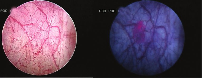
What is a cystoscopy?

A cystoscopy is a procedure that your urology
healthcare professional may use to examine your
bladder to help find the cause of symptoms, or
to treat or monitor conditions. This procedure
allows your healthcare professional (HCP) to look
directly inside your bladder and inspect the lining
very closely.
If during a cystoscopy any abnormal growths or
suspicious areas are seen, your HCP may remove
tissue samples (biopsy) and send them to the lab.
A cystoscopy can be done while you are asleep
under anesthesia or while you are awake with
moderate sedation and/or pain management.
Be sure to follow your healthcare professional’s
instructions about whether you should fast before
your procedure and how you should handle any
medications you take.

What is a cystoscope?

A cystoscope is a thin, tube-like telescope that is
carefully passed up the urethra (the tube through
which urine leaves your body) and into the bladder.
The cystoscope is a hollow tube that creates a path
for surgical instruments to pass through for use in
a cystoscopy.

                              Image of a
                              cystoscope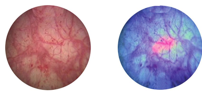
A standard cystoscopy
uses white light
During a cystoscopy procedure, the cystoscope
shines light inside the bladder to aid in visibility. In a
standard procedure, the light is regular white light—
the type we all use every day to
light a room.
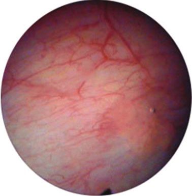
White light helps your
healthcare professional
visually assess the general
health of your bladder and
find irregularities to be
further evaluated.
Bladder image
under white light
A cystoscopy can also use
blue light with Cysview®
Your healthcare professional also has the option
of enhancing a cystoscopy by adding blue light
and Cysview® to the procedure. Called Blue Light
Cystoscopy (BLC®) with Cysview, this technology
significantly improves the detection of non-muscle
invasive bladder cancer (NMIBC).
With a standard cystoscopy
procedure, your HCP can see
some indicators of cancer
under white light. With the
addition of blue light and
Cysview, the procedure
offers significantly improved
detection of suspicious areas
compared to white light. The
Cysview causes cancerous
cells to glow bright pink under Same image
blue light, but it is not a dye.1 using blue light
and Cysview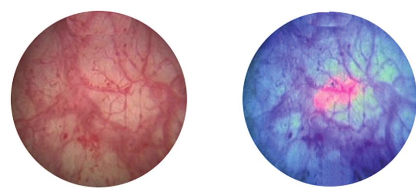
The Blue Light Cystoscopy with
Cysview® experience1

• professional
  One hour prior to a cystoscopy, a healthcare
               (HCP) uses a catheter to place about
 2 oz of Cysview in the bladder.

• thin
  For the procedure, your HCP inserts a long,
       tube (a cystoscope) and uses white light
 to examine the bladder

• Cysview
  When the HCP switches to blue light, the
          causes cancerous tumors to glow
 bright pink—making them more visible and
 possibly also revealing additional tumors not
 visible under white light

• bladder
  With Cysview, tumors stand out against normal
          tissue, so they are easier for your HCP
 to identify and remove completely

Is BLC® with Cysview® safe?1
Any procedure may have some risks. You should
consult your healthcare professional regarding the
risks and benefits of this procedure.
The most common patient complaints include:
 • Bladder spasms
 • Trouble urinating
 • Discomfort when urinating
 • Frequent urination
 • Blood in your urine
 • Bladder pain
Hypersensitivity reactions to hexaminolevulinate
may occur in some patients

Who can have BLC® with Cysview?®1
Anyone who is suspected of having or is known to
have bladder cancer (from a previous cystoscopy)
can have BLC with Cysview.
Bladder images
     under white and blue light*

   Standard White              Blue Light Cystoscopy
  Light Cystoscopy                 with Cysview

                         VS.

                         VS.

                         VS.

                         VS.

Blue Light Cystoscopy with Cysview has been
clinically proven to detect more bladder tumors
      than White Light Cystoscopy alone.1
*Side-by-side images are the same area of the bladder
 in white and blue light.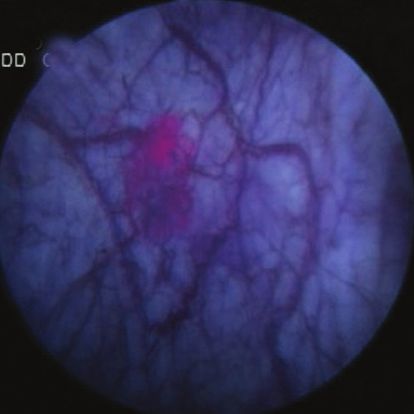
After the procedure
Here are some important things to keep in mind for
after your cystoscopy:
• any
  It’s a good idea to drink plenty of fluids after
       cystoscopy.

• Follow your discharge instructions carefully.
• Be  sure to contact your physician’s office with any
  issues or concerns.

Additional patient resources
Bladder Cancer Advisory Network (BCAN)
www.bcan.org
BCAN is the first national advocacy organization
dedicated to increasing public awareness about
bladder cancer; to advancing bladder cancer
research; and to providing educational and support
services for the bladder cancer community.
Founded in May 2005, BCAN is a cooperative effort
among bladder cancer survivors, their families and
caregivers, and the medical community.

  For more information about BLC with Cysview

            Visit Cysview.com

     If you have any questions about your
     Blue Light Cystoscopy with Cysview,
   ask your urology healthcare professional.
Important Risk & Safety Information
Cysview® (hexaminolevulinate HCl) is an optical imaging agent used
to detect non-muscle invasive bladder cancer in patients suspected
or known to have lesion(s) on the basis of a prior cystoscopy, or in
patients undergoing surveillance cystoscopy for bladder cancer.
Cysview is not a replacement for random bladder biopsies or other
procedures used in the detection of bladder cancer.

Anaphylactoid shock, hypersensitivity reactions, bladder pain,
bladder inflammation (cystitis), and abnormal urine tests have
been reported after administration of Cysview. The most common
adverse reactions seen in clinical trials were bladder spasm, trouble
urinating, discomfort when urinating, frequent urination, blood in the
urine, and bladder pain.

Cysview should not be used in patients with large amounts of
blood in their urine, any known allergy to Cysview or any derivative
of aminolevulinic acid, or porphyria, a condition that means you
already have high levels of porphyrins in your body. No specific
drug interaction studies have been performed.

Please see Full Prescribing Information enclosed.
Facts about bladder cancer

       82,501 each year
                                new cases of bladder cancer
                                             2

         Bladder cancer incidence
         is about four times higher
         in men than in women3

       712,614
                                     people living with a diagnosis
                                     of bladder cancer in 20174

References
1. Cysview® [prescribing information]. Princeton, NJ: Photocure
ASA; 2018. 2. Globocan. Incidence/mortality by population
2018. Available at: https://gco.iarc.fr/today/data/factsheets/
populations/840-united-states-of-america-fact-sheets.pdf.
Accessed February 26, 2020. 3. American Cancer Society.
Cancer Facts and Figures 2017. https://www.cancer.org/content/
dam/cancer-org/research/cancer-facts-and-statistics/annual-
cancer-facts-and-figures/2017/cancer-facts-and-figures-2017.
pdf. Accessed July 15, 2019. 4. National Cancer Institute.
SEER Stat Facts: Bladder Cancer 2017. https://seer.cancer.gov/
statfacts/html/urinb.html. Accessed April 16, 2020.

© 2020 Photocure Inc. All rights reserved.
Cysview is a registered trademark
of Photocure ASA.
December 2020 CYSC20200222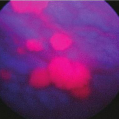
FULL PRESCRIBING INFORMATION: CONTENTS*
1 INDICATIONS AND USAGE
1.1 Limitations of Use
2 DOSAGE AND ADMINISTRATION
2.1 Recommended Dose
2.2 Reconstitution of Cysview
2.3 Bladder Instillation of Cysview
2.4 Use of the KARL STORZ D-Light C Photodynamic Diagnostic (PDD) System
2.5 Cystoscopic Examination
3 DOSAGE FORMS AND STRENGTHS
4 CONTRAINDICATIONS
5 WARNINGS AND PRECAUTIONS
HIGHLIGHTS OF PRESCRIBING INFORMATION 5.1 Anaphylaxis
5.2 Failed Detection
These highlights do not include all the information needed to use CYSVIEW safely and effectively. 5.3 False Fluorescence
See full prescribing information for CYSVIEW. 6 ADVERSE REACTIONS
Cysview (hexaminolevulinate hydrochloride), for Intravesical Solution 6.1 Clinical Study Experience
For bladder instillation only 6.2 Postmarketing Experience
Initial U.S. Approval: 2010 7 DRUG INTERACTIONS
8 USE IN SPECIFIC POPULATIONS
INDICATIONS AND USAGE
8.1 Pregnancy
Cysview is an optical imaging agent indicated for use in the cystoscopic detection of carcinoma of the bladder, 8.2 Lactation
including carcinoma in situ (CIS), among patients suspected or known to have lesion(s) on the basis of a prior 8.4 Pediatric Use
cystoscopy, or in patients undergoing surveillance cystoscopy for carcinoma of the bladder. Cysview is used with 8.5 Geriatric Use
the KARL STORZ D-Light C Photodynamic Diagnostic (PDD) system to perform Blue Light Cystoscopy (BLC®) as
10 OVERDOSAGE
an adjunct to the white light cystoscopy.
11 DESCRIPTION
Important Limitations of Use:
12 CLINICAL PHARMACOLOGY
• Not a replacement for random bladder biopsies or other procedures used in the detection of bladder 12.1 Mechanism of Action
cancer. (1.1, 5.2) 12.2 Pharmacodynamics
DOSAGE AND ADMINISTRATION 12.3 Pharmacokinetics
Training in blue light cystoscopy with the KARL STORZ D-Light C PDD system is essential prior to the use 13 NONCLINICAL TOXICOLOGY
of Cysview. (2.5) 13.1 Carcinogenesis, Mutagenesis, Impairment of Fertility
13.2 Animal Toxicology and/or Pharmacology
• Reconstitute Cysview powder with the supplied 50 mL DILUENT under aseptic conditions. (2.2)
14 CLINICAL STUDIES
• Use solution of Cysview shortly after reconstitution. If unable to use, the solution may be stored for up
to 2 hours in a refrigerator at 2°-8°C (36°-46°F) in the labeled syringe. Discard after 2 hours. (2.2, 16) 16 HOW SUPPLIED/STORAGE AND HANDLING
17 PATIENT COUNSELING INFORMATION
• Instill 50 mL of reconstituted solution of Cysview into the emptied bladder via an intravesical catheter.
*Sections or subsections omitted from the full prescribing information are not listed.
Retain in the bladder for 1 hour before evacuating and performing cystoscopic examination. (2.3, 2.5)
• First perform a complete cystoscopic examination of the entire bladder under white light and then
repeat the examination of the entire bladder under blue light. Record and document information about FULL PRESCRIBING INFORMATION
location and appearance of suspicious lesions and areas seen under both white and blue light. (2.5)
1 INDICATIONS AND USAGE
DOSAGE FORMS AND STRENGTHS Cysview is indicated for use in the cystoscopic detection of carcinoma of the bladder, including carcinoma in
Cysview (hexaminolevulinate hydrochloride) is supplied as a kit. The kit may be supplied as two options; situ (CIS), among patients suspected or known to have lesion(s) on the basis of a prior cystoscopy, or in patients
with or without a vial adapter: undergoing surveillance cystoscopy for carcinoma of the bladder. Cysview is used with the KARL STORZ D-Light
C Photodynamic Diagnostic (PDD) system to perform Blue Light Cystoscopy (BLC®) as an adjunct to the white
• One 10 mL glass vial containing 100 mg powder of Cysview. (hexaminolevulinate hydrochloride) for
light cystoscopy.
Intravesical Solution.
• One plastic prefilled syringe containing 50 mL DILUENT for Cysview. 1.1 Limitations of Use
Cysview is not a replacement for random bladder biopsies or other procedures used in the detection of bladder
• One Luer Lock catheter adapter.
cancer [see Warnings and Precautions (5.2)].
• One vial adapter for use during reconstitution (in the kit containing the vial adapter). (16)
2 DOSAGE AND ADMINISTRATION
Once reconstituted, the solution contains 2 mg/mL (8 mmol/L) of hexaminolevulinate hydrochloride.
2.1 Recommended Dose
CONTRAINDICATIONS
The recommended dose for adults is 50 mL of reconstituted solution of Cysview [see Dosage and Administration
Do not use Cysview in patients with: (2.2)], instilled into the bladder via a urinary catheter [see Dosage and Administration (2.3)].
• porphyria, 2.2 Reconstitution of Cysview
• gross hematuria, Cysview is supplied as a kit containing: a clear glass vial labeled as Cysview (hexaminolevulinate HCl) for
• known hypersensitivity to hexaminolevulinate or aminolevulinate derivatives. (4) Intravesical Solution containing 100 mg hexaminolevulinate hydrochloride as a powder, a prefilled syringe labeled
as DILUENT for Cysview, containing 50 mL of the diluent and a catheter adapter. The kit may be supplied as two
WARNINGS AND PRECAUTIONS
options; with or without a vial adapter for use during reconstitution.
• Anaphylaxis: have trained personnel and therapies available. (5.1). Perform all steps under aseptic conditions. Wear gloves during the reconstitution procedure; skin exposure to
• Failed Detection: Cysview may not detect all malignant lesions. Always perform white light cystoscopy hexaminolevulinate hydrochloride may increase the risk for sensitization to the drug.
followed by blue light cystoscopy. Do not biopsy with blue light only. (5.2)
• False fluorescence may occur due to inflammation, cystoscopic trauma, scar tissue, previous bladder
biopsy, recent BCG therapy or chemotherapy. (5.3)
ADVERSE REACTIONS
The most common adverse reaction reported in patients who received Cysview was bladder spasm,
occurring in 2% of patients, followed by dysuria, hematuria and bladder pain. (6.1)
To report SUSPECTED ADVERSE REACTIONS, contact Photocure Inc. at 1-855-297-8439 or FDA at
1-800-FDA-1088 or www.fda.gov/medwatch.
USE IN SPECIFIC POPULATIONS
Pediatric Use: Safety and effectiveness in pediatric patients have not been established. (8.4)
See 17 for PATIENT COUNSELING INFORMATION.
Cysview Cysview Plunger
Powder Diluent Rod
Figure 1.
Revised: 12/2019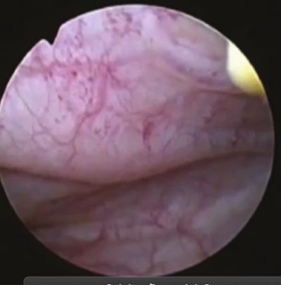
Reconstitution Using a Vial Adapter
1.	Fasten the plunger rod into the rubber stopper of the prefilled syringe by turning the plunger rod clockwise until
    it stops (Figure 1).

                                                                                                                           Figure 8.
                                                                                                                           3.	Without withdrawing the needle from the vial, hold the vial and syringe in a firm grip (Figure 8) and gently
Figure 2.                                                                                                                      shake to dissolve of the powder in the diluent. The powder normally dissolves almost immediately.
2.	Remove the plastic cap from the vial. Remove the TyveK® cover from the vial adapter blister package. Do not
    remove the vial adapter from the package. Place the Cysview vial on a flat surface. Using the blister package to
    hold the vial adapter, connect to the vial with a downward vertical motion. The vial adapter snaps onto the vial
    as the spike penetrates the rubber stopper of the vial. Remove the plastic blister package and discard it. Take
    care not to touch the exposed end of the vial adapter (Figure 2).

                                                                                                                           Figure 9.
Figure 3.                                                                                                                  4.	Turn the vial upside down and withdraw all of the dissolved solution from the vial back into the syringe (Figure 9).
3.	Remove the cap from the prefilled syringe and carefully retain it for subsequent reattachment to the syringe.
    Hold the prefilled syringe upright and carefully press the plunger rod upward to remove air. Connect the syringe
    to the vial adapter. Inject about 10 mL of the diluent from the prefilled syringe down into the vial. The vial
    should be about ¾ full (Figure 3).

Figure 4.                                                                                                                  Figure 10.
4.	Without disconnecting the vial adapter from the vial, hold the vial and syringe in a firm grip (Figure 4) and          5.	Remove the needle from the vial, disconnect the needle from the syringe tip and discard it. Plug the syringe
    gently shake to dissolve the powder in the diluent. The powder normally dissolves almost immediately.                      with the syringe cap (Figure 10). Gently mix the contents of the syringe. The reconstituted solution of Cysview
                                                                                                                               is colorless to pale yellow and clear to slightly opalescent, and free from visible particles.
                                                                                                                           6.	Peel off the detachable portion of the syringe label. On the syringe label, add two hours to the present time and
                                                                                                                               write the resulting expiration time and date.
                                                                                                                           Cysview is now reconstituted and ready for use. The solution of Cysview contains 2 mg/mL of hexaminolevulinate
                                                                                                                           hydrochloride. Instill the reconstituted solution of Cysview into the bladder [see Bladder instillation of Cysview
                                                                                                                           (2.3)]. If unable to administer the solution shortly after reconstitution, store the solution for up to 2 hours in a
                                                                                                                           refrigerator at 2°-8°C (36°- 46°F) in the labeled syringe. If not used within 2 hours, discard the solution [see How
                                                                                                                           Supplied/Storage and Handling (16)].

Figure 5.                                                                                                                  2.3      Bladder Instillation of Cysview
5.	Turn the vial upside down and withdraw all of the dissolved solution from the vial back into the syringe (Figure 5).   For bladder instillation of the solution of Cysview, use straight or intermittent, urethral catheters with a proximal
    Do not inject large amounts of air or diluent when vial is inverted as it may block the venting action of the vial     funnel opening that will accommodate the Luer Lock adapter. Use only catheters made of vinyl (uncoated or
    adapter. If this occurs, turn the vial upright and pull back on the plunger rod in the syringe.                        coated with hydrogel), latex (amber or red), and silicone to instill the reconstituted Cysview. Do not use catheters
                                                                                                                           coated or embedded with silver or antibiotics. In-dwelling bladder catheters (Foley catheters) may be used if the
                                                                                                                           catheters are inserted shortly prior to Cysview administration and are removed following the Cysview instillation.
                                                                                                                           Use the following steps for bladder instillation of Cysview:
                                                                                                                           1.	Using standard sterile catheterization technique, first insert the urethral catheter into the bladder of the patient
                                                                                                                               and use the catheter to completely empty the patient’s bladder before instillation of Cysview.

Figure 6.
6. Disconnect the empty vial with the vial adapter from the syringe tip and discard it. Plug the syringe with the
   syringe cap (Figure 6). Gently mix the contents of the syringe. The reconstituted solution of Cysview is colorless
   to pale yellow and clear to slightly opalescent, and free from visible particles.
7. Peel off the detachable portion of the syringe label. On the syringe label, add two hours to the present time and
   write the resulting expiration time and date.
Cysview is now reconstituted and ready for use. The solution of Cysview contains 2 mg/mL of hexaminolevulinate
hydrochloride. Instill the reconstituted solution of Cysview into the bladder [see Bladder instillation of Cysview
(2.3)]. If unable to administer the solution shortly after reconstitution, store the solution for up to 2 hours in a
refrigerator at 2°-8°C (36°- 46°F) in the labeled syringe. If not used within 2 hours, discard the solution [see How
Supplied/Storage and Handling (16)].                                                                                       Figure 11.
Reconstitution Without the Use of a Vial Adapter                                                                           2. To attach the syringe containing the solution of Cysview to the catheter, do the following:
1. Fasten the plunger rod into the rubber stopper of the prefilled syringe by turning the plunger rod clockwise until         • Remove the syringe cap from the syringe that contains the reconstituted solution of Cysview.
   it stops (Figure 1)                                                                                                         • Attach the Luer Lock end of the (provided) catheter adapter to the syringe.
                                                                                                                               • Insert the tapered end of the catheter adapter into the funnel opening of the catheter. See Figure 11, with the
                                                                                                                                  connection enlarged in the inset.
                                                                                                                           3.	Slowly instill the solution of Cysview into the bladder through the catheter (Figure 11), ensuring that the
                                                                                                                               complete volume of the syringe (50 mL) is administered.
                                                                                                                           4.	After the solution is instilled, remove the catheter and instruct the patient to retain the solution within the
                                                                                                                               bladder for at least 1 hour; do not exceed 3 hours [see Cystoscopic examination (2.5)]. Patients may stand, sit
                                                                                                                               and move about during the time period between instillation and start of the cystoscopic procedure.
                                                                                                                           5.	Evacuate the solution of Cysview from the bladder as part of routine emptying of the bladder immediately prior
                                                                                                                               to the initiation of the cystoscopic procedure (refer to the KARL STORZ D-Light C Photodynamic Diagnostic
Figure 7.
                                                                                                                               (PDD) System Manual for further details). Also, the patient may void and completely empty the bladder prior to
2.	Remove the plastic cap from the vial. Remove the cap from the prefilled syringe and carefully retain it for                the procedure.
    subsequent reattachment to the syringe. Attach a needle to the prefilled syringe. Hold the prefilled syringe
                                                                                                                           Avoid skin contact with Cysview. If skin does come in contact with Cysview, wash immediately with soap and
    upright and carefully press the plunger rod upward to remove air. Penetrate the stopper of the Cysview vial
                                                                                                                           water and dry off. After voiding the bladder of Cysview, routinely wash the patient’s perineal skin region with soap
    with the needle and inject about 10 mL of the diluent from the prefilled syringe down into the vial. The vial
                                                                                                                           and water and dry.
    should be about ¾ full (Figure 7).
2.4 Use of the KARL STORZ D-Light C Photodynamic Diagnostic (PDD) System 5.3 False Positive Fluorescence
Cysview imaging requires the use of the KARL STORZ D-Light C PDD system, which consists of either: Fluorescent areas detected during blue light cystoscopy may not indicate a bladder mucosal lesion. In
the controlled clinical studies, approximately 20% of the lesions detected only by blue light cystoscopy
• a light source, a camera head, a camera control unit, a light cable, and a rigid cystoscope for use with the Rigid
showed neither dysplasia nor carcinoma [see Clinical Studies (14)]. False positive fluorescence may result
PDD Cystoscope System, or
from inflammation, cystoscopic trauma, scar tissue or bladder mucosal biopsy from a previous cystoscopic
• a light source, a camera control unit, and a flexible video cystoscope for use with the Flexible PDD Video examination, and recent BCG immunotherapy or intravesical chemotherapy. In a study of patients treated with
Cystoscope System. recent BCG immunotherapy or intravesical chemotherapy, the rate of false positives with blue light was 55%
The light source enables both white light cystoscopy and blue light (wavelength 360 – 450 nm) fluorescence between 6 weeks to 90 days and 41% after 90 days; the false positive rate was 53% and 33% at the respective
cystoscopy. Familiarity with this system is essential before beginning the procedure and before instilling Cysview time intervals with white light.
into the bladder. For system set-up and general information for the safe use of the PDD system, refer to the KARL The presence of urine and/or blood within the bladder may interfere with the detection of tissue fluorescence.
STORZ instruction manual for the PDD system and the instruction manuals for each of the system components. To enhance the diagnostic utility of Cysview with the KARL STORZ D-Light C PDD System:
The PDD System is not for use by healthcare providers with green-red color blindness. • ensure the bladder is emptied of urine prior to the instillation of fluids at cystoscopy;
2.5 Cystoscopic Examination • biopsy/resect bladder mucosal lesions only following completion of both white light and blue light rigid
Training cystoscopy;
Training and proficiency in cystoscopic procedures are essential prior to the use of Cysview. Carefully review 6 ADVERSE REACTIONS
the instruction manuals provided with the KARL STORZ D-Light C Photodynamic Diagnosis (PDD) System. For Anaphylaxis has been reported following exposure to Cysview [see Warnings and Precautions (5.1)].
additional training in the use of the PDD System, contact the manufacturer’s representative.
6.1 Clinical Study Experience
Preparation for Cystoscopy Because clinical trials are conducted under widely varying conditions, adverse reaction rates observed in the
Initiate the cystoscopic examination within 30 minutes after evacuation of Cysview from the bladder, but no clinical trials of a drug cannot be directly compared to rates in the clinical trials of another drug and may not
less than 1 or more than 3 hours after Cysview is instilled in the bladder. If the patient did not retain Cysview in reflect the rates observed in practice.
the bladder for 1 hour, allow 1 hour to pass from the instillation of Cysview into the bladder to the start of the In seven clinical trials, safety data were obtained from 1,628 patients, aged 32 to 96 years with a median age
cystoscopic examination. The efficacy of Cysview has not been established when the solution was retained for of 70 years, all primarily Caucasian and approximately 75% male. All patients were evaluated after a single
less than 1 hour. instillation of 50 mL solution of Cysview, and 103 patients received a repeat administration of Cysview. Of these
Cystoscopic Examination patients, 170 (10.4%) patients reported at least one adverse reaction. The most common adverse reaction was
Empty the patient’s bladder and then fill the bladder with a clear fluid (standard bladder irrigation fluid) in order bladder spasm (reported in 2.0% of the patients) followed by dysuria, hematuria, and bladder pain. No patients
to distend the bladder wall for cystoscopic visibility. Ensure adequate irrigation during examination of the bladder; experienced anaphylaxis. In the randomized controlled clinical study, adverse reactions were similar in nature and
blood, urine or floating particles in the bladder may interfere with visualization under both white light and blue rate between the study drug group and the control group. In a controlled study using Cysview in the surveillance
light. setting, adverse reaction types were similar [see Clinical Studies (14)].
First perform a complete cystoscopic examination of the entire bladder under white light and then repeat the 6.2 Postmarketing Experience
examination of the entire bladder surface under blue light unless the white light cystoscopy reveals extensive The following adverse reactions have been identified during post-approval use of Cysview. Because these
mucosal inflammation. Do not perform the blue light cystoscopy if the white light cystoscopy reveals wide-spread reactions are reported voluntarily from a population of uncertain size, it is not always possible to reliably estimate
mucosal inflammation. Abnormalities of the bladder mucosa during blue light cystoscopy are characterized by their frequency or establish a causal relationship to drug exposure.
the detection of red, homogenous and intense fluorescence. The margins of the abnormal lesions are typically Anaphylactoid shock, hypersensitivity reactions, bladder pain, cystitis and abnormal urinalysis have been reported
well-demarcated and in contrast to the normal urothelium, which appears blue. Register and document (map) the during post-marketing use of Cysview.
location (as appropriate for the cystoscopy procedure) and appearance (e.g. papillary, flat) of suspicious lesions
and abnormalities seen under either white or blue light. 7 DRUG INTERACTIONS
During the cystoscopic examination, be aware that: No specific drug interaction studies have been performed.
• a red fluorescence is expected at the bladder outlet and the prostatic urethra; this fluorescence occurs in 8 USE IN SPECIFIC POPULATIONS
normal tissue and is usually less intense and more diffuse than the bladder mucosal fluorescence associated
8.1 Pregnancy
with malignant lesions.
Risk Summary
• tangential light may give false fluorescence. To help avoid false fluorescence, hold the endoscope
There are no available data on Cysview use in pregnant women to inform a drug associated risk of adverse
perpendicular and close to the bladder wall with the bladder distended.
developmental outcomes. Adequate reproductive and developmental toxicity studies in animals have not been
• false positive fluorescence may result from scope trauma from a previous cystoscopic examination and/or performed. Systemic absorption following administration of Cysview is expected to be minimal [see Clinical
bladder inflammation [see Warnings and Precautions (5.3)]. Pharmacology (12.3)].
• malignant lesions may not fluoresce following Cysview administration, particularly if the lesions are coated The background risk of major birth defects and miscarriage for the indicated populations is unknown. All
with necrotic tissue. Blue light may fail to detect tumors which have a tendency to be necrotic on the surface, pregnancies have a background risk of birth defect, loss, or other adverse outcomes. In the U.S. general
and necrotic cells generally do not fluoresce [see Warnings and Precautions (5.3)]. population, the estimated background risk of major birth defects and miscarriage in clinically recognized
• when performing the blue light cystoscopy, avoid prolonged blue light exposure. Studies have not evaluated the pregnancies is 2-4% and 15-20%, respectively.
potential for adverse effects from blue light. In the controlled clinical trials, the cumulative blue light exposure from
8.2 Lactation
bladder evaluation, mapping and resection did not exceed 32 minutes for any procedure [see Clinical Studies (14)].
Risk Summary
For rigid cystoscopy, perform biopsy and/or resection of suspicious lesions by transurethral resection of the
There are no data on the presence of hexaminolevulinate in human or animal milk, the effects on a breastfed
bladder (TURB) only after completing white and blue light cystoscopic examinations with bladder mapping. Using
infant, or the effects on milk production. Systemic absorption following administration of Cysview is expected
standard cystoscopic practices, obtain biopsies of abnormal areas identified during either white or blue light
to be minimal [see Clinical Pharmacology (12.3)]. The lack of clinical data during lactation precludes a clear
examination and perform resections. Always check for the completeness of the resections under both white light
determination of the risk of Cysview to an infant during lactation; therefore, the development and health benefits
and blue light before finalizing the TURB procedure.
of breastfeeding should be considered along with the mother’s clinical need for Cysview and any potential
3 DOSAGE FORMS AND STRENGTHS adverse effects on the breastfed infant from Cysview or from the underlying maternal condition.
Cysview (hexaminolevulinate hydrochloride) is supplied as a kit. The kit may be supplied as two options; with or 8.4 Pediatric Use
without a vial adapter, and contains: Safety and effectiveness in pediatric patients have not been established.
Cysview kit with a vial adapter 8.5 Geriatric Use
• Cysview (hexaminolevulinate hydrochloride) for Intravesical Solution, 100 mg, as a powder in a 10 mL clear glass vial. Of 2127 subjects in clinical studies of Cysview, 67% were 65 years and over. No clinically important differences in
• DILUENT for Cysview, 50 mL, in a plastic prefilled syringe. safety or efficacy have been observed between older and younger patients in the controlled study.
• One vial adapter for use during reconstitution.
• One Luer Lock catheter adapter (to connect the syringe containing the reconstituted solution of Cysview to the 10 OVERDOSAGE
urethral catheter for bladder instillation of Cysview). No adverse events were reported in a dose-finding study conducted among patients whose bladders were
Cysview kit without a vial adapter instilled with twice the recommended concentration (dose) of solution of Cysview.
• Cysview (hexaminolevulinate hydrochloride) for Intravesical Solution, 100 mg, as a powder in a 10 mL clear glass vial. 11 DESCRIPTION
• DILUENT for Cysview, 50 mL, in a plastic prefilled syringe. Cysview contains hexaminolevulinate hydrochloride, an optical imaging drug that in solution form is instilled
• One Luer Lock catheter adapter (to connect the syringe containing the reconstituted solution of Cysview to the intravesically for use with photodynamic blue light cystoscopy as an adjunct to white light cystoscopy.
urethral catheter for bladder instillation of Cysview).
Once reconstituted, the solution of Cysview contains 2 mg/mL of hexaminolevulinate hydrochloride. The chemical formula for hexaminolevulinate hydrochloride is C11H21NO3·HCl. Its molecular weight is 251.76
and it has the following structural formula:
4 CONTRAINDICATIONS
Cysview is contraindicated in patients with:
• porphyria,
• gross hematuria,
• known hypersensitivity to hexaminolevulinate or any derivative of aminolevulinic acid.
5 WARNINGS AND PRECAUTIONS Cysview (hexaminolevulinate hydrochloride) for Intravesical Solution is intended for intravesical administration
5.1 Anaphylaxis only after reconstitution with the supplied 50 mL DILUENT. Cysview (hexaminolevulinate hydrochloride) for
Anaphylaxis, including anaphylactoid shock, has been reported following administration of Cysview [see Adverse Intravesical Solution and DILUENT for Cysview are supplied together as a kit.
Reactions (6.2)]. Prior to and during use of the Cysview, have trained personnel and therapies available for the Cysview (hexaminolevulinate hydrochloride) for Intravesical Solution is supplied as a sterile, non-pyrogenic,
treatment of anaphylaxis. freeze-dried, white to off-white or pale yellow, powder containing 100 mg of hexaminolevulinate hydrochloride
5.2 Failed Detection (equivalent of 85 mg of hexaminolevulinate) in a 10 mL clear glass vial. The DILUENT for Cysview is a sterile,
Cysview may fail to detect some bladder tumors, including malignant lesions. Cysview is not a replacement non-pyrogenic solution (pH 6) containing 0.61 mg/ mL disodium hydrogen phosphate, 0.58 mg/mL of potassium
for random biopsies or any other procedure usually performed in the cystoscopic evaluation for cancer. Do dihydrogen phosphate, 7.02 mg/mL of sodium chloride, hydrochloric acid, sodium hydroxide, and water for
not perform cystoscopy with blue light alone as malignant lesions can be missed unless the bladder is initially injection. It is a clear, colorless solution, free from visible particles, and is provided in a 50 mL plastic prefilled
examined under white light [see Dosage and Administration (2.5) and Clinical Studies (14)]. syringe.
The reconstituted solution of Cysview contains 2 mg/ml of hexaminolevulinate hydrochloride and is colorless to
pale yellow. It is free from visible particles and has a pH between 5.7 and 6.2.12 CLINICAL PHARMACOLOGY Table 3 shows patient-level detection of malignancy suspected in cystoscopic surveillance stage that was
verified in the OR stage (n=103). Among the 103 patients, 63 patients had malignancy confirmed: 49 patients
12.1 Mechanism of Action
had malignancy detected by both WL and BL; 1 patient had malignancy detected by WL only; and 13 patients
Cysview is an ester of the heme precursor, aminolevulinic acid. After bladder instillation, Cysview enters the bladder
had malignancy detected by BL only [12.6% with 95% CI (7%, 21%), pYou can also read


