High potency STING agonists engage unique myeloid pathways to reverse pancreatic cancer immune privilege
←
→
Page content transcription
If your browser does not render page correctly, please read the page content below

Open access Original research
J Immunother Cancer: first published as 10.1136/jitc-2021-003246 on 2 August 2021. Downloaded from http://jitc.bmj.com/ on December 23, 2021 by guest. Protected by copyright.
High potency STING agonists engage
unique myeloid pathways to reverse
pancreatic cancer immune privilege
Casey R Ager,1,2,3 Akash Boda,1,2 Kimal Rajapakshe,4 Spencer Thomas Lea ,1,2
Maria Emilia Di Francesco,5 Priyamvada Jayaprakash,1 Ravaen B Slay,1
Brittany Morrow,1,2 Rishika Prasad,1,2 Meghan A Dean,1 Colm R Duffy,1,2
Cristian Coarfa,6 Philip Jones,5 Michael A Curran1,2
To cite: Ager CR, Boda A, ABSTRACT microenvironment (TME) in order to over-
Rajapakshe K, et al. High Background Intratumoral injection of cyclic dinucleotide come innate and adaptive immune tolerance.
potency STING agonists engage (CDN) agonists of the stimulator of interferon genes
unique myeloid pathways to
For ‘hot’ tumors possessing a sufficiently
(STING) pathway engages innate immune activation and dense but functionally suppressed T cell infil-
reverse pancreatic cancer
priming of adaptive immune effectors to foster local and trate, blockade of immune checkpoint recep-
immune privilege. Journal for
ImmunoTherapy of Cancer distal tumor clearance. Despite proven therapeutic efficacy
tors including CTLA-4 or PD-1 (ICB) can be
2021;9:e003246. doi:10.1136/ in preclinical models, a thorough understanding of how
CDNs reprogram suppressive myeloid stroma in mouse efficacious. In ‘cold’ tumors like pancreatic
jitc-2021-003246
and man is lacking. ductal adenocarcinoma (PDAC), however,
►► Additional supplemental Methods Here, we perform deep transcript-level and effector T cells are excluded or disabled by an
material is published online only. protein-level profiling of myeloid-derived suppressor cells immunosuppressive myeloid and fibroblast
To view, please visit the journal and M2 macrophages following stimulation with CDNs of stroma, leading to ICB failure.1 2 While vacci-
online (http://dx.d oi.org/10. ascending potency. Additionally, we leverage orthotopic nation can amplify peripheral antitumor T
1136/j itc-2021-0 03246). Kras+/G12DTP53+/R172HPdx1-Cre (KPC) derived models of cell frequencies,3 4 local desmoplasia remains
pancreatic adenocarcinoma (PDAC) to determine the capacity a major barrier to T cell entry, longevity, and
Accepted 09 July 2021 for locally administered CDNs to sensitize PDAC to immune
effector function at the malignant pancreatic
checkpoint blockade. We use bioluminescent in vivo imaging
and 30-parameter flow cytometry to profile growth kinetics
niche.5 6 We hypothesize that both proinflam-
and remodeling of the tumor stroma post-therapy. matory reprogramming of the tumor stroma
Results Highly potent synthetic STING agonists repolarize and enhanced immune priming via vacci-
suppressive myeloid populations of human and murine nation are required to unmask sensitivity to
origin in part through inhibition of Myc signaling, metabolic checkpoint blockade in PDAC.
modulation, and antagonism of cell cycle. Surprisingly, Direct intratumoral delivery of innate
high-potency synthetic agonists engage qualitatively unique immune adjuvants, in situ vaccination, can
pathways as compared with natural CDNs. Consistent with theoretically achieve both adaptive immune
our mechanistic observations, we find that intratumoral
priming and stromal remodeling.7 8 In the
injection of the highest activity STING agonist, IACS-
context of cancer, this requires two critical
8803, into orthotopic pancreatic adenocarcinoma lesions
unmasks sensitivity to checkpoint blockade immunotherapy. events: (1) tumor antigen cross-presentation
Dimensionality reduction analyses of high parameter flow by Batf3+ dendritic cells (DCs) to prime CD8
cytometry data reveals substantial contributions of both T cells and (2) inflammatory polarization of
myeloid repolarization and T cell activation underlying the in tumor- associated macrophages (TAM) and
vivo therapeutic benefit of this approach. myeloid- derived suppressor cells (MDSC)
Conclusions This study defines the molecular basis to phenotypes that facilitate, rather than
of STING-mediated myeloid reprogramming, revealing suppress, T cell mediated immunity. Type I
© Author(s) (or their
previously unappreciated and qualitatively unique interferons are essential to induce priming of
pathways engaged by CDNs of ascending potency during
employer(s)) 2021. Re-use antitumor T cells,9 10 and in cancer, the stim-
permitted under CC BY-NC. No functional repolarization. Furthermore, we demonstrate
commercial re-use. See rights the potential for high potency CDNs to overcome ulator of interferon genes (STING) pathway
and permissions. Published by immunotherapy resistance in an orthotopic, multifocal is considered essential for type I IFN elabo-
BMJ. model of PDAC. ration.11 12 Predictably, intratumoral admin-
For numbered affiliations see istration of STING agonists complements
end of article. ICB in many established preclinical tumor
BACKGROUND
Correspondence to
models.13–17 However, whether and how
Successful antitumor immunity requires
Dr Michael A Curran; STING agonists affect function of suppres-
proinflammatory activation of the tumor
mcurran@mdanderson.org sive tumor myeloid cells remains enigmatic.
Ager CR, et al. J Immunother Cancer 2021;9:e003246. doi:10.1136/jitc-2021-003246 1
Open access
J Immunother Cancer: first published as 10.1136/jitc-2021-003246 on 2 August 2021. Downloaded from http://jitc.bmj.com/ on December 23, 2021 by guest. Protected by copyright.
Several reports have associated in situ STING activation Cyclic dinucleotides (CDNs)
with inflammatory modulation of tumor myeloid pheno- CDG and 2′3′ cGAMP were purchased from InvivoGen
types, generally measured as conversion of ‘M2’ to ‘M1’ and reconstituted in water according to manufacturer’s
markers on TAMs.14 18 The molecular mechanisms driving instructions. ML-RR-CDA was synthesized by Wuxi AppTec
repolarization remain undescribed, and little is known and reconstituted in water. IACS-8803 was designed in
regarding effects of STING activation on MDSC popu- collaboration with the University of Texas MD Anderson
lations. Thus, a deeper understanding of how STING Cancer Center and synthesized by Wuxi AppTec19 and was
agonists modulate myeloid function is needed. reconstituted in water or PBS for in vitro or in vivo use.
In this study, we sought to dissect the underlying
molecular mechanisms by which STING agonists induce Antibodies and chemotherapy
proinflammatory conversion of the immunosuppressive Antibodies used in in vivo studies were purchased from
PDAC myeloid stroma. We evaluated four CDNs: cyclic BioXCell or Leinco Technologies and were administered
di-GMP (CDG), 2′3′-cGAMP (cGAMP), ML-RR-S2-CDA via intraperitoneal (IP) injection at the following concen-
(ADU-S100; abbreviated ML-RR), and a newly described trations: αCTLA-4 clone 9H10 (100 µg/mouse) and
CDN with high relative potency IACS-8803 (8803), for αPD-1 clone RMP1-14 (250 µg/mouse). For lymphocyte
efficacy and myeloid polarization efficiency in a novel depletion, 2.43 (CD8), GK1.5 (CD4), and PK136 (NK1.1)
murine model of PDAC.19 Also, we performed exten- antibodies were given at 250 µg/mouse every 3 days for
sive multiomic analysis of ex vivo generated MDSCs and the duration of the study. Chemotherapy agents gemcit-
M2-like macrophages of murine and human origin on abine (Gemzar; Eli Lily) and nab-paclitaxel (Abraxane;
stimulation with each agonist. Integration of these data Celgene) were obtained from the University of Texas MD
reveal unappreciated roles for STING agonists in coun- Anderson pharmacy and were reconstituted according to
teracting Myc signaling, altering cell cycle dynamics, and manufacturer’s instructions. Both agents were adminis-
modulating metabolic programming. Finally, we used our tered IP at 120 mg/kg. Antibodies used for flow cytom-
novel agonist 8803 to demonstrate that CDN-mediated in etry were obtained from BD Biosciences, BioLegend, or
situ vaccination can potentiate dual checkpoint blockade Roche.
in a multifocal, orthotopic model of pancreatic cancer.
STING reporter cell assays
By using 30- color flow cytometry and dimensionality-
All STING reporter cell assays were conducted according
reduction algorithms, we elucidate how each therapy
to manufacturer’s protocols. In short, CDN or control
remodels the tumor stroma and impacts anti-PDAC adap-
agonists were added at desired concentrations in 20 µL
tive immunity. These data provide a deeper mechanistic
PBS to desired wells of flat-bottom 96-well plates. HEK-
understanding of the molecular pathways engaged down-
Blue, J774-Dual, THP-1 Dual, or THP-1 parental cell lines
stream of STING activation to functionally repolarize
were harvested, washed, counted, and resuspended for
tumor myeloid cells and demonstrate how CDNs of the
addition of 5×10⁴ – 1×10⁵ cells per well in 180 µL recom-
highest potency can cooperate with T cell checkpoint
mended media. Cells were incubated with CDN for
blockade to enable effective immunotherapy of PDAC.
20–24 hours, when supernatants were harvested for Lucif-
erase or SEAP assays using QuantiLuc or QuantiBlue
solutions according to manufacturer’s instructions. For
MATERIALS AND METHODS ELISA, THP-1 parental cells were incubated with CDN
Mice for 72 hours, then supernatants were harvested, and IFNβ
Male C57BL/6 and B6(Cg)- Tyrc-2J/J albino mice were levels were quantified using the PBL Human IFNβ ELISA
purchased from The Jackson Laboratory, and STINGGt/ kit according to manufacturer’s protocols.
Gt
mice were a kind gift from Dr Kimberly Schluns. Mice
cMyc reporter cell assays
were used between 5 and 8 weeks of age.
The HCT116 cMyc reporter cell assays (BPS Bioscience)
were conducted according to manufacturer’s protocols.
Cell lines In short, 25 000 HCT116 cells were plated into flat bottom
The mT4- 2D cell line was a kind gift from Dr David 96-
well plates the night before the assay. Assays were
Tuveson20 and was maintained in complete Dulbecco′s performed with a threefold serial dilution of either the
Modified Eagle′s Medium (DMEM) containing 10% fetal provided b-catenin inhibitor (ICG-001), STING agonists
bovine serum (Gibco), 100 U/mL penicillin, 100 mg/mL IACS-8803 or cGAMP (Invivogen), or FSL-1 (Invivogen).
streptomycin sulfate, and 2 mM L-glutamine. THP-1 Dual After an overnight incubation, cMyc activity was assessed
and J774-Dual reporter cells were obtained from Invi- by Luciferase expression follow addition of One- Step
voGen and cultured according to manufacturer’s instruc- Luciferase substrate (Promega).
tions. HEK-Blue reporter cells were a kind gift from Dr
Simon Yu at the University of Texas MD Anderson Insti- MDSC generation, proliferation assays, and T cell suppression
tute for Applied Cancer Science and cultured in cDMEM assays
supplemented with zeocin (100 µg/mL) and blasticidin Bone marrow was isolated from naïve male C57BL/6 mice
(30 µg/mL) for antibiotic selection. through aspiration of the femur and/or tibia. Red blood
2 Ager CR, et al. J Immunother Cancer 2021;9:e003246. doi:10.1136/jitc-2021-003246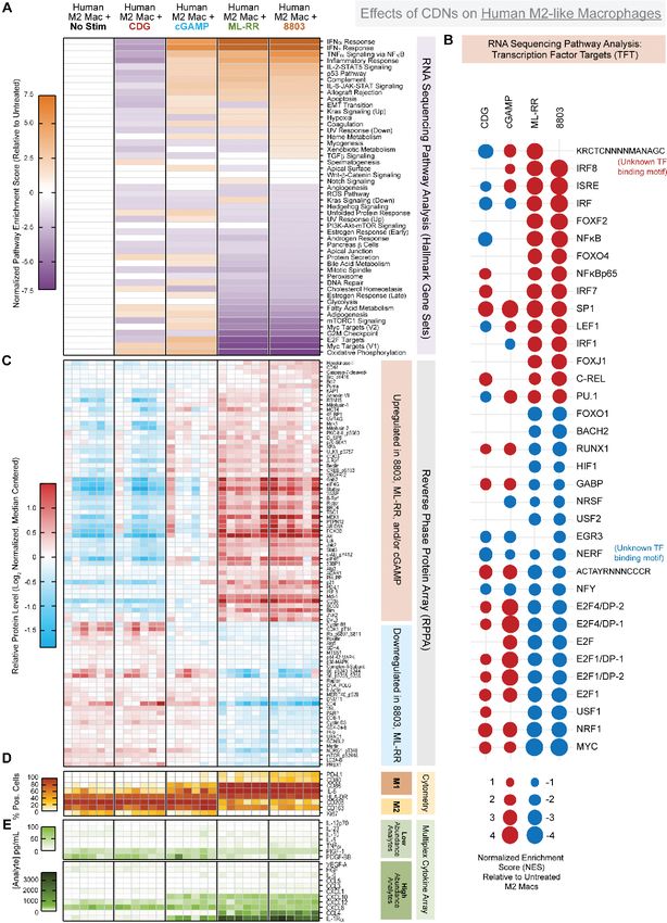
Open access
J Immunother Cancer: first published as 10.1136/jitc-2021-003246 on 2 August 2021. Downloaded from http://jitc.bmj.com/ on December 23, 2021 by guest. Protected by copyright.
cells were lysed using RBC lysis buffer (Sigma), and 4×10⁶ over 72 hours. Adherent macrophages were harvested
cells were plated in 10 mL in non-TC treated 10 cm petri with Detachin cell detachment solution (Genlantis) for
dishes (Falcon) in RPMI containing 10% Fetal Bovine downstream applications. All recombinant human cyto-
Serum (FBS), 100 U/mL penicillin, 100 mg/mL strep- kines were purchased from PeproTech and reconstituted
tomycin sulfate, recombinant mouse GM- CSF (40 ng/ according to manufacturer’s instructions.
mL; BioLegend) and IL-6 (40 ng/mL; BioLegend), and
2-Mercaptoethanol (β-ME; 55 µM) to induce MDSC differ- Luminex, RNA microarray, and RPPA sample preparation
entiation. After 4 days in culture, bone marrow derived Repolarized human M2c macrophages or murine
MDSC (BM- MDSC) suspension cells were harvested, BM- MDSC were generated as described. Supernatants
washed with cRPMI to remove cytokine, and replated in from repolarized cultures were isolated, spun to remove
10 mL cRPMI with no cytokine at 4×10⁵ cells/mL in 10 cm contaminating cells, then frozen at −80oC. All harvested
petri dishes. CDN were added at 1–10 µg/mL as indicated supernatants were thawed on ice and were analyzed with
for 24-hour repolarization. the Cytokine & Chemokine 36-Plex Mouse ProcartaPlex
or Cytokine/Chemokine/Growth Factor 45-Plex Human
MDSC proliferation assays and calculation of proliferation ProcartaPlex Panels (Invitrogen/ThermoFisher) using
index a Luminex MAGPIX machine according to manufac-
Bone marrow MDSCs were generated as above (GM-CSF turer’s instructions. For RNA, 4×10⁵ – 2×10⁶ repolarized
and IL-6 for 4 days) and treated with STING agonists cells were harvested, lysed, and homogenized for RNA
or vehicle control for 48 hours following CFSE labeling isolation with the QIAGEN RNeasy Mini Kit according to
prior to analysis on a flow cytometer. Proliferation index manufacturer’s protocols. RNA quality and concentration
is calculated by the average number of cell divisions that was determined by Bioanalyzer (Agilent), and RNAseq
a cell in the original population has undergone. It was was performed through Illumina. For RPPA, a minimum
obtained by the following formula that measures the ratio of 1×10⁶ cells were washed twice with cold PBS, and dry
of the number of cells that underwent division to the total pellets were stored at −80oC. Cell lysis and analysis was
∑i ( ) ∑ i conducted at the University of Texas MD Anderson Func-
number of cells in the system. PI = i∗ N
2i
i
/ Ni ,
2i tional Proteomics RPPA Core Facility.
i=0 i=0
where i=number of cell division peak as determined by
CFSE dilution, Ni=number of cells in that division peak. Orthotopic pancreatic tumor implantation and IVIS imaging
For suppression assays, naïve splenocytes were obtained Subconfluent cultures of mT4-2D, mT4-LA, or mT4-LS
from male C57BL/6 mice, and CD8 T cells were isolated were harvested with 0.05% trypsin, washed with PBS,
by negative selection with the Miltenyi Mouse CD8 T and resuspended at desired concentration in ice cold
Cell Isolation Kit. CD8 T cells were labeled with Cell- PBS containing 30% Matrigel (Corning) by volume for
Trace CFSE (ThermoFisher) as described. The 1×10⁵ implantation. Each mouse was anesthetized through
CFSE-labeled CD8 T cells were plated at indicated ratios isoflurane inhalation and was administered 3 µg
with unstimulated or repolarized BM-MDSC in 200 µL buprenorphine hydrochloride analgesic (Sigma) by IP
cDMEM containing 10% FBS, 50 U/mL IL-2, 27.5 µM injection. Following sterilization of the surgical field with
β-ME, and 2 µg/mL αCD28 (clone 37.51; BioLegend) in 70% isopropyl alcohol, a small (~1–2 cm) incision was
round bottom 96-well plates coated with αCD3 (BioXCell made on the left flank skin as well as the underlying peri-
clone 145–2 C11; coated overnight at 10 µg/mL in PBS toneal lining. Blunt-end forceps were used to access the
at 4°C). After 72–96 hours, CD8 T cells were analyzed for spleen and accompanying pancreas. Fifty microliters of
CFSE dilution by flow cytometry. cell suspension or CDN solution was administered into
the head of the pancreas using a U-40 insulin syringe with
Human macrophage generation 29.5-gage needle. The peritoneal wound and surrounding
Healthy donor buffy coats were obtained from the Univer- skin were cleaned with betadine antiseptic swabs, and
sity of Texas MD Anderson Cancer Center Blood Bank. the wound was closed using 4–0 absorbable sutures for
Peripheral blood mononuclear cells were isolated by the peritoneal incision and the Autoclip Wound Closing
ficoll gradient centrifugation (Histopaque 1077; Sigma), System for the skin (Braintree Scientific). Mice were
and CD14+ monocytes were isolated by negative selec- allowed to recover under a heat lamp until displaying
tion using the Miltenyi Classical Monocyte Isolation Kit. normal ambulance and were monitored daily for signs of
Monocytes were differentiated into M2c macrophages pain or discomfort. Wound clips were removed 10–14 days
as described.21 In short, monocytes were plated in T25 following application.
flasks (1×10⁶ cells/mL) in RPMI containing 20% FBS and Mice were imaged weekly to monitor tumor growth
100 ng/mL recombinant human M-CSF for 6 days, with using an IVIS Spectrum in vivo imaging system (Perki-
a media refresh on day 4. On day 6, media was replaced, nElmer) and randomized based on equivalent distribu-
with the addition of recombinant human TGF-β (10 ng/ tion of larger versus smaller tumors into groups prior to
mL) and IL-10 (10 ng/mL). On day 8, adherent cells were initiating therapy. Mice were anesthetized with isoflurane
washed with PBS and cultured with cRPMI + 100 ng/mL and received 200 µL D- Luciferin (15 mg/mL in PBS;
M-CSF + 10 µg/mL indicated CDN for repolarization GoldBio) by IP injection. Bioluminescent images were
Ager CR, et al. J Immunother Cancer 2021;9:e003246. doi:10.1136/jitc-2021-003246 3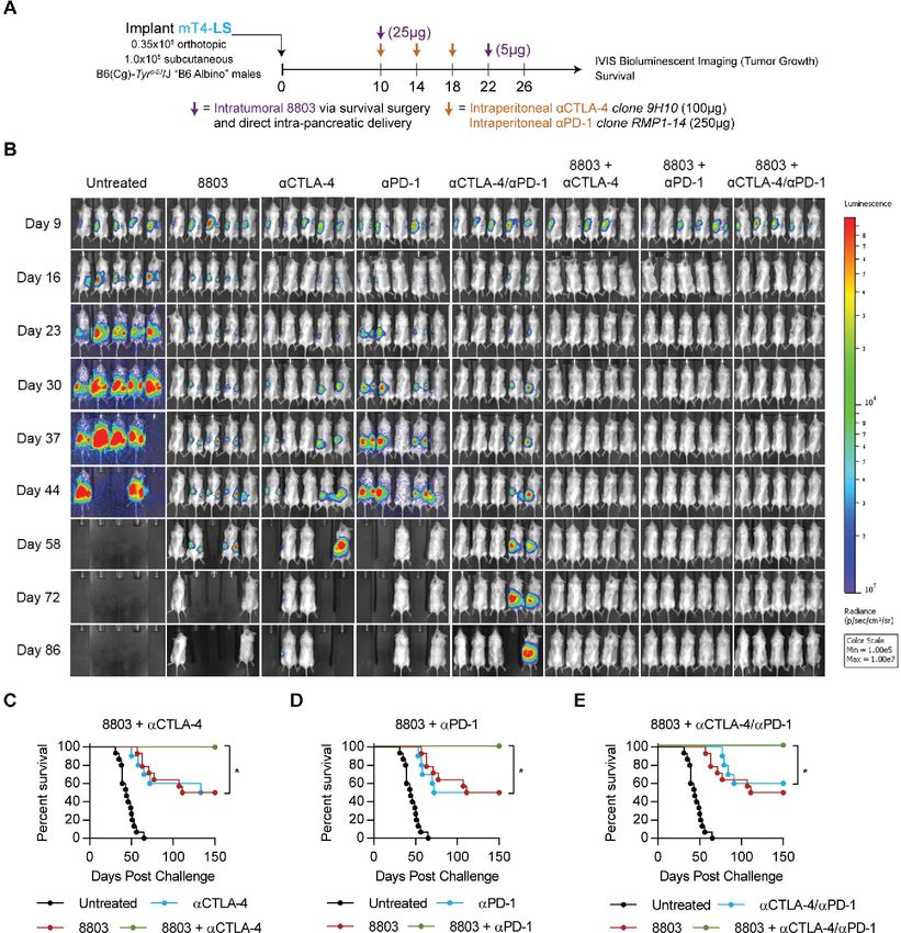
Open access
J Immunother Cancer: first published as 10.1136/jitc-2021-003246 on 2 August 2021. Downloaded from http://jitc.bmj.com/ on December 23, 2021 by guest. Protected by copyright.
acquired 11–12 min following administration of D-Lucif- flow cytometry (figure 1A). We found that, relative to
erin. All imaging was conducted in the University of Texas vehicle injection, ML-RR and 8803 induce regression of
MD Anderson Small Animal Imaging Facility. mT4-2D tumors, with 8803 demonstrating superior effi-
cacy (figure 1B). Local delivery of 8803 triggers expan-
Flow cytometry analysis of tumor immune infiltrates sion of the CD45+ immune infiltrate (figure 1C), which
mT4- 2D or mT4- LA cells were harvested with 0.05% is dominated by CD11b+Ly6G+Ly6CmidF4/80− granulo-
trypsin, washed with PBS, counted, and resuspended in cytes (figure 1D). This granulomatous response occurs
ice cold PBS containing 30% Matrigel for implantation at similar or reduced levels in tumors exposed to ML-RR
of 2.5×10⁵ cells in 100 µL subcutaneously or 3.5×10⁴ cells or CDG but is significantly less with cGAMP treatment.
in 50 µL orthotopically as described. Mice were treated CD8 T cells increase in number after 8803 exposure
with intratumoral CDN), checkpoint blockade anti- and express more Ki67 and Ly6C (figure 1D–F). Expan-
bodies, or chemotherapy according to treatment sched- sion of CD11b+Ly6C+Ly6G-F4/80mid/− monocytes with
ules included in associated figures. Following euthanasia, reduced arginase expression also occurs after 8803
primary tumors were harvested, massed, and finely diced (figure 1D,H). TAM (CD11b+Ly6C-Ly6G-F4/80+) do not
into 70 µm filters within 6 cm petri dishes. Diced tumors expand, but the expression of the M2 marker CD206 is
were enzymatically digested in a 37oC incubator for 30 min significantly reduced in the post-treatment TAM compart-
in digestion media consisting of X-Vivo15 media (Lonza) ment (figure 1G). Together, these data indicate 8803 is
supplemented with collagenase H (1 mg/mL; Sigma) the most potent therapeutic CDN among those tested in
and DNAse (160 µg/mL; Roche), then tumors were phys- ectopic KPC-derived tumors and that in situ vaccination
ically mashed through 70 µm filters to create single cell with 8803 precipitates inflammatory modulation of the
suspensions. Total cells were counted, then live immune PDAC TME including numeric and phenotypic enhance-
cells were purified by ficoll gradient centrifugation (Hist- ment of both lymphoid and myeloid populations. These
opaque 1119; Sigma). Samples were fixed overnight and data also suggest that therapeutic benefit in ‘cold’ TMEs
permeabilized using the FoxP3/Transcription Factor may scale proportional to the potency of the applied
Staining Buffer Set (eBioscience/ThermoFisher) for STING agonist. Of note, we compared both IACS-8802
staining with fluorescently labeled antibodies in 96-well and 8803 to cGAMP to determine to what extent higher
U-bottom plates. Stained samples were analyzed using a concentrations of the weaker agonist might generate
BD LSRII or X-30 prototype flow cytometer. functional equivalence and found that between a 10X
and 50X excess of cGAMP could achieve high level IRF3
Statistics activation (online supplemental figure 1). However, even
Unless otherwise indicated, all statistical analyses a 100X excess of cGAMP could not equal 8802/8803 level
comparing two independent groups were performed NFκB activation in short term assays, although equiva-
using Student’s t-test. For Kaplan-Meier survival curves, lence could be obtained over longer incubations.
Log-Rank Mantel Cox test was used to test for differ-
ences between treatment groups. For high dimensional High potency CDNs diminish proliferation and cMyc signaling
flow cytometry analyses comparing cluster frequencies in MDSC
between multiple treatment groups, two-way analysis of Given these observations, we sought a deeper under-
variance with Tukey’s correction for multiple compari- standing of the mechanisms by which synthetic CDNs
sons was used. alter the function, phenotype, and molecular program-
ming of the myeloid lineage. For this, we studied effects
of CDNs on in vitro differentiated suppressive myeloid
RESULTS populations. First, we generated bone marrow derived
Evaluating CDNs of ascending potency in a transplantable MDSC (BM-MDSC) using the prevalent protocol (GM-
model of PDAC CSF and IL-6) that yields a ~2:1 ratio of granulocytic to
The novel CDN STING agonist IACS-8803 exhibits monocytic MDSC, approximating their murine in vivo
enhanced potency relative to the clinical ML- RR prevalence (online supplemental figure 2A).22 These
compound in established in vitro and in vivo settings.19 We BM-MDSC cultures suppress T cell proliferation in vitro,
sought to leverage the diversity of STING-activating CDNs which we validated in a CFSE-based T cell suppression
available to probe whether higher potency STING activa- assay (online supplemental figure 2B). To determine
tion impacted the PDAC tumor stroma in unique ways. if STING activation modulates MDSC suppression, we
We first performed intratumoral injection of CDNs into stimulated BM-MDSC with CDNs for 24 hours, removed
mT4-2D PDAC tumors—a cell line derived from Kras+/ CDN from the culture, then evaluated BM-MDSC T cell
G12D
TP53+/R172HPdx1-Cre (KPC) organoid cultures.20 We suppression capacity. We find 8803- treated BM-MDSC
focused on four CDNs: natural agonists 3′-3′-cyclic di-GMP lose suppressive function in a dose-dependent manner
(CDG) and 2′3′-cGAMP (cGAMP), and synthetic agonists (online supplemental figure 2B). Comparing different
ML- RR and 8803 (figure 1A). Tumors were harvested CDNs, we find the relative effects mirror the therapeutic
following three injections with 5 µg of indicated CDN potencies observed in our in vivo studies (figure 2A and
for mass measurement and analysis by multiparameter online supplemental figure 2C). These data indicate
4 Ager CR, et al. J Immunother Cancer 2021;9:e003246. doi:10.1136/jitc-2021-003246
Open access
J Immunother Cancer: first published as 10.1136/jitc-2021-003246 on 2 August 2021. Downloaded from http://jitc.bmj.com/ on December 23, 2021 by guest. Protected by copyright.
Figure 1 In vivo characterization of 8803 in subcutaneous KPC-derived PDAC. (A) Mice received subcutaneous injection of
2.5×10⁵ mT4-2D cells in 30% matrigel, then were injected intratumorally with 5 µg CDN on days 15, 18, and 21 before tumor
harvest on day 23. Tumors were massed, counted, and processed for flow cytometry analysis as described in Methods. Data
shown represent (B) tumor mass, (C) CD45+ infiltration, (D) overall composition of analyzed CD45+ cells, (E) cellular densities,
(F) Ki67 and (G) Ly6C expression on tumor CD8 T cells, (H) normalized CD206 expression on macrophages, and (I) arginase
expression in MO-MDSC. Data are cumulative of two independent experiments with five mice per group. Statistical significance
was calculated using Student’s t-test. *P
Open access
J Immunother Cancer: first published as 10.1136/jitc-2021-003246 on 2 August 2021. Downloaded from http://jitc.bmj.com/ on December 23, 2021 by guest. Protected by copyright.
Figure 2 Profiling effects of CDNs on murine bone marrow derived MDSCs. (A) Summary of T cell suppression assay using
BM-MDSCs stimulated with the indicated CDN for 24 hours at 2.5 µg/mL. CFSE dilution at the 1:1 MDSC:T cell ratio is shown,
and per cent of live CD8 T cells proliferating at each indicated ratio is shown by heat map. For B–E, BM-MDSC were stimulated
by indicated CDN at 2.5 µg/mL for 24 hours, followed by RNA, whole cell pellet, or supernatant harvesting for downstream
multiomic analysis. (B) Gene set enrichment analysis results of RNA sequencing data represented as log2 normalized pathway
enrichment score for each condition as compared with untreated BM-MDSC. Focus on hallmark gene sets. (C) Same as in
figure part B), focused on transcription factor targets gene sets. (D) Relative protein levels as measured by reverse phase protein
array, following log2 normalization and median centering of the data. (E) Secreted analyte concentrations in culture supernatants
measured using Luminex multiplex cytokine/chemokine analysis. Data were manually segregated to display relevant low
abundance and high abundance analytes. Data are cumulative of MDSCs from 4 to 8 murine donors in two independent
batches. CDN, cyclic dinucleotide; MDSC, bone marrow derived MDSC.
6 Ager CR, et al. J Immunother Cancer 2021;9:e003246. doi:10.1136/jitc-2021-003246
Open access
J Immunother Cancer: first published as 10.1136/jitc-2021-003246 on 2 August 2021. Downloaded from http://jitc.bmj.com/ on December 23, 2021 by guest. Protected by copyright.
Hallmark gene set (figure 2B). While c-Myc has been identi- (figure 2C). These data suggest transcriptional signatures
fied as a regulator of MDSC cell cycle,23 this is to our knowl- engaged by CDNs of ascending potency can diverge in a
edge the first association between STING activation, Myc qualitative—not simply quantitative—manner.
inhibition, and reduced proliferation in MDSCs.
We additionally find CDNs promote secretion of High potency CDNs decrease proliferation and cMyc signaling
myeloid- homing chemokines by MDSCs. We report in human M2c-polarized macrophages
two ‘tiers’ of elicited chemokines based on relative To investigate how CDNs polarize human macrophages,
abundance following stimulation by ML-RR or 8803. we performed an analogous multiomic analysis of human
The high abundance chemokines are CCL2, CCL5, M2- like macrophages exposed to different CDNs. For
CXCL10, CCL4, and CCL7, which increase from 1000 this, we generated M2- like macrophages from human
to 2000-fold on stimulation (figure 2E). These chemo- CD14+ monocytes cultured in the presence of M- CSF,
kines share redundant and non-overlapping roles in IL-10, and TGF-β, yielding macrophages with high
recruitment of monocytes, macrophages, and select expression of CD68, CD163, CD206, CD204, and IRF4
T cell subsets. The lower abundance chemokines (online supplemental figure 4A,B).21 We stimulated these
CXCL2, CXCL1, and CCL3 also increase 10–100 fold M2c-like macrophages with CDNs for 3 days before anal-
on stimulation (figure 2E). In addition to chemok- ysis. We found unstimulated M2c macrophages to exhibit
ines, synthetic CDNs activate production and release classical M2-like morphology characterized by firmly
of diverse inflammatory factors, indicative of a shift adherent, elongated cellular architecture. In contrast,
away from an immunosuppressive phenotype. A total addition of CDNs results in a shift in morphology toward
of 8803 elicits robust production of IL-6 and Tumour a less adherent, ‘fried egg’ M1-like shape.24 25 The degree
Necrosis Factor alpha (TNFα), while 8803 and ML-RR to which this morphological shift occurred—as measured
both incite release of IL-18. To a lesser extent, synthetic approximately by side scatter characteristics on analysis
CDNs also induce IL-12 family members IL- 12p70, by flow cytometry—was correlated with CDN potency.
IL-23, and IL-27 (figure 2E). (online supplemental figure 4C,D).
These data also reveal a number of unexpected As previously mentioned, we quantified effects of
and unappreciated effects of CDNs on BM- MDSC. CDNs on human M2c-like macrophage transcription by
First, we report that cGAMP, ML-RR, and 8803 acti- RNAseq (figure 3A–B), translation by RPPA (figure 3C),
vate a transcriptional signature associated with oxida- and function by flow cytometry and multiplex cytokine
tive phosphorylation (OXPHOS), which correlates analysis (figure 3D–E). Consistent with previous findings,
with a reduction in the fatty acid oxidation (FAO) synthetic CDNs ML-RR and 8803 and the natural CDN
(figure 2B). Transcription factor targets analysis reveals cGAMP activate type I interferon and NFκB signaling in
modulation of unexpected transcription factors down- human M2-like macrophages (figure 3A). In agreement
stream of STING including enrichment of the CREB with our murine BM-MDSC data, synthetic CDNs uniquely
family member ATF3 and the PAX family of transcrip- inhibit proliferation in human M2-like macrophages as
tion factors, and negative enrichment for SRF, NRF1 evidenced by negative enrichment of E2F targets, G2M
and NRF2, and NF-Y. Intriguingly, this analysis also checkpoints, and mitotic spindle Hallmark gene sets
reveals 8803 induces enrichment of genes with an (figure 3A), protein-level downregulation of cyclin B1,
unknown transcription factor binding motif upstream cyclin D3, and phospho-CDK1 (pT14) (figure 3C), and
(KRCTCNNNNMANAGC). Whether this motif is an downregulation of Ki67 by flow cytometry (figure 3D).
unappreciated binding sequence of canonical down- Again, this antiproliferative state correlates with strong
stream STING targets in the Interferon Regulatory negative enrichment of Myc targets Hallmark gene sets.
Factor (IRF) or Nuclear Factor kappa- light-
chain- Myc has been implicated in supporting the prolifera-
enhancer of activated B cells (NFκB) families is an tive nature and phenotypic polarization of M2 macro-
area of active investigation. phages26 27; thus, we again discover a novel association
Finally, these integrated data suggest qualitative differ- between potent CDN stimulation, c-Myc inhibition, and
ences in signaling programs modulated by CDNs of reduced proliferation in cells of the myeloid lineage.
ascending potency. While highly potent synthetic CDNs By flow cytometry profiling, we report induction of
ML- RR and 8803 are largely overlapping in Hallmark M1 markers CD80, CD86, and HLA- DR, concomitant
gene sets, we find cGAMP elicits a unique—rather than with downregulation of M2 markers CD163 and CD206
quantitatively intermediate—signature. This is clear when following exposure to 8803, ML- RR, and cGAMP by
comparing E2F target, G2M checkpoint, mitotic spindle, flow cytometry (figure 3D). In supernatants from these
and Myc target gene sets, which are strongly downregu- cultures, we find synthetic CDNs achieve downregula-
lated by ML-RR and 8803 yet moderately upregulated by tion of known tumor- supportive or anti- inflammatory
cGAMP (figure 2B). Additionally, in our RPPA dataset, factors including CXCL8/IL-8, IL-10, VEGF- A, and
cGAMP stimulation promotes a strong positive feedback PDGF-BB (figure 3D). In contrast, 8803 and ML- RR
loop to increase STING protein levels that greatly exceeds induce moderate production of proinflammatory cyto-
the level of upregulation achieved by more potent kines TNFα and IL-6, together with substantial release of
agonists ML-RR or 8803, as well as the less potent CDG CCL4, CXCL12, and CXCL10 chemokines. Interestingly,
Ager CR, et al. J Immunother Cancer 2021;9:e003246. doi:10.1136/jitc-2021-003246 7Open access
J Immunother Cancer: first published as 10.1136/jitc-2021-003246 on 2 August 2021. Downloaded from http://jitc.bmj.com/ on December 23, 2021 by guest. Protected by copyright.
Figure 3 Profiling effects of CDNs on human M2c-polarized macrophages. Human M2c macrophages were differentiated
from PBMC monocytes as described in Methods, then stimulated by indicated CDN at 10 µg/mL in the presence of supportive
recombinant M-CSF for 72 hours. RNA, whole cell pellets, and/or supernatants were harvested for downstream multiomic
analysis. (A) Gene set enrichment analysis results of RNA sequencing data represented as log2 normalized pathway enrichment
score for each condition as compared with untreated M2c macrophages. Focus on hallmark gene sets. (B) Same as in figure
part A, focused on transcription factor targets gene sets. (C) Relative protein levels as measured by reverse phase protein
array, following log2 normalization and median centering. (D) Validation of phenotypic and functional marker expression at the
protein level by flow cytometry, with heatmap coloring representing percent cells expressing the indicated marker, relative to
isotype control gating. (E) Secreted analyte concentrations in culture supernatants measured using Luminex multiplex cytokine/
chemokine analysis. Data were manually segregated to display relevant low abundance and high abundance analytes. Data are
cumulative of macrophages from six unique donors in two independent batches. CDNs, cyclic dinucleotides.
8 Ager CR, et al. J Immunother Cancer 2021;9:e003246. doi:10.1136/jitc-2021-003246Open access
J Immunother Cancer: first published as 10.1136/jitc-2021-003246 on 2 August 2021. Downloaded from http://jitc.bmj.com/ on December 23, 2021 by guest. Protected by copyright.
of the 45 analytes detected in this assay, IL-1 decoy macrophage repolarization involves elements of the
receptor IL-1Rα is the most abundantly released factor in canonical M2/M1 transition yet extends beyond to
human M2-like macrophages exposed to synthetic CDNs include: (1) inhibition of Myc signaling, (2) metabolic
(figure 3D). In total, these observations confirm CDNs rewiring toward a hypometabolic, proautophagic state,
are capable of reprogramming the human M2-like macro- (3) enhanced secretion of proinflammatory factors
phage phenotype and secretome. and (4) activation of a diverse set of transcriptional
We additionally report CDNs alter human M2-like macro- enhancers of macrophage maturation and inflamma-
phage metabolic programs at the transcriptional level. tory function that include—but are not limited to—
We find OXPHOS to be the most downregulated gene NFκB and IRF family members.
set on ML-RR and 8803 stimulation in M2 macrophages
(figure 3A). Interestingly, we find exposure to ML-RR or Synthetic STING agonists induce cMyc downregulation
8803 induces a coordinate downregulation of mTORC1, Our multiomic data indicating cMyc suppression by
glycolysis, and FAO Hallmark gene sets (figure 3A), indic-
potent STING agonists reveal a novel mechanism of
ative of a transcriptionally ‘hypometabolic’ state. We did
action for these drugs with both proinflammatory immu-
find protein-level upregulation of proautophagic factors
noregulatory potential at the level of myeloid stroma and,
Beclin-1, phospho-ULK1 (pS757), ATG3, and FOXO3 by
in cases where STING remains intact, direct antitumor
RPPA (figure 3C), and positive enrichment of the KEGG
potential. To validate these findings, we generated MDSC
regulation of autophagy gene set on stimulation by 8803
from murine bone marrow, treated them with either
or ML-RR (NES=2.39 and 2.63). Our data indicate this is
restricted to synthetic CDNs, as cGAMP stimulation did cGAMP or 8803, and then measured cMyc transcript
not antagonize OXPHOS, mTORC1, or FAO at the tran- levels by RT-PCR. Consistent with the -omic data, we find
scriptional level (figure 3A). that IACS-8803 significantly suppressed MDSC expres-
A number of unexpected targets exist downstream sion of cMyc, while cGAMP did not (figure 4A). HCT116
of STING activation in human macrophages. The top is a human colorectal cancer line that retains sensitivity
positively enriched hit on ML-RR or 8803 stimulation to STING agonists and is available with an integrated
in Transcription Factor Target analysis is KRCTCNN- cMyc Luciferase reporter. Using this reporter system, we
NNMANAGC, the same unknown binding motif previ- found that both 8803 and cGAMP could induce cMyc
ously detected for murine BM-MDSC (figures 2C and reporter suppression, although not to the levels of the
3B). We found multiple Forkhead box transcription β-catenin inhibitor control, while the TLR6 agonist FSL-1
factor (FOX) family gene sets enriched on stimulation had no activity (TLR6 is highly expressed by HCT116)
with ML-RR or 8803 including FOXF2, FOXJ1, FOXO4, (figure 4B). Blockade of interferon-α/β receptor had no
and FOXO1 (figure 3B). Moreover, we report synthetic impact on Myc downregulation demonstrating that this
CDNs induce enrichment of genes regulated by was not a secondary effect of IFN release. While myeloid
BACH2 and RUNX1, coincident with downregulation cells appeared insensitive to cGAMP in terms of cMyc
of the RUNX1 repressor NERF/ELF-2 (figure 3B).28 levels, these tumor cells did respond but with less effi-
Synthesizing these data, we report that CDN-induced ciency than to 8803 (figure 4C).
Figure 4 Synthetic STING agonists induce cMyc downregulation. (A) Bone marrow derived MDSC were incubated with the
indicated drug at the concentration shown (μg/mL) for 48 hours, and then cMyc expression levels were measured using TaqMan
(Invitrogen) and are shown relative to the untreated control. (B) The HCT116 Myc reporter cell line (BPS Bioscience) was treated
overnight with the indicated drug at the concentration shown. Luciferase reporter expression was measured using the Promega
one-step system. The polyclonal PA1-24777 antibody (Invitrogen) was added at 10 µg/mL to block interferon-α/β receptor
engagement where indicated. (C) EC50s are shown for the triplicate repeats of IACS-8803 and 2’3’-cGAMP from the experiment
in (B). Statistical significance was calculated using analysis of variance (A) or Student’s t-test (C). *POpen access
J Immunother Cancer: first published as 10.1136/jitc-2021-003246 on 2 August 2021. Downloaded from http://jitc.bmj.com/ on December 23, 2021 by guest. Protected by copyright.
Combining local 8803 with checkpoint blockade cures treatment. These data demonstrate that local STING acti-
multifocal mT4-LS PDAC vation by 8803 within orthotopically PDAC can potentiate
Immunosuppressive macrophages and MDSC have been checkpoint blockade and induce robust curative immu-
associated with poor prognosis and resistance to check- nity against both injected and distal uninjected mT4-LS
point blockade in PDAC.29 30 Our data suggest that STING tumors.
agonists could provide therapeutic benefit in PDAC
through proinflammatory remodeling the suppressive 8803 potentiates checkpoint blockade against orthotopic
myeloid stroma to actively support CD8 T cell recruit- mT4-LA PDAC independent of chemotherapy
ment, activation, effector function and persistence. Given the cooperativity observed between intratumoral
Furthermore, by leveraging the known capacity for 8803 and systemic checkpoint blockade in the respon-
STING activation to expand CD8 T cells via enhance- sive mT4-LS model, we tested whether synergy would be
ment of DC antigen presentation, we hypothesized that observed in the aggressive, immune refractory mT4-LA
in situ CDN vaccination at a primary PDAC lesion would model. We implanted mice simultaneously with orthot-
promote systemic CD8 T cell responses that can be opic and subcutaneous mT4-LA to model primary and
protected by checkpoint inhibition for ‘abscopal’ control metastatic lesions and intratumorally injected 8803 or
of distal, uninjected PDAC metastases, as we previously vehicle on days 10 and 22 postimplantation. Mice received
demonstrated in a bilateral model of prostate cancer.15 αCTLA-4 and/or αPD-1 IP on days 10, 14, 18, 22, and 26
To evaluate these hypotheses, we again used mT4-2D, (figure 6A). In this model, local 8803 induced transient
a PDAC cell line derived from KPC tumor organoid regressions that moderately extend survival compared
cultures.20 As is common of KRAS-driven spontaneous with PBS- treated mice; however, no mice were cured.
murine tumor models, we found mT4- 2D to possess Monotherapy with αCTLA-4 or αPD-1 does not deliver
an extremely low mutational burden with similarly few significant survival benefit, and the combination of either
predicted neoantigenic epitopes (online supplemental αCTLA-4 or αPD-1 with 8803 does not significantly extend
figure 5A,B). Retroviral transduction of an optimized survival compared with monotherapies (figure 6B,C and
firefly luciferase gene facilitated longitudinal monitoring online supplemental figure 6A). Therefore, in contrast
of orthotopically implanted mT4-2D-luciferase (termed to the mT4-LS model, 8803 does not significantly syner-
mT4-LA) using IVIS bioluminescent imaging. We found gize with individual checkpoint blockade against aggres-
low doses ofOpen access
J Immunother Cancer: first published as 10.1136/jitc-2021-003246 on 2 August 2021. Downloaded from http://jitc.bmj.com/ on December 23, 2021 by guest. Protected by copyright.
Figure 5 Combining local 8803 with checkpoint blockade cures multifocal mT4-LS PDAC. (A) Implantation and treatment
schedule. Intrapancreatic delivery of 8803 involves survival surgery to inject 8803 directly into primary mT4-LS lesions. (B)
Representative longitudinal IVIS imaging of mice treated as in figure part A. (C) Survival data indicating therapeutic additivity
between 8803 and CTLA-4 or PD-1 (D) or the combination of CTLA-4 and PD-1 (E). Data are cumulative of two independent
experiments with 5–10 mice per group. Statistical significance was calculated using the log-rank Mantel-Cox test. *POpen access
J Immunother Cancer: first published as 10.1136/jitc-2021-003246 on 2 August 2021. Downloaded from http://jitc.bmj.com/ on December 23, 2021 by guest. Protected by copyright.
Figure 6 Intrapancreatic 8803 synergizes with dual checkpoint blockade to prolong survival in mT4-LA independent of
chemotherapy. (A) Implantation and treatment schedule. (B–D) Survival of mice bearing orthotopic and subcutaneous mT4-LA
treated as described in figure part A, with n=5–10 mice per group. (E) Mice bearing only orthotopic mT4-LA (0.35×10⁵ cells)
were treated as described in figure part A with additional standard of care chemotherapy consisting of gemcitabine (120 mg/
kg) and nab-paclitaxel (120 mg/kg) (Gem/nP) administered on days 10 and 22 in indicated groups. Longitudinal tumor growth
by IVIS imaging and overall survival are shown. (F) Survival data associated with images presented in figure part E. Data are
cumulative of two independent experiments each with 5–10 mice per group. (G) A single mouse cured by 8803 + αCTLA-4/
αPD-1 in figure part E was rechallenged with 0.35×10⁶ mT4-LA cells as previously described, in parallel with five naïve B6 albino
mice. Tumor regression is shown via IVIS imaging. Statistical significance was calculated using the Log-rank Mantel-Cox test.
*POpen access
J Immunother Cancer: first published as 10.1136/jitc-2021-003246 on 2 August 2021. Downloaded from http://jitc.bmj.com/ on December 23, 2021 by guest. Protected by copyright.
and αCTLA-4/αPD-1. To determine whether this ther- αCTLA-4/αPD-1 is most effective at expanding the CD8
apeutic approach engendered immunological memory, T cell compartment in aggressive orthotopic mT4- LA
this mouse was rechallenged with 0.35×10⁵ mT4-LA cells tumors.
again into the orthotopic site, and tumor growth was We next harnessed the high- dimensional nature of
monitored (figure 6G). Interestingly, this mouse spon- these data to more precisely define changes in specific
taneously rejected its tumor, whereas equivalent tumors immune subpopulations in response to 8803, checkpoint
grew rapidly in naïve mice. This case suggests robust blockade, and/or Gem/nP. For this, we manually gated
antitumor immunity elicited by in situ vaccination with and concatenated T cell (CD45+TCR-B+) and myeloid
8803 in combination with dual checkpoint inhibition is cell (CD45+TCR-B−CD11b+) compartments from each
capable of leading to fully protective immune memory. sample, computed tSNE projections of both compart-
ments in parallel, and used the Phenograph algorithm
Dimensionality reduction of high-parameter flow cytometry to define cellular subpopulations in an unsupervised
data reveals inflammatory remodeling of the PDAC stroma by fashion (figure 7C,E).
8803 and checkpoint blockade In the myeloid stroma, we find substantial changes
To deeply probe the immune microenvironment of PDAC occurring across the granulocyte compartment, as well
tumors at baseline and in response to 8803-mediated in as in a single putative macrophage cluster. We observe
situ vaccination and checkpoint blockade, we developed two granulocyte metaclusters that are largely divergent in
a 30-parameter immunophenotyping antibody panel for expression of LAP/TGF-β, CD40, and Ki67. The smaller
use with a BD FACSymphony A3. Orthotopic mT4-LA metacluster (composed of clusters 3, 7, and 17) encom-
tumors were harvested 20 days postimplantation, a thera- passes putative proinflammatory neutrophils, owing to
peutic response inflection point in our survival studies, in low LAP/ TGF-β expression, higher proliferative capacity,
mice receiving therapies outlined in figure 6E–F. At this and increased expression of CD40. These clusters are
timepoint, we observe significant reductions in overall
each expanded in mice receiving Gem/nP, and cluster
mass and increased CD45+ immune infiltration in tumors
7 is also significantly expanded relative to controls in
isolated from mice treated with 8803 and αCTLA-4/αPD-1
mice receiving 8803 and checkpoint blockade. Cluster 7
relative to checkpoint treated or untreated mice, but only
differs from clusters 3 and 17 by its increased expression
trending differences between mice receiving 8803 and
of PD-L1, possibly due to increased inflammation in the
αCTLA-4/αPD-1 versus those receiving concomitant
context of combination therapy. In contrast, clusters 19
Gem/nP chemotherapy (online supplemental figure
and 14 represent an ‘N2-like’ or PMN-MDSC phenotype
7A,B). Of note, antibody depletion of lymphocyte subsets
characterized by LAP/ TGF-β expression and reduced
suggests a critical dependence on CD8 T cells for ther-
CD40 and constitute roughly 20% of the immune infil-
apeutic benefit with lesser contributions of CD4 T cells
trate at baseline. These cells are reduced to 10% by
and NK cells (online supplemental figure 7C).
To visualize global changes in these data, we performed 8803 C/P and Gem/nP-8803- C/P. Similarly, a single
t stochastic neighbor embedding (tSNE) analysis of macrophage population (cluster 22) exhibiting LAP/
5×10⁴ CD45+ infiltrating immune cells concatenated from TGF-β exhibits a trending reduction in treated animals
each sample (figure 7A). Through retrospective marker relative to baseline, suggestive of possible in situ polar-
visualization and manual gating validation, we identified ization. Taken in the context of our in vitro studies, these
metaclusters in the master tSNE plot representing known data support that STING targeting in vivo can remodel
immune cell populations including CD8 and CD4 T cell the PDAC myeloid compartment and that effects of 8803
subsets, B cells, monocytes, macrophages, granulocytes, can be further augmented when combined with check-
and DCs (figure 7A and online supplemental figure 7D). point blockade and/or chemotherapy.
We report the combination of intratumoral 8803 with Next, focusing on the T cell compartment, we observe
systemic αCTLA-4/αPD-1 supports the greatest expansion few changes in CD4 T cells but substantial effects on
of tumor-infiltrating CD8 T cells, threefold and twofold the frequencies and phenotypes of tumor infiltrating
as a fraction of immune infiltrate relative to untreated CD8 T cells by our combination approach. After
and 8803 tumors, respectively (figure 7B). Unexpectedly, multiple testing correction, no statistically significant
intratumoral delivery of 8803 elicits a roughly fourfold changes are observed in the frequencies of two Treg
expansion of B220+MHC-II+CD11b−CD11c− B cells in the clusters 8 and 13, while effector CD4 T cell cluster 15 is
tumor. We find this is unique to 8803 monotherapy, as moderately expanded in mice treated with the combi-
relative B cell frequencies remain unchanged when 8803 nation of 8803 C/P. In contrast, of nine distinct CD8
is delivered in combination with systemic checkpoint T cell clusters identified by Phenograph, six exhibit
blockade (figure 7B). Furthermore, Gem/nP induces a statistically significant changes in frequency relative to
significant expansion of a CD11b+Ly6G+ population that baseline in multiple treatment groups. The majority
clusters distinctly from other granulocytes in tSNE space of CD8 clusters increase in frequency, with four of
(figure 7A,B). These data together reveal unique effects five clusters most highly expanded in mice receiving
of each therapeutic component on the PDAC TME and 8803 and αCTLA-4/αPD-1. These are: (1) cluster 9: a
suggest the combination of in situ 8803 and systemic highly activated blasting CD8 T cell with production
Ager CR, et al. J Immunother Cancer 2021;9:e003246. doi:10.1136/jitc-2021-003246 13Open access
J Immunother Cancer: first published as 10.1136/jitc-2021-003246 on 2 August 2021. Downloaded from http://jitc.bmj.com/ on December 23, 2021 by guest. Protected by copyright.
Figure 7 Dimensionality reduction of high-parameter flow cytometry reveals inflammatory remodeling of the PDAC stroma
following 8803 and checkpoint blockade. (A) Visual summary of tSNE analysis including validation of metaclusters via
manual population identification. (B) Visualized deconvolution of tSNE map by treatment group and quantified metacluster
frequencies as a percent of infiltrating CD45+ cells in each treatment group. (C) Phenograph clustering of all CD11b+ tumor
myeloid populations. (D) Summary of all myeloid cluster data including a phenotyping heat map of each cluster using median
fluorescence intensity of each marker normalized to the minimum and maximum expression levels of that marker, average
cluster frequencies as a percent of infiltrating CD45+ cells in each treatment group, log2 normalized fold change in reference to
untreated group frequencies, and a heat map summary of statistical significance calculations comparing each cluster frequency
(% of CD45) between each treatment group. (E) Phenograph clustering of all TCR-β+ tumor T cell populations. (F) Summary
of all T cell data as described for D. Data represent 5–10 mice in each treatment group and is representative of two individual
experiments. Statistical significance was calculated using a two-way analysis of variance with Tukey’s correction for multiple
comparisons. *POpen access
J Immunother Cancer: first published as 10.1136/jitc-2021-003246 on 2 August 2021. Downloaded from http://jitc.bmj.com/ on December 23, 2021 by guest. Protected by copyright.
of granzyme B, high expression of CD44, PD-1, and in a manner that could be more effectively exploited
non-canonical activation markers CD86, CD11c, and through precision medicine approaches. That −Myc is
MHC-II; (2) cluster 10: representing a CD103+ tissue amplified in 28% of all TCGA samples,32 yet is infa-
resident CD8 T cell; (3) cluster 6: defined by high mously ‘undruggable’,33 warrants further investiga-
expression of PD-1 and CD86; and (4) cluster 11: tion into the potential for CDNs to modulate tumor
defined by low PD-1 expression and increased levels cMyc activity clinically. Future work will be required to
of Ly6C, which can indicate central memory-like CD8 further understand the contributions of cMyc suppres-
T cells. A similar phenotype was observed in CD8s sion versus more direct immune activation to synthetic
from 8803-treated subcutaneous PDAC tumors earlier STING agonist potency against cancers sensitive to both
in this study (figure 1).31 These data demonstrate the modalities.
capacity for 8803-mediated in situ vaccination to coop- Second, we report a novel association between CDN
erate with checkpoint blockade to mobilize a pheno- stimulation and metabolic reprogramming within
typically diverse, numerically rich effector CD8 T suppressive myeloid cells. Interestingly, transcriptional
cell response in a highly aggressive, poorly antigenic signatures for metabolic pathways enriched in murine
orthotopic model of pancreatic cancer. This enhanced BM- MDSC by each CDN were distinct from those
CD8 response is accompanied by—and likely related observed in human M2 macrophages. This was clearly
to—a proinflammatory remodeling of the suppressive observed in the positive versus negative enrichment
PDAC myeloid stroma. In this fashion, potent STING of an OXPHOS gene signature in MDSC and macro-
agonists have utility in rendering PDAC sensitive to phages, respectively. It is likely that this incongruence
checkpoint blockade immunotherapy. is a cell type-specific phenomenon, as OXPHOS is a
key bioenergetic pathway for alternatively activated
macrophages,34 35 while MDSCs are thought to rely
DISCUSSION primarily on FAO.36 While MDSC appear to turn to
CDN agonists of the STING pathway are progressing OXPHOS in response to synthetic CDNs, macrophages
through the clinical sphere for use as in situ vaccine transcriptionally downregulate multiple metabolic
agents across many cancers; however, the molecular pathways: OXPHOS, glycolysis, FAO, and mTORC1
mechanisms underlying their well-documented ther- signaling. A recent study demonstrated that cMyc
apeutic potential are not fully understood. Here, activation can promote FAO, an observation that may
we sought to comprehensively describe how STING link the cMyc and FAO downregulation we observed
agonists of distinct potencies functionally repolarize in these myeloid compartments.37 We hypothesize
immunosuppressive myeloid lineages, as these cells autophagy becomes a central bioenergetic source for
play a critical role in establishment of tumor immune CDN-stimulated macrophages. Evolutionarily, STING
privilege. We integrated orthogonal transcriptional, is linked to autophagy. The proautophagic activity of
translational, and functional datasets to map the STING predates induction of IRF or NFκB signaling.38
myeloid response to CDNs at a heretofore unprece- Together, these observations open a novel avenue of
dented depth and in so doing identified key signaling study to understand how CDN- mediated metabolic
programs engaged during inflammatory polarization changes control myeloid cell function and phenotype
of murine MDSCs and human M2 macrophages. Our in the TME.
analysis of these data has generated at least four novel Third, we learned that transcriptional, protein, and
insights that merit further investigation. functional signatures of STING activation in myeloid
First, we report a novel association between synthetic cells via CDNs of ascending potency are not linearly
CDN stimulation and inhibition of Myc signaling, correlated with known in vitro potency. Specifically,
both in human M2 macrophages and murine MDSC. the cGAMP activation pattern diverged strongly
In macrophages, cMyc is induced by M2- polarizing from an intermediate state between weaker CDG
factors including IL-4, IL-13, IL-10, and TGF-β, can be and stronger ML- RR or 8803. This divergence was
highly expressed in TAM and regulates transcription observed at key nodes: in enrichment in Myc and E2F
of genes associated with alternative macrophage activa- target genes, in protein levels of STING in BM-MDSC,
tion including CD206, PPARγ, STAT6, TGF-β, VEGFα, in cellular morphology of macrophages poststimula-
and Hif1α.26 Additionally, interactome mapping of tion, and in the induced frequencies of granulocytes
RNAseq data from murine tumor infiltrating MDSC and monocytes post-intra-tumoral injection into mT4
implicates cMyc in the control of MDSC cell cycle tumors. We do not currently understand the mecha-
dynamics.23 Our data suggest that STING agonist nistic basis for this behavior, thus further investigation
driven proinflammatory repolarization of myeloid is warranted, as an ability to fine-tune the functional
stroma is, at least in part, linked to inhibition of cMyc. effects of STING activation by modifying CDN struc-
Beyond this novel association, we demonstrate that ture could be theoretically leveraged to optimize ther-
STING agonists are capable of inhibiting tumor cell- apeutic benefit in patients.
intrinsic cMyc activity and that may contribute to the In pancreatic cancer, deficient antitumor immunity
therapeutic effect of intratumorally delivered CDNs is a result of both numerically deficient CD8 T cell
Ager CR, et al. J Immunother Cancer 2021;9:e003246. doi:10.1136/jitc-2021-003246 15You can also read