Vascular Endothelial Growth Factor from Embryonic Status to Cardiovascular Pathology
←
→
Page content transcription
If your browser does not render page correctly, please read the page content below
Reports of Biochemistry & Molecular Biology
                                             Vol. 2, No. 2, Apr 2014

                                             Review article                                                                                        www.RBMB.net

                                                Vascular Endothelial Growth Factor from
                                               Embryonic Status to Cardiovascular Pathology
                                                                                    Mohsen Azimi-Nezhad*1,2

                                             Abstract
                                             Vascular endothelial growth factor (VEGF) is a multifunctional cytokine with distinct functions in
                                             angiogenesis, lymphangiogenesis, vascular permeability, and hematopoiesis. VEGF is a highly
                                             conserved, disulfide-bonded dimeric glycoprotein of 34 to 45 kDa produced by several cell types
                                             including fibroblasts, neutrophils, endothelial cells, and peripheral blood mononuclear cells,
                                             particularly T lymphocytes and macrophages. Six VEGF isoforms are generated as a result of
                                             alternative splicing from a single VEGF gene, consisting of 121, 145, 165, 183, 189, or 206 amino
                                             acids. VEGF121, VEGF145, and VEGF165 are secreted whereas VEGF183, VEGF189, and VEGF206 are
                                             cell membrane-bound. VEGF145 has a key role during the vascularization of the human ovarian
                                             follicle and corpus luteum, in the placentation and embryonic periods, and in bone and wound
                                             healing, while VEGF165 is the most abundant and biologically active isoform. VEGF has been
                                             linked with a number of vascular pathologies including cardiovascular diseases such ischemic heart
                                             disease, heart failure, stroke, and diabetes and its related complications. In this review we aimed to
                                             present some important roles of VEGF in a number of clinical issues and indicate its involvement in
                                             several phenomena from the initial steps of the embryonic period to cardiovascular diseases.

                                             Keywords: Vascular endothelial growth factor (VEGF), Vascular pathogenesis

                                             Introduction
                                             Vascular endothelial growth factor (VEGF) was first              roles of VEGF in a number of clinical issues and
                                             characterized as vascular permeability factor (VPF)              discuss its family, gene, and structure.
                                             by Senger et al. (1) in 1983. They reported that this
                                             protein promotes extravasation of proteins from                  VEGF family members, receptors and mode of
                                             tumor-associated blood vessels. VEGF was further                 action
                                             characterized in 1989 when two groups                            There are five VEGF variants including VEGF-A,
                                             independently identified a heparin-binding protein               VEGF-B, VEGF-C, VEGF-D, and placental growth
                                             acting as a mitogen specific for endothelial cells.              factor, all described in mammals, VEGF-E found in
                                             Subsequently, it was revealed that VPF and VEGF                  Parapoxviridae, and VEGF-F, also called svVEG-F,
[ Downloaded from rbmb.net on 2022-02-03 ]

                                             are the same protein encoded by a single gene (2, 3).            for snake venom VEGF found in viper venom, each
                                             VEGF is a potent mitogen with various functions                  with structurally similar proteins involved in the
                                             involved in early stages of embryogenesis to several             regulation and differentiation of the vascular system,
                                             pathologies through somatic angiogenesis with a                  particularly in the blood and lymph vessels (4-7).
                                             unique specificity for vascular endothelial cells (4, 5).        Table 1 shows similarities and differences between
                                             In this review we aimed to present some important                the human VEGF family members.

                                             1: Université de Lorraine, Unité de Recherche “Interactions Gène-Environnement en Physiopathologie CardioVasculaire” l’UMR
                                             INSERM U 1122, IGE-PCV, Nancy, France.
                                             2: Department of Medical Genetics, School of Medicine, Mashhad University of Medical Sciences, Mashhad, Iran.
                                             *Corresponding author: Mohsen Azimi-Nezhad; Tel: +98 88953005; Fax: +98 88953005; E-mail: Aziminm@mums.ac.ir
                                             Received: Nov 17, 2013; Accepted: Dec 22, 2014
Azimi-Nezhad M
Among these subtypes, VEGF-A has a main role associated with pathological angiogenesis, such as
in mediating angiogenic effects (4, 6). VEGF-A vascular network formation in tumors and diabetic
binds and activates two receptors on the cell retinopathy. VEGFR-1, however, has a dual role; in
membrane of endothelial cells, namely VEGF embryo it has a negative effect on angiogenesis via
receptor-1, also known as VEGFR-1 and Flt-1, and isolation of VEGF-A, while in adults it has a main
VEGF receptor-2, also known as VEGFR-2, FlK-1, influence on monocytes and endothelial cells that
and KDR. These receptors regulate physiologic and stimulate angiogenesis (4, 6, 9).
pathologic angiogenesis (6). VEGFR-2 is mainly
Table 1. The chromosomal localizations, similarities, and splice variants of VEGF family members (6, 8)
Number Chromosomal
Gene Sequence homology Splice variants
of exons localization
121,145,165*,1
VEGF-A 8 6p21.1
83*,189*,206*
VEGF-B 45% homology with VEGF-A 7 11q13 183,189,206
VEGF-C 30% homology with VEGF-A165 7 4q34 -
61% homology with VEGF-C;
VEGF-D 7 Xp22.31 -
31% with VEGF-A165
PlGF 42% homology with VEGF-A 14q24 131,152*,219
* Splice variants that bind heparin sulfate proteoglycans
VEGF-A gene, related isoforms, and proteins
The human VEGF-A gene is organized in eight VEGF165 has intermediate properties, because it
exons separated by seven introns and is located on is secreted, but a significant fraction remains bound
chromosome 6p21.1. The coding region spans to the cell surface and extracellular matrix. Several
approximately 14 kb. Alternative exon splicing studies suggest that VEGF165 has optimal
results in the generation of four isoforms with 121, characteristics of both bioavailability and biological
165, 189, and 206 amino acids, respectively, after potency (4, 6, 10).
signal sequence cleavage. The four isoforms are
referred to as VEGF121, VEGF165, VEGF189, and Vasculogenesis and angiogenesis
VEGF206. VEGF165, the predominant isoform, The formation of the vascular system is a
lacks the residues encoded by exon 6, whereas prerequisite for vertebrate embryogenesis and
VEGF121 lacks the residues encoded by exons 6 involves two fundamental processes:
and 7. Less frequent splice variants have been also vasculogenesis, defined as the differentiation of
reported, including VEGF145, VEGF183, VEGF121b, endothelial cell progenitors and their assembly into
VEGF145b,VEGF165b, and VEGF189b.The variants the primary capillary plexus, and angiogenesis, the
are reported to have, paradoxically, an inhibitory sprouting of new capillaries from pre-existing
effect on VEGF induced mitogenesis (6). Figure 1 vessels (6).
shows details of the VEGF gene, isoforms, and
protein. Vascular system in embryonic period
Solution of the crystal structure has shown that Induction by fibroblast growth factors of mesoderm
[ Downloaded from rbmb.net on 2022-02-03 ]
VEGF forms an antiparallel homodimer covalently during gastrulation leads to blood-forming tissue,
linked by two disulfide bridges (6, 10) (Figure 1). including angioblasts and hemopoietic cells, which
This mode of dimerization is similar to that of the together constitute the blood islands of the yolk sac.
PDGF monomers. VEGF121 is an acidic The differentiation of angioblasts from mesoderm
polypeptide that fails to bind heparin. VEGF189 and and the formation of primitive blood vessels from
VEGF206 are highly basic and bind heparin with angioblasts at or near the site of their origin are the
high affinity. VEGF121 is a freely diffusible protein. two distinct steps during the onset of vascularization
In contrast, VEGF189 and VEGF206 are almost that are defined as vasculogenesis. The central role
completely sequestered in the extracellular matrix. of VEGF in embryonic angiogenesis was illustrated
60 Rep. Biochem. Mol. Biol, Vol. 2, No. 2, Apr 2014Vascular Endothelial Growth Factor
in heterozygote knock-out mice suffering from fatal development, recruitment of angioblasts from bone
deficiencies in vascularization (5). Although marrow and peripheral blood in response to
vasculogenesis occurs mainly during fetal ischemic insult has been described in adults (12, 13).
Table 2. Properties of VEGFs
Phenotype of
Ligand Isoforms Receptor Solubility Source in adults Biological activities
knockout mouse
VEGF-A121,
VEGF-A165 Vasculogenesis, Loss of even single
Almost all vascularized
VEGF-A189, VEGFR-1 and R-2, VEGF121 soluble, angiogenesis, vascular VEGF allele leads to
tissues, especially
VEGF-A206 (also VEGF165 binds to longer forms bind homeostasis, vascular embryonic lethality due
VEGF-A fenestrated and sinusoidal
VEGF- neuroplin-1 and -2, heparin sulfates with permeability, and to impaired
endothelium, up-regulated
A138/145/162/165b VEGF145 neuroplin-2 increasing affinity recruitment of bone vasculogenesis and
by ischemia (via HIF-1α)
have been marrow-derived cells angiogenesis
described)
Almost-normal
PlGF131 (PlGF- PlGF131 and Angiogenesis, monocyte
VEGFR-1, PLGF152 phenotype and fertile
1), PlGF152 PlGF203 soluble, Placenta, thyroid, lung, and migration, recruitment of
PlGF binds neuroplin-1 and with minor defects in
(PlGF-2), PlGF152 binds goiter bone marrow-derived cells,
-2 vascular growth in
PlGF203 (PlGF-3) heparin sulfate up-regulation of VEGF-A
pathological conditions
Almost-normal
phenotype with minor
VEGF-B167 binds Heart, skeletal muscle, and Angiogenesis, recruitment possible defects:
VEGF-B167 and VEGF-1 and
VEGF-B heparin sulfates, vascular smooth muscle of bone marrow-derived reduced heart size,
VEGF-B186 neuroplin-1
VEGF186 soluble cells cells prolonged PQ-time,
impaired recovery from
ischemia
Unprocessed and VEGF-2, R-3, and Neuroendocrine organs, Development of
Lethal because of
VEGF-C proteolytically neuroplin-2, lung, heart, kidney, and lymphatics and
Soluble impaired development
(VEGF-2) processed mature processing increases vascular smooth muscle lymphangiogenesis,
of lymphatics
forms receptor affinity cells angiogenesis
Unprocessed and VEGF-2 and VEGF- Neuroendocrine organs,
proteolytically 3, processing lung, heart, skeletal muscle, Lymphangiogenesis and
VEGF-D Soluble Normal
processed mature increases receptor intestine, and vascular angiogenesis
forms affinity smooth muscle cells
VEGF-2 and
VEGF-E --- Soluble Virus-derived Angiogenesis ---
neuroplin-1
Binds to heparin Angiogenesis and vascular
VEGF-F --- VEGFR-2 Snake venom ---
sulfates permeability
HIF = hypoxia-inducible factor; PlGF = placental growth factor; VEGF = vascular endothelial growth factor
Vasculogenesis steps and angiogenesis forms or intussusceptive microvascular growth
Vasculogenesis consists of three major steps: induction (intussusception) (14). The sprouting process is based
[ Downloaded from rbmb.net on 2022-02-03 ]
of hemangioblasts and angioblasts (mediated mainly on endothelial cell migration, proliferation, and tube
through fibroblast growth factor (FGF)), assembly of formation. Intussusception divides existing vessel
primordial vessels (mediated mainly by vascular lumens by formation and insertion of tissue folds and
endothelial growth factor/vascular endothelial growth columns of interstitial tissue into the vessel lumen (14).
factor receptor system, VEGF/VEGFR), and transition Physiologic angiogenesis plays an important role in
from vasculogenesis to angiogenesis (9). Angiogenesis wound and fracture healing, endometrial growth,
represents the development of new vessels from pre- embryo implantation, and placentation. In contrast,
existing vessels. Two forms of angiogenesis have been pathologic angiogenesis underlies pathophysiology of
described: sprouting and non-sprouting angiogenesis the following conditions: tumor growth and metastasis,
Rep. Biochem. Mol. Biol, Vol. 2, No. 2, Apr 2014 61Azimi-Nezhad M
rheumatoid arthritis, retinopathies, chronic blood vessels, was developed for the treatment of
inflammation, and psoriasis (15). Therapeutic ischemic heart disease, cerebrovascular disease, and
angiogenesis, defined as the use of biological agents or delayed wound healing (16).
bioactive materials to stimulate the growth of new
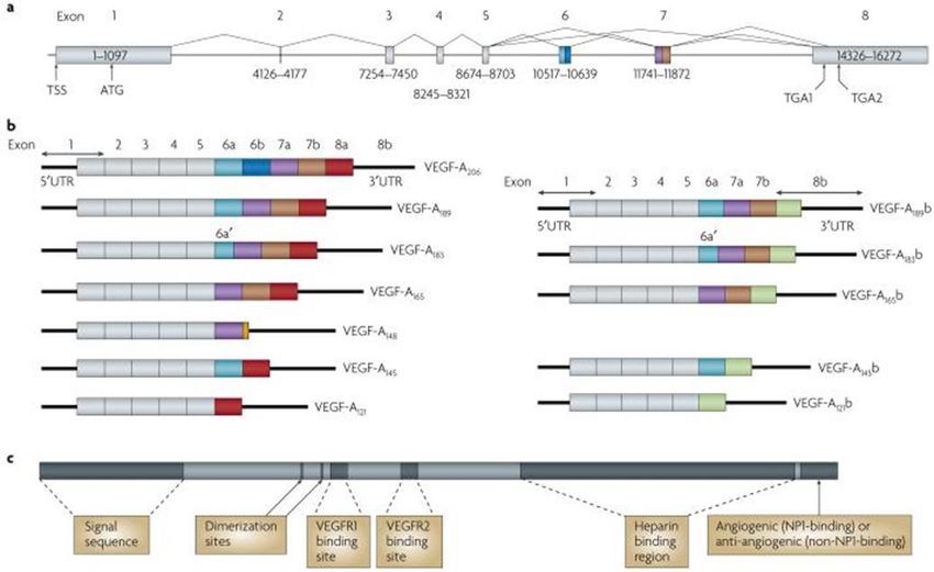
Fig. 2. Protein and mRNA products of human vascular endothelial growth factor A (VEGF-A)(11). a Gene structure of human VEGF-A. VEGF-A
spans 16,272 bp of chromosome 6p21.1 and consists of eight exons. Alternate 5′ and 3′ splice site selection in exons 6, 7, and 8 generate multiple
isoforms. Exons 6 and 7 encode heparin-binding domains. The transcriptional start site (TSS) and translational start site (ATG) in exon 1 are
indicated. Alternative stop codons within exon 8 are also indicated (TGA1 and TGA2). b | Alternative splicing can occur either at the 5′ donor splice
site (for example, VEGF-A189 versus VEGF-A206) or the 3′ acceptor splice site (for example, VEGF-A189 versus VEGF-A165). Two mRNA isoform
families are generated. The pro-angiogenic isoforms (VEGF-Axxx, left) are generated by proximal splice site (PSS) selection in exon 8 and the anti-
angiogenic family (VEGF-Axxxb, right) from exon 8 distal splice site (DSS) choice. Thus, VEGF-A165, formed by PSS selection in exon 8, has VEGF-
A165b as its DSS sister isoform, the DSS-selected mRNA encoding a protein of exactly the same length. Exon 6a’ occurs in VEGF-A183 as a result of a
conserved alternative splicing donor site in exon 6a and is 18 bp shorter than full-length exon 6a. VEGF-A148 is a truncated isoform splicing from exon
7a into exon 8a out of frame and resulting in a premature stop codon. VEGF-A206b has not yet been identified. c | Protein structure of VEGF-A
containing the dimerization sites and binding sites for heparin, VEGF-A receptor 1 (VEGFR1; encoded by exon 3) and VEGFR2 (encoded by exon
4), which are present in all isoforms. The six amino acids at the extreme carboxyl terminus of the protein can be either pro-angiogenic (CDKPRR,
encoded by exon 8a) or anti-angiogenic (SLTRKD, encoded by exon 8b). The epitopes recognized by most commercial antibodies are in the region
of the VEGF-A receptor-binding domains, present in VEGF-A isoforms of both families. UTR, untranslated region [ Adapted from (11)].
Angiogenesis regulatory factors hematopoietic effects, inducing colony formation by
Several regulatory factors (Table 3) play a role in mature subsets of granulocyte-macrophage progenitor
angiogenesis. Among them, during angiogenesis, cells, regulation of osteoclast differentiation, stimulation
VEGF interacts with several other angiogenic factors of surfactant production, and neurotrophic and
[ Downloaded from rbmb.net on 2022-02-03 ]
and plays an important role in cell proliferation, neuroprotective effects on neuronal and glial cells.
differentiation, migration, cell survival, nitric oxide (NO) Notably, VEGF infusion to adult mice inhibits dendritic
production, and release of other growth factors (7). cell development, leading to the hypothesis that VEGF
facilitates tumor growth by allowing evasion of tumors
Wide implications of VEGF from physiological from the host immune system. Also, VEGF increased
circumstance to pathological conditions production of B cells and the generation of immature
The ability of VEGF to promote monocyte chemotaxis myeloid cells. Some studies suggest that VEGF controls
was the earliest evidence that VEGF can affect blood hematopoietic stem cells survival during hematopoietic
cells. Subsequently, VEGF was reported to have repopulation (17).
62 Rep. Biochem. Mol. Biol, Vol. 2, No. 2, Apr 2014Vascular Endothelial Growth Factor
Table 2. Regulatory factors of angiogenesis including diabetes, cognitive decline and dementia,
Protein reproductive disorders such as polycystic ovary disease
a) Angiogenic Factors
and endometriosis, immunoallergic-inflammatory
FGF-β
FGF-α diseases such as asthma and rheumatoid arthritis,
Angiogenin psoriasis, ophthalmologic disorders such as macular
Transforming growth factor-α degeneration and diabetic retinopathy, chronic
Transforming growth factor-β obstructive pulmonary diseases, and several neoplastic
Tumor necrosis factor-α diseases (19).
Vascular endothelial growth factor
(VPF/VEGF)
Platelet-derived endothelial growth factor
Wound healing
Granulocyte colony-stimulating factor Skin-wound healing starts immediately after injury and
Placental growth factor consists of three phases: inflammation, proliferation,
Interleukin-8 and maturation. These phases proceed with
Hepatocyte growth factor complicated but well-organized interactions between
Proliferin various tissues and cells. Wounding can destroy blood
Angiopoietin-1
vessels and create a hypoxic environment because of
Leptin
hCG human chorionic gonadotropin poor perfusion, and therefore provides an appropriate
Estrogens environment for hypoxia inducible factor alpha
b) Angiostatic factors (natural) stabilization. At the first phase of wounding, induced
Angiostatin hypoxia leads to rapid infiltration of inflammatory cells
Thrompospodin including neutrophils, mast cells, lymphocytes, and
Endostatin macrophages. The formation of granulation tissue,
Tumor necrosis factor-α
which is necessary for the last phase of wound
Prolactin
Thromboxane A2 healing, starts at the wound space approximately four
c) Angiostatic factors (therapeutic) days after injury. Numerous new capillaries endow
Thalidomide the new stroma with its granular appearance.
Steroids Macrophages, fibroblasts, and endothelial cells move
FGF-β = basic fibroblast growth factor; FGF-α = acidic FGF; into the wound space at the same time. Macrophages
VPF = vascular permeability factor
not only augment inflammatory responses but also
secrete VEGF and FGF, eventually promoting
VEGF is also known as vascular permeability angiogenesis (20, 21).
factor, based on its ability to induce vascular leakage. Proangiogenic roles of macrophages in diabetic
The permeability-enhancing activity of this molecule wounds are similar to those in wounds inflicted by
underlies dominant roles in inflammation and other physical injuries. However, severe hypoxia in diabetic
pathological conditions. In accordance with a role in wounds often fails to directly trigger effective
the regulation of vascular permeability, VEGF angiogenesis because of significant cell death under
induces endothelial fenestration in some vascular such conditions. As a practical conclusion, the hypoxia
beds and cultured adrenal endothelial cells (6, 10). inducible factor pathway offers promising therapeutic
Some studies have shown a critical role for nitric targets to promote angiogenesis in wounds (20) (1).
oxide (NO) in VEGF-induced vascular permeability,
as well as angiogenesis. Fukumura et al. (18) verified
[ Downloaded from rbmb.net on 2022-02-03 ]
Diabetes and its related complications
the relative contribution of the NO synthase (NOS) Hiroaki Kakizawa et al. (22) reported that plasma
isoforms, inducible NOS (iNOS) and endothelial VEGF concentrations are higher in diabetic patients
NOS (eNOS), to these processes. Thereby, elevated who are hospitalized because of poor glycemic
circulating VEGF has been observed in vascular control than in healthy subjects. According to them,
diseases including ischemic heart disease, heart the increased plasma VEGF concentrations declined
failure, and stroke, and in various other disorders along with decreases in fasting plasma glucose and
Rep. Biochem. Mol. Biol, Vol. 2, No. 2, Apr 2014 63Azimi-Nezhad M
hemoglobin A1c (HbA1c) as a result of treatment. Hypertension
The significant and independent correlation between VEGF-A is highly expressed by renal glomerular
plasma VEGF concentrations and HbA1c suggests epithelial cells (podocytes) and plays an important role in
that chronic hyperglycemia may increase plasma the formation of the glomeruli during development (27),
levels of VEGF, and that reduction of high VEGF but curiously is also highly expressed within the adult
levels may be possible by improvement of glycemic glomerulus despite little or no angiogenesis occurring
control (23). Prolonged hyperglycemia activates the beyond development. VEGF isoform expression in
sorbitol pathway and induces intracellular anaerobic glomeruli is heterogeneous. Individual human glomeruli
conditions and hemodynamic change (24). These express one, two, or all three of these (VEGF121,
conditions may facilitate the production of VEGF that VEGF165, VEGF189) main isoforms at the mRNA level.
may contribute to diabetic vascular complications and VEGF189 and VEGF165 are most predominantly found
arteriosclerosis. Interestingly, the increase in VEGF in the renal glomerulus. Minor VEGF mRNA splice
production has a reverse correlation with the variants (VEGF206, VEGF183, VEGF148, and VEGF145)
reduction of hyperglycemia. Santilli et al. (25) have also been reported, but are less well characterized
suggested increased serum VEGF levels as a than the three main isoforms (28).
predicting risk factor for the development of
persistent microalbuminuria in young type 1 diabetic Obesity and metabolic syndrome
patients. Another study indicates that VEGF mRNA Adipose tissue is considered as the largest endocrine
and urinary excretion of VEGF are increased in gland because it produces free fatty acids, hormones,
diabetic nephropathy (26). growth factors, and cytokines such as leptin,
Most diabetic patients, especially those with poor adiponectin, resistin, VEGF, insulin growth factor
glycemic control, develop diabetic retinopathy, which (IGF-1), interleukin-6 (IL-6), and tumor necrosis
remains the major cause of new-onset blindness factor-alpha (TNF-α) (29). Epidemiological studies
among diabetic adults. Diabetic retinopathy is show that the visceral fat accumulation ”the
characterized by vascular permeability and increased predominant driving force behind the metabolic
tissue ischemia and angiogenesis. syndrome MetS “ (30) is the most important
VEGF has initially drawn much attention as an determining factor for VEGF circulating levels. During
important mediator of retinal ischemia– associated embryogenesis, adipose tissue development is spatially
intraocular neovascularization (4, 6, 10). VEGF is and temporally associated with microvessel growth. In
produced from many cell types within the eye and developing embryos, the formation of primitive fat
past studies have shown that VEGF levels are organs occurs at the perivascular site (29). Endothelial
markedly elevated in vitreous and aqueous fluids in cells isolated from different adipose tissues differ in
the eyes of individuals with proliferative diabetic their proliferative capacity, which suggests that
retinopathy (PDR). Recently there has been success adipocytes play both guidance and maintenance roles
in the treatment of diabetic retinopathy as well as in vascular development. A recent study suggests that
other ocular vasculopathies with anti-VEGF adipocytes and their accompanying endothelial cells
medication. Bevacizumab (Avastin, Genentech, might share a common progenitor that could
South San Francisco, CA) is a recombinant, differentiate into adipocytes or endothelial lineages
humanized monoclonal anti-VEGF antibody that depending upon exposure to different environments
binds all VEGF isoforms and exerts its neutralizing (29) (145). Accumulating evidence shows that
[ Downloaded from rbmb.net on 2022-02-03 ]
effect by inhibiting the VEGF–receptor interaction, capillary endothelial cells communicate with
thus blocking both increased vascular permeability adipocytes via paracrine signaling pathways,
and angiogenesis. The drug was approved by the extracellular components, and direct cell-cell
United States Food and Drug Administration initially interactions (31).
for intravenous use for metastatic colorectal cancer. Adipose tissue has been long known to promote
Bevacizumab has been administered off label for the wound healing and revascularize ischemic tissues
treatment of neovascular age-related macular including myocardium (31). These findings suggest
degeneration and other retinal vascular conditions that adipose tissue produces angiogenic molecules
with encouraging results (6). (31). Experimental angiogenesis assays show that
64 Rep. Biochem. Mol. Biol, Vol. 2, No. 2, Apr 2014Vascular Endothelial Growth Factor
conditioned media obtained from preadipocytes and Michaela Loebig et al. (37) reported a positive
tissue homogenates from omentum or subcutaneous correlation between plasma VEGF concentrations and
fat induce angiogenesis in chick chorioallantoic BMI over a large range of BMI groups in a healthy
membrane (CAM) and mouse cornea (31). It seems population. They also demonstrated significantly
that bone marrow-derived circulating endothelial higher concentrations of plasma VEGF in obese
precursor cells do not significantly participate in subjects than in normal and low weight individuals.
adipose neovascularization, although these cells are Lian-Yu Lin et al. (38), in their proposed model,
known to contribute to neovascularization in other demonstrated that obesity directs to an inflammatory
tissues. For instance, VEGF is a potent chemoattractant process, which could be the precursor of MetS
for inflammatory cells and contributes to mobilization components including insulin resistance, dyslipidemia,
of bone marrow-derived circulating endothelial and hypertension.
precursor cells, which are involved in tumor Gaby Kressel et al. (39) investigated the
neovascularization. Interestingly, expression levels of relationship between vascular and systemic markers of
VEGF are only moderately up-regulated in growing low-grade inflammation such as high sensitive c-
adipose tissue although VEGF is a main angiogenic reactive protein (hs-CRP ), soluble vascular adhesion
factor in omentum (29). molecule 1(sVCAM-1), soluble intracellular adhesion
In rapidly extending adipose tissue, hypoxia is molecule-1 (sICAM-1), plasminogen activating
another important factor for vascular growth and inhibitor-1(PAI-1), fibrinogen, and cardiovascular
remodeling. In response to hypoxia, adipose tissues traditional risk factors and the MetS.
produce hypoxia inducible factor 1a– induced It seems that the study of the association of
angiogenic factors such as VEGF, leptin, TNF-α, and circulating VEGF with MetS in human samples has
PAI-1, which regulate angiogenesis and only recently begun; therefore, documentable reports in
vasculogenesis (32). Thus it is reasonable to speculate this field are scarce (40-43). However, a few reports
that expansion of adipose tissue is associated with local have shown an association of circulating VEGF with
hypoxia, which contributes to angiogenesis by the MetS, as well as with a number of its components
induction of a number of growth factors. (40, 41). Some evidence indicates the associations of
Leptin is an adipocyte-derived hormone that some single nucleotide polymorphisms (SNPs) with
regulates food intake and energy homeostasis. MetS via a genome-wide association study (44) and
Functional impairment of leptin leads to severe obesity, candidate gene approach (45, 46). It is not yet clear
diabetes, and infertility. Interestingly, leptin is also whether the elevated VEGF level is a deleterious factor
defined as a potent angiogenic factor. In addition to its or a physiologic/negative feedback response to prevent
direct angiogenic activity, leptin up-regulates VEGF the progress of ischemia in metabolic linked ischemia
mRNA expression via activation of the Jak/Stat3 as well as MetS.
signaling pathway (33). Among all adipose tissues
examined in the body, omentum expresses the highest Atherosclerosis
level of VEGF (29). Localization studies have shown From endothelial dysfunction to plaque rupture, VEGF
that adipocytes are the primary source of VEGF, directly and indirectly attracts inflammatory cells into
which may act as an angiogenic and vascular survival the intima in various stages of atherogenesis. Human
factor for the omental vasculature. Additionally, monocytes express the receptor VEGFR-1 (flt-1), and
adipose- infiltrated inflammatory cells and adipose administration of VEGF has been shown to
[ Downloaded from rbmb.net on 2022-02-03 ]
stromal cells also significantly contribute to VEGF significantly increase the number of mature
production (29). Anatomically, visceral adipose tissue, macrophages in the arterial endothelium within weeks.
which is the main indicator of waist circumference, is This accumulation of macrophages was associated
present mainly in the mesentery and omentum, and with increased total plaque size. VEGF primes
drains directly through the portal circulation to the liver endothelial cells to secrete increased amounts of E-
(34). selectin, an adhesion molecule necessary for trans-
Several documents indicate that serum VEGF epithelial migration of leukocytes. Endothelial cells
concentrations are positively correlated with body ‘‘pretreated’’ with VEGF also produce significantly
mass index (BMI) and visceral obesity (35, 36). higher amounts of tissue factor after TNF-α exposure
Rep. Biochem. Mol. Biol, Vol. 2, No. 2, Apr 2014 65Azimi-Nezhad M
than cells exposed to TNF-α alone. If the plaque men with unstable angina and serum VEGF
contents later become exposed, the accumulation of concentrations ≥0.3 pg/L were 2.5 times more likely to
tissue factor could increase the risk of a thrombotic die of myocardial infarction within six months of
event. Not surprisingly then, serum VEGF level has cardiac event than those with serum VEGF levelsVascular Endothelial Growth Factor
References
1. Senger DR, Galli SJ, Dvorak AM, Perruzzi CA, mobilization of bone marrow-derived endothelial
Harvey VS, Dvorak HF. Tumor cells secrete a progenitor cells for neovascularization. Nat Med.
vascular permeability factor that promotes 1999 Apr;5(4):434-8.
accumulation of ascites fluid. Science. 1983 14. Folkman J, Shing Y. Angiogenesis. J Biol Chem.
Feb;219(4587):983-5. 1992 Jun;267(16):10931-4.
2. Keck PJ, Hauser SD, Krivi G, Sanzo K, Warren T, 15. Folkman J. Angiogenesis in cancer, vascular,
Feder J, et al. Vascular permeability factor, an rheumatoid and other disease. Nat Med. 1995
endothelial cell mitogen related to PDGF. Science. 1989 Jan;1(1):27-31.
Dec;246(4935):1309-12. 16. Hockel M, Schlenger K, Doctrow S, Kissel T,
3. Leung DW, Cachianes G, Kuang WJ, Goeddel Vaupel P. Therapeutic angiogenesis. Arch Surg. 1993
DV, Ferrara N. Vascular endothelial growth factor is Apr;128(4):423-9.
a secreted angiogenic mitogen. Science. 1989 17. Gerber HP, Malik AK, Solar GP, Sherman D,
Dec;246(4935):1306-9. Liang XH, Meng G, et al. VEGF regulates
4. Azimi-Nezhad M, Stathopoulou MG, Bonnefond haematopoietic stem cell survival by an internal
A, Rancier M, Saleh A, Lamont J, Fitzgerald P, et al. autocrine loop mechanism. Nature. 2002
Associations of vascular endothelial growth factor Jun;417(6892):954-8.
(VEGF) with adhesion and inflammation molecules 18. Fukumura D, Gohongi T, Kadambi A, Izumi Y,
in a healthy population. Cytokine. 2013 Ang J, Yun CO, et al. Predominant role of endothelial
Feb;61(2):602-7. nitric oxide synthase in vascular endothelial growth
5. Muller YA, Li B, Christinger HW, Wells JA, factor-induced angiogenesis and vascular permeability.
Cunningham BC, de Vos AM. Vascular endothelial Proc Natl Acad Sci U S A. 2001 Feb;98(5):2604-9.
growth factor: crystal structure and functional 19. Debette S, Visvikis-Siest S, Chen MH, Ndiaye
mapping of the kinase domain receptor binding site. NC, Song C, Destefano A, et al. Identification of cis-
Proc Natl Acad Sci U S A. 1997 Jul;94(14):7192-7. and trans-acting genetic variants explaining up to half
6. Ferrara N, Gerber HP, LeCouter J. The biology of the variation in circulating vascular endothelial growth
VEGF and its receptors. Nat Med. 2003 factor levels. Circ Res. 2011 Aug;109(5):554-63.
Jun;9(6):669-76. 20. Fong GH. Mechanisms of adaptive angiogenesis
7. Thanigaimani S, Kichenadasse G, Mangoni AA. to tissue hypoxia. Angiogenesis. 2008
The emerging role of vascular endothelial growth Mar;11(2):121-40.
factor (VEGF) in vascular homeostasis: lessons from 21. Kondo T, Ishida Y. Molecular pathology of
recent trials with anti-VEGF drugs. Curr Vasc wound healing. Forensic Sci Int. 2010 Dec;203(1-
Pharmacol. 2011 May;9(3):358-80. 3):93-8.
8. Tammela T, Enholm B, Alitalo K, Paavonen K. 22. Kakizawa H, Itoh M, Itoh Y, Imamura S, Ishiwata
The biology of vascular endothelial growth factors. Y, Matsumoto T, et al. The relationship between
Cardiovasc Res. 2005 Feb;15;65(3):550-63. glycemic control and plasma vascular endothelial
9. Flamme I, Frolich T, Risau W. Molecular growth factor and endothelin-1 concentration in diabetic
mechanisms of vasculogenesis and embryonic patients. Metabolism. 2004 May;53(5):550-5.
angiogenesis. J Cell Physiol. 1997 Nov; 173(2):206-10. 23. Zorena K, Mysliwska J, Mysliwiec M,
10. Ferrara N. Vascular endothelial growth factor: Rybarczyk-Kapturska K, Malinowska E, Wisniewski
[ Downloaded from rbmb.net on 2022-02-03 ]
basic science and clinical progress. Endocr Rev. 2004 P, et al. Association between vascular endothelial
Aug;25(4):581-611. growth factor and hypertension in children and
11. Harper SJ, Bates DO. VEGF-A splicing: the key adolescents type I diabetes mellitus. J Hum
to anti-angiogenic therapeutics? Nat Rev Cancer. Hypertens. 2010 Nov;24(11):755-62.
2008 Nov;8(11):880-7. 24. Tsilibary EC. Microvascular basement
12. Risau W, Flamme I. Vasculogenesis. Annu Rev membranes in diabetes mellitus. J Pathol. 2003
Cell Dev Biol. 1995;11:73-91. Jul;200(4):537-46.
13. Takahashi T, Kalka C, Masuda H, Chen D, Silver 25. Santilli F, Spagnoli A, Mohn A, Tumini S,
M, Kearney M, et al. Ischemia- and cytokine-induced Verrotti A, Cipollone F, et al. Increased vascular
Rep. Biochem. Mol. Biol, Vol. 2, No. 2, Apr 2014 67Azimi-Nezhad M
endothelial growth factor serum concentrations may obese individuals. Int J Obes (Lond). 2005
help to identify patients with onset of type 1 diabetes Nov;29(11):1308-14.
during childhood at risk for developing persistent 37. Loebig M, Klement J, Schmoller A, Betz S,
microalbuminuria. J Clin Endocrinol Metab. 2001 Heuck N, Schweiger U, et al. Evidence for a
Aug;86(8):3871-6. relationship between VEGF and BMI independent of
26. Murata T, Nagai R, Ishibashi T, Inomuta H, Ikeda insulin sensitivity by glucose clamp procedure in a
K, Horiuchi S. The relationship between homogenous group healthy young men. PLoS One.
accumulation of advanced glycation end products and 2010 Sep;5(9):e12610.
expression of vascular endothelial growth factor in 38. Lin LY, Kuo HK, Li HY, Hwang JJ, Lin JW.
human diabetic retinas. Diabetologia. 1997 Confirming a biological pathway in the metabolic
Jul;40(7):764-9. syndrome--insight from the NHANES 1999-2002.
27. Wakelin SJ, Marson L, Howie SE, Garden J, Obesity (Silver Spring). 2008 Dec;16(12):2676-81.
Lamb JR, Forsythe JL. The role of vascular 39. Kressel G, Trunz B, Bub A, Hulsmann O,
endothelial growth factor in the kidney in health and Wolters M, Lichtinghagen R, et al. Systemic and
disease. Nephron Physiol. 2004;98(3):73-9. vascular markers of inflammation in relation to
28. Foster RR, Hole R, Anderson K, Satchell SC, metabolic syndrome and insulin resistance in adults
Coward RJ, Mathieson PW, et al. Functional evidence with elevated atherosclerosis risk. Atherosclerosis.
that vascular endothelial growth factor may act as an 2009 Jan;202(1):263-71.
autocrine factor on human podocytes. Am J Physiol 40. Jialal I, Fadini GP, Pollock K, Devaraj S.
Renal Physiol. 2003 Jun;284(6):F1263-F1273. Circulating levels of endothelial progenitor cell
29. Cao Y. Angiogenesis modulates adipogenesis mobilizing factors in the metabolic syndrome. Am J
and obesity. J Clin Invest. 2007 Sep;117(9):2362-8. Cardiol. 2010 Dec;106(11):1606-8.
30. Grundy SM. Metabolic syndrome: a multiplex 41. Lieb W, Safa R, Benjamin EJ, Xanthakis V, Yin
cardiovascular risk factor. J Clin Endocrinol Metab. X, Sullivan LM, et al. Vascular endothelial growth
2007 Feb;92(2):399-404. factor, its soluble receptor, and hepatocyte growth
31. Sun K, Kusminski CM, Scherer PE. Adipose factor: clinical and genetic correlates and association
tissue remodeling and obesity. J Clin Invest with vascular function. Eur Heart J. 2009
2011;1217(6):2094-2101. May;30(9):1121-7.
32. Fitzpatrick TE, Graham CH. Stimulation of 42. Tarantino G, Lobello R, Scopacasa F, Contaldo F,
plasminogen activator inhibitor-1 expression in Pasanisi F, Cirillo M, et al. The contribution of
immortalized human trophoblast cells cultured under omental adipose tissue to adipokine concentrations in
low levels of oxygen. Exp Cell Res. 1998 patients with the metabolic syndrome. Clin Invest
Nov;245(1):155-62. Med. 2007;30(5):E192-E199.
33. Suganami E, Takagi H, Ohashi H, Suzuma K, 43. Wada H, Satoh N, Kitaoka S, Ono K, Morimoto
Suzuma I, Oh H, et al. Leptin stimulates ischemia- T, Kawamura T, et al. Soluble VEGF receptor-2 is
induced retinal neovascularization: possible role of increased in sera of subjects with metabolic syndrome
vascular endothelial growth factor expressed in retinal in association with insulin resistance. Atherosclerosis.
endothelial cells. Diabetes. 2004 Sep;53(9):2443-8. 2010 Feb;208(2):512-7.
34. Ibrahim MM. Subcutaneous and visceral adipose 44. Zabaneh D, Mackay IJ. Genome-wide linkage
tissue: structural and functional differences. Obes scan on estimated breeding values for a quantitative
[ Downloaded from rbmb.net on 2022-02-03 ]
Rev. 2010 Jan;11(1):11-8. trait. BMC Genet. 2003 Dec;4 Suppl 1:S61.
35. Miyazawa-Hoshimoto S, Takahashi K, Bujo H, 45. Kraja AT, Vaidya D, Pankow JS, Goodarzi MO,
Hashimoto N, Saito Y. Elevated serum vascular Assimes TL, Kullo IJ, et al. A bivariate genome-wide
endothelial growth factor is associated with visceral approach to metabolic syndrome: STAMPEED
fat accumulation in human obese subjects. consortium. Diabetes. 2011 Apr;60(4):1329-39.
Diabetologia. 2003 Nov;46(11):1483-8. 46. Povel CM, Boer JM, Reiling E, Feskens EJ. Genetic
36. Silha JV, Krsek M, Sucharda P, Murphy LJ. variants and the metabolic syndrome: a systematic
Angiogenic factors are elevated in overweight and review. Obes Rev. 2011 Nov;12(11):952-67.
68 Rep. Biochem. Mol. Biol, Vol. 2, No. 2, Apr 2014Vascular Endothelial Growth Factor
47. Armstrong AW, Voyles SV, Armstrong EJ, 50. Armstrong AW, Voyles SV, Armstrong EJ,
Fuller EN, Rutledge JC. Angiogenesis and oxidative Fuller EN, Rutledge JC. Angiogenesis and oxidative
stress: common mechanisms linking psoriasis with stress: common mechanisms linking psoriasis with
atherosclerosis. J Dermatol Sci. 2011 Jul;63(1):1-9. atherosclerosis. J Dermatol Sci. 2011 Jul;63(1):1-9.
48. Heeschen C, Dimmeler S, Hamm CW, Boersma 51. Celletti FL, Waugh JM, Amabile PG, Brendolan
E, Zeiher AM, Simoons ML. Prognostic significance A, Hilfiker PR, Dake MD. Vascular endothelial
of angiogenic growth factor serum levels in patients growth factor enhances atherosclerotic plaque
with acute coronary syndromes. Circulation. 2003 progression. Nat Med. 2001 Apr;7(4):425-9.
Feb; 4;107(4):524-30. 52. Stannard AK, Khurana R, Evans IM, Sofra V,
49. Holm PW, Slart RH, Zeebregts CJ, Hillebrands Holmes DI, Zachary I. Vascular endothelial growth
JL, Tio RA. Atherosclerotic plaque development and factor synergistically enhances induction of E-selectin
instability: a dual role for VEGF. Ann Med. by tumor necrosis factor-alpha. Arterioscler Thromb
2009;41(4):257-64. Vasc Biol. 2007 Mar;27(3):494-502.
[ Downloaded from rbmb.net on 2022-02-03 ]
Rep. Biochem. Mol. Biol, Vol. 2, No. 2, Apr 2014 69
Powered by TCPDF (www.tcpdf.org)You can also read