Using thermographic cameras to investigate eye temperature and clinical severity in depression
←
→
Page content transcription
If your browser does not render page correctly, please read the page content below
Using thermographic cameras to
                 investigate eye temperature and
                 clinical severity in depression

                 Jerome J. Maller
                 Shefin Sam George
                 Rekha Puzhavakkathumadom Viswanathan
                 Paul B. Fitzgerald
                 Paul Junor

Downloaded From: https://www.spiedigitallibrary.org/journals/Journal-of-Biomedical-Optics on 06 Feb 2022
Terms of Use: https://www.spiedigitallibrary.org/terms-of-use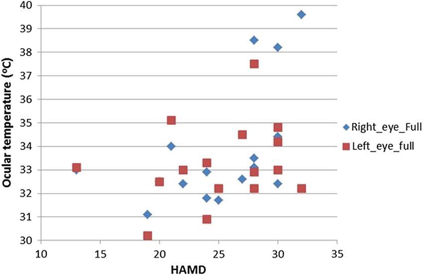
Journal of Biomedical Optics 21(2), 026001 (February 2016)

                Using thermographic cameras to investigate eye
                temperature and clinical severity in depression
                Jerome J. Maller,a,* Shefin Sam George,b Rekha Puzhavakkathumadom Viswanathan,b Paul B. Fitzgerald,a and
                Paul Junorb
                a
                 The Alfred and Monash University Central Clinical School, Monash Alfred Psychiatry Research Centre, Melbourne, Victoria, Australia
                b
                 LaTrobe University, School of Engineering and Mathematical Sciences, Department of Electronic Engineering, Melbourne, Victoria, Australia

                              Abstract. Previous studies suggest that altered corneal temperature may be a feature of schizophrenia, but the
                              association between major depressive disorder (MDD) and corneal temperature has yet to be assessed. The aim
                              of this study is to investigate whether eye temperature is different among MDD patients than among healthy
                              individuals. We used a thermographic camera to measure and compare the temperature profile across the cor-
                              neas of 16 patients with MDD and 16 age- and sex-matched healthy subjects. We found that the average corneal
                              temperature between the two groups did not differ statistically, although clinical severity correlated positively with
                              right corneal temperature. Corneal temperature may be an indicator of clinical severity in psychiatric disorders,
                              including depression. © 2016 Society of Photo-Optical Instrumentation Engineers (SPIE) [DOI: 10.1117/1.JBO.21.2.026001]
                              Keywords: depression; thermal imaging; ocular; corneal; psychiatry.
                              Paper 150440R received Jul. 3, 2015; accepted for publication Dec. 30, 2015; published online Feb. 1, 2016.

                1     Introduction                                                                 1.1     Methods of Measuring Core Body Temperature
                Thermoregulation is the most important and essential aspect of                     There is a definite correlation between body temperature and
                human homeostasis. The anterior hypothalamic nucleus and the                       diseases.5 Many previous studies have analyzed core body tem-
                adjacent preoptic area have been considered as the “seat of                        perature using rectal temperature although corneal, tympanic, or
                thermoregulation.”1,2 But in recent years, it has become clear                     axillary temperatures may also indicate core body temperature.
                that other regions of the central nervous system (CNS), espe-                      Even though rectal temperature is considered to be more accu-
                cially the spinal cord, are importantly involved in thermoregu-                    rate, the placement of a thermometer in the rectum is unpleasant
                latory processes.3 The anterior or preoptic hypothalamus                           and inconvenient. In humans, corneal temperature was found to
                contains thermosensitive neurons that coordinate to keep the                       be linearly related to body core temperature with corneal tem-
                core body temperature almost constant. Preoptic neurons also                       perature apparently plateauing at 36.5°C to 37.0°C. The corneal
                receive a somatosensory input from skin and spinal thermore-                       temperature of a healthy person is generally between 32°C and
                ceptors to indicate the peripheral temperature variations. In                      35°C, which is lower than the body temperature and higher than
                this way, preoptic neurons compare and integrate central and                       the atmospheric temperature. The axilla and tympanic mem-
                peripheral thermal information and coordinate the appropriate                      brane are the other easily accessible sites for temperature meas-
                efferent response. Typically, input from neurons located more                      urement.6 Among all the above mentioned methods, corneal
                proximally contribute more to autonomic and metabolic                              temperature measurements in humans can be easily taken
                responses than does input from the skin. The hypothalamus                          with relatively noninvasive procedures.
                is extensively connected with different parts of the CNS, includ-                      Continuous studies since 1970s have been performed to
                ing the limbic forebrain and thus to the dopaminergic and sero-                    study the effect of serotonin on the thermoregulation. An over-
                tonergic pathway. The hypothalamus is heavily innervated by                        view of the evidence supporting a central role for serotoninergic
                monoaminergic and cholinergic fibers and the role of these                         neurons in the control of body temperature by the diencephalon
                hypothalamic neural systems in thermoregulation has been                           was presented by Myers.7 Morphological analysis of the anterior
                extensively investigated.3 These studies suggest that proper                       hypothalamus manifested that hyperthermia was elicited in
                transmission in the dopaminergic pathway is crucial for the                        almost all species when 5-hydroxytryptophan was injected
                hypothalamus to maintain appropriate body temperature regula-                      locally into this thermosensitive zone. Moreover, the heat pro-
                tion to 37°C. Due to a disturbed and hyperactive central dopa-                     duction response during cold stress in animals such as rats and
                minergic transmission, patients with major depressive disorder                     monkeys was disrupted following damage to the serotoninergic
                (MDD) and schizophrenia (SCZ) are, therefore, at a higher risk                     neurons in the anterior hypothalamic preoptic area.7 A study
                of developing thermoregulatory disorders revealed by variations                    suggests that serotonin takes part in the control of the body tem-
                in core body temperature,4 as well as in superficial tissue such as                perature set-point.8 Some studies showed that efficacious
                cornea. Hence, classes of diseases involving abnormal dopami-                      treatment of MDD normalized the lowered amplitude of the
                nergic transmission are suitable for presenting the similar tem-                   temperature circadian rhythm and increased body temperature.
                perature variation.                                                                For example, it was found that disturbed circadian temperature
                                                                                                   rhythm in major depression patients was restored by

                *Address all correspondence to: Jerome J. Maller, E-mail: jerome.maller@
                 monash.edu                                                                        1083-3668/2016/$25.00 © 2016 SPIE

                Journal of Biomedical Optics                                              026001-1                                        February 2016   •   Vol. 21(2)

Downloaded From: https://www.spiedigitallibrary.org/journals/Journal-of-Biomedical-Optics on 06 Feb 2022
Terms of Use: https://www.spiedigitallibrary.org/terms-of-use
Maller et al.: Using thermographic cameras to investigate eye temperature and clinical severity in depression
electroconvulsive therapy (ECT). The temperature profile over a 1.4 Corneal Temperature in Psychiatric Disorders
period of 24 h was noticeably different in patients’ pre-ECT and
post-ECT and in controls. The 24-h profiles exhibited by post- The corneal temperature of drug-free SCZ patients has been
ECT patients were similar to healthy subjects. The amplitude of evaluated with a thermal imaging camera which demonstrated
circadian temperature rhythm was elevated after ECT. The mean a significantly higher corneal temperature (1.55°C higher) in
temperatures (both asleep and 24 hours) were substantially the patient group when compared to matched controls.21
higher in pre-ECT than post-ECT and healthy subjects.9 Moreover, the study indicated that the corneal temperature
Rausch10 presented a study which claims to be the first demon- has a positive correlation with the symptom severity assessed
stration of higher daytime body temperature in cases with major by the brief psychiatric rating scale (BPRS). Shortly afterward,
depression. The study showed that subjects with a corrected a study showed that the corneal temperature of neuroleptic-
temperature above 36.8°C were 2.6-fold more likely to be treated patients was significantly lower than that of the drug-
depressed. free patients.22 Another study then indicated that drug-free
SCZ patients have significantly higher corneal temperature
when compared to that of normal healthy controls or treated
1.2 Body Temperature in Psychiatric Disorders patients.23 Based on these findings, Shiloh et al. conducted fur-
Since 1930, a number of studies have been conducted to provide ther studies to validate their hypothesis that the mental status of
insight into thermoregulatory disorders in people with SCZ. A SCZ patients may be associated with their eye temperature. The
review of the relevant articles regarding thermoregulatory dys- stability of thermoregulatory variation in male treatment-resist-
function in SCZ patients was conducted.11 The studies by ant SCZ patients and healthy subjects was assessed over a period
Cameron12 and Buck et al.13 found that SCZ patients have of 6 weeks;6 (to avoid changes in body temperature due to men-
lower baseline temperature when compared to that of controls. strual cycle, women were not included in the study). The corneal
The initial skin temperature recorded by Hermesh et al.14 was temperature was evaluated two to three times a week using a
significantly lower in the patient group, but Douglass and thermal imaging camera in a designated room. Shiloh et al.
Toogood15 found that the baseline temperature in SCZ patients reported a significant correlation between SCZ patients’ mean
and controls were similar, and the study of Shiloh et al.16 showed weekly PANSS scores and their mean weekly corneal
a higher baseline temperature in patients. A longitudinal study temperature.24
examining the thermoregulatory alterations induced by acute Drugs may alter body temperature by acting on any compo-
antipsychotic drug (APD) administration in SCZ patients nent of the thermoregulatory system. These components include
showed that acute administration of APDs decreases body tem- heat production, heat conservation, and heat loss effectors and
perature in drug-free SCZ patients by ∼0.36°C, although the their efferent pathways, thermosensors and their afferent path-
absolute values remain within the normal range.17 ways, and neurons within the CNS that coordinate thermoreg-
It has been reported that major depression is associated with ulatory effector activities.25
increased core body temperature, reduced thermoregulatory The current study aims at recording and investigating corneal
cooling (e.g., sweating), and alterations in peripheral measures temperature in patients with MDD and comparing them to
of serotonin (5-HT) activity, all of which are expected manifes- healthy control subjects to study the relationship between cor-
tations of impaired activity in the skin-to-brain-to-skin thermo- neal temperature and depression. We aimed to study the temper-
regulatory circuit within which the ascending spinoparabrachial ature profile across the cornea of the eye and to investigate the
pathway and its CNS projections form core components.18 nature of corneal temperature difference in MDD patients and
Furthermore, large effect size correlation was observed between healthy controls. We also aimed to investigate if corneal temper-
reductions in Center for Epidemiologic Studies Depression ature correlates to the degree of symptom severity.
Scale scores and reductions in mean core body temperatures.19
2 Materials and Methods
1.3 Measurement of Corneal Temperature 2.1 Participants
Among the array of eye imaging systems, infrared (IR) imaging A total of 32 subjects aged 19 to 79 years [males mean ¼ 46.88
is relatively noninvasive, inexpensive, and harmless, and hence (SD ¼ 17.00); females mean ¼ 49.00 (SD ¼ 12.05)] compris-
widely used in medical diagnosis. Thermography (or thermal ing 16 subjects with MDD and 16 age- and sex-matched healthy
imaging) can be defined as an imaging procedure which traces controls underwent psychiatric and ocular thermographic
the thermal sequence with an IR camera. As a part of thermo- assessments (Table 1). Subjects were recruited either through
regulation, human body emits heat to the atmosphere. As the public notices or word of mouth from the clinical services of
body temperature rises, the amount of energy radiated at any The Alfred Hospital, Victoria, Australia. MDD subjects were
wavelength increases and the wavelength of peak emission recruited before their transcranial magnetic stimulation
decreases. An increased temperature profile indicates underly- (TMS) treatments began. Each was treated with either left-
ing venous flow changes.5 The temperature distribution on sided repetitive TMS (rTMS; 10 Hz) or right-sided slow
the surface of the body can be acquired by thermographic cam- (1 Hz) TMS. MDD subjects were required to have a diagnosis
era to check the health condition of a person. As the temperature of major depressive disorder as determined by a treating psy-
variation in the anterior segment of the eye rarely occurs, it has chiatrist and confirmed with the Mini-International
been used to study various eye problems.20 However, emissivity Neuropsychiatric Interview (MINI)26 and a score on the
may also change with the water content of the eye and of the Hamilton Depression Rating Scale (HAMD)27 of at least 13,
cornea. The thermographic image allows one to see variations representing moderate-to-severe depression. Exclusion criteria
in temperature. The visual and quantitative documentation of the included another current or previous DSM-IV axis I diagnosis,
temperature measurements can then be made using the appro- current active medical problem, and known neurological dis-
priate software. ease. In addition, control subjects were required to have no
Journal of Biomedical Optics 026001-2 February 2016 • Vol. 21(2)
Downloaded From: https://www.spiedigitallibrary.org/journals/Journal-of-Biomedical-Optics on 06 Feb 2022
Terms of Use: https://www.spiedigitallibrary.org/terms-of-useMaller et al.: Using thermographic cameras to investigate eye temperature and clinical severity in depression
Table 1 Demographics of the patient and control subjects. provide an accurate detailed analysis of the images stored into
the memory card. The analysis objects such as points, lines, and
Group
areas helped to create histograms and line profiles to present the
thermal information contained by the images. To study the ther-
Variable MDD Control mal information of the corneal surface, an elliptical area was
used because it resembles the oval shape of eye. This area
Number (M:F) 16 (8:8) 16 (8:8) was selected manually by comparing with the RGB image pro-
Years of age mean 48.31 12.72 47.56 16.57
vided by the camera and visual inspection of the temperature
profile of the eye. The area of the ellipse (the physical imaging
HAMD mean 25.06 5.08 N/A area) was fixed at 710 to 715 px2 (35;400.42 mm2 ) to ensure
Range: 13 to 32 uniform analysis in all participants; regional areas were one
Note: , SD; M, males; F, females; MDD, major depressive disorder; quarter of the total area (i.e., quadrants). An area histogram
N/A, not applicable; HAMD, Hamilton depression rating scale. showed the temperature distribution, indicating the minimum,
maximum, average, and radiation power values across the cor-
nea. The temperature profiles across the cornea were created
history of psychiatric illness as screened by the psychiatric using line objects in which the line profile (the length of which
assessment schedule.28 All subjects provided written informed depends upon the area of the full eye) showed temperatures of
consent on a form approved by the Alfred Hospital, La Trobe every point along it. This indicated whether the temperature pro-
University, and Monash University Human Subjects Research file across the cornea of a depression patient was different from
and Ethics Committees. healthy controls.
Patients were taking a variety of medications, including
SSRIs, SNRIs, MAOIs, and atypical antipsychotics.
No subjects had diagnosed eye disease (e.g., glaucoma and 2.4 Procedure
cataracts).
Each participant participated in a single 1.5 h session which con-
2.2 Thermographic Camera sisted of a cognitive and symptom rating session (including ad-
ministration of the DSM-IV, MINI, and HDRS to patients with
Because the normal core temperature of a human body ranges major depression disorder) of ∼45 min, and a thermographic
between 32°C and 38°C, the temperature measurement range assessment of ∼25 to 30 min. For the thermographic component
required for this study is small. We used an NEC F30S of the study, participants were required to sit in a comfortable
Thermo Shot (NEC Corporation, Japan) which has a spatial chair for ∼15 min with their chin stabilized in a chin-holder and
resolution of 160 × 120 pixels, thermal resolution of 100 mK eyes wide open. Participants were asked not to blink during the
(0.1°C), and accuracy of ∼2000 mK (2°C). Emissivity image acquisition periods. The thermographic camera (mounted
was set to 0.98 based on previous corneal temperature on a tripod) then imaged various sections of each cornea to esti-
studies.29–31 mate their temperatures. The images were stored in JPEG format
containing the radiometric data for analysis. The ambient tem-
2.3 Room Temperature, Humidity and Lux perature, humidity, and light intensity of the room were recorded
Monitoring, and Corneal Thermographic so that they could be considered when calculating the ocular
Acquisition temperatures.
All thermographic assessments were performed in a specified
designated room with a temperature of 23.2°C to 26.4°C and
relative humidity of 42% to 47%. Based on the specifications 2.5 Statistical Analysis
of our camera, the best measurement distance to image each cor- GORATEC Thermography Studio and LABView were used for
nea separately was evaluated to be 20 cm. We constructed appa- detailed analysis and evaluation of thermal images. SPSS for
ratus to measure and record the temperature, humidity, and Windows version 22.0 (SPSS, Illinois) was used to conduct stat-
intensity of light at all times. istical analyses between the groups. Analyses were two-tailed
A National Instruments USB-6009 data acquisition device and were evaluated for significance at the 0.05 alpha level.
was used to acquire differential mode data from temperature
Simple chi-square and analysis of variance (ANOVA) were
(LM35; Texas Instruments), humidity (HIH 4000; Honeywell
employed to compare demographics and ocular temperatures,
S&C), and light sensors (ORP12; Silonex, UK). LabVIEW
and Pearson correlations were used to investigate the association
(National Instruments, Texas), and an open standard graphical
between clinical severity and ocular temperature.
programming environment was used to access and control the
data acquisition hardware.
Both eyes were imaged separately. Using a protractor, the 3 Results
images were taken at an angle of 15 deg between the eye
and the camera to avoid variations in surface emissivity at 3.1 Demographics
the given observation angle. Moreover, the human eye has an
ellipsoidal-type shape, so some variations and errors can There was no significant age or gender difference among the
occur in the IR image due to variation in the observation angle. groups, hence combined male and female results are presented.
By fixing the viewing angle, variations due to surface emissivity Mean HAMD was 25.06 (SD ¼ 5.08); two patients had scores
and observation angle were avoided. GORATEC Thermography below 20 (moderate depression), while the remaining 14
Studio (GORATEC Technology, Erding, Germany) was used to patients had scores beyond 20 (major depression).
Journal of Biomedical Optics 026001-3 February 2016 • Vol. 21(2)
Downloaded From: https://www.spiedigitallibrary.org/journals/Journal-of-Biomedical-Optics on 06 Feb 2022
Terms of Use: https://www.spiedigitallibrary.org/terms-of-useMaller et al.: Using thermographic cameras to investigate eye temperature and clinical severity in depression
3.2 Corneal Temperature in Major Depressive all ocular temperatures measurements were negative and none
Disorder and Controls were statistically significant (all p > 0.05). There were sta-
tistically significant positive correlations between right eye tem-
There were no significant differences in mean corneal temper- peratures and clinical depression severity (Table 3 and Fig. 2).
atures between the MDD patients and control subjects [right Controlling for age made very little difference to these results.
cornea: Fð1;30Þ ¼ 0.624, p ¼ 0.436; left cornea: (Fð1;30Þ ¼
0.664, p ¼ 0.664; Table 2]. There was no significant difference
in temperature between the left eye and right eye of patients with 3.4 Corneal Temperature and Medication
MDD or in control subjects. Controlling for age made no differ- Each patient was taking different medications, so statistical
ence to these results. Figure 1 shows an example of temperature analysis of the potential effect of medication could not be clearly
measurement. considered. However, two patients who were taking lithium car-
bonate had corneal temperatures that were among the lowest
3.3 Corneal Temperature and Clinical Severity within the clinical group.
There was no significant correlation between HAMD and age 4 Discussion
[rð16Þ ¼ 0.87, p ¼ 0.748]. All correlations between age and This study reports a unique finding of right-sided ocular temper-
ature being correlated positively with increased depression
Table 2 Mean ocular temperatures of the patient and control groups.
severity.
Our patients with MDD were recruited before their TMS
Group treatments began. The protocols used have shown that those
with MDD usually have an overactive (hyperactive) right-
Variable MDD Control sided prefrontal cortex and underactive (hypoactive) left-sided
prefrontal cortex.32 Hence, right-sided perfusion would be
Right
expected to be increased and left-sided perfusion to be decreased
Cornea 33.86 2.58 34.606 2.33 before treatment. Therefore, right-sided perfusion of the vessel
feeding the eye (i.e., ophthalmic artery) would also be increased,
Range: 30.80 to 39.40 Range: 31.30 to 39.50 resulting in increased ocular temperature. Our finding of right-
sided ocular temperature being positively correlated with
Quadrant A 33.83 2.60 34.53 2.31
increased depression severity supports this assumption. This
Quadrant B 33.66 2.60 34.39 2.37 finding is consistent with previously reported findings21
which indicated that the corneal temperature has a positive cor-
Quadrant C 33.73 2.57 34.44 2.35 relation with the symptomatic SCZ severity assessed by the
BPRS and PANSS.24
Quadrant D 33.74 2.57 34.51 2.34 The mean ocular temperatures in this study are well within
Entire eyeball 33.58 2.56 34.26 2.31 the range previously reported in large samples.20,33 Our findings
are consistent with a previous study which reported a large effect
Left size correlation between reductions in depression scores and
reductions in mean core body temperatures.19 In line with
Cornea 33.23 1.74 33.03 1.19 this, it has been suggested that cold showers be adapted as a
Quadrant A 33.18 1.76 32.99 1.22
potential treatment for depression.34
It has also been reported that nocturnal temperature is signifi-
Quadrant B 33.06 1.78 32.84 1.19 cantly higher in depressed patients compared to controls, and
decreases appreciably on recovery.35 In that study, the nocturnal
Quadrant C 33.09 1.78 32.93 1.21 sweat rates of depressed patients did not differ significantly from
those of controls, but decreased significantly with recovery.
Quadrant D 33.11 1.76 32.89 1.48
Vasodilatation and vasoconstriction after local application of
Entire eyeball 32.92 1.78 32.68 1.23 vasoactive substances have shown an increase or a decrease in
corneal temperature, respectively.30 Peripheral blood flow may
Note: , SD.
show parallel changes with blood flow variation in the eye.36,37
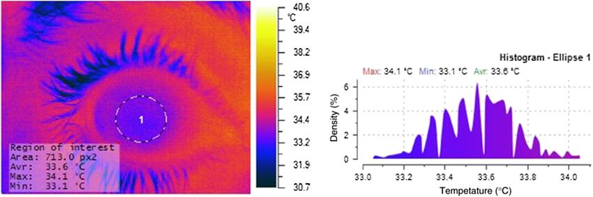
Fig. 1 Example of thermographic software used to estimate corneal temperature.
Journal of Biomedical Optics 026001-4 February 2016 • Vol. 21(2)
Downloaded From: https://www.spiedigitallibrary.org/journals/Journal-of-Biomedical-Optics on 06 Feb 2022
Terms of Use: https://www.spiedigitallibrary.org/terms-of-useMaller et al.: Using thermographic cameras to investigate eye temperature and clinical severity in depression
Table 3 Correlations between HAMD and ocular temperatures.
Right Left
Full Q-A Q-B Q-C Q-D ROI Full Q-A Q-B Q-C Q-D ROI
* * * * * *
0.537 0.538 0.537 0.535 0.539 0.539 0.260 0.256 0.237 0.244 0.240 0.212
Note: *denotes p < 0.05. Q, Quadrant.
account for this diurnal variability. Furthermore, body temper-
ature (and, therefore, ocular temperature) also varies with men-
strual cycle.41 These factors may have impacted upon the
reliability of our conclusions. Hence, we will endeavor to con-
trol for these variables in any future corneal temperature
investigations.
As this was a cross-sectional study, we could not establish a
direct cause–consequence relationship between corneal temper-
ature and depression severity.
5 Conclusion
This study, conducted in a room with a controlled environment,
showed no difference between mean corneal temperatures of
MDD patients and matched controls. The main finding is
that corneal temperature is significantly related to depression
Fig. 2 Relationship between HAMD scores and ocular temperatures
severity, and future research in larger samples will facilitate
(whole eye averages).
our understanding of this association. The possible effects of
medication on thermoregulatory functions can be studied by
This preliminary study indicated that in a controlled environ- analyzing drug-free and -treated depression patients separately.
ment, the average corneal temperature of patients with MDD Awareness of the medication history of the treated patients can
was no different than that of healthy control subjects. inform a better understanding of the effects of antidepressants
However, we observed that the corneal temperature of patients on their body temperature.
undergoing lithium medication were among the lowest of all the
subjects. While it is difficult to conclude that reduced corneal Acknowledgments
temperatures were related to the effect of lithium medication, The authors thank all the patients and volunteers who agreed to
it does lend support to the premise that lithium treatment is participate in this study.
hypothermic.38 Future investigations with larger groups of sub-
jects may be able to address the extent to which medication
impacts upon corneal temperature. References
Future investigations should consider multimodality imag- 1. B. Monge-Roffarello et al., “The medial preoptic nucleus as a site of the
ing. For example, merging MRI/CT with thermography may en- thermogenic and metabolic actions of melanotan II in male rats,” Am. J.
able better quality and fidelity, especially for medical Physiol. 307(2), R158–R166 (2014).
applications in which the temperature changes are clinically 2. D. Martelli et al., “The direct cooling of the preoptic-hypothalamic area
elicits the release of thyroid stimulating hormone during wakefulness
significant.39 but not during REM sleep,” PLoS One 9(2), e87793 (2014).
3. R. M. Lopachin and T. A. Rudy, “The thermoregulatory effects of nor-
adrenaline, serotonin and carbachol injected into the rat spinal subarach-
4.1 Limitations noid space,” J. Physiol. 333, 511–529 (1982).
4. S. Elsenga and R. H. Van den Hoofdakker, “Body core temperature and
The study was limited by the number of participants, hence the depression during total sleep deprivation in depressives,” Biol.
results must be interpreted with caution. The study was also lim- Psychiatry 24(5), 531–540 (1988).
ited by the accuracy of the thermographic camera which may be 5. S. Bagavathiappan et al., “Infrared thermal imaging for detection of
insufficient for this study, noting that the temperature difference peripheral vascular disorders,” J. Med. Phys. 34(1), 43–47 (2009).
between MDD and normal groups may be beyond the thermal 6. C. G. Blainey, “Site selection in taking body temperature,” Am. J.
Nursing 74(10), 1859–1861 (1974).
resolution of our camera. Therefore, the average corneal temper- 7. R. D. Myers, “Serotonin and thermoregulation: old and new views,” J.
ature of the patients with MDD may be found to be different Physiol. 77(2–3), 505–513 (1981).
from that of the healthy controls in more extensive studies 8. V. D. Sitnikov et al., “[Effect of serotonin on thermoregulation in nor-
recruiting higher numbers of subjects and using cameras of mothermic hibernators and during arousal from deep hypothermia],”
higher thermal resolution. Corneal temperature evaluation Biull. Eksp. Biol. Med. 101(1), 5–7 (1986).
may then potentially serve as a noninvasive diagnostic tool to 9. M. P. Szuba, B. H. Guze, and L. R. Baxter Jr., “Electroconvulsive
therapy increases circadian amplitude and lowers core body temperature
discriminate MDD patients from normal healthy people. in depressed subjects,” Biol. Psychiatry 42(12), 1130–1137 (1997).
Measurements were not acquired at the same time of day for 10. J. L. Rausch et al., “Depressed patients have higher body temperature:
each subject, and ocular temperatures fluctuate during the day.40 5-HT transporter long promoter region effects,” Neuropsychobiology
We conducted only one measurement per person, so we did not 47(3), 120–127 (2003).
Journal of Biomedical Optics 026001-5 February 2016 • Vol. 21(2)
Downloaded From: https://www.spiedigitallibrary.org/journals/Journal-of-Biomedical-Optics on 06 Feb 2022
Terms of Use: https://www.spiedigitallibrary.org/terms-of-useMaller et al.: Using thermographic cameras to investigate eye temperature and clinical severity in depression
11. T. W. Chong and D. J. Castle, “Layer upon layer: thermoregulation in 33. L. Tan, Z. Q. Cai, and N. S. Lai, “Accuracy and sensitivity of the
schizophrenia,” Schizophrenia Res. 69(2–3), 149–157 (2004). dynamic ocular thermography and inter-subjects ocular surface temper-
12. D. E. Cameron, “Heat production and heat control in the schizophrenic ature (OST) in Chinese young adults,” Contact Lens Anterior Eye 32(2),
reaction,” Arch. Neurol. Psychiatry 32, 704–711 (1934). 78–83 (2009).
13. C. W. Buck, H. B. Carscallen, and G. E. Hobbs, “Temperature regula- 34. N. A. Shevchuk, “Adapted cold shower as a potential treatment for
tion in schizophrenia. I. Comparison of schizophrenic and normal sub- depression,” Med. Hypotheses 70(5), 995–1001 (2008).
jects. II. Analysis by duration of psychosis,” AMA Arch. Neurol. 35. D. H. Avery et al., “Nocturnal sweating and temperature in depression,”
Psychiatry 64(6), 828–842 (1950). Acta Psychiatr. Scand. 100(4), 295–301 (1999).
14. H. Hermesh et al., “Heat intolerance in patients with chronic schizo- 36. J. Flammer et al., “The probable involvement of factors other than ocu-
phrenia maintained with antipsychotic drugs,” Am. J. Psychiatry lar pressure in the pathogenesis of glaucoma,” in Pharmacology of
157(8), 1327–1329 (2000). Glaucoma S. M. Drance et al., Eds., pp. 273–283, Williams &
15. A. B. Douglass and R. W. Toogood, “Temperature regulation and dop- Wilkins, Baltimore (1992).
amine in schizophrenia,” Biol. Psychiatry 22(8), 1048–1050 (1987). 37. U. Guthauser, J. Flammer, and F. Mahler, “The relationship between
16. R. Shiloh et al., “Abnormal thermoregulation in drug-free male schizo- digital and ocular vasospasm,” Graefes Arch. Clin. Exp. Ophthalmol.
phrenia patients,” Eur. Neuropsychopharmacolog. 11, 285–288 (2001). 226(3), 224–226 (1988).
17. R. Shiloh et al., “Acute antipsychotic drug administration lowers 38. A. J. Salerian, N. G. Saleri, and J. A. Salerian, “Brain temperature may
body temperature in drug-free male schizophrenic patients,” Eur. influence mood: a hypothesis,” Med. Hypotheses 70(3), 497–500
Neuropsychopharmacolog. 10(6), 443–445 (2000). (2008).
18. M. W. Hale, C. L. Raison, and C. A. Lowry, “Integrative physiology of 39. M. Abreu de Souza et al., “3D thermal medical image visualization tool:
depression and antidepressant drug action: implications for serotonergic integration between MRI and thermographic images,” Annual Int. Conf.
mechanisms of action and novel therapeutic strategies for treatment of of the IEEE Engineering in Medicine and Biology Society. IEEE
depression,” Pharmacol. Ther. 137(1), 108–118 (2013). Engineering in Medicine and Biology Society. Annual Conf. 5583–
19. K. U. Hanusch et al., “Whole-body hyperthermia for the treatment of 5586 (2014).
major depression: associations with thermoregulatory cooling,” Am. J. 40. A. Wirz-Justice, “Diurnal variation of depressive symptoms,” Dialogues
Psychiatry 170(7), 802–804 (2013). Clin. Neurosci. 10(3), 337–343 (2008).
20. R. Acharya et al., “Analysis of normal human eye with different age 41. M. Matsuda-Nakamura, S. Yasuhara, and K. Nagashima, “Effect of
groups using infrared images,” J. Med. Syst. 33, 207–213 (2009). menstrual cycle on thermal perception and autonomic thermoregulatory
21. R. Shiloh et al., “Increased corneal temperature in drug-free male responses during mild cold exposure,” J. Physiol. Sci. 65(4), 339–347
schizophrenia patients,” Eur. Neuropsychopharmacolog. 13(1), 49–52 (2015).
(2003).
22. R. Shiloh et al., “Lower corneal temperature in neuroleptic-treated vs. Jerome J. Maller is a neuroscientist and senior research fellow at the
drug-free schizophrenia patients,” Neuropsychobiology 48(1), 1–4 Monash Alfred Psychiatry Research Centre, Alfred and Monash
(2003). University Central Clinical School, Melbourne, Victoria, Australia.
23. R. Shiloh et al., “Corneal temperature in schizophrenia patients,” Int. J. With a background in brain imaging, neurostimulation, head injury,
Neuropsychopharmacolog. 8(4), 537–547 (2005). and psychiatry, he applies his abilities to develop new diagnostic
24. R. Shiloh et al., “Association between corneal temperature and mental tools and treatment strategies for psychiatric disorders and traumatic
status of treatment-resistant schizophrenia inpatients,” Eur. brain injury.
Neuropsychopharmacolog. 19(9), 654–658 (2009).
25. W. G. Clark, “Changes in body temperature after administration of Shefin Sam George is a biomedical engineer with expertise in neuro-
amino acids, peptides, dopamine, neuroleptics and related agents,” scientific applications. Her current research focuses on enhancing
Neurosci. Biobehav. Rev. 3(4), 179–231 (1979). psychiatric diagnosis through biomedical innovations and investigat-
26. D. V. Sheehan et al., “The Mini-International Neuropsychiatric ing focusing stimulation techniques for cochlear implants using elec-
Interview (M.I.N.I.): the development and validation of a structured trophysiological studies.
diagnostic psychiatric interview for DSM-IV and ICD-10,” J. Clin.
Psychiatry 59(Suppl. 20), 22–33 (1998). Rekha Puzhavakkathumadom Viswanathan is an electronics engi-
27. M. Hamilton, “A rating scale for depression,” J. Neurol. Neurosurg. neer with a background in diagnosis and assessment of neurodegen-
erative disorders such as Parkinson’s disease. Her current research
Psychiatry 23, 56–62 (1960).
focuses on the use of thermographic technology to investigate its
28. L. Gask, The Psychiatric Assessment Schedule (revised), Department of
potential to explore psychiatric conditions.
Psychiatry, Manchester University, Manchester (1988).
29. R. Mapstone, “Determinants of corneal temperature,” Br. J. Ophthal- Paul B. Fitzgerald is a professor and deputy director of the Monash
mol. 52(10), 729–741 (1968). Alfred Psychiatry Research Centre. He has over 20 years of experi-
30. F. Girardin et al., “Relationship between corneal temperature and finger ence in clinical psychiatry and innovation in the fields of neuroimaging
temperature,” Arch. Ophthalmol. 117(2), 166–169 (1999). and brain stimulation technologies.
31. P. Y. Hwang, P. M. Lewis, and J. J. Maller, “Use of intracranial and
ocular thermography before and after arteriovenous malformation exci- Paul Junor is a senior lecturer at La Trobe University in Victoria,
sion,” J. Biomed. Opt. 19(11), 110503 (2014). Australia, and an electronics engineer with substantial experience
32. P. B. Fitzgerald et al., “A meta-analytic study of changes in brain acti- in the field of biomedical science, particularly sensor technology.
vation in depression,” Hum. Brain Mapp. 29(6), 683–695 (2008).
Journal of Biomedical Optics 026001-6 February 2016 • Vol. 21(2)
Downloaded From: https://www.spiedigitallibrary.org/journals/Journal-of-Biomedical-Optics on 06 Feb 2022
Terms of Use: https://www.spiedigitallibrary.org/terms-of-useYou can also read