Use of Sodium Hydroxide DNA Extraction Methods for Nested PCR Detection of Bretziella fagacearum in the Sapwood of Oak Species in Minnesota
←
→
Page content transcription
If your browser does not render page correctly, please read the page content below
Plant Health Progress 䉬 XXXX 䉬 XX:XX–XXX                                                                    https://doi.org/10.1094/PHP-03-21-0057-RS

                                                                             Research

              Use of Sodium Hydroxide DNA Extraction Methods for Nested
                PCR Detection of Bretziella fagacearum in the Sapwood of
                               Oak Species in Minnesota
Melanie J. Moore,1 Jennifer Juzwik,1,† Olga Saiapina,2 Snober Ahmed,2 Anna Yang,1 and Abdennour Abbas2
1
    United States Department of Agriculture Forest Service, Northern Research Station, St. Paul, MN 55108
2
    Department of Bioproducts and Biosystems Engineering, University of Minnesota, St. Paul, MN 55108

Accepted for publication 8 November 2021.

                                                                               Abstract

      Oak wilt caused by Bretziella fagacearum is an important disease               of 48 branch segments assayed) were found for northern pin oak
      of Quercus spp.; however, its diagnosis may be confused with                   using the two NaOH-based and the CK methods. Detection rates
      damage resulting from other diseases, insects, or abiotic factors.             were similar but lower for bur oak (ranged from 58 to 79%) and
      Laboratory diagnosis is important in such situations and when                  white oak (ranged from 54 to 71%) regardless of DNA extraction
      disease control action is desired. PCR tests can provide accurate              method. Using our alternative DNA extraction protocols may
      lab diagnosis within 2 days. Two variations of a simple DNA                    reduce total time and cost of B. fagacearum detection in PCR-
      extraction protocol using sodium hydroxide (NaOH) were com-                    based diagnosis and other downstream applications.
      pared with that of the proprietary protocol of a commercially
      available kit (CK) for nested PCR to detect the pathogen in oak                Keywords: Bretziella fagacearum, DNA extraction, oak wilt fungus,
      sapwood. High frequencies of pathogen detection (98 to 100%                    Quercus

   Bretziella fagacearum (Bretz) Z. W. deBeer, Marinc., T. A.                        part of the annual growth ring) from the site of pathogen intro-
Duong & M. J. Wingf. (syn. Ceratocystis fagacearum (Bretz) J.                        duction to the upper crown. Once infected, red oak may succumb
Hunt) causes one of the most important diseases of oak (Quercus                      to complete wilt in as few as 4 to 6 weeks. In contrast, with
spp.) in the eastern United States and Texas. Oak wilt, the sys-                     members of the white oak group (Quercus Section Quercus), the
temic vascular disease caused by the pathogen, has been reported                     internal spread of B. fagacearum endoconidia is slower and
in more than 825 counties in 24 states (Juzwik et al. 2011).                         infection compartmentalized due to host response. Once infected,
Recently, the disease range has expanded to include New York,                        bur oak may not experience complete crown wilt for 2 to 4 years
with new oak wilt centers documented in upstate New York since                       while infected white oak may live for many years (Pokorny
2008 (Jensen-Tracy et al. 2009) and on Long Island in southeast-                     2015).
ern New York since 2016 (New York State Department of Envi-                             Diagnosis of oak wilt is generally straightforward in red oak
ronmental Conservation 2020). Significant portions of Michigan,                      species, where field diagnosis is often possible for arborists and
Minnesota, Texas, and Wisconsin have experienced ongoing epi-                        foresters familiar with the disease. Characteristics used for field
demics in recent decades, although disease suppression programs                      diagnosis of the disease in red oak include bronzing or water-
have also been underway in most of these states since the early                      soaking appearance of the leaves, pattern of wilt progression in
1990s (Juzwik et al. 2011). Oak wilt can dramatically alter both                     the tree crown, and pattern of disease spread on the affected land
urban and natural ecosystems if left untreated (Appel 1995).                         parcel. However, field diagnosis in red oak may be confused, in
   In red oak (Quercus Section Lobatae), the endoconidia of the                      particular, with symptoms of bacterial leaf scorch caused by
fungus are carried relatively quickly in the sapstream through                       Xylella fastidiosa subsp. multiplex (Gould and Lashomb 2005)
large-diameter, springwood vessels (i.e., the vessels in the early                   and the damage resulting from outbreaks of the two-lined chest-
                                                                                     nut borer (Agrilus bilineatus) (Haack and Acciavatti 1992). Field
                                                                                     diagnosis of oak wilt in white oak group species is more prob-
†
    Corresponding author: J. Juzwik; jennifer.juzwik@usda.gov
                                                                                     lematic and symptoms vary by species (Juzwik and Appel 2016).
                                                                                     For example, affected branches in bur oak are generally scattered
Funding: This work is supported by the Minnesota Environment and Natural             throughout the crown of an infected tree whereas affected white
Resource Trust Fund as recommended by the Legislative Citizen Commission on          oak may exhibit only one or two wilting branches or wilt of a
Minnesota’s Resources, through the Minnesota Invasive Terrestrial Plants and Pests
Center, and by the United States Department of Agriculture–National Institute of     main fork. Leaf symptoms also are more irregular in white oak
Food and Agriculture (Hatch project 1006789).                                        species. In the Upper Midwest, oak wilt in bur oak may be con-
                                                                                     fused with symptoms caused by the bur oak blight pathogen
The author(s) declare no conflict of interest.
                                                                                     Tubakia iowenensis (Harrington et al. 2012). The overall decline
                                                                                     that occurs over multiple years in an oak-wilt-affected white oak
This article is in the public domain and not copyrightable. It may be freely
reprinted with customary crediting of the source. The American Phytopatho-           may be confused with gradual decline attributable to a number
logical Society, 2022.                                                               of other biotic agents or to abiotic or human-caused damage.

                                                 PLANT HEALTH PROGRESS 䉬 XXXX, Vol. XX, No. X 䉬 Page 1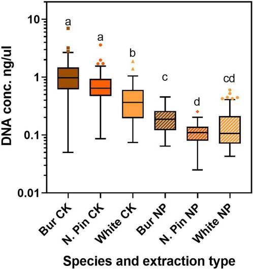
Thus, laboratory diagnosis is required for accurate diagnosis in      proprietary extraction protocol of a CK commonly used for
many cases, particularly if disease control action is planned. Suc-   nested PCR based diagnosis of oak wilt in the laboratory. An
cess in oak wilt management is greatest when control actions are      alternative method for DNA purification and concentration in
taken soon after early detection and timely diagnosis occur (Juzwik   addition to a previously published NaOH protocol also was
et al. 2011).                                                         developed for comparison. The main objective of this study was
   Due to the spotty or discontinuous colonization pattern of the     to compare detection rates of B. fagacearum in red and white
pathogen in white oak species, the University of Minnesota Plant      oak species using the current standard molecular protocol (DNA
Disease Clinic often assays two times the number of sapwood           extraction and nested PCR amplification using a CK) with those
chips from bur and white oak samples compared with red oak            obtained using DNA extracted following two NaOH protocols
ones when conducting traditional isolation assays (J. Flynn, per-     and then subjected to nested PCR amplification using the same
sonal communication). To maximize chances of successful               CK. The standard isolation protocol for B. fagacearum was
detection and avoid false-negative results, arborists and others      included for further comparison. The second objective was to
are advised to remove several symptomatic branches from a sus-        compare the amount of total DNA obtained by each extraction
pected tree and cut several segments from each branch for sub-        method.
mission to a diagnostic laboratory (Pokorny 2015; Yang and
Juzwik 2017).                                                         Materials and Methods
   Currently, laboratory diagnosis of oak wilt is based on results       Sampling sites and branch sampling protocols. In early
from standard pathogen isolation (Pokorny 2015) or molecular          September 2018, one location near Stacy, MN, in the Carlos
assays. Nested and real-time PCR protocols with B. fagacearum-        Avery Wildlife Refuge, with actively wilting northern pin oak
specific primers were first developed by Wu et al. (2011). These      (Quercus ellipsoidalis) was selected for sampling. Three
protocols were recently modified and evaluated by Yang and            branches (3.0 to 7.0 cm in diameter) were cut from each of four
Juzwik (2017). At least one commercial diagnostic laboratory          trees exhibiting classic oak wilt symptoms (leaf discoloration
also uses proprietary molecular procedures for processing sus-        and xylem staining in branches) per the protocol of Yang and
pect oak wilt samples (Research Associates Laboratory, Allen,         Juzwik (2017). Nonsymptomatic branches from healthy northern
TX, U.S.A.). Commonly used molecular tests (arguably, the cur-        pin oak were collected to use as controls. Four segments
rent “gold standard”) involve extraction and purification of DNA      (approximately 30 cm long) were cut from each branch, placed
followed by PCR amplification of the DNA using primers from           in plastic bags, stored on ice during transport to the laboratory,
a selected gene region. The PCR product is then visualized using      and placed in cold storage (4 C).
gel electrophoresis protocols. The nested PCR protocol reported          Between mid-July and early September 2019, multiple white
by Yang and Juzwik (2017) involves extraction of DNA from             oak (Q. alba) trees with scattered branches exhibiting foliar
sapwood drill shavings taken from actively wilting branch sam-        symptoms typical of oak wilt and located in street and park set-
ples or lower stem sections of completely wilted trees. The           tings were selected for sampling from seven trees on several
amplification step is a two-part process starting with the general    sites in Eagan, Apple Valley, and Minneapolis, MN. Between
fungal primer pair internal transcribed spacer (ITS)1F/ITS4 and       mid-June late August 2020, bur oak (Q. macrocarpa) branches
followed by further amplification with species-specific primers       were obtained from six bur oak trees in Stacy, St. Paul, and
(CF01 and CF02) developed by Wu et al. (2011). Resulting              Becker, MN. Nonsymptomatic branches from healthy bur and
bands for B. fagacearum appear at approximately 280 bp on the         white oak were sampled to use as controls. Segments (approxi-
gel. DNA extraction can be completed using a commercially             mately 30 cm long) were obtained from each harvested branch
available kit (CK) that contains a proprietary solution for PCR       and handled and stored as described above.
inhibitors in plant or soil extracts (QIAmp DNA Stool Kit; Qia-          Branch sample processing and standard isolation protocols. In
gen, Venlo, The Netherlands). The total time required for sample      the laboratory, the bark was carefully peeled from each segment
processing (from obtaining drill shavings through gel visualiza-      with a sterile drawknife to reveal the presence of outer xylem
tion) is approximately 2 days. We estimate that the materials         staining. In a laminar flow hood, four or five small wood chips
cost for using the CK for DNA extraction is U.S.$6.47 per sam-        were excised and placed on two 100-mm-diameter Petri plates
ple. Previously published methods involving use of a strong base      containing oak wilt identification agar (Barnett 1953) per Yang
(sodium hydroxide [NaOH]) to extract DNA from plant samples           and Juzwik (2017) protocols. The agar plates were incubated for
could potentially be used to modify the above-referenced nested       14 days at room temperature (approximately 24 C) under ambi-
PCR protocol to reduce both cost and time for processing sam-         ent lighting and checked daily after 7 days for the presence of
ples in plant diagnostic laboratories. In a comparative study of      suspected colonies. Subcultures were made as necessary onto
rapid and dependable methods for extracting DNA from environ-         half-strength potato dextrose agar plates to obtain pure cultures.
mental samples, Osmundson et al. (2013) found that NaOH               Resulting isolates of B. fagacearum were identified based on col-
extraction protocols of Wang et al. (1993) worked well in terms       ony morphology, characteristic odor, and presence of endoconi-
of detection success rate, cost, simplicity, and speed. The NaOH      dia. Isolation was also attempted from branches cut from healthy
methods, as described by Wang et al. (1993) and Xin et al.            oak trees. All branch samples from northern pin oak were proc-
(2003), use NaOH to extract DNA from plant samples, then neu-         essed within 7 days of collection, while samples from bur and
tralize it with Tris buffer. The obtained DNA is then amplified       white oak were processed within 12 days of branch harvest.
using a typical PCR procedure with additives to suppress any          Only those trees which produced B. fagacearum-positive cultures
PCR inhibitors carried from the extract. However, Osmundson           (four trees of each species) were used in the DNA extraction
et al. (2013) offered several cautions when using NaOH extrac-        phase of the study.
tion, particularly for substrates with low amounts of the target         At the same time that the above isolations were performed, drill
fungus present or that are rich in lignin or humic acids.             shaving samples were collected for DNA-based assays. Shavings
   The purpose of this research was to evaluate the potential for     were obtained using previously published protocols (Yang and
substituting an NaOH-based DNA extraction protocol for the            Juzwik 2017). Approximately 2 ml of shavings was placed in each

                                      PLANT HEALTH PROGRESS 䉬 XXXX, Vol. XX, No. X 䉬 Page 2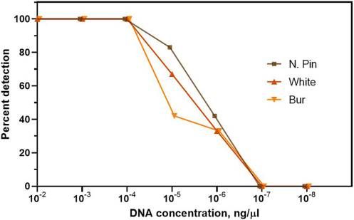
of two 2-ml microcentrifuge tubes per segment. When branches NaOH crude protocol from sapwood shavings from healthy
were too small for making drill shavings, thin strips of outer sap- northern pin, bur, and white oak branches). The extracted patho-
wood exhibiting staining characteristic of oak wilt infection were gen DNA for water controls (n = 4) and for the pathogen DNA
shaved and then cut into small pieces. All tubes were stored at suspended in crude pathogen-free DNA (n = 12) were subjected
−20 C until DNA was extracted. to nested PCR amplification and gel visualization as described
DNA extraction. Two general approaches were used to extract below.
DNA from the drill shaving samples: (i) the protocol of a CK Nested PCR and DNA sequencing. The DNA extracts ob-
(QIAmp DNA Stool Kit; Qiagen) and (ii) modified versions of tained (CK, NaOH crude, and NaOH purified) were subjected to
published protocols using NaOH (Lemke et al. 2011; Xin nested PCR as per the protocol described by Yang and Juzwik
et al. 2003). (2017), with two modifications. The PCR mixture was modified
For extractions using the kit, samples were processed as by adding 0.1% bovine serum albumen (BSA) and 1% polyvinyl
described by Yang and Juzwik (2017). In short, drill shavings pyrroliodone-40 (PVP) (Sigma-Aldrich, St. Louis, MO, U.S.A.),
(approximately 110 mg) were placed in a 2-ml microcentrifuge as described by Xin et al. (2003). These additions were used to
tube with two sterilized metal beads (4.5 mm in diameter). Lysis increase efficiency of amplification in the PCR assays using crude
buffer (750 ll) from the kit was added and the sample was DNA extracts obtained from pine needles and cotton leaves in a
homogenized, then heated at 70 C for 5 min. Extraction of DNA previously published report (Xin et al. 2003). In each full-plate
was then completed according to the manufacturer’s instructions reaction, PCR was performed with two negative (water) controls
for the kit (mixing with ethanol, spin filtering, washing, and elut- that lacked template DNA and one positive control with DNA
ing), except that that buffer quantities were reduced to 75%. extracted from known B. fagacearum.
For extraction of “crude” DNA using NaOH, drill shavings For all experiments, products from the second round of PCR
(approximately 110 mg) were placed in a microcentrifuge tube were visualized on 1.5% agarose gels with ethidium bromide
and 1.0 ml of 0.5 N NaOH (buffer A as described by Xin et al. staining. A nested PCR product was deemed positive if it pro-
[2003]) was added to sufficiently immerse the shavings (propor- duced a gel band of 280 bp (i.e., amplicon size). In general, the
tion used was 1 ml of NaOH to 110 mg of wood shavings). Vig- PCR assays were performed in two technical replicates (i.e., two
orous agitation with a vortex mixer was performed for 2 min, PCR assays with the same batch of extracted DNA). A sample
then several times for 2 to 3 s during 10 min of soaking in was deemed positive if either one or both samples produced the
NaOH. The mixture was then centrifuged (13,000 rpm for appropriate gel band. This approach has been used for other
2 min) and the resulting supernatant was mixed with 100 mM PCR-based diagnostic tests (Parra et al. 2020; Pilotti et al. 2012).
Tris-HCl (pH 4.0) (buffer B as described by Xin et al. [2003]) at Representatives of amplicon size-based “positive” samples were
a ratio of 1 to 9 (supernatant to buffer). The DNA extract confirmed as pathogen positive via DNA sequencing using pro-
obtained in this manner is referred to as “NaOH crude.” tocols described by Yang and Juzwik (2017). Obtained sequen-
To obtain a purified and concentrated DNA extract, the NaOH ces were evaluated for quality and trimmed as appropriate.
crude extract was subjected to a spin filter step that is similar to BLASTn searches in GenBank (https://blast.ncbi.nlm.nih.gov/
that used with the CK. Specifically, two volumes of the crude Blast.cgi) were performed with the resultant sequences to iden-
extract (1,000 ll) were mixed with one volume of absolute etha- tify B. fagacearum based on closely matched fungal accessions
nol (500 ll) and added to a mini-spin column (Econospin; Epoch in the database (using 98% or higher identity for sequences
Life Sciences, Missouri City, TX, U.S.A.). The column was greater than 200 bp).
spun at 8,000 rpm for 1 min followed by two rinses with a wash- Statistical analyses. A generalized mixed-effects model was used
ing buffer (“home-made AW2”) (described as washing buffer I to identify differences in the probability of a B. fagacearum-pos-
by Lemke et al. [2011]). After a 5-min drying period, 100 µl of itive PCR result between the three DNA extraction methods and
elution buffer (10 mM Tris and 0.5 mM EDTA, pH 8) was between different oak species. Branch segments were treated as
added to the column, incubated at room temperature for 2 min, the experimental unit. The mixed-effects model was fit using
and then spun (6,000 rpm for 1 min) to obtain the final DNA DNA extraction method and oak species as fixed effects and
extract. The DNA extract obtained in this manner is referred to with two random effects—one at the branch level and one at the
as “NaOH purified” This process resulted in a DNA extract that tree level—to account for correlation between segments sampled
was theoretically 20× more concentrated than that of the NaOH from the same branch or tree (Bates et al. 2020; R Core Team
crude extracts. 2019). Posthoc pairwise comparisons and Tukey’s honestly signifi-
Total concentration of extracted DNA was estimated for the cant difference (HSD) test were conducted using the emmeans
CK and the NaOH crude and purified extracts using a fluorome- packages in R to identify where detection rates differed between
ter (Qubit 3; Thermo Fisher Scientific, Hampton, NH, U.S.A.) species (Lenth 2019). A two-way analysis of variance (ANOVA)
following the manufacturer’s instructions. A subset was also ana- of transformed data (log10) was conducted to examine the effect of
lyzed by a microvolume spectrophotometer (NanoDrop 2000; extraction method (CK and NaOH purified only) and oak species
Thermo Fisher Scientific, Waltham, MA, U.S.A.) for both DNA on the concentration of extracted DNA (R Core Team 2019).
concentration and purity calculations (ratios of absorbance at
260 and 28 nm [A260/A280] and 260 and 230 nm [A260/A230]). Results
The limit of detection of B. fagacearum DNA present in Pathogen detection by standard isolation method. For actively
extracted DNA samples from oak were determined using a dilu- wilting northern pin oak trees (n = 4), B. fagacearum was iso-
tion series. For the positive control, a series was made using lated from 90% of assayed branch segments (n = 48) using stan-
pathogen DNA extract from a pure culture and obtained using dard pathogen isolation techniques (Table 1). In comparison,
the QIAmp Stool Kit, standardized for DNA concentration, and 40% of branch segments (n = 48) from 12 branches of actively
added in serial dilution to molecular-grade water. An equivalent wilting bur oak trees (n = 4) and 54% of segments (n = 48) from
dilution series was made by adding the pure pathogen DNA 12 branches of wilting white oak (n = 4) yielded the fungus. Dif-
extract to a crude background solution (DNA extracted using the ferences among oak species were significant based on glm model
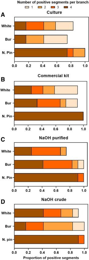
PLANT HEALTH PROGRESS 䉬 XXXX, Vol. XX, No. X 䉬 Page 3analysis (P = 0.0098). When the proportion of positive segments HSD lower and upper confidence limits of −0.56 and 2.42,
is viewed graphically, the frequency of four segments per branch respectively, for bur oak; −0.93 and 2.09 for white oak; and 2.54
yielding the pathogen was high (75%) for northern pin compared and 7.62 for northern pin oak). Based on graphical presentation
with similar, low frequency (17%) of this occurrence in bur and of branch segment results (four per branch), lower frequencies of
white oak (Fig. 1A). positive segments per branch were found for bur and white oak
Pathogen detection by nested PCR using different DNA compared with northern pin regardless of DNA extraction
extraction methods. For the three extraction methods evalu- method used for the nested PCR assay (Fig. 1B to D).
ated, resulting DNA was subjected to the same nested PCR pro- Total DNA obtained. Total DNA obtained from the drill shav-
tocol as described by Yang and Juzwik (2017), with the addition ings using the CK, NaOH crude, and NaOH purified extraction
of BSA and PVP. For DNA extraction completed using the CK, methods were compared using fluorometric analysis. However,
100% of the assayed northern pin oak branch segments (n = 48) inconsistent results were obtained with the crude DNA extracts
were positive for the pathogen based on the presence of a 280- and are not included in this report. When the concentration data
bp band on agarose gels produced from PCR products. In addi- were analyzed by both species and extraction protocol (two-factor
tion, all were positive on each of two technical PCR replicates. ANOVA), extractions using the CK yielded higher mean concen-
In comparison, the pathogen detection rates as determined by the trations of total DNA than those from the NaOH purified protocol
CK were lower for bur oak (67%) and white oak (54%) com- for all three oak species (P < 0.0001) (Table 3). The overall mean
pared with the rate for northern pin oak (Table 1). Nested PCR DNA concentration using the CK was 0.81 ng/ll (standard devia-
results using DNA extracted via the NaOH crude and NaOH tion [SD] = 0.511), while the mean concentration using NaOH
purified protocols varied by oak species. A high pathogen detec- purified was 0.16 ng/ll (SD = 0.109). Interactions between factors
tion rate (98%) was obtained for 48 branch segments from north- were significant. DNA concentrations were lower for white oak
ern pin oak by either method, while lower rates were found for samples than those from bur and northern pin oak with the CK
bur (58 and 79%) and white oak (71 and 62%) of 48 assayed extractions (Fig. 3). Northern pin oak extractions obtained using
samples per oak species by the crude or purified method, respec- the NaOH purified protocol yielded lower DNA concentrations
tively (Table 1). A representative gel (Fig. 2) using nested PCR than bur oak.
products of the three different bur oak extractions shows the typ- Concentrations were within the published sensitivity limits for
ical scattered pattern of positive samples. No differences in the the fluorimeter high-sensitivity assay (lower detection limit 0.01
likelihood of a positive detection by PCR were found among the ng/ll; Qubit 3) but were generally below the threshold for the
different DNA extraction methods based on model estimates microvolume spectrophotometer (lower detection limit 2 ng/ll;
(P = 0.361) (Table 2). However, the likelihood of such detection Nanodrop 2000). Therefore, any attempt to determine DNA
did differ by oak species; for example, for northern pin oak com- purity with the later instrument from A260/A280 or A260/A230
pared with bur oak (P < 0.001). Molecular assay-based detection ratios would be unreliable.
rates for white and bur oak samples were similar and signifi- Calculating limit of detection. Because pathogen DNA con-
cantly lower than detection rates for northern pin oak based on centration in the crude extract could not be determined by the
P values resulting from posthoc means comparisons (Tukey’s instruments available, the limit of B. fagacearum DNA detection
TABLE 1
Detection of Bretziella fagacearum in branches from actively wilting crowns of northern pin, bur, and white oak trees using
standard isolation and nested PCR amplification of extracted DNA obtained by using three different protocolsa
Branch segmentsc Branch leveld
b
Oak species, detection approach Method Assayed Positive Assayed Positive
Northern pin
Isolation – 48 43 12 12
PCR Commercial kit 48 48 12 12
PCR NaOH purified 48 47 12 12
PCR NaOH crude 48 47 12 12
Bur
Isolation – 48 19 12 9
PCR Commercial kit 48 32 12 11
PCR NaOH purified 48 38 12 12
PCR NaOH crude 48 28 12 12
White
Isolation – 48 26 12 10
PCR Commercial kit 48 26 12 11
PCR NaOH purified 48 30 12 9
PCR NaOH crude 48 34 12 11
a
Data shown are total number branch segments assayed and number found to be positive, as numbers for when segment data were compiled by
branch; − indicates not applicable.
b
DNA extraction methods. Commercial kit = QIAmp DNA Stool Kit, Qiagen; NaOH purified and NaOH crude extractions of DNA are based on
modifications of protocol by Xin et al. (2003) and Lemke et al. (2011).
c
PCR positive based on results of two technical replicate PCR runs.
d
Results of branch segment assays were composited by branch.
PLANT HEALTH PROGRESS 䉬 XXXX, Vol. XX, No. X 䉬 Page 4was estimated by adding pathogen DNA (obtained from pure
cultures using the CK method) in a dilution series utilizing crude
pathogen-free extract as a background. The latter was obtained
using the NaOH protocol with drill shavings obtained from oak-
wilt-free northern pin, bur, or white oak branches. The lower
limit (approximately 50% detection) found was pathogen DNA
at 10−6 ng/µl, with no detection occurring at 10−7 ng/µl based on
nested PCR amplification (12 replicates/concentration) and gel
visualization. Similar data were obtained for all three species of
wood extracts (Fig. 4).
Discussion
In general, substitution of either the crude NaOH or purified
NaOH protocol for DNA extraction in the nested PCR procedure
performed as well as or better than the CK DNA extraction
method in detecting B. fagacearum (i.e., success rate) in actively
wilting branches of a red oak species (northern pin oak) and two
white oak group species (bur oak and white oak). This is compa-
rable with results reported by others who have used a similar
NaOH extraction technique. Osmundson et al. (2013) were suc-
cessful in extracting and identifying DNA from lyophilized cul-
tures and sporocarps, and for amplification of microsatellite loci.
In fact, in several applications, the NaOH method performed as
well or better than traditional cetyltrimethylammonium bromide
methods.
Our detection results for northern pin oak samples were better
than or similar to those reported by Yang and Juzwik (2017) for
northern pin oak or northern red oak for nested PCR based on
proportion of B. fagacearum-positive branch segments derived
from total number of segments assayed from a branch. Using
this same measure of detection frequency, our results were worse
than those of Yang and Juzwik (2017) for CK use for DNA
extraction for both bur and white oak in our study. These differ-
ences may be due to (i) the high variability of distribution of the
fungus in the white and bur oak samples used in both studies or
(ii) the skill and experience of the individual taking the drill
shaving subsamples in the earlier study compared with the cur-
rent one. For best results, drill shavings should be obtained from
portions of the exposed xylem exhibiting characteristic vascular
staining characteristic of B. fagacearum colonization.
The costs for performing the two NaOH extraction methods in
this study were estimated to be $0.62/sample for NaOH crude
and $1.60/sample for NaOH purified. This represents a reduction
in cost when compared with that for the CK used (i.e., estimated
to be $6.47/sample). Additionally, time and cost savings of
NaOH protocol use with single or small numbers of samples
would not be as great as for high-throughput scenarios such as
those developed by Lemke et al. (2011) and Xin et al. (2003).
The NaOH procedure has been recommended by other researchers
for barcoding, genotyping, and disease diagnostics (Osmundson
et al. 2013) because of its speed, economy, and waste reduction.
Waste reduction is reflected in the number of tubes needed (only
one transfer is needed for the crude extract) and the lower amount
of toxic wastes compared with traditional chloroform or phenol
extractions. The potential also exists for our DNA extraction meth-
ods to be coupled with a PCR alternative procedure (e.g., gold
nanoparticle enhanced chemiluminescence) (Singh et al. 2017),
FIGURE 1 and the total amount of time to assay a small number of samples is
Detection frequencies for Bretziella fagacearum in branches of actively wilt- reduced to less than 1 h in the laboratory.
ing northern pin, bur, and white oak. Numbers of positive branch segments One potential disadvantage of the NaOH crude extract is the
per branch of each oak species are shown for A, a standard isolation assay, presence of potential PCR inhibitors in the extract. The addition
and for molecular PCR assays using three DNA extractions methods: B, of BSA and PVP to the nested PCR mixture used in our studies
commercial kit; C, purified NaOH protocol; and D, crude NaOH protocol. did result in improved sharpness and brightness of bands on
PLANT HEALTH PROGRESS 䉬 XXXX, Vol. XX, No. X 䉬 Page 5agarose gels during visualization of the nested PCR products in Another potential disadvantage of the NaOH procedure is the
all cases. Their use with DNA from the crude NaOH protocol low DNA concentration obtained. The amount of total DNA
made further purification (i.e., NaOH purified protocol) unneces- obtained using either NaOH protocol was less than that obtained
sary in the case of northern pin and white oak. Xin et al. (2003) using the CK; however, the NaOH-obtained DNA was sufficient
reported more efficient amplification of PCR mixtures in their to give generally comparable results in terms of success (i.e.,
work with crude DNA preparations from cotton leaves and from detection of B. fagacearum DNA following PCR amplification).
pine needles. Koonjul et al. (1999) suggested that BSA and PVP Crude extracts tended to have a light-brown or orange color, and
suppress certain substances in wood extracts (e.g., tannins and assays performed by the fluorimeter were inconsistent and proba-
other polyphenols) that inhibit PCR assays. BSA but not PVP bly reflected contaminants and not true DNA concentrations.
was used in the protocol evaluated by Osmundson et al. (2013) DNA concentrations below those detectable by the microvolume
for multiple substrates. spectrophotometer hampered our ability to address any DNA
FIGURE 2
Agarose gel image showing results of nested PCR amplification of DNA extracted from bur oak using three different methods: CK = commercial kit, NP =
NaOH purified, and NCrude = NaOH crude extract. Lane L: molecular size marker, 100 bp per line; lanes 1 to 12: DNA extracts from individual bur oak branch
segments, each column from the same segment sample; lane 13: positive control, diluted Bretziella fagacearum DNA; lanes 14 and 15: negative control
extract from known healthy bur oak wood; and lane 16: water control.
TABLE 2
Generalized linear mixed effects model of the interactions of actively wilting branches of northern pin, bur and white oak
trees and DNA extraction methods used for nested PCR detection of Bretziella fagacearuma
Variable Level Estimate SE Z value P value
Intercept 0.8996 0.6545 1.375 0.1693
DNA extraction NaOH purified 0.3255 0.3569 0.912 0.3618
Commercial kit −0.2243 0.3433 −0.653 0.5137
Oak species Northern pin 4.1502 1.2261 3.385 0.0007
White −0.3569 0.8868 −0.402 0.6873
a
Extraction method NaOH crude and bur oak species are the reference levels. NaOH extraction were methods based on modification of protocols
by Xin et al. (2003) and Lemke et al. (2011). Commercial kit = QIAmp DNA Stool Kit, Qiagen; SE = standard error.
TABLE 3
F test for fixed effects from analysis of variance of DNA concentration determined using two DNA extraction methods on
sapwood drill shavings from Bretziella fagacearum infected northern pin, bur, and white oak treesa
Fixed effects dfb Mean square F Pr (> F)
Oak species 2 2.165 24.39quality and quantity questions. Nevertheless, the nested PCR Statistically, the frequencies of pathogen detection using DNA
technique allows one to start with a mixture containing minute extracted by the CK were no different than those obtained with
quantities of target DNA, amplify the general fungal ITS DNA first, NaOH purified DNA extracts for three oak species. Based on results
then amplify the specific target DNA for successful detection. of our limit of detection investigation, the lowest pathogen detection
was found with extracts containing pathogen DNA at 10−6 ng/ll.
Therefore, by inference, it is likely that the DNA present in most
positive samples was 10−6 ng/ll or greater. We have occasionally
encountered bur oak crude extract samples that required four techni-
cal replications of PCR runs in order to detect a positive sample
(unpublished data). This suggests that B. fagacearum DNA concen-
tration of those samples was probably at or below its lower level of
detection. We hypothesize that the NaOH crude protocol performs
best with oak sapwood samples with relatively high concentrations
of the target B. fagacearum DNA concentrations.
Conclusions and Significance
In summary, substituting NaOH-based DNA extraction proto-
cols for those in a CK resulted in similar detection rates for the
oak wilt fungus in sapwood samples of three oak species when
amplified using the same nested PCR protocols. Statistically, the
crude extracts for all three species performed as well as the
extracts that had been subjected to purification in spin filters.
The clear advantages are reductions in time, expense, and waste.
Potential disadvantage may be the limit of detection of the path-
ogen at very low concentrations, especially in the bur oak, or
potential PCR inhibitors in the crude extract, either of which
may be improved by the spin filter purification procedure and
addition of BSA and PVP to the PCR. The alternative DNA
extraction protocols may prove useful in future development of
improved diagnostic methods for B. fagacearum detection in
FIGURE 3 oak-wilt-suspect trees.
Box plots showing the concentration of total DNA obtained from Bretziella
fagacearum-colonized sapwood of northern pin, bur, and white oak Acknowledgments
branches using two DNA extraction protocols: commercial kit (CK) or NaOH We thank M. Mendez Carrejo and T. Garrison for their help with lab
purified (NP). DNA concentration was measured using a fluorometer (Qubit 3).
and field support on this project; P. Castillo for field support; the cities
of Eagan, Apple Valley, and Minneapolis for personnel and equipment;
Different letters above plot columns indicate statistical difference at P < 0.05
D. Samac for manuscript review and access to and guidance in use of
based on analysis of variance and Tukey’s method for means separation analy- the microvolume spectrophotometer in her laboratory; two anonymous
ses of log10-transformed data. reviewers for helpful comments; and The Institute for Research in Sta-
tistics at the University of Minnesota and, in particular, L. Liu, for help
with the design and analysis of the experiments for this study.
Literature Cited
Appel, D. N. 1995. The oak wilt enigma: Perspectives from the Texas
epidemic. Annu. Rev. Phytopathol. 33:103-118.
Barnett, H. L. 1953. Isolation and identification of the oak wilt fungus. W.
Va. Exp. Stn. Bull. 359T.
Bates, D., M€achler, M., and Walker, S. 2020. Fitting linear mixed-effects
models using lme4. J. Stat. Softw. 67:1-48.
Gould, A. B., and Lashomb, J. H. 2005. Bacterial leaf scorch of shade trees.
APSnet Features. https://www.apsnet.org/edcenter/apsnetfeatures/Pages/
BacterialLeafScorch.aspx
Haack, R. A., and Acciavatti, R. E. 1992. Twolined chestnut borer. USDA
Forest Service Forest Insect and Disease Leaflet 168.
Harrington, T. C., McNew, D., and Yun, H. Y. 2012. Bur oak blight, a new
disease on Quercus macrocarpa caused by Tubakia iowensis sp. nov.
Mycologia 104:79-92.
Jensen-Tracy, S., Kenaly, S., Hudler, G., Harrington, T., and Logue, C.
FIGURE 4 2009. First report of the oak wilt fungus, Ceratocystis fagacearum, in
Limit of detection via nested PCR of Bretziella fagacearum DNA in NaOH New York State. Plant Dis. 93:428.
crude extracts. Serial dilutions of pathogen DNA (extracted from pure cul- Juzwik, J., and Appel, D. N. 2016. Oak wilt. Pages 129-133 in: Diseases of
Trees in the Great Plains. Gen. Tech. Rep. RMRS-GTR-335. A. Bergdahl
ture using a commercial kit) were made in a matrix of NaOH crude extract
and A. Hill, tech. cords. USDA Forest Service, Rocky Mountain Research
of healthy northern pin, bur, and white oak wood samples. Percent detec- Station, Fort Collins, CO, U.S.A.
tion is based on number of positive gel bands from 12 replicates per con- Juzwik, J., Appel, D. N., MacDonald, W. L., and Burks, S. 2011. Challenges and
centration level. successes in managing oak wilt in the United States. Plant Dis. 95:888-900.
PLANT HEALTH PROGRESS 䉬 XXXX, Vol. XX, No. X 䉬 Page 7Koonjul, P. K., Brandt, W. F., Lindsey, G. G., and Farrant, J. M. 1999. Pilotti, M., Lumia, V., Di Lernia, G., and Brunetti, A. 2012. Development of
Inclusion of polyvinylpyrrolidone in the polymerase chain reaction real-time PCR for in wood-detection of Ceratocystis platani, the agent of
reverses the inhibitory effects of polyphenolic contamination of RNA. canker stain of Platanus spp. Eur. J. Plant Pathol. 134:61-79.
Nucleic Acids Res. 27:915-916. Pokorny, J. D. 2015. How to Recognize Common Diseases of Oaks in the
Lemke, L., Rex, M., Zyprian, E., and T€opfer, R. 2011. A simple, Midwest: A Quick Guide. NA-FR-01-15. USDA Forest Service,
inexpensive and environmentally friendly method for high throughput Northeastern Area State and Private Forestry, St. Paul, MN, U.S.A.
DNA extraction from grapevine (Vitis spp.). Vitis 50:7-10. R Core Team. 2019. R: A Language and Environment for Statistical
Lenth, R. 2019. emmeans: Estimated marginal means, aka least-square Computing. R Foundation for Statistical Computing, Vienna, Austria.
means. R package version 1.4. https://cran.r-project.org/web/packages/ http://www.R-project.org
emmeans Singh, R., Feltmeyer, A., Saiapina, O., Juzwik, J., Arenz, B., and Abbas, A.
New York State Department of Environmental Conservation. 2020. Oak wilt. 2017. Rapid and PCR-free DNA detection by nanoaggregation-enhanced
Division of Lands and Forests, Department of Environmental chemiluminescence. Sci. Rep. 7:14011.
Conservation, Albany, NY, U.S.A. https://www.dec.ny.gov/lands/46919. Wang, H., Qi, M., and Cutler, A. J. 1993. A simple method of preparing
html plant samples for PCR. Nucleic Acids Res. 21:4153-4154.
Osmundson, T. W., Eyre, C. A., Hayden, K. M., Dhillon, J., and Garbelotto, Wu, C. P., Chen, G. Y., Li, B., Su, H., An, Y. L., Zhen, S. Z., and Ye, J. R.
M. M. 2013. Back to basics: An evaluation of NaOH and alternative rapid 2011. Rapid and accurate detection of Ceratocystis fagacearum from
DNA extraction protocols for DNA barcoding, genotyping and disease stained wood and soil by nested and real-time PCR. For. Pathol. 41:15-21.
diagnostics from fungal and oomycete samples. Mol. Ecol. Resour. 13: Xin, Z., Velten, J. P., Oliver, M. J., and Burke, J. J. 2003. High-throughput
66-74. DNA extraction method suitable for PCR. Biotechniques 34:820-826.
Parra, P. P., Dantes, W., Sandford, A., de la Torre, C., Perez, J., Hadziabdic, Yang, A., and Juzwik, J. 2017. Use of nested and real-time PCR for the
D., Schaffer, B., and Gazis, R. 2020. Rapid detection of the laurel wilt detection of Ceratocystis fagacearum in the sapwood of diseased oak spe-
pathogen in sapwood of Lauraceae hosts. Plant Health Prog. 21:356-364. cies in Minnesota. Plant Dis. 101:480-486.
PLANT HEALTH PROGRESS 䉬 XXXX, Vol. XX, No. X 䉬 Page 8You can also read