Unusual intramuscular locations as a first presentation of hydatid cyst disease in children: a report of two cases - BMC Pediatrics
←
→
Page content transcription
If your browser does not render page correctly, please read the page content below
Khasawneh et al. BMC Pediatrics     (2021) 21:371
https://doi.org/10.1186/s12887-021-02843-5

 CASE REPORT                                                                                                                                        Open Access

Unusual intramuscular locations as a first
presentation of hydatid cyst disease in
children: a report of two cases
Ruba A. Khasawneh1* , Ziyad M. Mohaidat2, Rawand A. Khasawneh3, Sohaib B. Zoghoul4 and Yousef M. Henawi1

  Abstract
  Background: Hydatid disease is an endemic disease in many countries of the world including the Middle East. It
  mainly affects the liver and lungs. Intramuscular hydatid disease is rarely reported in children. Such uncommon
  localization of hydatid cyst may pose difficulties in the clinical and radiological diagnosis; hence affecting patient’s
  management and outcome even in endemic areas.
  Case presentation: We herein describe intramuscular hydatid cysts in 2 different children. The first case is a 5-year-
  old boy who presented with a painless palpable lump over the right lumbar paraspinal region. His history was
  remarkable for sheep contact. His laboratory results revealed a mild increase in white blood cell (WBC) count and C-
  reactive protein. The lesion showed typical features of a hydatid cyst on ultrasound. Further imaging including
  ultrasound of the abdomen and CT of the chest, abdomen, and pelvis showed infestation of the liver and lung as
  well. The lesions were resected surgically without complications. The patient received Albendazole preoperatively
  and after surgery for 3 months. No evidence of recurrence was seen during follow-up.
  The second case is a 6-year-old girl who presented with an incidental palpable lump in her left thigh during her
  hospital admission for recurrent meningitis. Ultrasound and MRI imaging were performed demonstrating a
  unilocular cystic lesion in the left proximal rectus femoris muscle. A provisional diagnosis of hematoma vs. myxoma
  was given. Biopsy was performed and yielded blood products only. The lesion was resected surgically with a
  postoperative diagnosis of hydatid cyst. Blood tests performed afterward showed a positive titer for Echinococcus.
  The patient received Albendazole for 3 months. No evidence of recurrence was seen during follow-up.
  Conclusions: Despite its rarity; skeletal muscle hydatid cyst should always be considered in the differential
  diagnosis of cystic muscle lesions in children in endemic areas even if imaging studies did not show any of the
  typical signs. This will improve patient outcome by preventing unnecessary cystic puncture which might lead to
  serious complications, such as anaphylaxis and local dissemination.
  Keywords: Hydatid cyst, Pediatric, Intramuscular, Paraspinal muscle, Thigh hydatid cyst, Case report

* Correspondence: rakhasawneh2@just.edu.jo
1
 Department of Diagnostic Radiology and Nuclear Medicine, Faculty of
Medicine, King Abdullah University Hospital, Jordan University of Science and
Technology, Irbid 22110, Jordan
Full list of author information is available at the end of the article

                                        © The Author(s). 2021 Open Access This article is licensed under a Creative Commons Attribution 4.0 International License,
                                        which permits use, sharing, adaptation, distribution and reproduction in any medium or format, as long as you give
                                        appropriate credit to the original author(s) and the source, provide a link to the Creative Commons licence, and indicate if
                                        changes were made. The images or other third party material in this article are included in the article's Creative Commons
                                        licence, unless indicated otherwise in a credit line to the material. If material is not included in the article's Creative Commons
                                        licence and your intended use is not permitted by statutory regulation or exceeds the permitted use, you will need to obtain
                                        permission directly from the copyright holder. To view a copy of this licence, visit http://creativecommons.org/licenses/by/4.0/.
                                        The Creative Commons Public Domain Dedication waiver (http://creativecommons.org/publicdomain/zero/1.0/) applies to the
                                        data made available in this article, unless otherwise stated in a credit line to the data.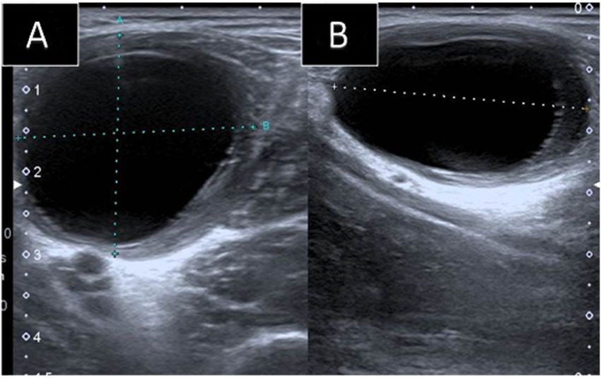
Khasawneh et al. BMC Pediatrics       (2021) 21:371                                                                                        Page 2 of 6

Background                                                                   within it (Fig. 1a). Multiple small cysts (daughter cysts)
Echinococcus Granulosus is a well-known tapeworm                             and hyperemia of its capsule on Doppler images were
causing hydatid disease which is an endemic and a com-                       also demonstrated (Fig. 1b). A provisional diagnosis of
mon public health problem in many countries of the                           intramuscular hydatid cyst was made. Further history re-
Middle East, Mediterranean region, Africa, Asia, South                       vealed that the patient had close contact with sheep. Ab-
America, and Australia [1–8]. Liver and lung hydatid                         dominal ultrasound showed multiple hydatid cysts in the
disease constitute the majority in affected patients [1, 3–                  liver. Computed Tomography (CT) scan of the chest, ab-
7, 9–17]. Still, hydatid disease can occur rarely in other                   domen, and pelvis re-demonstrated the liver (Fig. 2a)
viscera and much rarer in skeletal muscles [2, 3, 6].                        and right paraspinal muscle hydatid cysts. In addition, a
Intramuscular infestation can pose difficulties in the                       well-defined multi-loculated cystic lesion was seen at the
diagnosis and the management as it lacks the typical                         medial segment of the right middle lung lobe with an
clinical appearance especially if isolated [7]. Further-                     adjacent area of collapse consolidation, representing a
more, intramuscular hydatid cyst can mimic abscess,                          complicated hydatid cyst (Fig. 2b). Enhanced lumbar
hematoma, lymphatic malformation, synovial cyst as well                      spine Magnetic Resonance Imaging (MRI), was ordered
as necrotic malignant soft tissue mass lesions [18–20].                      for preoperative planning. It showed the right paraspinal
Scarce studies have reported intramuscular hydatid cyst                      hydatid cyst as a cystic lesion spanning the levels of L2
in children [7, 8, 15, 18, 21–24]. Herein we report rare                     to L3 vertebrae with a peripheral thick enhancing cap-
localizations of hydatid cyst disease in the paraspinal                      sule, containing Water Lilly sign and daughter cysts.
and proximal thigh muscles in two different children;                        There was a reactive enhancement of the involved mus-
one of whom presented with an isolated primary intra-                        cles as well (Fig. 3a, b). No deeper extension of the cyst
muscular hydatid cyst.                                                       was seen.
  The case reports in this study are presented in line                          Upon radiological diagnosis, the patient was started on
with the CARE criteria [25].                                                 Albendazole 170 mg for 1 week before surgery. Blood
                                                                             serology showed a negative antibody titer for the Echino-
Case (1) presentation                                                        coccus Granulosus (8.1) En-bloc surgical excision of the
A 5-year-old male presented to the pediatric surgery                         paraspinal cyst was performed. The liver and lung hyda-
clinic with a slowly growing painless lump in the right                      tid cysts were subsequently excised in two separate pro-
side of the lumbar region over 6-months. The lump was                        cedures through open laparotomy and thoracotomy. R
bothering him while walking and sitting. No history of                       vacuum drain was placed postoperatively for 3 days after
trauma at the site of the lump was recalled. The systemic                    each procedure. The patient received Albendazole (170
review was negative for any other complaints. On clin-                       mg) for 3 months postoperatively.
ical examination, the mass was firm with no elicitable                          Histopathology confirmed the diagnosis of hydatid cyst
tenderness. No overlying skin changes were noticed.                          disease. No recurrence was observed in the 16 months
  His blood tests were all normal except for a mild in-                      follow-up period.
crease in white blood cell count (WBC) (12 × 10*3/
mm*3) and C-reactive protein (48 mg/L). Ultrasound                           Case (2) presentation
exam showed a well-defined thick encapsulated cystic                         A 6-year-old female presented to the pediatric emer-
mass lesion in the right paraspinal muscle measuring                         gency room with a decrease in the level of conscious-
about 2.4 × 1.7 cm. Water Lilly sign membrane was seen                       ness, inability to walk, and fever. The past medical

 Fig. 1 Ultrasound exam of the right Paraspinal region. a: A well-defined thick encapsulated cystic lesion is seen in the right paraspinal muscle
 measuring about 2.4 × 1.7 cm with water Lilly sign (arrow) consistent with hydatid cyst, b Doppler ultrasound of the same lesion showing
 hyperemia of the capsule and the daughter cysts (arrow)
Khasawneh et al. BMC Pediatrics (2021) 21:371 Page 3 of 6 Fig. 2 Enhanced axial CT scan of the chest and abdomen. a: showing multiple liver cysts (arrows). b: showing a Multi-loculated cystic lesion in the medial segment of the right middle lung lobe with a surrounding area of collapse consolidation (arrow); representing complicated hydatid cyst history was remarkable for documented recurrent men- for this incidental isolated intramuscular cyst was ingitis secondary to dental problems. Physical examin- chronic hematoma versus intramuscular myxoma, des- ation showed positive meningeal signs. Her lab results pite its rarity in the pediatric age group. including lumbar puncture showed a new attack of acute A biopsy was advised. Aspirate of the cyst was per- meningitis. Enhanced brain MRI was performed and was formed and yielded blood cells only. The orthopedic sur- unremarkable. During her physical exam, a non-tender geon decided to proceed with wide local excision. The incidental lump was noted in the anterior aspect of her mass was excised through an anterior proximal thigh in- left proximal thigh. The mass was soft measuring about cision overlying the mass. After isolating the femoral 5 cm. The patient did not recall any trauma to the af- vessels, the mass was dissected out of the surrounding fected region. No discoloration of the overlying skin or tissues and excised without rupture. palpable lymphadenopathy was noted. Surprisingly, histopathology came back as an intra- Ultrasound showed a well-defined cystic lesion in the muscular hydatid cyst. No history of animal contact was left rectus femoris muscle, with acoustic enhancement found upon further questioning. In a retrospective re- measuring about 2.7 × 5.2 × 2.4 cm. The cyst abutted the view of the MRI images, a faint water Lilly sign mem- superficial femoral vessels. No internal vascularity, sur- brane on STIR images was recognized (Fig. 5a). Further rounding hyperemia, or soft tissue component was seen laboratory exams showed a positive antibody titer for (Fig. 4a, b). The rest of the rectus femoris muscle fibers the Echinococcus Granulosus (30.1). CT scan of the appeared edematous. A provisional diagnosis of chronic chest, abdomen, and pelvis did not reveal any other hematoma was given. Follow-up ultrasound after a organ infestation. The patient was started on oral Alben- month showed the same findings with no evidence of in- dazole (170) mg for 3 months. No recurrence was noted volution. Hence, an enhanced MRI of the left thigh was during the 2 years follow-up period. recommended to exclude other serious conditions. MRI revealed a well-defined oval-shaped intramuscular Discussion and conclusions homogenous cystic lesion with no perceptible wall en- Hydatid disease of the musculoskeletal system is rare hancement. (Fig. 5a, b). The given differential diagnosis with a reported incidence of about 1–5% [3, 5, 11, 12]. Fig. 3 MRI of the lumbar spine in the axial planes. a: Axial T2 weighted images demonstrating the right Paraspinal hydatid cyst with water Lilly sign (arrow). b: Axial T1 postcontrast MRI with fat sat showing thick enhancement of the cyst capsule (arrow)
Khasawneh et al. BMC Pediatrics       (2021) 21:371                                                                                        Page 4 of 6

 Fig. 4 Ultrasound images of the left thigh. a: Trans, b: longitudinal: Showing a well-defined homogenous cystic lesion in the left rectus femoris
 muscle with posterior acoustic enhancement seen

Primary intramuscular hydatid disease without liver and                      children (Table 1). Both of the cases presented here were
lung involvement is even rarer with an incidence of                          seen in the pediatric age group, adding to the challenge
about 2–3% [4, 10, 14]. Few cases of lower limb isolated                     in diagnosis and treatment.
intramuscular hydatid cysts were reported in the litera-                       Intramuscular hydatid disease is frequently asymptom-
ture in the pediatric age group (Table 1). None of the re-                   atic [15]. Painless slow-growing mass with normal over-
ported cases, however, were in the left rectus femoris                       lying skin is the most commonly reported complaint in
muscle as seen in this study (case (2) presentation)                         the literature [7, 10, 12, 13]. This is similar to the clinical
(Table 1). Paraspinal muscles are much more rarely af-                       presentation of the patients presented in this report.
fected in hydatid disease [14]. Herein, we also report an                      Imaging plays a major role in diagnosing intramuscu-
unusual localization of hydatid cyst in the right para-                      lar hydatid cysts when typical imaging features are
spinal muscle, which was not reported before in                              present. Ultrasonography is a major non-invasive tool to
pediatric patients (Fig. 1a, b and Fig. 3a, b). (Table 1).                   confirm the diagnosis of hydatid disease [7]. Typical
The reason behind the rare occurrence of hydatid cyst in                     ultrasound imaging features include the pathognomonic
skeletal muscles is their frequent contractility and high                    daughter cysts [16]. Double line sign is another charac-
lactic acid content [1, 3–5, 7, 9, 10, 13–16, 19].                           teristic sign in Ultrasound of hydatid cysts [7].
   The average age for most of the cases reported in the                       The CT appearance of the hydatid cyst is variable [5].
literature was adult to middle age groups [1]. Scarce                        Hydatid cyst on CT may mimic tumor if presented as a
cases of intramuscular hydatid cyst were reported in                         solid mass secondary to inflammatory changes [5].

 Fig. 5 MRI of the thighs in coronal planes. a: STIR MRI image showing a well-defined cystic lesion with faint water Lilly sign (arrow) seen in the
 left rectus femoris muscle. b: T1 postcontrast image with fat saturation showing no perceptible enhancement of the lesion
Khasawneh et al. BMC Pediatrics         (2021) 21:371                                                                                  Page 5 of 6

Table 1 Literature review of pediatric intramuscular hydatid cysts
Author                 Age• Sex         Location                      Presenting             Isolated vs.          Uniloculated vs.
                                                                      symptom                Multiple              Multiloculated.
Tekin et al [7]        10      Female Thigh M                         Painless mass          Isolated              NA
                       6       Female Thigh M                         Painless mass          Isolated              NA
Ghoroobi et al [15] 5          Male     Left Posterior distal thigh   Painless swelling      Isolated              Uniloculated
Atalar et al [8]       4       Female Left Vastus Medialis M          Painless mass          Isolated              Uniloculated
Erol et al [21]        11      Male     Right Medical Gastroc. M      Painless mass          Isolated              Uniloculated
Dudkiewez et al        14      Male     Right Vastus Medialis M       Painless mass          Multiple              NA
[22]
Duygulu et al [20]     8       Female Left Sartorius M                Painless mass          Isolated              Multiloculated
Landolsi et al [24]    8       Male     Left semitendinosus M         Painless mass          Isolated              Multiloculated
Kerimoglu et al        8       Female Left flexor halluces longus M   Painful swelling       Isolated              Multiloculated
[18]
Cankorkmaz et al       4       Female Between left adductor M and     Painless mass          Isolated              Uniloculated
[23]                                  iliopsoas M
M Muscle, Gastroc. Gastrocnemius, NA not available
•: Years

However, the presence of daughter cysts, germinal epi-                infestation, while it was positive in the second case al-
thelium detachment, and wall calcifications may confirm               though it was an isolated intramuscular hydatid cyst.
the diagnosis [7]. CT is also superior for the assessment               There are multiple treatment options for hydatid cyst.
of bone invasion by the cysts [7].                                    En bloc surgical resection is considered the treatment of
   MRI has a major diagnostic role in assessing the extent            choice with lower complications and recurrence rates [2,
of skeletal muscle infestation, exclude other possible eti-           4, 14]. Other Options include PAIR (percutaneous aspir-
ologies, and for surgical planning [1, 10, 14, 16]. MRI               ation injection-re-aspiration); which showed promising
can reveal a cystic mass containing daughter cysts, rim               results lately; medical treatment (Albendazole), and
sign, and Water Lilly sign [6, 17]. The rim sign is consid-           watch and wait [2]. Both patients in this series were
ered a characteristic sign in muscular hydatidosis [5, 8].            treated with en bloc surgical resection along with Alben-
It is defined as a low signal intensity rim seen around               dazole before and after surgery in the first case, while
the cyst, likely formed by the peri-cyst, most evident on             only after surgery in the second case, as the diagnosis
T2 weighted images (WI’s).                                            was revealed only after surgery.
   The diagnosis of hydatid cyst was made easily by im-                 In conclusion; pediatric intramuscular hydatid cyst is
aging in the first case despite its atypical clinical presen-         rare. A painless lump is the usual clinical presentation.
tation. Daughter cysts and Water Lilly sign were seen in              Radiology plays a crucial role in making the diagnosis of
all imaging modalities. The diagnosis was also supported              hydatid cyst when typical imaging features are seen. Sur-
by the patient’s positive history of sheep contact and co-            gery, and chemotherapy are the standard treatment op-
incident liver and lung cysts upon further imaging.                   tions. Atypical clinical and radiological findings may
   The diagnosis in the second case, however, was more                result in mismanagement of the patient leading to ser-
challenging. The possibility of an intramuscular hydatid              ious complications. Intramuscular hydatid cyst should be
cyst was not raised neither clinically nor radiologically.            kept in the differential diagnosis for any cystic muscular
Biopsy was also performed and was not diagnostic. The                 lesion in endemic areas.
final diagnosis came out only after surgical resection and
histopathological analysis. Its ultrasound imaging ap-                Abbreviations
                                                                      WBC: White Blood Cell; CT: Computed Tomography; MRI: Magnetic
pearance as a unilocular mostly homogenous cystic le-                 Resonance Imaging; STIR: Short Tau Inversion Recovery; WI’s: Weighted
sion (Fig. 4a, b) may reflect on the initial stage of                 images; IHA: Indirect Hemagglutination Assay; PAIR: Percutaneous Aspiration
parasitic infestation in muscles [5].                                 Injection Re-aspiration
   Serological tests, like the indirect hemagglutination
assay test (IHA), can be valuable in making the diagnosis             Supplementary Information
when they are positive [16]. Unfortunately half of the                The online version contains supplementary material available at https://doi.
primary skeletal muscle hydatidosis gives false -negative             org/10.1186/s12887-021-02843-5.

results in serology [8, 14, 16, 19]. The serology in the
                                                                       Additional file 1. Timeline for case 1.
first presented case was negative despite multi-organ
Khasawneh et al. BMC Pediatrics (2021) 21:371 Page 6 of 6
Additional file 2. Timeline for case 2. 7. Tekin R, Avci A, Tekin RC, Gem M, Cevik R. Hydatid cysts in muscles: clinical
manifestations, diagnosis, and management of this atypical presentation.
Rev Soc Bras Med Trop. 2015;48(5):594–8. https://doi.org/10.1590/0037-8682-
Acknowledgments 0197-2015.
Not applicable. 8. Atalar MH, Cankorkmaz L, Koyluoglu G, Salk I. Imaging characteristics of
three primary muscular hydatid cyst cases with various patterns. Kafkas J
Authors’ contributions Med Sci. 2012;2:74–6. https://doi.org/10.5505/kjms.2012.76486.
RK: Design of the work, acquisition of Data, wrote the manuscript, 9. Alaskar FA, Aldameg M. Hydatid disease with water lily sign manifesting as
corresponding author. ZM: Design of the work, wrote the manuscript, a soft-tissue mass in the soleus muscle of an adult woman : case report.
revision of the manuscript. RAK: Acquisition of Data, wrote the manuscript, Egypt J Hosp Med. 2018;71(3):2788–91. https://doi.org/10.12816/0045845.
revision of the manuscript. SZ: Acquisition of Data, wrote the manuscript, 10. Alatassi R, Koaban S, Alshayie M, Almogbil I. Solitary hydatid cyst in the
revision of the manuscript. YH: Acquisition of Data, wrote the manuscript, forearm: a case report. Int J Surg Case Rep. 2018;51:419–24. https://doi.org/1
revision of the manuscript. All authors have read and approved the final 0.1016/j.ijscr.2018.09.038.
manuscript. 11. Merkle EM, Schulte M, Vogel J, Tomczak R, Rieber A, Kern P, et al.
Musculoskeletal involvement in cystic echinococcosis: report of eight cases
and review of the literature. AJR Am J Roentgenol. 1997;168(6):1531–4.
Funding https://doi.org/10.2214/ajr.168.6.9168719.
No funding was obtained for this manuscript. 12. García-Alvarez F, Torcal J, Salinas JC, Navarro A, García-Alvarez I, Navarro-
Zorraquino M, et al. Musculoskeletal hydatid disease: a report of 13 cases.
Availability of data and materials Acta Orthop Scand. 2002;73(2):227–31. https://doi.org/10.1080/000164702
All data generated or analyzed during this study are included in this 753671858.
manuscript and its supplementary information files. 13. Geramizadeh B. Unusual locations of the hydatid cyst: a review from Iran.
Iran J Med Sci. 2013;38(1):2–14.
Declarations 14. El Harroudi T, Souadka A, Tijami F, El Otmany A, Jalil A. Primary paraspinal
hydatid cyst: a case report. Internet J Orthop Surg. 2012;13:13–6.
Ethics approval and consent to participate 15. Ghoroobi J, Mohajerzadeh L, Mirshemirani A, Mahdavi A. Primary hydatid
Ethics approval and consent to participate are waived for case reports per cyst of thigh: a case report in child. J Krishna Inst Med Sci Univ. 2017;6:111–
our Jordan University of Science and Technology institutional regulations. 3.
16. Durakbasa M, Kose O, Islam N, Kilicoglu G. A primary hydatid cyst of the
Consent for publication gracilis: a case report. J Orthop Surg. 2007;15(1):118–20. https://doi.org/10.11
Written informed consent was obtained from the patients’ parents for 77/230949900701500127.
publication of these case reports and accompanying images. Copies of the 17. Sener R, Calli C, Kitis O, Yalman O. Multiple, primary spinal–paraspinal
consent forms are available for review by the Editor-in-chief of this journal hydatid cysts. Eur Radiol. 2001;11(11):2314–6. https://doi.org/10.1007/s0033
upon request. 00000771.
18. Kerimoglu U, Kapicioglu S, Emlik D, Arazi M, Ural O. Case 161: hydatid
disease with water lily sign manifesting as a soft-tissue mass in the calf of a
Competing interests
child. Radiology. 2010;256(3):1007–10. https://doi.org/10.1148/radiol.10081
The authors declare that they have no competing interests.
066.
19. Adıyeke L, Cakır T, Duymus TM, Aydogmus S. Unexpected diagnosis in
Author details
1 gluteal region -a primary intramuscular hydatid cyst: a case report. J Orthop
Department of Diagnostic Radiology and Nuclear Medicine, Faculty of
Case Rep. 2018;8(2):104–6. https://doi.org/10.13107/jocr.2250-0685.1072.
Medicine, King Abdullah University Hospital, Jordan University of Science and
20. Duygulu F, Karaoǧlu S, Erdoǧan N, Yildiz O. Primary hydatid cyst of the
Technology, Irbid 22110, Jordan. 2Orthopedic Division, Special Surgery
thigh: a case report of an unusual localization. Turk J Pediatr. 2006;48(3):
Department, Faculty of Medicine, Jordan University of Science and
256–9.
Technology, Irbid 22110, Jordan. 3Department of Clinical Pharmacy, Faculty
21. Erol B, Tetik C, Altun E, Soysal A, Bakir M. Hydatid cyst presenting as a soft-
of Pharmacy, Jordan University of Science and Technology, Irbid 22110,
tissue calf mass in a child. Eur J Pediatr Surg. 2007;17(1):55–8. https://doi.
Jordan. 4Radiology Department, Hamad Medical Corporation (HMC), Doha,
org/10.1055/s-2007-964949.
Qatar 00000.
22. Dudkiewicz I, Salai M, Apter S. Hydatid cyst presenting as a soft-tissue thigh
mass in a child. Arch Orthop Trauma Surg. 1999;119(7-8):474–5. https://doi.
Received: 22 September 2020 Accepted: 17 August 2021
org/10.1007/s004020050025.
23. Cankorkmaz L, Ozturk H, Koyluoglu G, Atalar MH, Arslan MS. Intermuscular
hydatid cyst in a 4-year-old child: a case report. J Pediatr Surg. 2007;42(11):
References 1946–8. https://doi.org/10.1016/j.jpedsurg.2007.07.051.
1. Comert RB, Aydingoz U, Ucaner A, Arikan M. Water-lily sign on MR imaging 24. Maha L, Daib A, Hella Y, Boughdir M, Ben Abdallah R, Trabelsi F, et al. A rare
of primary intramuscular hydatidosis of sartorius muscle. Skelet Radiol. 2003; etiology of muscular mass in infant: a case report. J Heal Med Sci. 2019;2:
32(7):420–3. https://doi.org/10.1007/s00256-003-0661-x. 275–8.
2. Kurz K, Schwabegger A, Schreieck S, Zelger B, Weiss G, Bellmann-Weiler R. 25. Riley DS, Barber MS, Kienle GS, Aronson JK, von Schoen-Angerer T, Tugwell
Cystic echinococcosis in the thigh: a case report. Infection. 2019;47(2):323–9. P, et al. CARE guidelines for case reports: explanation and elaboration
https://doi.org/10.1007/s15010-018-1255-9. document. J Clin Epidemiol. 2017;89:218–35. https://doi.org/10.1016/j.
3. Sreeramulu PN. Krishnaprasad, Girish gowda SL. Gluteal region jclinepi.2017.04.026.
musculoskeletal hydatid cyst: case report and review of literature. Indian J
Surg. 2010;72(S1):302–5. https://doi.org/10.1007/s12262-010-0096-2.
4. Din DPMU, Anjum WA, Ahmad ML, Rehman KA, Ahmad GB, Gulshan WN. Publisher’s Note
Primary para-vertebral hydatid cyst in the sub-occipital area of the neck: an Springer Nature remains neutral with regard to jurisdictional claims in
unusual case of echinococcosis. Egypt J Neurosurg. 2018;33(1):16. https:// published maps and institutional affiliations.
doi.org/10.1186/s41984-018-0015-6.
5. García-Díez AI, Ros Mendoza LH, Villacampa VM, Cózar M, Fuertes MI. MRI
evaluation of soft tissue hydatid disease. Eur Radiol. 2000;10(3):462–6.
https://doi.org/10.1007/s003300050077.
6. Alam W, Shah F, Ali MA. Hydatid cystic disease of lumbar paraspinal
muscles. Biomed J Sci Tech Res. 2017;1(6). https://doi.org/10.26717/BJSTR.2
017.01.000495.You can also read