Unexpected occurrence of Aeromonas species in the cutaneous mucus of Barbour's seahorses (Hippocampus barbouri)
←
→
Page content transcription
If your browser does not render page correctly, please read the page content below
Unexpected occurrence of Aeromonas species in the cutaneous mucus of
Barbour's seahorses (Hippocampus barbouri)
Rose Chinly Mae Huerte Ortega ( rosechinlymae.ortega@g.msuiit.edu.ph )
MSU-Iligan Institute of Technology https://orcid.org/0000-0003-1176-2361
Sharon Rose Tabugo
MSU-IIT: MSU-Iligan Institute of Technology
Joey Genevieve Martinez
MSU-IIT: MSU-Iligan Institute of Technology
José Balcázar
University of Girona: Universitat de Girona
Short Report
Keywords: Aeromonas, Illumina sequencing, H. barbouri, seahorse
Posted Date: February 2nd, 2022
DOI: https://doi.org/10.21203/rs.3.rs-1294475/v1
License: This work is licensed under a Creative Commons Attribution 4.0 International License. Read Full License
Page 1/6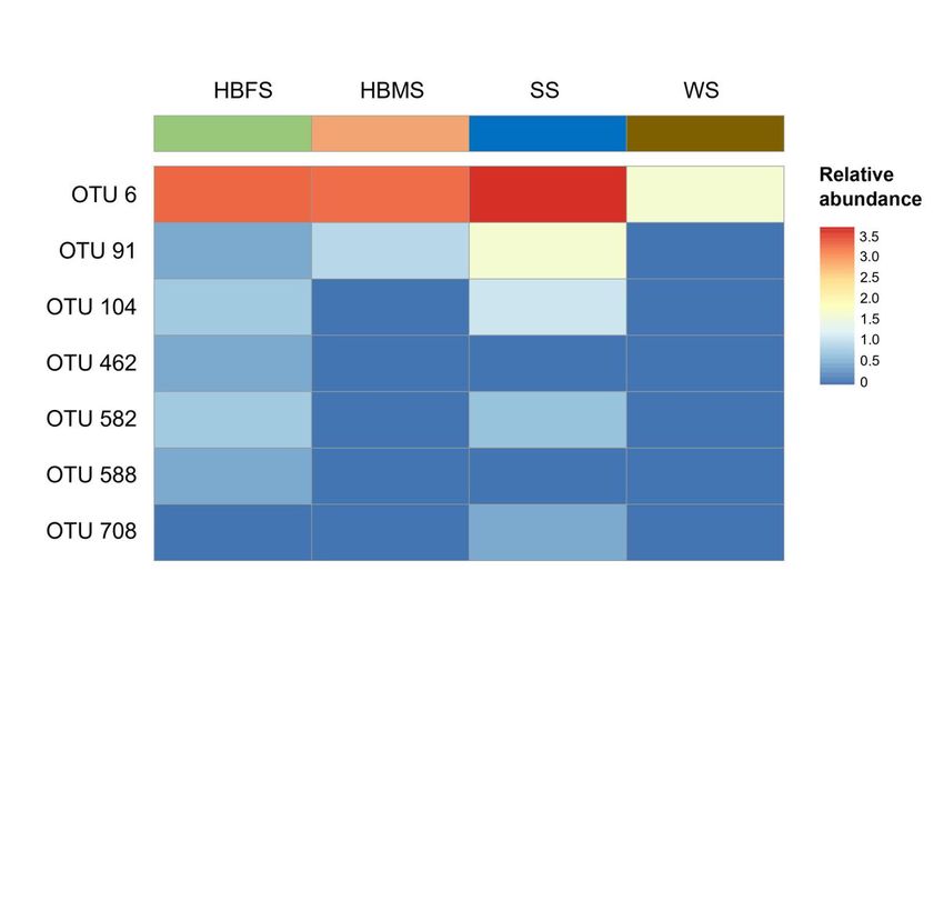
Abstract
Because the skin microbiota is valuable for elucidating health status and common disorders in the host, this study aimed to describe the bacterial community
composition of the skin of Barbour’s seahorse ( Hippocampus barbouri ) using high-throughput sequencing of 16S ribosomal RNA genes. Water and sediment
samples from the surrounding environment were also analyzed for comparative purposes. The results revealed the presence of operational taxonomic units
(OTUs) affiliated to the Aeromonas genus in the skin, whose abundance was slightly similar to the sediment samples. Given that some Aeromonas species
are known to be important pathogens in humans and fish, these results may provide information to show the dependency of the skin microbial composition
on the environment, as well as determine whether the presence of Aeromonas species has implications on seahorse health.

Introduction
Although seahorses (Hippocampus) play important roles in the ecosystem, they are constantly at risk because of trade for medicinal and ornamental
purposes (IUCN 2019). Moreover, seahorses are not exempt from diseases, which could make the situation even more critical. In fact, some studies have
reported skin diseases of diverse etiology in wild and farmed seahorses (Balcázar et al. 2010a, 2011; LePage et al. 2014). In a recent study carried out by our
research team, we observed the presence of Aeromonas in the cutaneous mucus of healthy Hippocampus barbouri (Ortega et al. 2021). It should be noted that
Aeromonas species are ubiquitous bacteria primarily recovered from aquatic ecosystems (Janda and Abbot 2010), as they are commonly found in freshwater
bodies, estuaries (Chaix et al. 2017), and even seawater (Chenia and Duma 2017). Despite that some Aeromonas species are potential pathogens of
fish (Vivekanandhan et al. 2005), there is limited information on the prevalence of these microorganisms in seahorses. Thanks to the implementation of
cutting-edge sequencing technologies, a full picture of bacterial community composition can be obtained, thereby increasing our knowledge on the role of skin
microbiota in health and disease. Here we used the Illumina sequencing technology to explore the bacterial community structure and composition within the
skin ecosystem of Barbour’s seahorses, with particular emphasis on members belonging to the Aeromonadaceae family.

Materials And Methods
Eleven healthy Barbour’s seahorses and samples from the surrounding environment (water and sediment) were collected from coral reefs off the coast of
Cantiasay Island, San Pedro, Surigao del Norte. Samples were immediately transported to the Oceanography Laboratory of the Premier Research Institute of
Science and Mathematics, Mindanao State University-Iligan Institute of Technology for further analysis. Seahorses were gently washed with sterile seawater
twice to remove debris without compromising the microbial community on the skin (Nurul et al. 2018). Skin mucus samples were then collected by scraping
the seahorse’s dorsal surface using a sterile swab (Balcazar et al. 2010b), which were placed on a 2 ml microcentrifuge tube and stored at -65℃ until DNA
extraction.

Genomic DNA was extracted from the skin microbial community of six female Barbour’s seahorses (HBFS) and five male Barbour’s seahorses (HBMS), as well
as from water (WS) and sediment (SS) samples. Samples were pooled according to their origin and HiPurA™ DNA Purification Kit (HIMEDIA; Mumbai, India)
was used for DNA extraction, according to the manufacturer’s instructions. Universal primers targeting the V1-V3 regions of the 16S rRNA gene were used for
PCR amplification, whose purified products were sent to Macrogen Inc. (Seoul, South Korea) for high-throughput sequencing on the Illumina MiSeq platform.
Raw data were processed using the Quantitative Insights Into Microbial Ecology (QIIME 2) pipeline (Bolyen et al. 2019). Operational taxonomic units (OTUs)
were defined at 99% sequence similarity of 16S rRNA genes. This value was used to define a core set of representative sequences, which were used for
phylogenetic analyses. The weighted UniFrac test was applied to determine whether two or more communities have the same structure (Lozupone et al. 2007).
A heatmap was also generated showing the relative abundance of OTUs assigned to the Aeromonadaceae family across the samples, which were classified
using the EzBioCloud database (Yoon et al. 2017). Phylogenetic analyses were performed by using MEGA version 6.0 (Tamura et al. 2013). Distances
(distance options according to the Kimura 2-parameter model) and clustering with the neighbor-joining method were determined by using bootstrap values for
1,000 replications.

Results And Discussion
After normalizing to avoid any bias due to the difference in the total number of sequences, bacterial community structure was analyzed using the weighted
UniFrac test (sensitive to abundances of taxa), whose results demonstrated that the relative abundance of OTUs (defined at 99% similarity) was significantly
different (p
consumption such as tilapia and salmonids (Figueras and Beaz-Hidalgo 2014), bivalve mollusks (Galbis et al. 2007), whereas other Aeromonas species have
been reported in clinical and environmental samples (Carnahan et al. 1991; Demarta et al. 2008; Alperi et al. 2010; Marti et al. 2015).

The relative abundance of each OTUs classified as Aeromonas species in the skin of H. barbouri was compared to sediment and water samples through a
heatmap (Fig. 2). The map showed that the abundance of Aeromonas species from the skin of H. barbouri was more related to the sediments compared to
water. This is consistent with the overall OTU representations. Aeromonas species detected in the skin of H. barbouri samples were greatly influenced by the
environment where they thrive. Based on Larsen et al. (2013), the structure and composition of the skin microbiota are likely to be impacted by several
variables including abiotic factors linked to geographic locality and season, as well as biotic factors related to the nutrient potential or antimicrobial
components of fish mucus. Given the interface influencing this skin microbiota, water and sediments were considered (Chiarello et al. 2015), and in this case,
the sediments had a higher influence on skin microbiota. Our results were also supported by recent findings, where the skin-microbiome assemblage of marine
organisms is strongly associated with the surrounding sediments (Kawamoto et al. 2021).

The genus Aeromonas is a member of the Aeromonadaceae family, which consists of Gram-negative bacteria commonly found in aquatic environments,
some of which are capable of causing disease in humans, fish, and other aquatic animals (Abu-Elala et al. 2015; Bravo and Figueras 2020). As inhabitants of
marine environments, fish and other seafood are the most common sources for isolating these microorganisms (Pessoa et al. 2019), supporting their
occurrence in seahorses. Moreover, Aeromonas spp. are known to be opportunistic pathogens for fish, and under stressful conditions such as an increase in
water temperature, poor water quality, excessive handling, etc., they can cause epidemic outbreaks (Beaz-Hidalgo et al. 2010). However, the severity of disease
cases depends on the concentration of these microorganisms (Hu et al. 2012). Some changes of the skin microbiota based on its phylogenetic composition
may affect its functions, thereby upsetting its homeostatic interactions with the host and eventually favoring disease development (Chiarello et al. 2015).
Considering that H. barbouri specimens in this study were directly collected from the wild with no trace of skin disease, they can be considered apparently
healthy. As a result, further research is needed to understand the differences in the composition of a healthy and diseased H. barbouri.

Conclusions
High-throughput sequencing technology revealed the presence of sequences affiliated to Aeromonas species in the skin of Hippocampus
barbouri. Interestingly, Aeromonas species were also observed in the sediments, which seem to be the most probable source of these species. Although
some Aeromonas species are known to be important pathogens, the presence of these species in this study may not have been affecting the health status of
Hippocampus barbouri yet. However, further studies are required to explore the implications of these species on seahorse health.

Declarations
Acknowledgement
The authors would like to thank DOST-ASTHRDP, BFAR, MSU-IIT PRISM, LGU Surigao and locals for supporting this study.

Author contributions
RCMHO: Conceptualization, methodology, investigation, formal analysis, visualization, writing - (both original draft and review and editing), funding
acquisition. SRMT: Conceptualization, methodology, supervision, funding acquisition. JGTM: Conceptualization, methodology, supervision. JLB:
Methodology, investigation, formal analysis, visualization, supervision, writing - (review and editing).

Funding
This study is funded by DOST under DOST-ASTHRDP scholarship grant to RCMHO.

Data availability
All sequences obtained in this study are available and will be submitted to GenBank at the NCBI.

Consent to Participate
Not applicable.

Consent for Publication
All authors consent to the publication of this article.

Conflict of Interest
The authors declare no competing interests.
                                                                             Page 3/6
Ethical Statement
Legal requirements from the BFAR national office under a project “Seahorses and Pipefishes with pharmaceutical potentials in Mindanao” was acquired and
granted and their guidelines for the care and use of animals have been followed.
References
1. Abu-Elala N, Abdelsalam M, Marouf S, Setta A (2015) Comparative analysis of virulence genes, antibiotic resistance and gyrB-based phylogeny of motile
Aeromonas species isolates from Nile tilapia and domestic fowl. Let Appl Microbiol 61(5):429–436. https://doi.org/10.1111/lam.12484
2. Alperi A, Martinez-Murcia AJ, Ko WC, Monera A, Saavedra MJ, Figueras MJ (2010) Aeromonas taiwanensis sp. nov. and Aeromonas sanarellii sp. nov.,
clinical species from Taiwan. Int J Syst Evol Microbiol 60:2048-2055. https://doi.org/10.1099/ijs.0.014621-0
3. Balcázar JL, Planas M, Pintado J (2011) Novel Mycobacterium species in seahorses with tail rot. Emerg Infect Dis 17:1770–1772.
https://doi.org/10.3201/eid1709.101289
4. Balcázar JL, Gallo-Bueno A, Planas M, Pintado J (2010a) Isolation of Vibrio alginolyticus and Vibrio splendidus from captive-bred seahorses with disease
symptoms. Antonie van Leeuwenhoek 97:207-210. https://doi.org/10.1007/s10482-009-9398-4
5. Balcázar J, Loureiro S, Da Silva Y, Pintado J, Planas M (2010b) Identification and characterization of bacteria with antibacterial activities isolated from
seahorses (Hippocampus guttulatus). J Antibiot 63:271–274. https://doi.org/10.1038/ja.2010.27
6. Beaz-Hidalgo R, Alperi A, Buja´n N, Romalde JL, Figueras MJ (2010) Comparison of phenotypical and genetic identification of Aeromonas strains isolated
from diseased fish. Syst Appl Microbiol 33(3):149-153. https://doi.org/10.1016/j.syapm.2010.02.002
7. Bolyen E, Rideout JR, Dillon MR et al (2019) Reproducible, interactive, scalable and extensible microbiome data science using QIIME 2. Nat Biotechnol
37:852–857. https://doi.org/10.1038/s41587-019-0209-9
8. Bravo A, Figueras MJ (2020). An Update on the Genus Aeromonas: Taxonomy, Epidemiology and Pathogenicity.
Microorganisms 8(1):129. https://doi.org/10.3390/microorganisms8010129
9. Carnahan A, Joseph SW (2005) Order XII. Aeromonadales ord. nov. In: Brenner D.J., Krieg N.R., Staley J.T., Garrity G.M., editors. Bergey’s Manual of
Systematic Bacteriology 2:556–578.
10. Carnahan A, Fanning GR, Joseph SW (1991) Aeromonas jandaei (formerly genospecies DNA group 9 A. sobria), a new sucrose-negative species isolated
from clinical specimens. J Clin Microbiol 29:560-564. https://doi.org/10.1128/jcm.29.3.560-564.1991
11. Chaix G, Roger F, Berthe T, Lamy B, Jumas-Bilak E, Lafite R, Forget-Leray J (2017). Distinct Aeromonas Populations in Water Column and Associated with
Copepods from Estuarine Environment (Seine, France). Front Microbiol 8:1259. https://doi.org/10.3389/fmicb.2017.01259
12. Chenia HY, Duma S (2017) Characterization of virulence, cell surface characteristics and biofilm-forming ability of Aeromonas spp. isolates from fish and
sea water. J Fish Dis 40:339–350. https://doi.org/10.1111/jfd.12516
13. Chiarello M, Villéger S, Bouvier C, Bettarel Y, Bouvier T (2015). High diversity of skin-associated bacterial communities of marine fishes is promoted by
their high variability among body parts, individuals and species. FEMS Microbiol Eco 91(7): fiv061. https://doi.org/10.1093/femsec/fiv061
14. Demarta A, Küpfer M, Riegel P, Harf-Monteil C, Tonolla M, Peduzzi R, Monera A, Saavedra MJ, Martinez-Murcia A (2008) Aeromonas tecta sp. nov., isolated
from clinical and environmental sources. Syst Appl Microbiol 31:278-286. https://doi.org/10.1016/j.syapm.2008.04.005
15. Figueras M J, Beaz-Hidalgo R (2014) Aeromonas: Introduction. Encyclopedia of Food Microbiol 24–30. doi:10.1016/b978-0-12-384730-0.00004-5
16. Galbis D, Farfán M, Fusté MC, Lorén JG (2007) Aeromonas bivalvium sp. nov., isolated from bivalve molluscs. Int J Syst Evol Microbiol 57(3):582-
587. https://doi.org/10.1099/ijs.0.64497-0
17. Hu M, Wang N, Pan ZH, Lu CP, Liu YJ (2012) Identity and virulence properties of Aeromonas isolates from diseased fish, healthy controls and water
environment in China. Lett Appl Microbiol 55(3):224-233. https://doi.org/10.1111/j.1472-765x.2012.03281.x
18. IUCN Report (2019) Why care about seahorses? Seahorses, Pipefishes and Seadragon, Specialist
Group. https://static1.squarespace.com/static/5734f2a086db43e3f6fb1bbc/t/5d97c107cfcd403eddf462c1/1570226441051/Why+care+about+seahorse
Accessed 23 Oct 2021.
19. Janda J M, Abbott SL (2010) The genus Aeromonas: taxonomy, pathogenicity, and infection. Clin Microbiol Rev 23:35–73.
https://doi.org/10.1128/CMR.00039-09
20. Kawamoto Y, Kato H, Nagata Y, Urabe J (2021) Microbial communities developing within bulk sediments under fish carcasses on a tidal flat. PLOS ONE
16(2): e0247220. https://doi.org/10.1371/journal.pone.0247220
21. Larsen A, Tao Z, Bullard SA, Arias CR (2013) Diversity of the skin microbiota of fishes: evidence for host species specificity. FEMS Microbiol Ecol 85:483-
494 https://doi.org/10.1111/1574-6941.12136
22. LePage V, Young J, Dutton CJ, Crawshaw G, Pare JA, Kummrow M, McLelland DJ, Huber P, Young K, Russell S, Al-Hussinee L, Lumsden
JS (2014) Diseases of captive yellow seahorse Hippocampus kuda Bleeker, pot-bellied seahorse Hippocampus abdominalis Lesson and weedy
seadragon Phyllopteryx taeniolatus (Lacépède). J Fish Diseases 38(5):439–450. https://doi.org/10.1111/jfd.12254
23. Lozupone C, Knight R (2005) UniFrac: a new phylogenetic method for comparing microbial communities. Appl Environ Microbiol 71:8228–8235.
https://doi.org/10.1128/AEM.71.12.8228-8235.2005
24. Marti E, Balcázar JL (2015) Aeromonas rivipollensis sp. nov., a novel species isolated from aquatic samples. J Basic Microbiol 55(12):1435-9.
https://doi.org/10.1002/jobm.201500264.
Page 4/625. Nurul ANA, Muhammad DD, Okomoda VT, Nur AAB (2019) 16S rRNA-Based metagenomic analysis of microbial communities associated with wild
Labroides dimidiatus from Karah Island, Terengganu, Malaysia. Biotech Rep 21:e00303. https://doi.org/10.1016/j.btre.2019.e00303
26. Ortega RCMH, Tabugo SRM, Martinez JGT, Padasas CS, Balolong MP, Balcazar JL (2021) High-throughput sequencing-based analysis of bacterial
communities associated with Barbour's seahorses (Hippocampus barbouri) from Surigao del Norte, Philippines. Lett Appl Microbiol 73:280–285.
https://doi.org/10.1111/lam.13511
27. Pessoa RB, de Oliveira WF, Marques DSC, Dos Santos Correia MT, de Carvalho EVMM, Coelho LCBB (2019) The genus Aeromonas: A general approach.
Microb Pathog 130:81-94.https://doi.org/10.1016/j.micpath.2019.02.036
28. Tamura K, Stecher G, Peterson D, Filipski A, Kumar S (2013). MEGA6: Molecular Evolutionary Genetics Analysis version 6.0. Mol Biol Evol 30(12): 2725–
2729. doi: 10.1093/molbev/mst197
29. Vivekanandhan G, Hatha AAM, Lakshmanaperumalsamy P (2005) Prevalence of Aeromonas hydrophila in fish and prawns from the seafood market of
Coimbatore, South India. Food Microbiol 22:133–137.https://doi.org/10.1016/j.fm.2004.01.015
30. Yoon SH, Ha SM, Kwon S, Lim J, Kim Y, Seo H, Chun J (2017) Introducing EzBioCloud: a taxonomically united database of 16S rRNA gene sequences and
whole-genome assemblies. Int J Syst Evol Microbiol 67(5):1613-1617. https://doi.org/10.1099/ijsem.0.001755
Figures
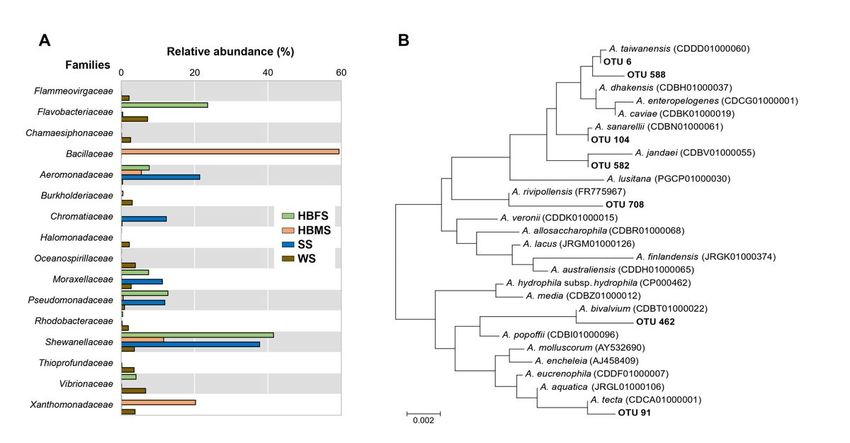
Figure 1
(A) Relative abundance of dominant bacterial families found in the skin samples from Barbour’s seahorses, as well as in water and sediment samples from
the surrounding environment. (B) Phylogenetic dendrogram of selected OTUs with the most closely related Aeromonas species, based on 16S rRNA gene
sequences and constructed by the neighbor-joining method. Bootstrap percentages (>50 %) based on 1000 replications are shown at branch nodes. Bar, 0.002
estimated substitutions per site.
Page 5/6Figure 2
Heatmap of OTUs assigned to the Aeromonadaceae family, which were compared with their abundances in each sample. The color intensity (log2 scale) in
each panel shows the percentage of each OTU.
Page 6/6You can also read