UC Davis UC Davis Previously Published Works
←
→
Page content transcription
If your browser does not render page correctly, please read the page content below
UC Davis
UC Davis Previously Published Works

Title
Chromoanagenesis from radiation-induced genome damage in Populus.

Permalink
https://escholarship.org/uc/item/4qk795dm

Journal
PLoS genetics, 17(8)

ISSN
1553-7390

Authors
Guo, Weier
Comai, Luca
Henry, Isabelle M

Publication Date
2021-08-25

DOI
10.1371/journal.pgen.1009735

Peer reviewed

 eScholarship.org                             Powered by the California Digital Library
                                                              University of California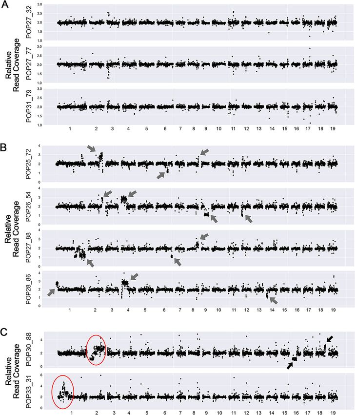
PLOS GENETICS

                                                       RESEARCH ARTICLE

                                                       Chromoanagenesis from radiation-induced
                                                       genome damage in Populus
                                                       Weier Guo ID, Luca Comai ID, Isabelle M. Henry ID*
                                                       Genome Center and Dept. Plant Biology, University of California Davis, Davis, California, United States of
                                                       America

                                                       * imhenry@ucdavis.edu

a1111111111
a1111111111                                            Abstract
a1111111111
a1111111111                                            Chromoanagenesis is a genomic catastrophe that results in chromosomal shattering and
a1111111111                                            reassembly. These extreme single chromosome events were first identified in cancer, and
                                                       have since been observed in other systems, but have so far only been formally documented
                                                       in plants in the context of haploid induction crosses. The frequency, origins, consequences,
                                                       and evolutionary impact of such major chromosomal remodeling in other situations remain
    OPEN ACCESS                                        obscure. Here, we demonstrate the occurrence of chromoanagenesis in poplar (Populus
Citation: Guo W, Comai L, Henry IM (2021)
                                                       sp.) trees produced from gamma-irradiated pollen. Specifically, in this population of siblings
Chromoanagenesis from radiation-induced                carrying indel mutations, two individuals exhibited highly frequent copy number variation
genome damage in Populus. PLoS Genet 17(8):            (CNV) clustered on a single chromosome, one of the hallmarks of chromoanagenesis.
e1009735. https://doi.org/10.1371/journal.
                                                       Using short-read sequencing, we confirmed the presence of clustered segmental rearrange-
pgen.1009735
                                                       ment. Independently, we identified and validated novel DNA junctions and confirmed that
Editor: Ian Henderson, University of Cambridge,
                                                       they were clustered and corresponded to these rearrangements. Our reconstruction of the
UNITED KINGDOM
                                                       novel sequences suggests that the chromosomal segments have reorganized randomly to
Received: May 7, 2021
                                                       produce a novel rearranged chromosome but that two different mechanisms might be at
Accepted: July 22, 2021                                play. Our results indicate that gamma irradiation can trigger chromoanagenesis, suggesting
Published: August 25, 2021                             that this may also occur when natural or induced mutagens cause DNA breaks. We further
Peer Review History: PLOS recognizes the
                                                       demonstrate that such events can be tolerated in poplar, and even replicated clonally, pro-
benefits of transparency in the peer review            viding an attractive system for more in-depth investigations of their consequences.
process; therefore, we enable the publication of
all of the content of peer review and author
responses alongside final, published articles. The
editorial history of this article is available here:
https://doi.org/10.1371/journal.pgen.1009735             Author summary
Copyright: © 2021 Guo et al. This is an open             Plant breeders often use radiation treatment to produce variation, with the goal of identi-
access article distributed under the terms of the        fying new varieties with superior traits. We studied a population of poplar trees produced
Creative Commons Attribution License, which
                                                         by gamma irradiation of pollen, and asked what kind of DNA changes were associated
permits unrestricted use, distribution, and
reproduction in any medium, provided the original        with this variation. We found many changes, most often in the form of added (insertions)
author and source are credited.                          or removed (deletions) pieces of DNA. We also found two lines with much more drastic
                                                         changes. In those lines, we observed massive reorganization. We characterized these two
Data Availability Statement: The sequences
reported in this paper have been deposited in the        lines in detail and found that catastrophic pulverization and random reassembly only
National Center for Biotechnology Information            occurred on a single chromosome. Looking closely at how the pieces were put back
BioProject database (BioProject ID:                      together suggest that the rearrangements in these two lines may have resulted from two
PRJNA723573).

PLOS Genetics | https://doi.org/10.1371/journal.pgen.1009735 August 25, 2021                                                                                   1 / 22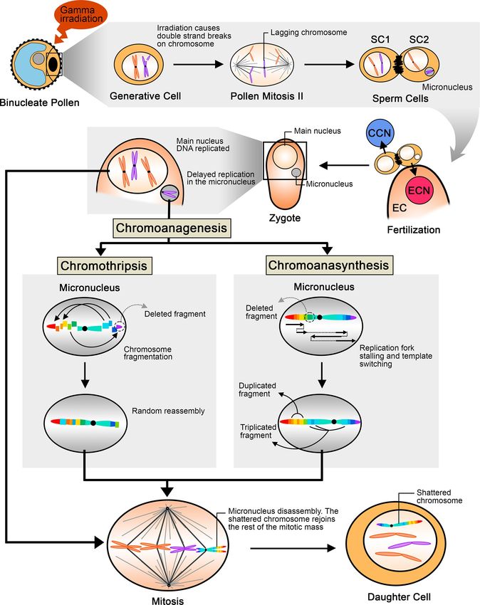
PLOS GENETICS                                                                                                                   Chromoanagenesis in Populus

Funding: This research was supported by the US
Department of Energy Office of Science, Office of        slightly different mechanisms. This type of rearrangement is commonly observed in
Biological and Environmental Research, Grant DE-         human cancer cells, but has rarely been observed in plants. We demonstrated here that
SC0007183 to LC (https://www.energy.gov/
                                                         they can be induced by gamma irradiation, indicating this type of event might be more
science/ber/biological-and-environmental-
research). The DNA Technologies and Expression
                                                         widespread than we expected. Characterizing such genome restructuring instances helps
Analysis Core at the UC Davis Genome Center is           to understand how genome instability can remodel chromosomes and affect genome
supported by National Institute of Health (NIH)          function.
Shared Instrumentation Grant 1S10OD010786-01
(https://www.nih.gov/grants-funding). The funders
had no role in study design, data collection and
                                                       Introduction
analysis, decision to publish, or preparation of the
manuscript.                                            Genomic structural variation (SV) includes various types of chromosomal rearrangements,
Competing interests: The authors have declared
                                                       such as insertion, deletion (INDEL), copy number variation (CNV), inversion and transloca-
that no competing interests exist.                     tion. Structural variation can produce evolutionary significant variation, because it can affect
                                                       large regions of the genome, and influence multiple traits at once. In one extreme scenario,
                                                       restructuring of the genome results in clustered CNV affecting a single or a few chromosomes,
                                                       a syndrome called chromoanagenesis. Chromoanagenesis results from a single triggering
                                                       event that leads to highly complex segmental rearrangements [1,2]. The extreme restructuring
                                                       of a single chromosome (or rarely two or more) results from two distinct processes: (i) in chro-
                                                       mothripsis dsDNA breaks and Non Homologous End Joining rearrange tens to hundreds seg-
                                                       ments, with oscillations between two copy number states (occasionally three) [3,4], and (ii) in
                                                       chromoanasynthesis, replication forks stalled at DNA breaks switch templates, resulting in seg-
                                                       mental duplication and triplication events combined with complex chromosomal rearrange-
                                                       ment of the implicated and intervening segments [5]. Chromothripsis and
                                                       chromoanasynthesis are associated with missegregation of chromosomes, followed by micro-
                                                       nucleus formation around a single chromosome, leading to a single, catastrophic pulverization
                                                       event [6]. A third type of restructuring classified under chromoanagenesis differs in mecha-
                                                       nism and outcome: during chromoplexy, chromosomes are broken in pieces, shuffled together
                                                       and reassembled, resulting in rearranged chromosomes. Chromoplexy always affects more
                                                       than one chromosome [7]. Chromoplexy can occur sequentially and may be originally related
                                                       to DNA breaks caused by transcription factor binding [8]. In plants, chromoplexy-like events
                                                       have been observed in natural variants in camelina [9], and also as a consequence of plant
                                                       transformation in arabidopsis, rice and maize [10].
                                                           Chromothripsis and chromoanasynthesis were originally identified in human cancerous cells
                                                       [1]. To distinguish them from indels, precise criteria are applied [11,12]. In plants, there are multi-
                                                       ple cases of extensive genomic rearrangements [10,13], but when applying the important criterion
                                                       of highly frequent and clustered (at least 10) rearrangements within a single chromosome, only
                                                       haploid induction crosses in Arabidopsis thaliana display catastrophic chromosomal reconstruct-
                                                       ing patterns [14]. In these haploid induction crosses, both chromothripsis and chromoanasynth-
                                                       esis were detected, and the early zygotic divisions are often also accompanied by the formation of
                                                       micronuclei [15], another diagnostic feature of chromothripsis [1,12].
                                                           A critical step in the plant life cycle is pollen production and fertilization. Pollen is prone to
                                                       natural mechanisms that break DNA [16,17] and it is also a classical target for chemical and
                                                       radiation mutagenesis [18]. While traditional chromosomal rearrangements have been
                                                       described, the range of variation resulting from these mechanisms, however, has not been
                                                       determined. We decided to address this question in a poplar F1 population that we previously
                                                       developed from an interspecific cross using gamma-irradiated pollen. This population was
                                                       characterized genetically and harbors >650 unique large-scale insertions and deletions, rang-
                                                       ing from a few hundred kbp to entire chromosomes. Cumulatively, these indels cover the
                                                       genome multiple times [19]. To investigate whether gamma irradiation could have also

PLOS Genetics | https://doi.org/10.1371/journal.pgen.1009735 August 25, 2021                                                                            2 / 22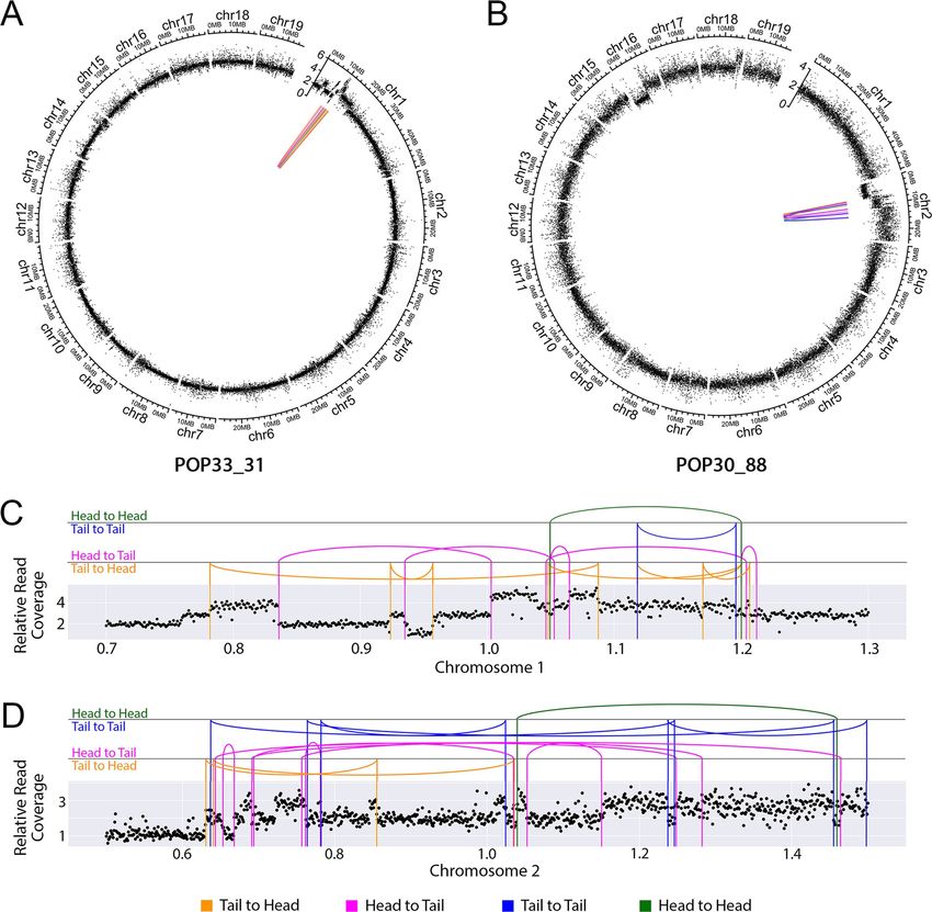
PLOS GENETICS                                                                                                      Chromoanagenesis in Populus

                                            resulted in more severe genome reorganization events, we screened this population for signs
                                            of clustered copy number variation patterns. We identified two individuals with genomic pat-
                                            terns reminiscent of chromoanagenesis, which we characterized in detail. Our results indicate
                                            that DNA breaks induced by irradiation triggered single chromosome fragmentation and
                                            restructuring patterns consistent with chromoanagenesis. These results suggest that pollen
                                            DNA breaks, either natural or induced, can produce extreme structural variations that may
                                            provide evolutionary innovation and, in perennial plants such as poplar, where we were able
                                            to preserve the chromoanagenesis outcomes by vegetative propagation, provide an attractive
                                            system for long-term investigation of the outcome of chromoanagenesis.

                                            Results
                                            Gamma irradiation can result in chromoanagenesis in poplar
                                            A poplar P. deltoides x P. nigra F1 hybrid population was developed previously [19], and char-
                                            acterized using low-coverage illumina genome sequencing. In this population, ~58% of the
                                            lines carried large-scale genomic insertions and deletions (indels) [20], induced by gamma-
                                            irradiation of pollen grains before fertilization. Each F1 line was characterized by a unique set
                                            of indels randomly distributed along the 19 chromosomes of the poplar genome [19,20]. Inter-
                                            estingly, two of these lines exhibited dosage variation consistent with chromoanagenesis. Spe-
                                            cifically, they displayed multiple clustered CNVs on a single chromosome. To investigate the
                                            mechanisms that resulted in these extreme genomic rearrangements, we selected 9 lines for
                                            further analysis: the 2 lines exhibiting extreme rearrangements (Shattering Group, Fig 1C), 4
                                            lines with limited number of indels (Lesion Group, Fig 1B), and 3 lines with no apparent dos-
                                            age variation (No-lesion Group, Fig 1A). Genomic DNAs from these 9 lines were sent for
                                            higher coverage Illumina genomic sequencing (coverage 25–50), with the goal of characteriz-
                                            ing dosage variation in detail, especially those lines with shattered chromosomes.
                                                The dosage variation patterns obtained using the deep-sequencing reads were consistent
                                            with their corresponding low-coverage data. Also consistent with previous results [19], paren-
                                            tal allele frequencies from our high-coverage data indicated that all indels in the genome of the
                                            9 selected individuals originated from loss or gain of the paternal P. nigra copy (Fig 2), con-
                                            firming that the irradiated P. nigra pollen caused dosage variation.
                                                Both lines in the Shattering Group fit our definition of clustered changes (>10 events per chro-
                                            mosome arm) (Fig 2A and 2B and S2 Table). POP33_31 exhibited 21 CNVs on Chromosome 1,
                                            including 2 deletions and 19 insertions, of sizes ranging from 10kb to 5.7Mb (Fig 2A and S2
                                            Table). Among the clustered CNVs, we observed multiple copy number states, ranging from 1 to
                                            5 (Fig 2A and S2 Table), suggesting that some fragments had been lost, while others went through
                                            duplication, triplication or even quadruplication. The second individual in the Shattering Group,
                                            POP30_88, only exhibited single-copy dosage variation. Specifically, 11 CNVs were found in this
                                            line, including 3 deletions and 8 duplications, all localized on Chromosome 2. These fragments
                                            ranged in size, from 80kb to 10.7Mb (Fig 2 and S1 and S2 Table). Taken together, these results
                                            suggested that chromoanagenesis is a possible outcome of gamma-irradiation. The different copy
                                            number variation patterns observed in these two lines further suggest that these two rearranged
                                            genomes might have been shaped by different rearrangement mechanisms.

                                            Novel DNA junctions can be detected using high-coverage short-read
                                            sequencing
                                            To further confirm the hypothesis that these two lines underwent chromoanagenesis, we
                                            aimed to characterize their genome structure in detail (Fig 3). Specifically, we sought to

PLOS Genetics | https://doi.org/10.1371/journal.pgen.1009735 August 25, 2021                                                              3 / 22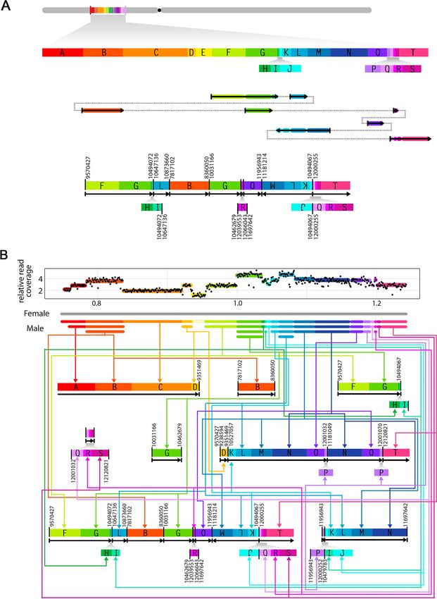
PLOS GENETICS                                                                  Chromoanagenesis in Populus

PLOS Genetics | https://doi.org/10.1371/journal.pgen.1009735 August 25, 2021                         4 / 22
PLOS GENETICS Chromoanagenesis in Populus
Fig 1. Dosage variation detection. Dosage variation was detected by displaying relative read coverage. Each data point represents the mean read coverage in non-
overlapping 100kb bins, standardized to the mean read depth across all 9 lines. The expected value for a diploid line is a relative read coverage of 2. Values around 1
suggest deletions and values around 3 suggest insertions. (A) Dosage plots for 3 lines exhibiting no obvious instances of dosage variation. (B) Dosage plots for 4 lines
containing a small number of indels. The arrows point to the randomly distributed indels identified. (C) Dosage plots for the 2 lines exhibiting shattering patterns.
The red circles represent the regions displaying highly clustered copy number variation.
https://doi.org/10.1371/journal.pgen.1009735.g001
characterize the patterns of genome restructuring by searching for novel DNA junctions cre-
ated with the observed rearrangements. To identify these novel junctions, we searched for
sequencing reads with ends that mapped to two different positions within the genome, sug-
gesting that these two sequences are now adjacent in the reconstructed genome. Because these
rearrangements are expected to occur randomly, these junctions should be unique to each
line. The boundaries of the indels described above provide prime candidates for the localiza-
tion of novel junctions, but other locations in the genome are possible as well. Once potential
junctions were identified, the exact position of the breakpoints were determined through de
novo assembly of the corresponding sequencing reads.
Consistent with our expectations, multiple potential junctions were identified from both of
the lines exhibiting shattered chromosomes, but overall fewer were identified for the other
lines (Table 1). We next validated the presence of these junctions using PCR amplification fol-
lowed by Sanger sequencing, and using sibling F1 lines as negative controls. For the two lines
in the Shattering Group, 26/33 assembled potential junctions were validated by PCR. On the
other hand, none of the potential junctions from the Lesion Group (0/22) and No-lesion
Group (0/11) were validated. Junctions were determined as invalid if they could be amplified
from the genome of other sibling lines as well, or if the Sanger sequencing results were not
consistent with the expectation. In total, we identified 26 novel DNA junctions, all of which
originated from the two shattered lines (S3 Table).
Extreme genomic rearrangements are associated with intra-chromosomal
junctions
By using the junction detection approach mentioned above, we observed multiple novel DNA
junctions in the lines containing a shattered chromosome (Fig 4A). We next seeked to charac-
terize them further and attempted to reconstruct the rearranged sequences.
First, we documented the genomic localization of the validated junctions in each line. Junc-
tions and dosage variation data were plotted together on Circos Plots (Fig 5). For both of the
lines exhibiting shattering, all of the junctions were located on a single chromosome, whether
they corresponded to a shift in dosage variation or not (Fig 5A and 5B). In POP33_31, only 2
breakpoints (each junction consisted of two breakpoints) occurred on regions with no
detected dosage variation, while the other 22 overlapped with CNV boundaries (Fig 5C and S3
Table). However, in POP30_88, only 12/28 breakpoints corresponded to CNV regions (Fig 5D
and S3 Table). This suggests that the mechanisms underlying the rearrangements in these two
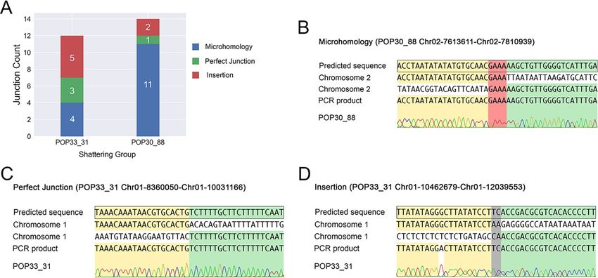
lines might differ. Based on the orientation of two junction ends, we observed that 17% and
36% of the junctions involved an inverted fragment in POP33_31 and POP30_88, respectively
(S3 Table). Finally, we observed three different types of junctions based on the sequence struc-
ture of each junction: microhomology, perfect joining, and insertion (Fig 4B, 4C and 4D).
Both shattered lines exhibited all three junction types (Fig 4A).
With the exact genomic position and orientation of two breakpoints in each junction, we
were able to partially reconstruct the structure of the rearranged sequences in the two shattered
lines. Specifically, we were able to construct 9 and 12 rearranged chromosomal pieces for
POP33_31 (Chromosome 1) and POP30_88 (Chromosome 2), respectively (Figs 6 and S1). In
PLOS Genetics | https://doi.org/10.1371/journal.pgen.1009735 August 25, 2021 5 / 22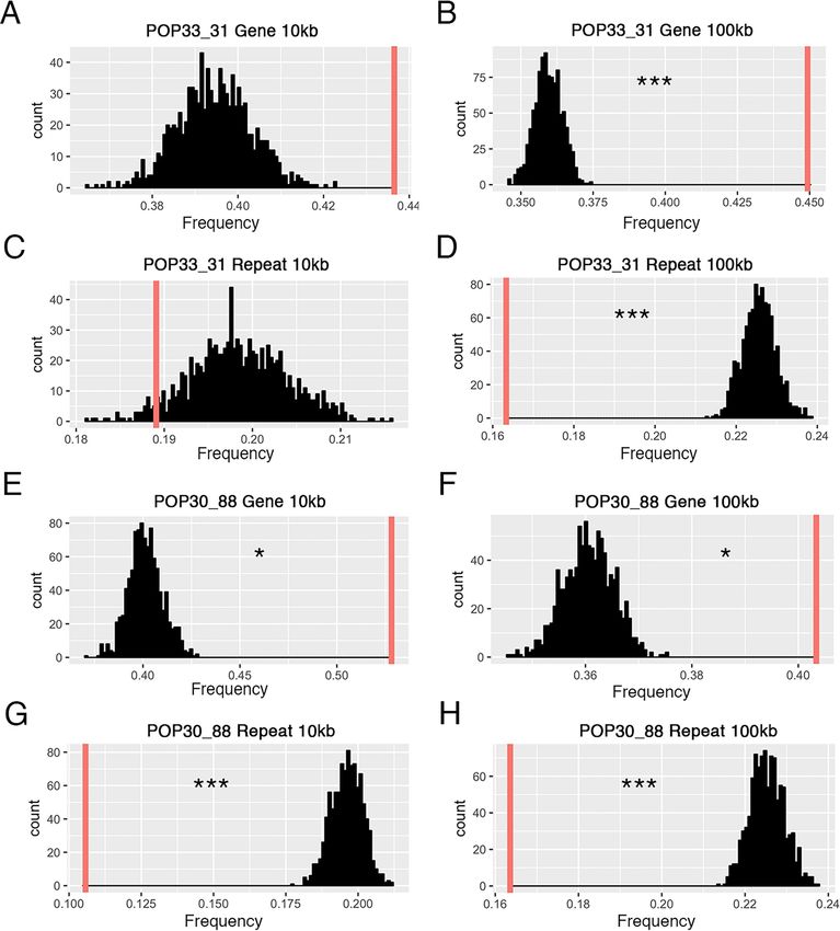
PLOS GENETICS                                                                  Chromoanagenesis in Populus

PLOS Genetics | https://doi.org/10.1371/journal.pgen.1009735 August 25, 2021                         6 / 22
PLOS GENETICS Chromoanagenesis in Populus
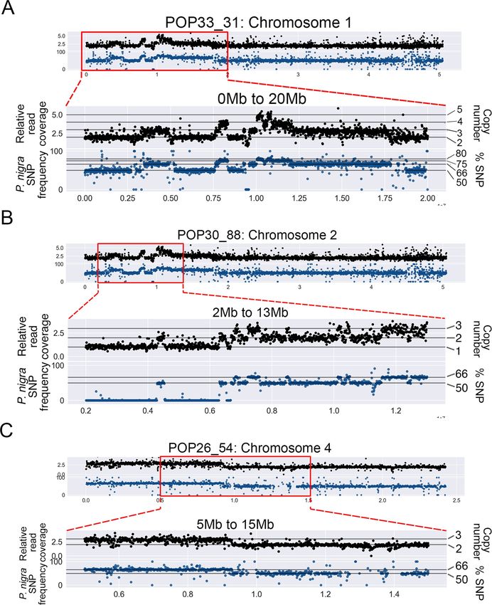
Fig 2. Association of dosage variation patterns with SNP frequency. To obtain a detailed view of the shattered regions, the genome was divided into
narrower bins (10kb bins). Additionally, to confirm the origin of indels, P. nigra (male) SNPs frequencies were calculated for 10kb bins. Black scatterplots: each
black dot represents the relative read coverage for a 10kb bin. Blue scatterplots: each blue dot represents the average P. nigra SNP frequency for a 10kb bin.
Horizontal lines indicate the expected SNP frequency for different copy number states, as indicated on the right. (A) Chromosome 1 of POP33_3 displayed
extremely clustered dosage variation within the first 20Mb, and all variation patterns were associated with P. nigra SNP frequency shifts. (B) Chromosome 2 of
POP30_88 displayed extremely clustered dosage variation in the region between 3Mb and 13Mb, and all CNVs were associated with expected P. nigra SNP
frequency shifts. (C) One of the large-scale lesions on the POP26_54 genome is shown, providing a comparison between larger randomly distributed indels
and the observed shattering patterns in the other two lines.
https://doi.org/10.1371/journal.pgen.1009735.g002
both cases, our results suggest that the restructured region underwent extreme fragmentation,
with chromosomal fragments joined together in a seemingly random order, some fragments
lost altogether, and others copied multiple times (Figs 6 and S1).
The novel DNA junctions are enriched in gene-rich regions
To investigate the DNA context around the novel junctions identified in the shattered lines,
we asked whether the junctions occurred more often in genic or repeated regions of the
genome. Every validated novel junction contained two breakpoints. For each breakpoint, we
calculated the enrichment ratio (see Material and Methods) of genomic features. We used two
different window sizes, 10kb and 100kb, for investigating gene contents and repeated elements.
For both of the shattered lines, breakpoints occurred significantly more often in gene rich
regions and significantly less often near repeated elements (Fig 7 and S6 Table). These results
were consistent with previous studies of aneuploid Arabidopsis thaliana individuals carrying
shattered chromosomes, which also indicated that breakpoints were more likely to occur in
gene-rich regions [14]. Additionally, 26 breakpoints (50%) occurred within a gene coding
sequence (11/24 for POP33_31; 15/28 for POP30_88, S4 Table), and 14 of these involved gene
to gene fusion (S5 Table). Breakpoints were found on different genic elements, including cod-
ing region, introns and untranslated regions. Genes of various functions were affected by these
breakpoints (meiosis-specific proteins, dynamin, etc, see S4 Table). These results indicate that
novel DNA junctions induced by irradiation had the potential to dramatically influence the
function of multiple genes at once.
Discussion
We identified and characterized two instances of highly clustered CNVs on a single chromo-
some in poplar F1 hybrids that resulted from interspecific crosses using gamma-irradiated pol-
len. To investigate the structure of these extreme genome rearrangements, we characterized
the candidate chromosomes from two individuals, and identified localized shattering and
rejoining of DNA in each. Specifically, we identified and characterized 12 and 14 novel DNA
junctions in these two lines, which were clustered on a single chromosome, and always
appeared in the shattered genomic region. These observations are consistent with the charac-
teristics of chromoanagenesis, which is a catastrophic event creating large numbers of complex
rearrangements on one or a few chromosomes [1]. They also suggest that gamma-irradiation
of pollen can result in chromoanagenesis-like patterns in poplar. In our population, we
observed shattered chromosomes in 2/592 individuals. The two poplar lines carrying the shat-
tered chromosomes did not exhibit significant phenotypic differences compared to their sib-
lings. One of the two was sufficiently robust to be selected amongst the F1 individuals that
were clonally propagated and transferred to a field for a population-wide phenotyping experi-
ment [20,21], and did not exhibit extreme phenotypic behaviors in the traits analyzed.
To date, extreme chromosomal rearrangement have only been reported in a few plant spe-
cies, including in aneuploid Arabidopsis thaliana individuals originating from haploid
PLOS Genetics | https://doi.org/10.1371/journal.pgen.1009735 August 25, 2021 7 / 22PLOS GENETICS Chromoanagenesis in Populus
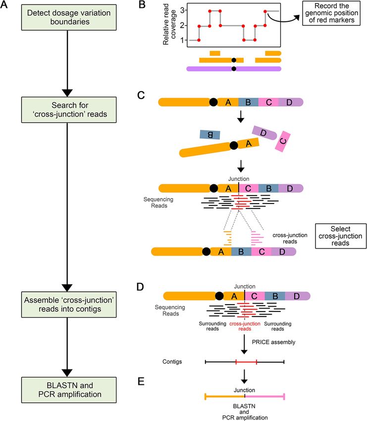
Fig 3. Process of novel DNA junctions selection and validation. (A) Flow chart illustrating the steps involved in novel DNA junction detection, selection, and
validation. (B-E) Diagram illustrating the approach involved in each step. (B) A schematic dosage plot showing a genomic region containing many instances of
dosage variations. The red dots highlight the boundaries of every indel and constituting potential breakpoint positions. (C) Schematic diagram illustrating the
origin and mapping behavior of cross-junction reads. After chromosomal rearrangement, fragment A and C joined together and formed a novel DNA junction.
PLOS Genetics | https://doi.org/10.1371/journal.pgen.1009735 August 25, 2021 8 / 22PLOS GENETICS Chromoanagenesis in Populus
The sequencing reads (in red) that crossed this novel DNA junction are called cross-junction reads. These cross-junction reads map onto two different locations
on the reference genome. (D) Assembly of the novel DNA junctions. Cross-junction reads are assembled into one contig using the PRICE assembler. (E) Each
newly assembled scaffold is compared to the reference genome using BLASTN to: (i) find out the exact alignment positions of two breakpoints of the novel
junction; (ii) confirm the uniqueness of contigs.
https://doi.org/10.1371/journal.pgen.1009735.g003
induction crosses [14], in maize and rice individuals that have undergone biolistic transforma-
tion [10], and in somatic variants of grape [9]. But, except for Tan’s reports in Arabidopsis,
which reported the observation of extreme DNA damage on a single chromosome, other
reports described genomic restructuring involving multiple chromosomes and thus fitting
chromoplexy [7]. Our study and Tan’s study are the only two that showed evidence of clus-
tered, single chromosomal rearrangement in plants, thus fitting the definition of chromothrip-
sis and chromoanasynthesis [4,5].
The two shattered poplar lines both carry highly clustered breakpoints but differ in other
ways, suggesting that the mechanisms underlying these events might be different. The line car-
rying a shattered Chromosome 1 (POP33_31), exhibits a wide variation in copy number states,
ranging from 1 to 5, which indicates segmental duplication and triplication during the geno-
mic remodeling. This is consistent with the replication-based complex rearrangements of
chromoanasynthesis [5]. During chromoanasynthesis, the replication fork stops, and the poly-
merizing strand switches to a proximate template with micro-homologous sequences, and
finally causes the formation of a complex chromosomal rearrangement involving multiple
copy number states [22]. On the other hand, several features of the shattered chromosome of
POP30_88 suggest that it is more likely to be the result of chromothripsis, the fragmentation
and random reorganization of one or a few chromosomes [23]. First, the shattered chromo-
some of POP30_88 only exhibits three copy number states (1, 2 or 3), which is consistent with
the limited copy number states observed in chromothripsis. Chromothripsis usually exhibits
two copy number states: the lower one represents fragment deletion, and the higher one repre-
sents fragment retention [3]. Occasionally, it can also carry three copy number states. This can
be caused by the partial duplication of the rearranged chromosome after experiencing chro-
mothripsis [3]. The oscillation of three copy number states in POP33_88 Chromosome 2 sug-
gests that it may have undergone chromothripsis, followed by a segmental duplication.
Second, the novel DNA junctions observed in POP30_88 cover all four types of the orienta-
tions (H-T, T-H, H-H, T-T), and the rearranged fragments order appears random. This feature
can also be potential evidence for chromothripsis, since the randomness of fragments
Table 1. Summary of DNA junction validation frequencies.
Type Lines In silico Assembled PCR validated Validation Assembled Junction in Validated Junction in Validation Frequency
Junction Junction Frequency Group Group in Group
Shattering POP33_31 16 12 0.75 33 26 0.788
Group POP30_88 17 14 0.824
Lesion Group POP25_72 5 0 0 22 0 0
POP26_54 1 0 0
POP27_88 14 0 0
POP28_86 2 0 0
No-lesion POP27_32 10 0 0 11 0 0
Group POP27_77 1 0 0
POP31_79 0 0 0
The validation frequencies represent the percentage of PCR-validated junctions out of the total number of in silico predicted junctions.
https://doi.org/10.1371/journal.pgen.1009735.t001
PLOS Genetics | https://doi.org/10.1371/journal.pgen.1009735 August 25, 2021 9 / 22PLOS GENETICS Chromoanagenesis in Populus
Fig 4. Types of novel DNA junctions. (A) Number and types of validated DNA junction identified in each line. Different colors represent the three junction types.
(B) Microhomology: presence of 1–11 bp of overlap between the two reconstructed fragment ends. (C) Perfect junction: the two fragment ends are perfectly joined
together, with neither overlapping bps nor inserted bps. (D) Insertion: 1–18 bp of novel nucleotide sequence is inserted between two fragment ends.
https://doi.org/10.1371/journal.pgen.1009735.g004
orientation and order is a representative property for this type of catastrophic event as well [4].
Altogether, our results suggest that the two chromosomal rearrangements observed might
have originated from two different mechanisms: chromoanasynthesis for POP33_31 and chro-
mothripsis for POP30_88.
Ionizing radiation has a long-standing role in plant mutation breeding [24]. The genomic
consequences of ionizing mutations depend on tissue type [25], radiation dosage, and type of
ionizing mutations, and can produce many different types of mutations [19,26–28], including
the creation of variants exhibiting potentially favorable characteristics [20,21]. Ionizing radia-
tion has also been proposed as a potential trigger of chromoanagenesis [3,29,30]. Finally, local-
ized ionizing radiation targeting the nuclei of tumor cells was shown to induce
chromoanagenesis-like patterns in those cells [31]. In this experiment, the authors used a
microbeam system to precisely target the nuclei and induce double strand breaks in some
chromosomes. Their study reported 14 de novo junctions involving four chromosomes, and
proposed that targeted irradiation induced chromothripsis on a few chromosomes. Based on
the features of the three types of chromoanagenesis events, Morishita’s results suggest that
their lines underwent chromoplexy, since the novel junctions are sparsely distributed on sev-
eral chromosomes. Yet, it is also possible that, if the beam only targets a portion of the nuclei,
only the chromosomes located in the affected area underwent rearrangement.
In contrast, our study reported highly clustered novel DNA junctions in a limited genomic
region, while the initial irradiation treatment targeted whole desicated pollen grains [19]. For-
mation of extreme rearranged chromosomes by chromoanagenesis occurs over at least two
mitotic divisions: during the first mitosis, a broken chromosome lags during anaphase and is
incorporated into a micronucleus. During the following interphase, DNA replication of the
micronucleus chromosome is delayed compared with the chromosomes in the major nucleus.
PLOS Genetics | https://doi.org/10.1371/journal.pgen.1009735 August 25, 2021 10 / 22PLOS GENETICS Chromoanagenesis in Populus
Fig 5. Distribution of the genomic location of the validated DNA junctions. (A-B) DNA junctions in the two shattered lines (POP33_31 (A) and POP30_88
(B)). The outermost layer displays each chromosome. The next layer displays relative reads coverage, averaged over 10kb non-overlapping bins. In the center,
colored lines connect the original genomic locations of each pair of sequences found in novel DNA junctions. (C-D) Close-up view of DNA junctions
distribution on the shattered regions of chromosome 1 in POP33_31 (C) and chromosome 2 in POP30_88 (D). The scatter plots show average relative read
coverage per 10kb bins, and the colored vertical lines represent exact breakpoints. The arc connecting two vertical lines illustrate the novel junctions
connecting vertical lines that represent the breakpoints. All panels: Magenta and orange lines represent sequences that connect in the same direction (Head to
Tail in magenta and Tail to Head in orange). Blue and green lines represent sequences that connect in opposite directions (Tail to Tail in blue and Head to
Head in green).
https://doi.org/10.1371/journal.pgen.1009735.g005
During the second mitotic divisions, the replicating micronucleus chromosome pulverizes and
reassembles randomly, forming a shattered chromosome, which is then incorporated into the
normal set [1].
PLOS Genetics | https://doi.org/10.1371/journal.pgen.1009735 August 25, 2021 11 / 22PLOS GENETICS Chromoanagenesis in Populus PLOS Genetics | https://doi.org/10.1371/journal.pgen.1009735 August 25, 2021 12 / 22
PLOS GENETICS                                                                                                                        Chromoanagenesis in Populus

                        Fig 6. Unraveling the structure of the shattered chromosomes. Schematic diagrams illustrating the breakpoints rearrangement in one of
                        the genome shattered lines (POP33_31). (A) The reference chromosome 1 is shown in grey and the regions engaged in rearrangement are
                        labeled in alphabetical order. Each labeled block has a unique color and represents a genomic fragment with validated breakpoints on its
                        flanking ends. The small blocks are enlarged below. All block sizes are proportional to genomic coordinates. In the middle is the potential
                        junctions creating one of the rearranged fragments. Solid arrows with colors represent the corresponding blocks, and the dashed lines
                        illustrate the order of blocks reconstruction. At the bottom is the new structure of that same fragment. Novel junctions are highlighted with
                        bold vertical lines, and are labeled with their original genomic positions on two sides. Black arrows below blocks indicate the orientation of
                        reconstructed fragments. Small blocks are enlarged below proportionally. Fragment duplications are linked and pointed out with the same
                        color. (B) Summary of the fragments reconstructed based on the data obtained from line POP33_31. The dosage plot on top displays relative
                        read coverage of the shattering region in chromosome 1. Each DNA block is labeled with the same color used in (A). A schematic
                        representation of chromosome is shown below the dosage plot, with female (P. deltoides, WT) inherited chromosome colored in grey, and
                        male (P. nigra, pollen irradiated) inherited chromosome illustrated in their corresponded colors and copy number states. For each DNA
                        block, the same color arrows guide to the corresponding fragments on the reconstructed chromosome pieces.
                        https://doi.org/10.1371/journal.pgen.1009735.g006

                                                   In poplar, mature pollen is binucleate, and must undergo the second pollen mitosis, in
                                               which the generative cell divides into two sperm cells, just before fertilizing the egg cell. It is
                                               thus possible that the radiation-induced DNA breaks remained unrepaired, causing chromo-
                                               some missegregation during the generative cell division, possibly resulting in the formation of
                                               a micronucleus in one of the sperm cells (Fig 8). After fertilization of the egg cell by the micro-
                                               nucleus-carrying sperm, during the first zygotic mitotic division, damage, such as incomplete
                                               replication, results in catastrophic DNA pulverization of the chromosome in the micronucleus
                                               [1,6]. The rearranged chromosome is reincorporated in the normal set during the subsequent
                                               mitosis. If the centromere is present, the shattered chromosome can segregate normally in the
                                               main nucleus, fixing the rearrangement.
                                                   Our study shows that novel DNA junctions were significantly enriched in gene-rich
                                               regions, which is consistent with Tan’s results in Arabidopsis [14]. Similar outcomes have also
                                               been demonstrated in human breast cancer, where high density of DSBs occurred on chromo-
                                               some 17, one of the human chromosomes with high gene content [32]. Further, open chroma-
                                               tin may be more available for recombination. In our analysis, 14/26 breakpoints formed
                                               junctions between genes, suggesting the potential of these events for genic innovation.
                                                   Our analysis used Illumina short reads to identify and assemble novel DNA junctions. In
                                               the shattered lines, this approach was successful as 78% of novel DNA junctions could be vali-
                                               dated in vitro. Based on the number of copy number variation boundaries found in these two
                                               lines, and the number of novel breakpoints (each junction contains two breakpoints) that
                                               match these boundaries, we can estimate that we successfully identified 68% of the novel
                                               breakpoints. The false negative breakpoints could be caused by poor read coverage across
                                               those genomic regions, by the presence of repeated sequences complicating the read mapping
                                               process, or by differences between the reference genome used for read mapping (P. tricho-
                                               carpa) and that of the male parent (P. nigra). When applying this approach to sibling lines con-
                                               taining sparse indels along the genome, we did not identify any novel breakpoints despite the
                                               presence of seven large-scale insertions in these lines, which indicates that at least 7 new break-
                                               points should be present. As a result, it is still unclear where the duplicated fragments detected
                                               in these lines are located. Given that the probability of identifying real breakpoints in the two
                                               lines displaying shattering was 0.68 (34 breakpoints / 50 copy number shifts), our failure to
                                               find any real breakpoint out of 7 copy number shifts for the normal indels is surprising (p-
                                               value of Bootstrap hypothesis testing = 0.0081). It is thus possible that breaks giving rise to
                                               indels may result from a different DNA damage mechanism. For instance, unlike the junctions
                                               detected in the shattered lines, those present in the other lines may not be located within gene
                                               space and might therefore be more difficult to detect using short reads. Nevertheless, our anal-
                                               ysis using short reads was successful at identifying enough novel junctions to confirm the ran-
                                               domly reorganized state of the shattered chromosomes.

PLOS Genetics | https://doi.org/10.1371/journal.pgen.1009735 August 25, 2021                                                                                    13 / 22
PLOS GENETICS Chromoanagenesis in Populus
Fig 7. Sequence context surrounding the breakpoints of novel DNA junctions. The frequency of genes and repeated elements surrounding novel junctions is
compared to the corresponding frequencies in randomly selected pseudo junctions. For each panel: 1,000 pseudo-junction were selected at random and the mean
percentage of gene or TE space in these 1,000 junctions was calculated. This process was repeated 1,000 times and the distribution of these means are represented in
black. The red vertical line represents the mean of enriched frequency for the observed validated novel junctions. Breakpoints of novel junctions in POP33_31
(A-D) occur significantly in gene-rich, repeats-deficient regions under 100kb window size (p-value < 0.001), but do not show statistical significance in 10kb
PLOS Genetics | https://doi.org/10.1371/journal.pgen.1009735 August 25, 2021 14 / 22PLOS GENETICS Chromoanagenesis in Populus
window size. Results were similar for POP30_88 (E-H). The observed junctions are significantly enriched with genes (p-value < 0.05), and have the lack of repeated
elements (p-value < 0.001) regardless of window size.
https://doi.org/10.1371/journal.pgen.1009735.g007
In conclusion, our study demonstrated that chromoanagenesis can be induced in plants by
ionizing radiation of pollen, indicating that extreme chromosomal rearrangements can be
more widespread, and more tolerated than expected. Notably, natural mechanisms can also
produce dsDNA breaks in pollen [16,17]. This type of cataclysmic outcomes is thus possible in
a natural setting and can contribute evolutionary innovations, similarly to chromosomal inver-
sions [33]. They may also mediate gene amplification [34], which has been detected in glypho-
sate-resistant weeds [35]. Because poplar is vegetatively propagated, we were able to produce
several clones from each chromoanagenetic line and maintain some of these extreme chromo-
somal rearrangements in the field for at least five years. Finally, our results show that the
observed chromosomal rearrangements directly affected the sequence of multiple genes and,
in some cases, have the potential to produce new chimeric proteins. While most of these ran-
dom events will probably result in non- or dys-functional proteins, it is an interesting avenue
for the creation of new gene functions.
Materials and methods
Genomic sequencing and dosage variation analysis
Genomic DNA was extracted from leaf samples and prepared for deep-sequencing using Illu-
mina technology, as previously described [19]. Sequencing reads (150 PE) were demultiplexed
into individual libraries based on their barcodes, using a custom Python script (http://
comailab.genomecenter.ucdavis.edu/index.php/Barcoded_data_preparation_tools), as
described in previous studies [19]. Next, reads were aligned to the poplar reference P. tricho-
carpa v3.0 [36], using a custom Python script based on mapping using BWA [37] (http://
comailab.genomecenter.ucdavis.edu/index.php/Bwa-doall). This generated an alignment file
(sam file) for each line, which was used for further analysis.
To detect dosage variation, we calculated relative read coverage values across the genome
for each line, as described previously [38]. Specifically, the genome was divided into a series of
non-overlapping consecutive bins of 100kb or 10kb, depending on sequence coverage (see
results). Next, for each bin, relative read coverage was calculated by taking the fraction of
aligned reads in a particular bin for that line, and dividing it by the mean fraction of reads
aligning to the same bin in all lines, and multiplying by 2, the background ploidy of poplar. A
custom Python script was used to achieve these calculations (http://comailab.genomecenter.
ucdavis.edu/index.php/Bin-by-sam). The relative coverage values obtained were then plotted
according to the corresponded genomic region of their belonging bins. Values around 2 indi-
cate the expected two copies, while values closer to 3 and 1 suggest the presence of insertions,
or deletions, respectively.
Detection of novel genomic junctions
To detect novel genomic junctions, we first searched for indels boundaries, which represented
the potential breakpoints of reorganized genomic fragments. Based on the dosage variation
plots obtained using 10kb bins, we recorded potential junctions using the following criteria:
bins where relative read coverage decreased or increased by >0.7 compared to their adjacent
forward bin, and instances where this trend was true for at least three consecutive bins. Addi-
tionally, potential breakpoints were only retained if they were unique to a single line. These
potential breakpoints became the most likely locations for forming novel DNA junctions. To
PLOS Genetics | https://doi.org/10.1371/journal.pgen.1009735 August 25, 2021 15 / 22PLOS GENETICS Chromoanagenesis in Populus PLOS Genetics | https://doi.org/10.1371/journal.pgen.1009735 August 25, 2021 16 / 22
PLOS GENETICS                                                                                                                         Chromoanagenesis in Populus

              Fig 8. Proposed model illustrating the steps leading to chromoanagenesis following pollen irradiation. Gamma irradiation of binucleate pollen
              induces double stranded DNA breaks in the generative cell, and results in chromosome lagging or in bridge formation [12] during the second pollen
              mitosis. The lagging chromosome is excluded from the main nucleus and forms a micronucleus. The sperm cell carrying the micronucleus undergoes
              karyogamy with the egg cell, and produces a zygote with a (2n-1) nucleus and a micronucleus containing a single paternal chromosome. DNA replication
              in micronuclei is delayed and leads to chromoanagenesis via two possible mechanisms, chromothripsis and chromoanasynthesis, which were both
              observed in our poplar lines. Chromothripsis involves fragmentation and random reassembly, while chromoanasynthesis results from replication fork
              stalling and template switching. The highly rearranged chromosome is eventually released from the micronucleus and reunites with the main nuclear
              genome during mitotic division. The shattered chromosome is thereafter retained in the main nucleus. SC: sperm cell; EC: egg cell; ECN: egg cell nucleus;
              CCN: central cell nucleus.
              https://doi.org/10.1371/journal.pgen.1009735.g008

                                                 characterize novel junctions in more detail, we next searched for reads mapping to two distant
                                                 genomic locations, and therefore crossed the targeted junctions. A custom Python script
                                                 (https://github.com/guoweier/Poplar_Chromoanagenesis) was used. Specifically, the script
                                                 divided the genome into non-overlapping consecutive 10kb bins and, for each combination of
                                                 two non-consecutive bins that were at least 2,000 bp apart, the number of reads mapping to
                                                 both bins was recorded for each line. Numbers were then compared between lines to identify
                                                 pairs of bins with high coverage in a single line compared to the others, suggesting the pres-
                                                 ence of a novel junction. In order to set a minimum threshold of coverage to eliminate false
                                                 positives, we needed to calculate the expected average coverage over each junction. To do so,
                                                 we created artificial non-overlapping 5 kb bins throughout the genome, considered the bound-
                                                 ary between two consecutive bins as pseudo-junctions and recorded the average reads cover-
                                                 age at these pseudo-junctions. These values were then divided by 2, to account for the fact that
                                                 these pseudo-junctions are expected to be present in two copies in the diploid poplar genome,
                                                 while the indels and other novel junctions are expected to only affect one copy of the genome.
                                                 We used these line-specific thresholds as minimum coverage thresholds for the identification
                                                 of potential novel junctions. Second, to ensure that junctions were specific to a single line, we
                                                 discarded bin-pairs that were positive in more than one line. Specifically, a potential in-pair
                                                 was only retained if none of the other lines exhibited reads that mapped to those two bins.

                                                 Novel junction validation
                                                 To assemble potential novel junctions, we searched the alignment file (sam file) of each line
                                                 and extracted the cross-junction reads identified at the selected bins, using a custom Python
                                                 script (https://github.com/guoweier/Poplar_Chromoanagenesis). Next, the PRICE genome
                                                 assembler was used to assemble the cross-junction reads into contigs [39]. The assembly
                                                 parameters and input data can be found in our github repository (https://github.com/
                                                 guoweier/Poplar_Chromoanagenesis). To confirm the junction genomic composition, we
                                                 aligned the output contigs to the P. trichocarpa genome by using blast+ package [40] by using
                                                 a custom bash script (https://github.com/guoweier/Poplar_Chromoanagenesis). When the
                                                 two ends of the contig aligned to the expected regions, we considered that the novel junction
                                                 was confirmed in silico.
                                                     To validate these potential junctions in vitro, PCR primers were designed using Primer3
                                                 [37] (S1 Table). PCR were run using the GoTaq Green Mastermix (Promega Corporation,
                                                 Madison, WI) with 1ng sample gDNA. The obtained PCR products were purified using gel
                                                 extraction (QIAquick Gel Extraction Kit, Qiagen) and sent for Sanger sequencing.

                                                 SNP frequency analysis
                                                 We used parental SNP allelic percentage to identify the parental origin of the lesions. Single
                                                 nucleotide polymorphism (SNP) between P. deltoides (female) and P. nigra (male) were identi-
                                                 fied previously [19]. We genotyped each line as described previously [19]. In short, to calculate

PLOS Genetics | https://doi.org/10.1371/journal.pgen.1009735 August 25, 2021                                                                                    17 / 22
PLOS GENETICS Chromoanagenesis in Populus
the percentage of P. nigra and P. deltoides alleles at each position, we created an mpileup file
containing every base allele and coverage for all examined lines, using a custom Python pack-
age based on Samtools [41] and built-in mpileup function (http://comailab.genomecenter.
ucdavis.edu/index.php/Mpileup). The mpileup file was then simplified by converting a parsed-
mpileup file, using the custom Python package described above. Next, the parsed-mpileup file
was used to search for the preselected SNPs position. Finally, to obtain robust allele percent-
ages, SNP allele calls were pooled within consecutive bins, and the percentage of P. nigra
parental alleles were calculated for each bin. According to this approach, a diploid chromo-
some exhibited 50% P. nigra alleles. A deletion on one chromosome is expected to exhibit 0%
or 100% P. nigra alleles, depending on which parental chromosome was lost. An increase of
copy number states is expected to exhibit allelic ratio bias between two parents, with 1:2 repre-
sented DNA fragment duplication, 1:3 represented DNA fragment triplication, and so on.
Genome restructuring analysis
To reconstruct each mutant genome based on the identified validated junctions, we searched
for fragments with the same breakpoints and strung them together manually, with the expecta-
tion that each breakpoint should be involved in two junctions, one on each side of the break-
point. With this logic, we manually looked for paired fragment end locations among the
junctions, and arranged them into longer pieces. We then built the rearranged chromosomes,
while taking junction orientation and fragments copy number into account.
Enrichment ratio analysis
The poplar genome annotation file, including the genomic positions of gene and repeatmasked
(GFF-Version3.0) was downloaded from Phytozome (http://phytozome.jgi.doe.gov/pz/portal.
html). Next, we used a custom python script to calculate genes/repeats density around each of
the novel breakpoints (https://github.com/guoweier/Poplar_Chromoanagenesis). Specifically,
each potential breakpoint was set as the center of a 10kb or 100kb window, and the nucleotide
number of typical genomic features within these windows was recorded. Next, to provide a
random set of junctions, we used the previously constructed pseudo-junction pool, and ran-
domly selected 1,000 of these pseudo breaks for genomic feature density calculation. For each
line, this type of random pseudo-break datasets were established 1,000 times for every exam-
ined genomic feature. Enrichment ratios were calculated by taking the means of genomic fea-
ture density at real breakpoints, divided by the means of the corresponded features density at
random pseudo breakpoints datasets. Significance was assessed by comparing the density of
real breakpoints and 1,000 randomized datasets using one sample t-test.
Supporting information
S1 Fig. Summary of the fragments reconstructed on line POP30_88 Chromosome 2. The
diagram follows the same criteria as in Fig 6B. The rearranged fragments were constructed
based on the data of novel junction observation in POP30_88.
(TIF)
S2 Fig. Detailed view of the P. nigra SNP frequency pattern in the shattered regions. The
genome was divided into consecutive non-overlapping 10kb bins. Each blue dot represents the
average P. nigra SNP frequency for a 10kb bin. Horizontal lines exhibit the expected frequency
levels for different copy number states, with their numbers labeled on the right.
(TIF)
PLOS Genetics | https://doi.org/10.1371/journal.pgen.1009735 August 25, 2021 18 / 22PLOS GENETICS Chromoanagenesis in Populus
S1 Table. PCR primers used in novel junction validation. List of primers used for PCR
amplification.
(XLSX)
S2 Table. Summary of indels in 2 shattered lines. Large-scale indels in two shattering lines
(POP33_31 = 21, POP30_88 = 11) were identified based on dosage variation patterns and SNP
frequency. The locations and copy number states of indels are indicated, as well as the parental
genotype they originated from. D: P. deltoides; N: P. nigra.
(XLSX)
S3 Table. Summary of all validated novel DNA junctions. List of validated novel DNA junc-
tions in the two shattering lines (POP33_31 = 12, POP30_88 = 14). Each junction is indicated
with its junction type, two breakpoints positions, orientation, and its correlation with CNV
edges.
(XLSX)
S4 Table. DNA context at the breakpoints. List of all breakpoints identified, as well as infor-
mation about the affected genes when the breakpoints occurred within a gene.
(XLSX)
S5 Table. Summary of the possible gene fusion events at novel DNA junctions. List of junc-
tions containing gene to gene fusion within the two shattering lines. Instances where two
genes are fused in the same direction are labelled in green, indicating that these fusions might
form novel gene products.
(XLSX)
S6 Table. DNA context surrounding the novel junctions. The novel DNA junctions identi-
fied in the two lines (POP33_31 = 12, POP30_88 = 14) exhibiting clustered patterns are prefer-
entially located in regions that are rich in gene sequences and poor in repeated sequences,
compared to the rest of the genome. Enrichment ratio represents the comparison between the
means of genomic feature density at real breakpoints and the means of a similar set of ran-
domly selected features. Ratio >1 indicates validated breakpoints that have a higher density of
features than the genome average, while ratioPLOS GENETICS Chromoanagenesis in Populus
Project administration: Isabelle M. Henry.
Software: Weier Guo.
Visualization: Weier Guo.
Writing – original draft: Weier Guo.
Writing – review & editing: Weier Guo, Luca Comai, Isabelle M. Henry.
References
1. Holland AJ, Cleveland DW. Chromoanagenesis and cancer: mechanisms and consequences of local-
ized, complex chromosomal rearrangements. Nat Med. 2012; 18:1630–8. https://doi.org/10.1038/nm.
2988 PMID: 23135524
2. Pellestor F. Chromoanagenesis: Cataclysms behind complex chromosomal rearrangements. Mol Cyto-
genet. Molecular Cytogenetics; 2019; 12:1–12. https://doi.org/10.1186/s13039-018-0413-1 PMID:
30647775
3. Stephens PJ, Greenman CD, Fu B, Yang F, Bignell GR, Mudie LJ, et al. Massive genomic rearrange-
ment acquired in a single catastrophic event during cancer development. Cell. Elsevier Inc.; 2011;
144:27–40. https://doi.org/10.1016/j.cell.2010.11.055 PMID: 21215367
4. Korbel JO, Campbell PJ. Criteria for inference of chromothripsis in cancer genomes. Cell. Elsevier Inc.;
2013; 152:1226–36. https://doi.org/10.1016/j.cell.2013.02.023 PMID: 23498933
5. Pellestor F, Gatinois V. Chromoanasynthesis: Another way for the formation of complex chromosomal
abnormalities in human reproduction. Hum Reprod. 2018; 33:1381–7. https://doi.org/10.1093/humrep/
dey231 PMID: 30325427
6. Crasta K, Ganem NJ, Dagher R, Lantermann AB, Ivanova EV, Pan Y, et al. DNA breaks and chromo-
some pulverization from errors in mitosis. Nature. Nature Publishing Group; 2012; 482:53–8. https://doi.
org/10.1038/nature10802 PMID: 22258507
7. Shen MM. Chromoplexy: a new category of complex rearrangements in the cancer genome. Cancer
Cell. 2013. p. 567–9. https://doi.org/10.1016/j.ccr.2013.04.025 PMID: 23680143
8. Baca SC, Prandi D, Lawrence MS, Mosquera JM, Romanel A, Drier Y, et al. Punctuated evolution of
prostate cancer genomes. Cell. Elsevier; 2013; 153:666–77. https://doi.org/10.1016/j.cell.2013.03.021
PMID: 23622249
9. Carbonell-Bejerano P, Royo C, Torres-Pérez R, Grimplet J, Fernandez L, Franco-Zorrilla JM, et al. Cat-
astrophic Unbalanced Genome Rearrangements Cause Somatic Loss of Berry Color in Grapevine.
Plant Physiol. 2017; 175:786–801. https://doi.org/10.1104/pp.17.00715 PMID: 28811336
10. Liu J, Nannas NJ, Fu F-F, Shi J, Aspinwall B, Parrott WA, et al. Genome-Scale Sequence Disruption
Following Biolistic Transformation in Rice and Maize. Plant Cell. 2019; 31:368–83. https://doi.org/10.
1105/tpc.18.00613 PMID: 30651345
11. Pellestor F, Gatinois V. Chromoanagenesis: a piece of the macroevolution scenario. Mol Cytogenet.
2020; 13:3. https://doi.org/10.1186/s13039-020-0470-0 PMID: 32010222
12. Umbreit NT, Zhang C-Z, Lynch LD, Blaine LJ, Cheng AM, Tourdot R, et al. Mechanisms generating can-
cer genome complexity from a single cell division error. Science [Internet]. 2020; 368. Available from:
https://doi.org/10.1126/science.aba0712 PMID: 32299917
13. Mandáková T, Pouch M, Brock JR, Al-Shehbaz IA, Lysak MA. Origin and Evolution of Diploid and Allo-
polyploid Camelina Genomes Were Accompanied by Chromosome Shattering. Plant Cell. 2019;
31:2596–612. https://doi.org/10.1105/tpc.19.00366 PMID: 31451448
14. Tan EH, Henry IM, Ravi M, Bradnam KR, Mandakova T, Marimuthu MPA, et al. Catastrophic chromo-
somal restructuring during genome elimination in plants. Elife. 2015; 4:1–16. https://doi.org/10.7554/
eLife.06516 PMID: 25977984
15. Marimuthu MPA, Maruthachalam R, Bondada R, Kuppu S, Tan E-H, Britt A, et al. Biased removal and
loading of centromeric histone H3 during reproduction underlies uniparental genome elimination [Inter-
net]. bioRxiv. 2021 [cited 2021 Apr 17]. p. 2021.02.24.432754. Available from: https://www.biorxiv.org/
content/10.1101/2021.02.24.432754v1.abstract
16. Li X, Meng D, Chen S, Luo H, Zhang Q, Jin W, et al. Single nucleus sequencing reveals spermatid chro-
mosome fragmentation as a possible cause of maize haploid induction. Nat Commun. nature.com;
2017; 8:991.
PLOS Genetics | https://doi.org/10.1371/journal.pgen.1009735 August 25, 2021 20 / 22PLOS GENETICS Chromoanagenesis in Populus
17. Nasuda S, Friebe B, Gill BS. Gametocidal genes induce chromosome breakage in the interphase prior
to the first mitotic cell division of the male gametophyte in wheat. Genetics. 1998; 149:1115–24. PMID:
9611219
18. van Harten AM. Mutation breeding: theory and practical applications. Cambridge University Press;
1998.
19. Henry IM, Zinkgraf MS, Groover AT, Comaia L. A system for dosage-based functional genomics in pop-
lar. Plant Cell. 2015; 27:2370–83. https://doi.org/10.1105/tpc.15.00349 PMID: 26320226
20. Bastiaanse H, Zinkgraf M, Canning C, Tsai H, Lieberman M, Comai L, et al. A comprehensive genomic
scan reveals gene dosage balance impacts on quantitative traits in Populus trees. Proc Natl Acad Sci U
S A. 2019; 116:13690–9. https://doi.org/10.1073/pnas.1903229116 PMID: 31213538
21. Bastiaanse H, Henry I, Tsai H, Lieberman M, Canning C, Comai L, et al. A systems genetics approach
to deciphering the effect of dosage variation on leaf morphology in Populus. Plant Cell [Internet]. 2020;
Available from: https://academic.oup.com/plcell/advance-article-abstract/doi/10.1093/plcell/koaa016/
6007531
22. Liu P, Erez A, Nagamani SCS, Dhar SU, Kołodziejska KE, Dharmadhikari AV, et al. Chromosome
catastrophes involve replication mechanisms generating complex genomic rearrangements. Cell. 2011;
146:889–903. https://doi.org/10.1016/j.cell.2011.07.042 PMID: 21925314
23. Forment JV, Kaidi A, Jackson SP. Chromothripsis and cancer: Causes and consequences of chromo-
some shattering. Nat Rev Cancer. Nature Publishing Group; 2012; 12:663–70. https://doi.org/10.1038/
nrc3352 PMID: 22972457
24. Brunner H. Radiation induced mutations for plant selection. Appl Radiat Isot. 1995; 46:589–94.
25. Hase Y, Satoh K, Kitamura S, Oono Y. Physiological status of plant tissue affects the frequency and
types of mutations induced by carbon-ion irradiation in Arabidopsis. Sci Rep. 2018; 8:1394. https://doi.
org/10.1038/s41598-018-19278-1 PMID: 29362368
26. Belfield EJ, Gan X, Mithani A, Brown C, Jiang C, Franklin K, et al. Genome-wide analysis of mutations
in mutant lineages selected following fast-neutron irradiation mutagenesis of Arabidopsis thaliana.
Genome Res. 2012; 22:1306–15. https://doi.org/10.1101/gr.131474.111 PMID: 22499668
27. Henry IM, Nagalakshmi U, Lieberman MC, Ngo KJ, Krasileva KV, Vasquez-Gross H, et al. Efficient
Genome-Wide Detection and Cataloging of EMS-Induced Mutations Using Exome Capture and Next-
Generation Sequencing. Plant Cell. 2014; 26:1382–97. https://doi.org/10.1105/tpc.113.121590 PMID:
24728647
28. Sakamoto AN, Lan VTT, Fujimoto S, Matsunaga S, Tanaka A. An ion beam-induced Arabidopsis
mutant with marked chromosomal rearrangement. J Radiat Res. 2017; 58:772–81. https://doi.org/10.
1093/jrr/rrx024 PMID: 28637346
29. Lieber MR. The mechanism of double-strand DNA break repair by the nonhomologous DNA end-joining
pathway. Annu Rev Biochem. annualreviews.org; 2010; 79:181–211. https://doi.org/10.1146/annurev.
biochem.052308.093131 PMID: 20192759
30. Tsai AG, Lieber MR. Mechanisms of chromosomal rearrangement in the human genome. BMC Geno-
mics. bmcgenomics.biomedcentral.com; 2010; 11 Suppl 1:S1. https://doi.org/10.1186/1471-2164-11-
S1-S1 PMID: 20158866
31. Morishita M, Muramatsu T, Suto Y, Hirai M, Konishi T, Hayashi S, et al. Chromothripsis-like chromo-
somal rearrangements induced by ionizing radiation using proton microbeam irradiation system. Onco-
target. 2016; 7:10182–92. https://doi.org/10.18632/oncotarget.7186 PMID: 26862731
32. Przybytkowski E, Lenkiewicz E, Barrett MT, Klein K, Nabavi S, Greenwood CM, et al. Chromosome-
breakage genomic instability and chromothripsis in breast cancer. BMC genomics. 2014; 15(1):1–5.
https://doi.org/10.1186/1471-2164-15-579 PMID: 25011954
33. Huang K, Rieseberg LH. Frequency, Origins, and Evolutionary Role of Chromosomal Inversions in
Plants. Front Plant Sci. 2020; 11:296. https://doi.org/10.3389/fpls.2020.00296 PMID: 32256515
34. Shoshani O, Brunner SF, Yaeger R, Ly P, Nechemia-Arbely Y, Kim DH, et al. Chromothripsis drives the
evolution of gene amplification in cancer. Nature [Internet]. nature.com; 2020; Available from: http://dx.
doi.org/10.1038/s41586-020-03064-z PMID: 33361815
35. Koo D-H, Molin WT, Saski CA, Jiang J, Putta K, Jugulam M, et al. Extrachromosomal circular DNA-
based amplification and transmission of herbicide resistance in crop weed Amaranthus palmeri. Proc
Natl Acad Sci U S A. 2018; 115:3332–7. https://doi.org/10.1073/pnas.1719354115 PMID: 29531028
36. Tuskan GA, Difazio S, Jansson S, Bohlmann J, Grigoriev I, Hellsten U, et al. The genome of black cot-
tonwood, Populus trichocarpa (Torr. & Gray). Science. 2006; 313:1596–604. https://doi.org/10.1126/
science.1128691 PMID: 16973872
PLOS Genetics | https://doi.org/10.1371/journal.pgen.1009735 August 25, 2021 21 / 22You can also read