Treatment and outcome of tenosynovial giant cell tumor/ pigmented villonodular synovitis patients receiving radiotherapy in Taiwan: a ...
←
→
Page content transcription
If your browser does not render page correctly, please read the page content below
Original Article
                                                                                                                                       Page 1 of 9

Treatment and outcome of tenosynovial giant cell tumor/
pigmented villonodular synovitis patients receiving radiotherapy in
Taiwan: a single-center experience
Chi-Shuo Lin1^, Yu-Mei Kang1,2,3, Keng-Li Lan1,4, Ling-Wei Wang1,2, Yu-Ming Liu1,2, Pin-I Huang1,2, Yu-
Wen Hu1,2, I-Chun Lai1,2, Yuan-Hung Wu1,2, Tzu-Yu Lai1,2, Wan-Chin Yang1, Jia-Cheng Lee1,
Cheng-Ying Shiau1
1
Division of Radiation Oncology, Department of Oncology, Taipei Veterans General Hospital, Taipei, Taiwan; 2Faculty of Medicine, National Yang-
Ming University, Taipei, Taiwan; 3Institute of Clinical Medicine, National Yang-Ming University, Taipei, Taiwan; 4Institute of Traditional Medicine,
School of Medicine, National Yang-Ming University, Taipei, Taiwan
Contributions: (I) Conception and design: CS Lin, YM Kang; (II) Administrative support: CY Shiau, KL Lan, YM Liu; (III) Provision of study
materials or patients: CY Shiau, KL Lan, LW Wang, YM Liu, JC Lee; (IV) Collection and assembly of data: CS Lin, PI Huang, YW Hu, IC Lai,
YH Wu, TY Lai, WC Yang, JC Lee, YM Kang; (V) Data analysis and interpretation: All authors; (VI) Manuscript writing: All authors; (VII) Final
approval of manuscript: All authors.
Correspondence to: Cheng-Ying Shiau, MD. Division of Radiation Oncology, Department of Oncology, Taipei Veterans General Hospital, 201, Sec.2,
Shih-Pai Road, Taipei 11217, Taiwan. Email: cyshiau@vghtpe.gov.tw.

                Background: Tenosynovial giant cell tumor (TGCT), historically known as pigmented villonodular
                synovitis (PVNS), is a rare and benign proliferative disease of joints. Diffuse form TGCT/PVNS has higher
                local recurrence rate that requires surgery and adjuvant treatment such as radiotherapy (RT). We reported
                our experience in treating patients with advanced TGCT/PVNS by surgery and RT or RT alone.
                Methods: We included 48 patients of TGCT/PVNS who received RT between January 2001 and
                December 2018. Patients demographics, treatment parameters, complications and recurrences were evaluated.
                The Kaplan-Meier curves of local control (LC) were estimated, and variables affecting LC were analyzed by
                the univariate and multivariate Cox proportional hazard model.
                Results: All patients had diffuse form TGCT/PVNS. Knee joint was affected in 34 patients (70.8%). Eight
                patients (16.7%) were inoperable so definitive RT was delivered. Most of the patients (85.4%) received
                RT dose of 30 Gy in 15 fractions. The median follow-up time was 4.3 years, and seven patients (14.6%)
                suffered from recurrence. Obesity (body mass index >27 kg/m2) was a significant prognostic factor for local
                recurrence in univariate and multivariate Cox regression analyses. The overall 1-, 3- and 5-year LC rate was
                100%, 92.8% and 87.2%, respectively. There was no significant surgical complication. RT-related acute
                toxicities were mild, including grade 1 dermatitis, joint effusion and decrease in joint range of motion.
                Conclusions: RT in TGCT/PVNS patients revealed good LC and low treatment toxicities. Obesity was
                associated with higher recurrence rate. Definitive RT may be a treatment option for inoperable patients.

                Keywords: Giant cell tumor of tendon sheath; obesity; radiotherapy (RT); synovitis, pigmented villonodular

                Received: 24 May 2020; Accepted: 20 July 2020; Published: 30 September 2020.
                doi: 10.21037/tro-20-41
                View this article at: http://dx.doi.org/10.21037/tro-20-41

^ ORCID: 0000-0002-1750-0736.

© Therapeutic Radiology and Oncology. All rights reserved.                       Ther Radiol Oncol 2020;4:14 | http://dx.doi.org/10.21037/tro-20-41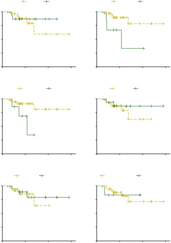
Page 2 of 9                                                                              Therapeutic Radiology and Oncology, 2020

Introduction                                                        our hospital. We present the following article in accordance
                                                                    with the STROBE reporting checklist (available at http://
Tenosynovial giant cell tumor (TGCT), historically
                                                                    dx.doi.org/10.21037/tro-20-41).
known as pigmented villonodular synovitis (PVNS), is a
rare, benign proliferative disorder arising from synovial
cells of joint capsules, bursae, and tendon sheaths most            Methods
commonly affecting young adults. The estimated incidence
                                                                    Patient recruitment
of TGCT/PVNS is about 1.8 per million population (1).
It is characterized histologically by the presence of               A retrospective study of orthopedic patients treated at
inflammation, hemosiderin deposition, multinucleated                Taipei Veterans General Hospital (VGHTPE) between
giant cells, and lipid-laden macrophages (2). There is no           January 2001 and December 2018 was performed by review
consensus on the etiology and pathogenesis of the disease.          of the electronic RT registry. The study was conducted in
Some risk factors have been recognized such as trauma,              accordance with the Declaration of Helsinki (as revised
chronic inflammation, and abnormal lipid metabolism (3).            in 2013). The study was approved by institutional ethics
Erosion of cartilage and bone will occur, causing deformity         board of VGHTPE (NO.: 2020-06-006AC) and a request
and limited range of motion in the affected joint if left           to waive of informed consent was approved because it was
untreated.                                                          no more than minimal risk. Eligibility included: (I) clinical
    TGCT/PVNS is typically a monoarticular process that             or pathological diagnosis of TGCT/PVNS, (II) receiving
usually affects large joints. Its clinical course is slow and       operation with adjuvant RT or definitive RT alone, (III)
insidious onset of pain, swelling, stiffness and reduced            at least one year of follow-up after treatment. Table 1
range of motion in the involved joint (4). It occurs in two         summarizes the characteristics of the 48 patients recruited.
histopathologic forms: the diffuse form, and the localized
form. The localized form is characterized by single, nodular
                                                                    Operation and RT
or pedunculated masses surrounded by normal synovial
tissue, while the diffuse form involves the entire synovium         For patients who received operations in our study, they
and accounts for the majority of cases (5). Magnetic                received open synovectomy, arthroscopic synovectomy
resonance imaging (MRI) is key to establishing the correct          or both, depending on orthopedic surgeons’ evaluation.
diagnosis showing nodular synovial proliferation with               Treatment was individualized according to patient
low signal on T1- and T2-weighted images, secondary to              characteristics and lesion extension. Adjuvant RT and
hemosiderin deposition (6-8).                                       definitive RT were delivered by linear accelerators with
    The first-line treatment of choice for all types of TGCT/       mega-voltage X-rays (4, 6, and 10 MV) using three-
PVNS is radical resection of the involved tissue, but the risk      dimensional conformal RT or intensity-modulated RT. The
of recurrence is high after the operation, especially in diffuse    median dose of irradiation was 30 Gy (range, 30–40 Gy),
TGCT/PVNS (9,10). Post-operative radiotherapy (RT) is               in 15 daily fractions of 2 Gy. The clinical target volume
not needed for localized TGCT/PVNS but it is associated             of RT included whole synovial space with TGCT/PVNS
with a reduced rate of recurrence for diffuse TGCT/PVNS             involvement as well as the cartilage and bony structures
(11,12). The planned treatment volume includes the whole            invaded by proliferative tissue. An expansion of at least
synovial space and eventually all invasive components of the        0.5 cm was delineated as the planning target volume.
disease as the synovial cells are the radiation target and an       An example of clinical target delineation and treatment
inhibition of its proliferation is suggested as RT mechanism        planning was illustrated in Figure 1.
(9,13,14).
    Due to small number of cases, studies in diffuse form
                                                                    Assessment of treatment results
TGCT/PVNS receiving RT are scarce. To the best of our
knowledge, this is the first article to discuss the local control   Patients were followed every 3 months in the first year,
(LC) rate via Kaplan-Meier estimates and prognostic factors         every 6 months in the second year and annually thereafter.
with a relatively large number of TGCT/PVNS patients.               Disease control was assessed by physical examination and
In this study, we retrospectively evaluated the treatment           MRI at 3, 6, 9, 12, 18 and 24 months and annually after
and outcome of TGCT/PVNS patients who received RT in                treatment to at least 5 years in all patients. If associated

© Therapeutic Radiology and Oncology. All rights reserved.             Ther Radiol Oncol 2020;4:14 | http://dx.doi.org/10.21037/tro-20-41
Therapeutic Radiology and Oncology, 2020 Page 3 of 9
Table 1 Demographic characteristics and treatment parameters A
Characteristic Number of patients (N=48)
Sex, n (%)
Male 19 (39.6)
Female 29 (60.4)
Age (years), median [range] 39 [15–76]
BMI (kg/m2), median [range] 23.1 [15.8–40.6]
Site, n (%)
B
Shoulder 1 (2.1)
Elbow 2 (4.2)
Hip 4 (8.3)
Knee 34 (70.8)
Ankle 7 (14.6)
Laterality, n (%)
Right 25 (52.1)
Left 23 (47.9)
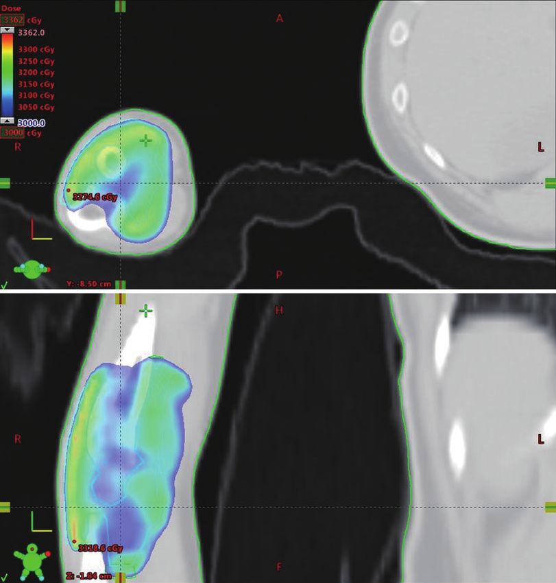
Figure 1 An example of radiotherapy planning of elbow
Previous trauma, n (%) 6 (12.5) tenosynovial giant cell tumor/pigmented villonodular synovitis
Symptoms, n (%) with axial (A) and coronal (B) view. A cast was made to stabilize
the patient position. Pre-operative magnetic resonance imaging
Pain 41 (85.4)
was co-registered with image fusion to help clinical target volume
Swelling 26 (54.2)
delineation (light cyan line). Intensity-modulated radiotherapy
Stiffness 4 (8.3) with five portals were used to create conformal plan.
No. of surgeries†, n (%)
0 29 (60.4)
symptoms occurred, additional MRI would be arranged.
1 12 (25.0) Treatment results were retrospectively assessed by post-
2 7 (14.6) treatment MRI with clinical correlation. Recurrence
Presenting status, n (%) was defined as the new appearance of a tumor lesion or
enlargement of residual soft tissue in the primary site,
Primary 29 (60.4)
assessed by experienced orthopedic radiologist. Persistent
Recurrence 19 (39.6)
thickening of synovium without interval change was
RT type, n (%) regarded as stable disease. Common Terminology Criteria
Definitive 8 (16.7) for Adverse Events (version 4.03) was used to evaluate
Post-operative 40 (83.3) adverse treatment effects.
‡
Deferred RT (>40 days) , n (%) 27 (56.3)
RT dose, n (%) Statistical analysis
30 Gy/15 Fx 41 (85.4) Descriptive statistics, including medians and proportions,
36 Gy/18 Fx 2 (4.2) were used to characterize the patients and treatment
40 Gy/20 Fx 5 (10.4)
results. The Kaplan-Meier method was used to determine
†
the LC rate. The duration of LC was defined as the time
, number of surgeries in previous courses, excluding surgery in
current course of treatment; ‡, deferred RT is defined as interval from
from the first date of intervention (operation or RT) to the
surgery to adjuvant RT or from diagnosis to definitive RT >40 days. date of any evidence of recurrence or the last follow-up.
BMI, body mass index; RT, radiotherapy; Fx, fraction(s). The Kaplan-Meier curves of LC with different variables
© Therapeutic Radiology and Oncology. All rights reserved. Ther Radiol Oncol 2020;4:14 | http://dx.doi.org/10.21037/tro-20-41Page 4 of 9 Therapeutic Radiology and Oncology, 2020
were compared by log-rank test. Variables affecting LC after RT and all of them were from the group of surgery
were analyzed by the univariate and multivariate Cox followed by post-operative RT while patients receiving
proportional hazard model. All the analyses were performed definitive RT were all free of recurrence. The median time
using R project (version R-3.6.3; http://www.r-project. of treatment to recurrence was 3.3 years (range, 1.7–6.9
org). A P value less than 0.05 was considered statistically years). Recurrences were treated by salvage operation alone
significant. in one patient, operation with adjuvant RT in one patient
and close follow-up in five patients due to slow progression
of symptoms.
Results
In univariate Cox regression analysis, obesity [body
Baseline characteristics mass index (BMI) >27 kg/m2] (hazard ratio =4.61; 95% CI,
1.08–19.67; P=0.039) as well as ankle joint affected (hazard
Twenty-nine women (60.4%) and 19 men (39.6%) were
ratio =4.46; 95% CI, 1.08–18.42; P=0.039) were found to be
identified, giving a gender ratio (female/male) of 1.53,
significant prognostic factors for local recurrence (Table 2).
with a median age of 39 years (range, 15–76 years). Among Sex, disease status (primary or recurrence), timing of RT
our patients, 46 patients (95.8%) received pre-treatment and RT dose were not significant factors for recurrence
evaluation via MRI, while only 2 patients (4.2%) were (Figure 2). However, only obesity remained significance in
determined by plain film and clinical findings. Forty- multivariate Cox proportional hazard analysis (hazard ratio
seven patients (97.9%) had pathology confirmation of the =5.32; 95% CI, 1.07–26.50; P=0.041) (Table 2).
disease. The knee joint was affected in 34 patients (70.8%). The overall 1-, 3- and 5-year LC rate was 100%, 92.8%
All patients had diffuse form TGCT/PVNS. Six patients and 87.2%, respectively (Figure 3). The obese group (BMI
(12.5%) reported a prior history of trauma to the involved >27 kg/m2) had a 1-, 3- and 5-year LC rate of 100%, 66.7%
joint. Most common clinical symptoms were pain (85.4%), and 66.7% while the non-obese group had a 1-, 3- and
followed by swelling (54.2%) and stiffness (8.3%). Other 5-year LC rate of 100%, 97.0% and 89.4%, respectively
baseline characteristics were summarized in Table 1. (Figure 2B).
Treatment modalities Toxicities and compliance
A total of 19 patients (39.6%) were treated as recurrent No significant surgical complication was found. In terms of
disease and had received one or two surgeries in previous RT-related acute toxicities, there were 12 patients (25.0%)
courses of treatment. Eight patients (16.7%) were not reporting grade 1 dermatitis, 11 patients (22.9%) with grade
suitable for surgery because of probable function loss 1 joint effusion and two patients (4.2%) with grade 1 joint
of the affected joint so definitive RT (dose 30–40 Gy) range of motion decrease. There was no interruption of RT
was delivered. Of the 40 patients having surgery in and all the patients completed treatment by schedule. No
current course, 37 patients (92.5%) were treated with late toxicity or secondary malignancy was found during the
open synovectomy while two patients (5.0%) received follow-up period. Total joint arthroplasty was required in
arthroscopic synovectomy and one patient (2.5%) five patients (10.4%) because of symptomatic osteoarthritis.
underwent both. The interval between surgery and post- The median time of treatment to arthroplasty was 4.5 years
operative RT or diagnosis to definitive RT was greater (range, 0.9–11.7 years). None of the five patients had
than 40 days in 23 patients (47.9%) and it was regarded as disease recurrence.
deferred RT. Most of the patients (85.4%) received RT dose
of 30 Gy in 15 fractions (Table 1). Higher dose was given in
Discussion
patients with high risk of recurrence or when gross residual
tumor was highly suspected by the radiation oncologists. In our study, we found that obesity (BMI >27 kg/m 2) is
a prognostic factor via multivariate Cox proportional
hazard analysis. Possible explanation includes greater
Outcomes and factors
loading of weight-bearing joints as well as abnormal lipid
The median follow-up time was 4.3 years (range, 1.2– metabolism which is suspected to be a possible etiology
16.4 years). Seven patients (14.6%) had disease recurrence of TGCT/PVNS (15). Obesity is also associated with
© Therapeutic Radiology and Oncology. All rights reserved. Ther Radiol Oncol 2020;4:14 | http://dx.doi.org/10.21037/tro-20-41Therapeutic Radiology and Oncology, 2020 Page 5 of 9
Table 2 The hazard ratio and P values of each factor in univariate and multivariate Cox model for disease recurrence
Univariate analysis Multivariate analysis
Factor
Hazard ratio (95% CI) P value Hazard ratio (95% CI) P value
Sex
Male 0.60 (0.10–3.54) 0.575 0.42 (0.05–3.35) 0.417
Female Reference – Reference –
BMI
>27 kg/m2 4.61 (1.08–19.67) 0.039* 5.32 (1.07–26.50) 0.041*
2
≤27 kg/m Reference – Reference –
Site
Ankle 4.46 (1.08–18.42) 0.039* 2.76 (0.46–16.69) 0.269
Sites other than ankle Reference – Reference –
Status
Recurrence 0.55 (0.10–2.88) 0.478 0.59 (0.13–2.75) 0.502
Primary Reference – Reference –
†
Deferred RT >40 days
Yes 0.75 (0.17–3.34) 0.705 0.45 (0.07–3.00) 0.413
No Reference – Reference –
RT dose
>30 Gy 1.02 (0.10–10.10) 0.987 2.67 (0.26–27.55) 0.409
30 Gy Reference – Reference –
†
, deferred RT is defined as interval from surgery to adjuvant RT or from diagnosis to definitive RT >40 days; *, P valuePage 6 of 9 Therapeutic Radiology and Oncology, 2020
A Female Male B BMI ≤27 BMI >27
1.00 1.00
0.75 0.75
Local control rate
Local control rate
0.50 0.50
P=0.55 P=0.028
0.25 0.25
0.00 0.00
0 5 10 15 0 5 10 15
Follow-up time (years) Follow-up time (years)
C Other sites Ankle D Primary Recurrence
1.00 1.00
0.75 0.75
Local control rate
Local control rate
0.50 0.50
P=0.033 P=0.47
0.25 0.25
0.00 0.00
0 5 10 15 0 5 10 15
Follow-up time (years) Follow-up time (years)
E Early RT Deferred RT F RT dose =30 Gy RT dose >30 Gy
1.00 1.00
0.75 0.75
Local control rate
Local control rate
0.50 0.50
P=0.71 P=0.99
0.25 0.25
0.00 0.00
0 5 10 15 0 5 10 15
Follow-up time (years) Follow-up time (years)
Figure 2 Kaplan-Meier curves of local control rate by sex (A), body mass index (BMI) (kg/m2) (B), sites (ankle or other sites) (C), status
(primary or recurrence) (D), timing of radiotherapy (RT) (early or deferred) with cut-off value of 40 days from surgery to RT or diagnosis to
definitive RT (E), and RT dose (F). The P values were calculated by log-rank test.
© Therapeutic Radiology and Oncology. All rights reserved. Ther Radiol Oncol 2020;4:14 | http://dx.doi.org/10.21037/tro-20-41Therapeutic Radiology and Oncology, 2020 Page 7 of 9
1.00 shown robust response in TGCT/PVNS cases not amenable
to improvement with surgery in a phase 3 trial (21).
0.75
However, those target agents were not available in our
institute.
Local control rate
Our study has several limitations. Firstly, the number
0.50 of the participants is not large enough that association
between some factors and outcome may be obscured.
0.25
However, it is a rare disease and we still manage to find
significant factors from our cases. Secondly, we didn’t
obtain the data of inflammatory markers such as C-reactive
0.00 protein and neutrophil-lymphocyte ratio which were found
0 5 10 15
Follow-up time (years) to be significant predictors in a previous study (23). Thirdly,
Figure 3 Kaplan-Meier curve of local control rate for all patients.
absence of quality-of-life information and functional score
evaluation makes it difficult to present a comprehensive
outcome of our treatment. In addition, the follow-up time is
The outcomes of our study were consistent with historical not long enough for late recurrence and radiation-induced
data. A meta-analysis published in 2015 reported recurrence secondary malignancy.
rate of 12.0% in the peri-operative RT group of diffuse In conclusion, our results revealed good LC and low
TGCT/PVNS patients (11) which is compatible to our treatment toxicities in patients with TGCT/PVNS who
results with crude recurrence rate of 14.6% and 5-year LC received RT. Obesity (BMI >27 kg/m2) was associated with
rate of 87.2%. A study from German Cooperative Group higher recurrence rate among these patients. Definitive RT
on Radiotherapy in Benign Diseases revealed a dose range may be a treatment option for inoperable patients.
of 30–50 Gy (median, 36 Gy) as adjuvant RT (18). Our data
showed that 30 Gy in 15 fractions was sufficient for most Acknowledgments
of the patients and higher dose did not reduce the risk for
recurrence. We would like to express our gratitude to all the members
It is challenging to manage patients with inoperable in Department of Oncology and Orthopedic Surgery in
diffuse TGCT/PVNS. Eight patients who were unfit for Taipei Veterans General Hospital.
surgery underwent definitive RT alone (dose 30–40 Gy). Funding: None.
None of them had recurrence at the end of follow-up.
Although the case number is small, moderate dose definitive
Footnote
RT seems to be an effective treatment option for inoperable
patients. Such treatment modality may be investigated in Reporting Checklist: The authors have completed the
the future. As for persistent or recurrent disease, further STROBE reporting checklist. Available at http://dx.doi.
surgical intervention such as total synovectomy, subtotal org/10.21037/tro-20-41
resection, joint replacement, or amputation is recommended
first. Since TGCT/PVNS is a non-fatal disease, functional Conflicts of Interest: All authors have completed the ICMJE
outcome, and quality of life after surgery should be uniform disclosure form (available at http://dx.doi.
considered carefully. Another treatment of choice for this org/10.21037/tro-20-41). LWW serves as an unpaid
population is colony stimulating factor 1 (CSF1) receptor editorial board member of Therapeutic Radiology and
inhibitors such as pexidartinib, nilotinib, emactuzumab Oncology from May 2020 to Apr 2022. YML serves as an
which have been studied in clinical studies (19-21). In unpaid editorial board member of Therapeutic Radiology and
TGCT/PVNS, a translocation in CSF1-COL6A3, t(1;2) Oncology from Jun 2020 to May 2022. The other authors
(p13;q35) causes overexpression of CSF1, attracting non- have no conflicts of interest to declare.
neoplastic cells which express CSF1 receptor (22). The
accumulation of those non-neoplastic cells creates tumorous Ethical Statement: The authors are accountable for all
mass. Inhibition of CSF1 receptor by pexidartinib has aspects of the work in ensuring that questions related
© Therapeutic Radiology and Oncology. All rights reserved. Ther Radiol Oncol 2020;4:14 | http://dx.doi.org/10.21037/tro-20-41Page 8 of 9 Therapeutic Radiology and Oncology, 2020
to the accuracy or integrity of any part of the work are malignant disorders: part III: hyperproliferative disorders.
appropriately investigated and resolved. The study was Strahlenther Onkol 2015;191:541-8.
conducted in accordance with the Declaration of Helsinki (as 10. Korim MT, Clarke DR, Allen PE, et al. Clinical and
revised in 2013). The study was approved by institutional oncological outcomes after surgical excision of pigmented
ethics board of VGHTPE (NO.: 2020-06-006AC) and a villonodular synovitis at the foot and ankle. Foot Ankle
request to waive of informed consent was approved because Surg 2014;20:130-4.
it was no more than minimal risk. 11. Mollon B, Lee A, Busse JW, et al. The effect of surgical
synovectomy and radiotherapy on the rate of recurrence of
Open Access Statement: This is an Open Access article pigmented villonodular synovitis of the knee: an individual
distributed in accordance with the Creative Commons patient meta-analysis. Bone Joint J 2015;97-B:550-7.
Attribution-NonCommercial-NoDerivs 4.0 International 12. Guo Q, Shi W, Jiao C, et al. Results and recurrence
License (CC BY-NC-ND 4.0), which permits the non- of pigmented villonodular synovitis of the ankle: does
commercial replication and distribution of the article with diffuse PVNS with extra-articular extension tend to
the strict proviso that no changes or edits are made and the recur more often? Knee Surg Sports Traumatol Arthrosc
original work is properly cited (including links to both the 2018;26:3118-23.
formal publication through the relevant DOI and the license). 13. Heyd R, Seegenschmiedt MH, Micke O. The role of
See: https://creativecommons.org/licenses/by-nc-nd/4.0/. external beam radiation therapy in the adjuvant treatment
of pigmented villonodular synovitis. Z Orthop Unfall
2011;149:677-82.
References
14. Duan Y, Qian J, Chen K, et al. Necessity of adjuvant
1. Myers BW, Masi AT. Pigmented villonodular synovitis and postoperative radiotherapy for diffuse pigmented
tenosynovitis: a clinical epidemiologic study of 166 cases villonodular synovitis of the knee: A case report and
and literature review. Medicine (Baltimore) 1980;59:223-38. literature review. Medicine (Baltimore) 2018;97:e9637.
2. Goldman AB, DiCarlo EF. Pigmented villonodular 15. Steinmetz S, Rougemont AL, Peter R. Pigmented
synovitis. Diagnosis and differential diagnosis. Radiol Clin villonodular synovitis of the hip. EFORT Open Rev
North Am 1988;26:1327-47. 2017;1:260-6.
3. Ottaviani S, Ayral X, Dougados M, et al. Pigmented 16. Karczewski J, Śledzińska E, Baturo A, et al. Obesity and
villonodular synovitis: a retrospective single-center study inflammation. Eur Cytokine Netw 2018;29:83-94.
of 122 cases and review of the literature. Semin Arthritis 17. Griffin AM, Ferguson PC, Catton CN, et al. Long-
Rheum 2011;40:539-46. term outcome of the treatment of high-risk tenosynovial
4. Gelhorn HL, Tong S, McQuarrie K, et al. Patient- giant cell tumor/pigmented villonodular synovitis with
reported Symptoms of Tenosynovial Giant Cell Tumors. radiotherapy and surgery. Cancer 2012;118:4901-9.
Clin Ther 2016;38:778-93. 18. Heyd R, Micke O, Berger B, et al. Radiation therapy for
5. Abdul-Karim FW, el-Naggar AK, Joyce MJ, et al. Diffuse treatment of pigmented villonodular synovitis: results of
and localized tenosynovial giant cell tumor and pigmented a national patterns of care study. Int J Radiat Oncol Biol
villonodular synovitis: a clinicopathologic and flow Phys 2010;78:199-204.
cytometric DNA analysis. Hum Pathol 1992;23:729-35. 19. Cassier PA, Italiano A, Gomez-Roca CA, et al. CSF1R
6. Bhimani MA, Wenz JF, Frassica FJ. Pigmented inhibition with emactuzumab in locally advanced diffuse-
villonodular synovitis: keys to early diagnosis. Clin Orthop type tenosynovial giant cell tumours of the soft tissue: a
Relat Res 2001;(386):197-202. dose-escalation and dose-expansion phase 1 study. Lancet
7. Cheng XG, You YH, Liu W, et al. MRI features of Oncol 2015;16:949-56.
pigmented villonodular synovitis (PVNS). Clin Rheumatol 20. Gelderblom H, Cropet C, Chevreau C, et al. Nilotinib
2004;23:31-4. in locally advanced pigmented villonodular synovitis: a
8. Dürr HR, Stäbler A, Maier M, et al. Pigmented multicentre, open-label, single-arm, phase 2 trial. Lancet
villonodular synovitis. Review of 20 cases. J Rheumatol Oncol 2018;19:639-48.
2001;28:1620-30. 21. Tap WD, Gelderblom H, Palmerini E, et al. Pexidartinib
9. Seegenschmiedt MH, Micke O, Niewald M, et al. versus placebo for advanced tenosynovial giant cell
DEGRO guidelines for the radiotherapy of non- tumour (ENLIVEN): a randomised phase 3 trial. Lancet
© Therapeutic Radiology and Oncology. All rights reserved. Ther Radiol Oncol 2020;4:14 | http://dx.doi.org/10.21037/tro-20-41Therapeutic Radiology and Oncology, 2020 Page 9 of 9
2019;394:478-87. 23. Zhao G, Wang J, Xia J, et al. The predictive value of
22. West RB, Rubin BP, Miller MA, et al. A landscape effect preoperative neutrophil-lymphocyte ratio (NLR) on the
in tenosynovial giant-cell tumor from activation of CSF1 recurrence of the local pigmented villonodular synovitis of
expression by a translocation in a minority of tumor cells. the knee joint. BMC Musculoskelet Disord 2018;19:339.
Proc Natl Acad Sci U S A 2006;103:690-5.
doi: 10.21037/tro-20-41
Cite this article as: Lin CS, Kang YM, Lan KL, Wang LW,
Liu YM, Huang PI, Hu YW, Lai IC, Wu YH, Lai TY, Yang
WC, Lee JC, Shiau CY. Treatment and outcome of tenosynovial
giant cell tumor/pigmented villonodular synovitis patients
receiving radiotherapy in Taiwan: a single-center experience.
Ther Radiol Oncol 2020;4:14.
© Therapeutic Radiology and Oncology. All rights reserved. Ther Radiol Oncol 2020;4:14 | http://dx.doi.org/10.21037/tro-20-41You can also read


