Tomato Divinyl Ether-Biosynthesis Pathway Is Implicated in Modulating of Root-Knot Nematode Meloidogyne javanica's Parasitic Ability
←
→
Page content transcription
If your browser does not render page correctly, please read the page content below
ORIGINAL RESEARCH
                                                                                                                                           published: 25 August 2021
                                                                                                                                      doi: 10.3389/fpls.2021.670772

                                             Tomato Divinyl Ether-Biosynthesis
                                             Pathway Is Implicated in Modulating
                                             of Root-Knot Nematode Meloidogyne
                                             javanica’s Parasitic Ability
                                             Payal Sanadhya 1 , Anil Kumar 1 , Patricia Bucki 1 , Nathalia Fitoussi 1,2 ,
                                             Mira Carmeli-Weissberg 3 , Menachem Borenstein 4 and Sigal Brown-Miyara 1*
                                             1
                                              Department of Entomology, Nematology and Chemistry Units, Agricultural Research Organization, The Volcani Center, Bet
                                             Dagan, Israel, 2 Department of Plant Pathology and Microbiology, The Faculty of Agriculture, Food and Environment, The
                                             Hebrew University of Jerusalem, Rehovot, Israel, 3 Metabolomics, Institute of Plant Sciences, Agricultural Research
                                             Organization, The Volcani Center, Bet Dagan, Israel, 4 Department of Plant Pathology and Weed Research, Agricultural
                          Edited by:
                                             Research Organization (ARO), The Volcani Center, Bet Dagan, Israel
              Michael V. Kolomiets,
  Texas A&M University, United States

                        Reviewed by:         The role of the 9-lipoxygenase (9-LOX)-derived oxylipins in plant defense is mainly known
                       Sofia R. Costa,
                                             in solanaceous plants. In this work, we identify the functional role of the tomato divinyl
         University of Minho, Portugal
                 Ana López Sánchez,          ether synthase (LeDES) branch, which exclusively converts 9-hydroperoxides to the
     National Center of Biotechnology        9-divinyl ethers (DVEs) colneleic acid (CA) and colnelenic acid (CnA), during infection
                          (CSIC), Spain
                 Carmen Castresana,
                                             by the root-knot nematode Meloidogyne javanica. Analysis of LeDES expression in
     National Center of Biotechnology        roots indicated a concurrent response to nematode infection, demonstrating a sharp
   (CSIC), Spain, in collaboration with
                                             increase in expression during the molting of third/fourth-stage juveniles, 15 days after
                            reviewer AS
                                             inoculation. Spatiotemporal expression analysis using an LeDES promoter:GUS tomato
                  *Correspondence:
                   Sigal Brown-Miyara        line showed high GUS activity associated with the developing gall; however the GUS
            sigalhor@volcani.agri.gov.il     signal became more constricted as infection progressed to the mature nematode feeding
                                             sites, and eventually disappeared. Wounding did not activate the LeDES promoter, but
                    Specialty section:
          This article was submitted to      auxins and methyl salicylate triggered LeDES expression, indicating a hormone-mediated
          Plant Pathogen Interactions,       function of DVEs. Heterologous expression of LeDES in Arabidopsis thaliana rendered
                a section of the journal
             Frontiers in Plant Science
                                             the plants more resistant to nematode infection and resulted in a significant reduction
         Received: 22 February 2021
                                             in third/fourth-stage juveniles and adult females as compared to a vector control and
            Accepted: 21 July 2021           the wild type. To further evaluate the nematotoxic activity of the DVEs CA and CnA,
          Published: 25 August 2021          recombinant yeast that catalyzes the formation of CA and CnA from 9-hydroperoxides
                               Citation:     was generated. Transgenic yeast accumulating CnA was tested for its impact on
        Sanadhya P, Kumar A, Bucki P,
     Fitoussi N, Carmeli-Weissberg M,        M. javanica juveniles, indicating a decrease in second-stage juvenile motility. Taken
   Borenstein M and Brown-Miyara S           together, our results suggest an important role for LeDES as a determinant in the defense
                  (2021) Tomato Divinyl
         Ether-Biosynthesis Pathway Is
                                             response during M. javanica parasitism, and indicate two functional modes: directly via
Implicated in Modulating of Root-Knot        DVE motility inhibition effect and through signal molecule-mediated defense reactions to
   Nematode Meloidogyne javanica’s           nematodes that depend on methyl salicylate.
                        Parasitic Ability.
           Front. Plant Sci. 12:670772.      Keywords: divinyl ether synthase, Meloidogyne javanica, plant defense signaling, oxylipins, hormone signaling,
        doi: 10.3389/fpls.2021.670772        innate immunity

Frontiers in Plant Science | www.frontiersin.org                                  1                                         August 2021 | Volume 12 | Article 670772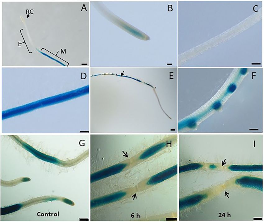
Sanadhya et al.                                                                                                    LeDES Regulates RKN Parasitism

INTRODUCTION                                                                    Infection of potato leaves by the pathogens Phytophthora
                                                                            infestans and Pseudomonas syringae leads to elevated
Root knot nematodes (RKNs) are devastating pathogens of                     transcription of DES and accumulation of the 9-divinyl
a wide variety of plants, causing substantial annual losses in              ethers (DVEs) colneleic acid and colnelenic acid (CA and
yield worldwide (McCarter, 2009). They also incur the intensive             CnA, respectively; Weber et al., 1999; Stumpe et al., 2001).
use of toxic nematicides that pose a great threat to human                  The involvement of CA and CnA in plant defense was also
and environmental health (Hague and Gowen, 1987; Johnson                    demonstrated by Fammartino et al. (2007), who reported high
and Feldmesser, 1987; Thomason, 1987; Desaeger et al., 2020).               concentrations of CA and CnA in wild-type (WT) tobacco roots
The consequent withdrawal of frontline nematicides has put a                challenged with P. parasitica var. nicotianae, whereas silencing
significant dent in our ability to control RKNs, as well as all other       of NtLOX1 downregulated the production of CA and CnA. The
plant-parasitic nematodes. The Meloidogyne life cycle consists              DES genes involved in the synthesis of CA and CnA in potato
of six stages: eggs, J1 (first-stage juvenile), J2 (second-stage            and tomato have been cloned and characterized (Itoh and Howe,
juvenile), J3 (third-stage juvenile), J4 (fourth-stage juvenile), and       2001; Stumpe et al., 2001). 9-Divinyl ethers have an important
adults (female and male). The J2, which are motile and infective,           role in defense mechanisms against different plant pathogens
pierce the elongation zone of the plant root near its tip and               and can act as antimicrobial agents (Weber et al., 1999; Göbel
migrate intercellularly through the root apex to the vascular               et al., 2001; Stumpe et al., 2001; Granér et al., 2003; Fammartino
cylinder (Bird et al., 2009). Once they reach the differentiation           et al., 2007) or signaling components that induce further
zone in the vascular bundle, they become sessile and induce the             defense responses. Although much literature is available on the
transformation of 4–8 root cells into coenocytic giant cells that           structural diversity of DVEs (Göbel and Feussner, 2009) their
operate as feeding sites, supplying the RKNs with the nutrients             occurrence in different plants and their role as antimicrobial
required for their development and reproduction (Wyss et al.,               agents (Weber et al., 1999; Prost et al., 2005; Fammartino et al.,
1992; Davis et al., 2004; Bird et al., 2009).                               2007); information on transcriptional regulation of DES and its
    Transcriptomic studies of Arabidopsis thaliana, Glycine max             potential role in mediating defense responses is rather scarce,
(soybean), and Solanum lycopersicum (tomato) plants in response             with no knowledge of its function in the tomato–RKN system.
to RKN infection have revealed complicated multistep signaling              Here, we investigated the functional role of DES from tomato
cascades that are instigated by pathogen attack (Bar-Or et al.,             (S. lycopersicum) in response to nematode infection. To further
2005; Jammes et al., 2005; Alkharouf et al., 2006; Szakasits et al.,        study the spatial and temporal expression of LeDES, we generated
2009). Among them, fatty acid signaling produces oxygenated                 pLeDES::GUS transgenic hairy root lines which show induced
fatty acids known as oxylipins, which are involved in defense               expression of LeDES in response to early and middle–late stages
against pathogen attack (Feussner and Wasternack, 2002).                    (15 days) of Meloidogyne javanica infection. In addition, the
Studies have shown that various oxylipin-biosynthesis pathway               full-length LeDES was functionally characterized by heterologous
genes are induced in plants by pathogen attack (Deboever et al.,            expression in Arabidopsis plants. Characterization of transgenic
2020; Estelle et al., 2020), along with a spike in the concentration        lines indicated a significant reduction in the number of M.
of related oxylipins as shown in oxylipin-profiling studies (Weber          javanica developmental stages and suggests a role for the DVEs
et al., 1999; Göbel et al., 2001; Mariutto et al., 2014; Fitoussi           in modulating M. javanica disease development in the host root.
et al., 2021). Several studies on mutant or transgenic plants with
altered synthesis or signaling of oxylipins have suggested a direct
                                                                            MATERIALS AND METHODS
signaling role for oxylipins in the plant’s response to pathogen
attack (Deboever et al., 2020), acting as antimicrobial (Croft              Plant Materials and Growth Conditions
et al., 1993; Weber et al., 1999; Granér et al., 2003; Prost et al.,        Tomato (Lycopersicon esculentum) cv Avigail 870 was used as the
2005) or nematicidal agents (Naor et al., 2018). Overexpression             background line for the transformation in experiments involved
of jasmonic acid carboxyl methyltransferase in Arabidopsis or               tomato plants. For the transformation protocol, tomato seeds
9-lipoxygenase (9-LOX) in tobacco conferred resistance against              were treated with 1.6% NaOCl for 10 min with continuous
the necrotrophic pathogen Botrytis cinerea (Seo et al., 2001) and           shaking, washed with sterile water for 5 min, and placed on
the oomycete pathogen Phytophthora parasitica (Mène-Saffrané                standard-strength Gambourg’s B5 medium supplemented with
et al., 2003). Ozalvo et al. (2014) also demonstrated the role              2% sucrose and 0.8% Gelrite agar (Duchefa, Haarlem, The
of 13-LOXs in Arabidopsis; they found that LOX3 and LOX4                    Netherlands). The plates were kept in darkness at 26◦ C for
are involved in the response to nematodes, and that LOX4 is                 2 days, followed by 2 weeks under a 16/8-h photoperiod
required for resistance to nematode infection. High expression              until cotyledons emerged. They then were used immediately
levels of the divinyl ether synthase (DES) and 9-LOX genes was              for cocultivation as described by Chinnapandi et al. (2017).
also detected in pepper (Capsicum annuum) leaves inoculated                 Similarly, tomato roots sections were subculture, by placing one
with Obuda pepper virus (Gullner et al., 2010; Juhász et al., 2015).        root per Petri dish (Miniplast, Ein Shemer, Israel), containing
Moreover, disruption of LOX1 in pepper plants renders them                  B5 medium. Branching roots were used for inoculation as
more susceptible to Xanthomonas campestris pv. vesicatoria and              described below. For experiments conducted on Arabidopsis,
Colletotrichum coccodes infection, further emphasizing the role             A. thaliana WT and transgenic lines were grown in a
of LOX-biosynthesis pathway products in pepper plants defense               growth chamber at 24◦ C on MS medium (Murashige and
response (Hwang and Hwang, 2010).                                           Skoog, 1962) or in pots filled with cocopeat. The plants

Frontiers in Plant Science | www.frontiersin.org                        2                                   August 2021 | Volume 12 | Article 670772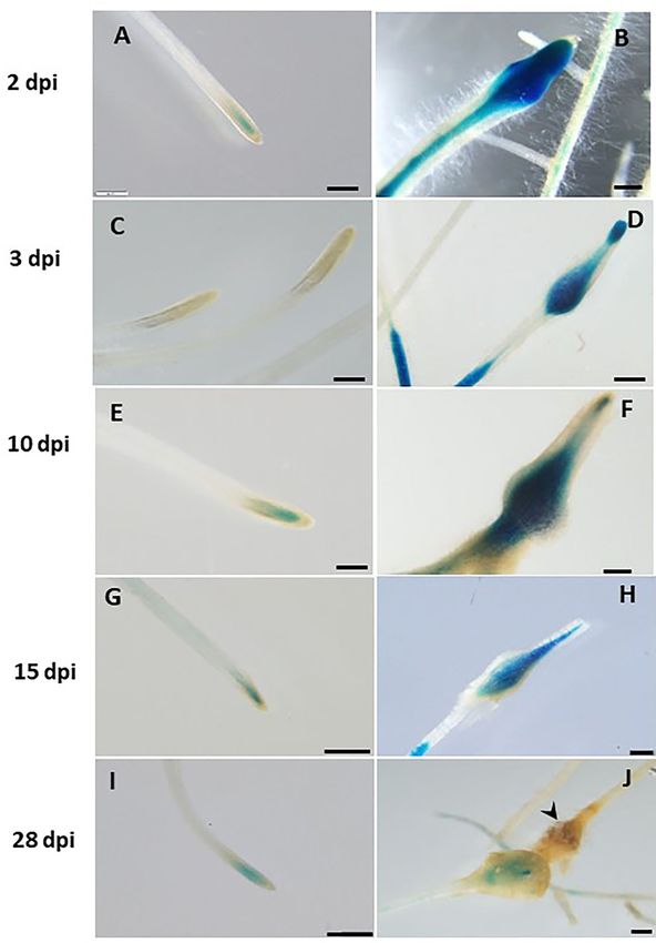
Sanadhya et al.                                                                                                 LeDES Regulates RKN Parasitism

were maintained under a 14 h/10 h day/night cycle at a light              RNA was extracted using Trizol reagent (Invitrogen, Carlsbad,
intensity of 50–60 µmol/m2 s. Arabidopsis thaliana ecotype                CA, USA) and subjected to Turbo DNase (Ambion, Thermo
Columbia-0 (Col-0) was used as the genetic background for all             Fisher Scientific, Waltham, MA, USA). One microgram of total
transgenic lines.                                                         RNA was reverse-transcribed to cDNA using the Verso cDNA
                                                                          Synthesis Kit (Thermo Scientific, Waltham, MA, USA) according
Nematode Inoculation and Infection of                                     to the manufacturer’s instructions; qRT-PCR was carried out in
Tomato and Arabidopsis Plants                                             a StepOne Real Time PCR system (Applied Biosystems, Foster
Meloidogyne javanica was propagated on greenhouse-grown                   City, CA, USA) in 10 µl reaction mix comprised of 3.4 µl
tomato “Avigail” (870) plants. Nematode egg masses were                   cDNA, SYBR-Green ROX Mix (Abgene, Portsmouth, NH, USA),
extracted from roots with 0.05% (v/v) sodium hypochlorite                 150 nM forward primer and 150 nM reverse primer as described
followed by sucrose flotation (Hussey and Baker, 1973).                   in Chinnapandi et al. (2019). Reactions were performed in
For sterilization, eggs were placed on a cellulose–acetate                MicroAmp 96-well plates (Applied Biosystems) and PCR cycles
filter membrane (Sartorius Stedim Biotech GmbH, Goettingen,               consisted of initial denaturation at 95◦ C for 10 min; 40 cycles
Germany, pore size 5 µm) in a sterile Whatman R filter holder             at 95◦ C for 15 s, 56◦ C for 20 s, and 72◦ C for 20 s, and a
(Whatman International Ltd., Dassel, Germany). Eggs on the                final extension at 72◦ C for 2 min. Reactions were conducted in
filter were exposed for 10 min to 0.01% (w/v) mercuric chloride           triplicate, with a control with no template, and the presented
(Sigma-Aldrich, St Louis, MO, USA), followed by 0.7% (v/v)                results are the means of two independent biological experiments.
streptomycin solution (Sigma-Aldrich), and three washing steps            The qRT-PCR results were analyzed and interpreted using the
with 50 ml sterilized distilled water (Jansen van Vuuren and              2−11Ct method (Livak and Schmittgen, 2001) integrated into
Woodward, 2001). The sterilized eggs were collected from the              StepOne v2.3 of the StepOne Plus (Applied Biosystems) real-time
membrane and placed on 25-µm-pore sieves in 0.01 M 2-                     PCR instrument. Tomato β-tubulin gene (GenBank accession
morpholinoethanesulfonic acid buffer (Sigma-Aldrich) under                number NM_001247878.1) was used as the internal reference to
aseptic dark conditions for 3 days, allowing J2s to hatch. Freshly        normalize the expression of LeDES.
hatched preparasitic J2s were collected in a 50 ml falcon tube.
For nematode infection, 1-week-old transgenic tomato root hairy           Plasmid Construction and Generation of
root lines, growing on standard-strength Gamborg’s B5 salt                pLeDES::GUS Reporter Hairy-Root
medium, were inoculated with 200 sterile freshly hatched M.
javanica preparasitic J2s. Plates were left uncovered in a laminar        Cultures
flow hood until water had completely soaked into the medium               Genomic DNA was isolated from soil-grown tomato seedlings,
(Sijmons et al., 1991). The inoculated and non-inoculated roots           cv. Avigail 870, according to the cetyltrimethylammonium
were incubated horizontally in the dark, and root samples were            bromide (CTAB) method (Murray and Thompson, 1980).
taken for GUS bioassay at the designated time points after                A promoter–GUS construct, to study the expression
wounding or inoculation. To test the response of transgenic               pattern of LeDES, was generated by Gateway Technology
Arabidopsis plants to nematode infection, three transgenic lines          (Invitrogen). A 1,602-bp sequence upstream of the ATG
were used, 10 plants per line in each experiment. Seeds of                start codon of LeDES was amplified with Platinum Taq
WT, vector-only and transgenic Arabidopsis lines were surface             DNA polymerase (Invitrogen) using the primers LeDESprom
sterilized and germinated on standard-strength GB media with              (attB1)     5′ -GGGGACAAGTTTGTACAAAAAAGCAGGCT-
2% (w/v) sucrose and 0.8% (w/v) Gelrite as described previously           GTATAGTGTAGCTACGCGCTT-3′ and LeDESprom (attB2) 5′ -
by Joshi et al. (2019). One-week-old seedlings grown in vitro were        GGGGACCACTTTGTACAAGAAAGCTGGGT-TTTTCTTA
gently transferred with a forceps to a petri dish containing GB           ACAAGTTTTGG-3′ and tomato genomic DNA as the template.
media, one plant per dish. Each seedling was inoculated with 300          The purified product was first cloned into the pDONR221
sterile freshly hatched M. javanica J2s applied near the roots; the       vector (Invitrogen) by BP reaction, generating the entry vector.
plates were left open until the solution was completely absorbed          The entry vector was then cloned by LR Clonase II (Thermo
into the media (Sijmons et al., 1991). The infected seedlings were        Fisher Scientific) into the destination vector pKGWFS7 (VIB-
kept vertically and root samples were gently harvested 28 days            Ugent Center for Plant System Biology, Ghent, Belgium)
post-infection (dpi).                                                     upstream of GFP::GUS-coding sequences, generating the
                                                                          pKGWFS7:prom-LeDES (pLeDES::GUS) construct. The resulting
                                                                          promoter construct was confirmed by sequencing prior to
LeDES Transcript Accumulation, RNA                                        Rhizobium rhizogenes transformation. The pLeDES::GUS
Isolation, and Quantitative RT-PCR                                        construct was mobilized to Rhizobium rhizogenes ATCC 15834
(qRT-PCR) Analysis                                                        by freeze–thaw procedure as described by Xu and Li (2008).
Expression of LeDES in infected tomato roots (non-transgenic)             The transformed R. rhizogenes colonies were confirmed by
at different time points was analyzed by qRT-PCR using gene-              colony PCR using LeDES promoter-specific primers (pLeDESF:
specific primers LeDes-rtF: 5′ -CCGGATGAGTTTGTACCTGA-                     5′ -GTATAGTGGAGCTCCGCGCTT-3′ and pLeDESR: 5′ -
3′ and LeDes-rtR: 5′ -ATCTTTGCCTGGACATTGCT-3′ (López-                     CCCCCGGGTTTTCTTAACAAGTTTTGG-3′ ) and kanamycin
Ráez et al., 2010). Ten root systems (100 mg) from each time              primers (kanF: 5′ -GCTCTTCGTCCAGATCATCC-3′ and
point were pooled from uninfected and infected roots. Total               kanR: 5′ -GCGTTCAAAAGTCGCCTAAG-3′ ).

Frontiers in Plant Science | www.frontiersin.org                      3                                  August 2021 | Volume 12 | Article 670772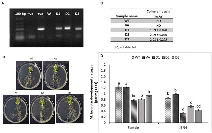
Sanadhya et al.                                                                                                 LeDES Regulates RKN Parasitism

   R. rhizogenes-mediated transformation of tomato cotyledons             (v/v) formaldehyde in 50 mM sodium phosphate buffer pH
was carried out according to the protocol described by                    7.2, dehydrated, and embedded in Technovit 7100 (Heraeus
Chinnapandi et al. (2017). Two weeks after transformation,                Kulzer, Wehrheim, Germany) according to the manufacturer’s
cotyledons with emerging kanamycin-resistant roots were                   instructions. Semi-thin sections (3 µm) were cut, mounted
transferred to Gamborg’s B5 salts medium (GB; Duchefa                     in DePeX (Sigma-Aldrich) and observed under a Nikon DS
Biochemie, Haarlem, The Netherlands) containing 0.8% (w/v)                Ri2-equipped microscope with dark-field illumination (Leica
Gelrite + cefotaxime (200 mg/L) + kanamycin (50 mg/L). Roots              Microsystems GmbH).
were excised from the cotyledons once they were 1.0 cm long. The
excised roots were transferred to individual GB medium plates             Plasmid Construction and Agrobacterium
(with 200 mg/L cefotaxime + 50 mg/L kanamycin). After two
rounds of subculture, cefotaxime was eliminated from the media
                                                                          tumefaciens-Mediated Transformation of
and transgenic roots were maintained on GB medium plates                  Arabidopsis Plants
(with 50 mg/L kanamycin) for further analysis. The genomic                For amplification of LeDES, PCR was performed with
DNA of the hairy roots was isolated and used as a template                cDNA from 1-month-old tomato plants as the template.
for identification of the positive transgenic lines with promoter-        The full-length LeDES (1,437 bp) coding sequence
specific (pLeDESF and pLeDESR) and kanamycin (kanF and                    was amplified using gene-specific primers (LeDESF 5′ -
kanR) primers.                                                            GTATGGGTACCATGGATACAAA CTTGG-3′ and LeDESR
                                                                          5′ -CCTAGAAGCTTTTATATAATTTTTTGCATTTGA-3′ ),
                                                                          with KpnI and HindIII restriction enzyme cutting sites in
Histochemical Localization of GUS Activity                                the forward and reverse primers, respectively. The PCR
and Microscopic Analysis                                                  products were digested with KpnI/HindIII and ligated into
For the wounding treatment, transgenic roots were subcultured             the pHANNIBAL vector. The expression cassette (3,700 bp)
in GB media for 1 week. Roots were mechanically wounded at                containing LeDES flanked by the CaMV35S promoter and
several points across their length using sterile forceps. Wounded         OCS terminator was obtained as fallout after digestion with
roots were kept on the GB plates in the dark and collected                NotI restriction enzyme, which was subsequently cloned into
after 6 and 24 h. GUS activity was assessed histochemically by            the pART27 binary vector to generate expression plasmid
incubating root samples in GUS staining buffer: 50 mM sodium              pART27::LeDES. The identity, orientation, and vector-insert
phosphate (pH 7.0), 10 mM EDTA, 5 mM K4 [Fe2 (CN)6 ], 5 mM                junctions for pART27::LeDES were checked by restriction
K3 [Fe2 (CN)6 ], 0.2% (v/v) Triton X-100, and 2 mM 5-bromo-4-             enzyme digestion and sequencing. The pART27::LeDES and
chloro-3-indolyl ß- D-glucuronide (X-Gluc) overnight at 37◦ C             pART27 empty vector were transferred into Agrobacterium
as described previously (Iberkleid et al., 2015). The stained roots       tumefaciens strain GV3101 by freeze–thaw method as described
were washed twice with distilled water, and then suspended in             in Xu and Li (2008). Positive Agrobacterium transformants
water in petri plates or mounted on slides and observed under             containing pART27::LeDES and pART27 were transformed into
a Leica DMLB light microscope (Leica Microsystems GmbH,                   Arabidopsis Col-0 plants (5-week-old flowering plants) by floral
Wetzlar, Germany) and photographed with a Nikon Eclipse                   dipping method (Clough and Bent, 1998). The T1 transformants
90i (Nikon Corporation, Tokyo, Japan), or observed under a                were screened on agar plates containing half-strength MS
stereomicroscope (Leica MZFLIII, Leica Microsystems GmbH)                 medium supplemented with 50 mg/L kanamycin. Segregation
equipped with a Nikon DS-Fi1 camera.                                      ratios of T2 lines with kanamycin resistance were calculated and
   To study the effect of hormones on the expression of LeDES,            lines showing an insertional event were selected. Independent T3
1-week-old pLeDES::GUS roots were subcultured on a GB                     plants homozygous for kanamycin resistance were used for the
plate supplemented with indoleacetic acid (IAA; 1 and 5 µM),              infection experiments. The presence of LeDES was confirmed
indolebutyric acid (IBA; 1 and 10 µM), methyl jasmonate (Me-              by PCR on genomic DNA isolated from the transgenic lines.
JA; 0.01 and 0.1 mM), or methyl salicylate (Me-SA; 1 and 5 mM)            Expression of LeDES for each homozygous line was determined
for 16 h. One-week-old roots grown on GB medium were used                 by RT-PCR using cDNA.
as a control. Treated roots were harvested and analyzed by GUS
staining as described above.                                              DVE Extraction and Analysis by LC–MS
   Prior to the infection assay with M. javanica J2s, the                 Analysis for the presence of CnA and CA in transgenic
established root cultures were transferred to antibiotic-free GB          Arabidopsis roots was performed according to method described
media for 1 week. Control and pLeDES::GUS root cultures                   by Kuroda with minor modifications (Kuroda et al., 2005).
were inoculated with 300 J2s per culture previously sterilized            Production of CnA and CA from (9S)-hydroperoxy linoleic acid
as described above. Non-infected transgenic roots served as the           (9-HPODE; Cayman Chemicals, Ann Arbor, MI, USA) by root
control. The inoculated and non-inoculated transgenic roots               lysate of transgenic Arabidopsis lines (D1, D2, and D3) and
were placed in the dark, and the roots were gently harvested              control lines was measured by LC–MS as follows. Arabidopsis
for analysis of histochemical GUS activity at designated time             root samples were harvested 2 weeks after germination and
points post-inoculation.                                                  homogenized in Bead Ruptor Elite (OMNI International,
   For tissue localization of GUS signal within galls, stained            Kennesaw, GA, USA) and 500 µl of 50 mM sodium phosphate
galls were dissected, fixed in 1% (w/v) glutaraldehyde and 4%             buffer pH 7. Samples were centrifuged at 15,000 g and the

Frontiers in Plant Science | www.frontiersin.org                      4                                  August 2021 | Volume 12 | Article 670772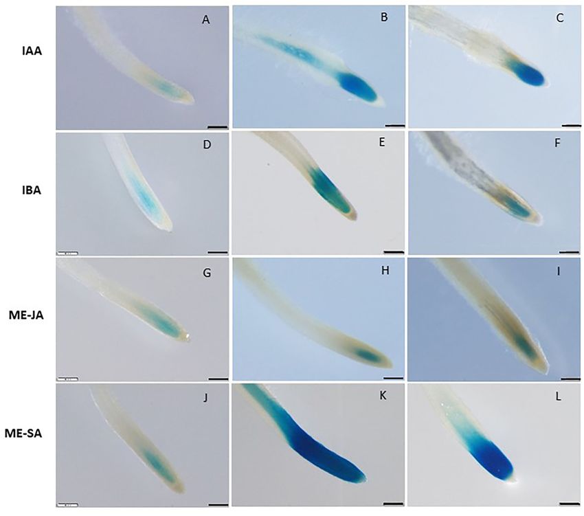
Sanadhya et al.                                                                                                   LeDES Regulates RKN Parasitism

supernatant was collected and incubated with 5 µg/ml 9-HPODE               and DesNotR: 5′ -AAGCGGCCGCCTATTTACTTGCTTTAGT-
at 30◦ C overnight. The reaction was terminated by adding                  3′ ) and cloned into the pFL61 vector. Saccharomyces cerevisiae
an equal amount of 1 ppm 2-hydroxydecanoic acid (Cayman                    INVSc1 (MATa his311 leu2 trp1-289 ura3-52/MATα his311
Chemicals) in ethanol. LC–MS analyses were conducted using                 leu2 trp1-289 ura3-52), procured from Invitrogen, was used
a UPLC-Triple Quadrupole mass spectrometer (Waters Xevo                    as the host strain for yeast transformation. S. cerevisiae was
TQ MS, Waters Corp., Milford, MA, USA). Separation was                     transformed with the empty pFL61 vector (negative control)
performed on a Waters Acquity UPLC BEH C18 1.7 µm, 2.1                     and with the pFL61::LeDES construct using the lithium
× 100 mm column with a VanGuard precolumn (BEH C18                         acetate/polyethylene glycol transformation method (Sanadhya
1.7 µm, 2.1 × 5 mm). Chromatographic and MS parameters were                et al., 2015). Positive colonies were selected on synthetic dropout
as follows: the mobile phase consisted of water (phase A) and              (SD) plates with synthetic defined medium lacking uracil (SD/-
acetonitrile (phase B), both containing 0.1% (v/v) formic acid             Ura; Clontech, Mountain View, CA, USA), and verified by PCR
in gradient elution mode. The flow rate was 0.3 ml/min, and                using gene-specific primers. Expression of LeDES in transgenic
the column temperature was kept at 35 ◦ C. LC–MS analysis was              yeast clones was further confirmed by RT-PCR using cDNA.
performed using the ESI source in positive ion mode for CA and
CnA and in negative ion mode for 2-hydroxydecanoic acid with               Evaluating the Immotility Effect of
the following settings: capillary voltage 3.1 kV, cone voltage 30 V,
desolvation temperature 350◦ C, desolvation gas flow 650 L/h,
                                                                           Transgenic Yeast on J2s
                                                                           The method used in this study to analyze the impact of
source temperature 150◦ C. Quantification was performed using
                                                                           the transgenic yeast harboring LeDES was adapted from Naor
MRM acquisition by monitoring the 293/275, 293/93 [retention
                                                                           et al. (2018) with some minor modifications. S. cerevisiae
time (RT) = 9.50, dwell time of 100 ms for each transition]
                                                                           cells transformed with pFL61 and pFL61::LeDES were grown
for CnA, 295/81, 295/277 (RT = 10.08, dwell time of 100 ms)
                                                                           overnight at 28◦ C from individual colonies in 5 ml of SD/-
for CA, 187/141, 187/169 (RT = 6.51, dwell time of 161 ms)
                                                                           Ura medium. The precultured yeast cells were pelleted by
for 2-hydroxydecanoic acid. Acquisition of LC–MS data was
                                                                           centrifugation and resuspended in 10 ml of induction medium:
performed using MassLynx v4.1 software (Waters).
                                                                           SD/-Ura with or without 9-HPODE or 13-HPODE to a final
                                                                           optical density at 600 nm of 2.0. After overnight incubation at
Evaluation of Arabidopsis Plant Response                                   28◦ C with the respective oxylipin, 300 sterile J2s suspended in
to RKN Infection                                                           0.01 M MES buffer were added to the cultures and incubated
To study the extent of nematode infection in WT, vector-                   for additional 24 h at 25◦ C with shaking at 30 rpm. After
only, and transgenic Arabidopsis lines, infected roots grown               24 h incubation, the J2s in the suspensions were collected and
in monoxenic culture were collected at 28 dpi and analyzed                 subjected to 30-µm filtering (AD Sinun Technologies, Petach
following the procedure describe by Joshi et al. (2019, 2020)              Tikvah, Israel) for 2 h (Sanadhya et al., 2018). J2s that actively
with minor modifications. Roots were gently harvested from                 passed through the filter were counted with nematode-counting
the plates, agar was removed, and they were weighed. After                 slides (Chalex LLC, Portland, OR, USA) under the microscope
cleaning, the roots were stained with acid fuchsin (Sigma-                 (Wilovert Standard microscope, Helmut Hund GmbH, Wetzlar,
Aldrich) solution (17.5 mg acid fuchsin, 500 ml ethanol, 500 ml            Germany). To measure % motility, J2s counting in treatments
acetic acid) overnight. Stained roots were washed three times              included pFL61 or pFL61::LeDES plasmids and the oxylipins 9-
with distilled water, then mounted in water, and roots and galls           HPODE or 13-HPODE were divided by J2s counts following
were manually dissected under a stereomicroscope (Olympus                  incubation with respective plasmids but without the respective
SZX12, Tokyo, Japan). Different nematode development stages                oxylipins. Thus, the pure nematotoxic activity of LeDES could
(J3/J4 and adult females) embedded in the roots and galls were             be assessed. Ten replicates were used for each treatment and
manually dissected and counted for each root. Each treatment               experiments were performed three times independently. A
(Arabidopsis line) included 10 individual plates and the infection         general linear mixed model ANOVA test was applied to the data
assays were repeated three times. Infection assay data were fitted         with similar results. Different letters above the columns indicate
with general linear mixed model in SPSS, by keeping samples                significant difference (P ≤ 0.05) among the different treatments
as fixed factors and repeated assays as random factor. Further,            analyzed by Tukey post-hoc range test.
Tukey post-hoc range test was applied to check significant
differences (p < 0.05) among different samples.                            Sample Preparation From Transgenic Yeast
                                                                           for DVE Extraction and Analysis by LC–MS
Heterologous Expression of LeDES in                                        colnelenic acid (CnA) and colneleic acid (CA) were produced
Yeast                                                                      from 9-HPODE by yeast lysate of transgenic and control yeast
Full-length LeDES cDNA was cloned into the yeast expression                lines according to the method described by Kuroda with minor
vector pFL61 under the control of the constitutive yeast                   modifications (Kuroda et al., 2005). Yeast strains carrying empty
phosphoglycerate kinase (PGK) promoter (Minet et al., 1992).               vector and LeDES were precultured in SD/-Ura liquid medium
To generate yeast expression of the construct, LeDES cDNA was              at 28◦ C. After 16 h, the starter culture was added to a 250-ml
amplified from pART27::LeDES using NotI-containing primers                 flask containing 50 ml SD/-Ura liquid medium and incubated
(DesNotF: 5′ -TTGCGGCCGCATGTCTTCTTATTCAGAG-3′                              at 28◦ C for 24 h. The yeast cultures were centrifuged at 2,744 g

Frontiers in Plant Science | www.frontiersin.org                       5                                   August 2021 | Volume 12 | Article 670772
Sanadhya et al. LeDES Regulates RKN Parasitism
FIGURE 1 | Expression pattern of LeDES in tomato roots during the infection time course. To investigate the transcript levels of LeDES upon nematode infection, total
RNA was isolated from each sample at 1, 2, 3, 10, 15, and 28 dpi or without infection; RNA was subjected to qRT-PCR and normalized to β-tubulin as the reference
gene. Two biological replicates were taken and three independent qRT-PCRs were performed per sample, resulting in a total of six replicates for statistical analyses.
The graph shows the mean and SD of the amount of LeDES transcript relative to non-inoculated samples. Error bars correspond to SD n = 2 while different letters
above the bars denote a significant difference (P ≤ 0.05, ANOVA) between different treatments as analyzed by Tukey–Kramer multiple comparison test.
for 10 min, and the pellet was weighed and then homogenized 28 dpi, suggesting that LeDES has a role in the early defense
in 500 µl 50 mM phosphate buffer pH 7 by Bead Ruptor Elite mechanism induced by nematode penetration and migration,
(OMNI International, Kennesaw, GA, USA) and centrifuged. with expression peaking when the young developing galls form.
The lysate was collected and incubated with 5 µg/ml 9-HPODE at
30◦ C overnight. The reaction was terminated by adding an equal Spatiotemporal Expression and
amount of 1 ppm 2-hydroxydecanoic acid in ethanol. LC–MS Distribution of LeDES Transcripts
analyses of the lysates were conducted as described above. In order to investigate the spatiotemporal activity of LeDES
promoter, the 1,602-bp sequence upstream of the ATG start
RESULTS site of LeDES was fused to the GUS reporter gene, generating
a pLeDES::GUS construct. The pLeDES::GUS reporter construct
Nematode-Induced LeDES Transcript was then transformed into tomato via R. rhizogenes-mediated
Accumulation root transformation. Transgenic hairy roots that emerged from
To study the expression of LeDES in roots of tomato challenged the cotyledons were confirmed to be carrying the pLeDES::GUS
with M. javanica J2s, qRT-PCR was performed with cDNA from construct. pLeDES-driven GUS expression was monitored
root tissues collected 1, 2, 3, 10, 15, and 28 dpi. The nematodes following wounding, exogenous application of defense-related
induced activation of LeDES, as shown by a moderate course signaling molecules, and in response to nematode infection
of increase in expression at all-time points upon inoculation conducted at specific time points after induction, in transgenic
(Figure 1). The relative fold changes in LeDES transcript in hairy roots by histochemical staining. In the absence of any
infected roots at 1, 2, 3, and 10 dpi were approximately 1.9, stimulus, very intense GUS staining was typically observed in the
5.3, 3.6, and 4.3, respectively, compared to non-inoculated roots. root maturation zone (Figures 2A,D), whereas no GUS activity
Transcript abundance of LeDES was dramatically induced at 15 was found in the elongation zone (Figures 2A,C); notably, a very
dpi (17.9-fold), and was the highest for all-time points considered faint GUS signal could be observed in the root apex (Figure 2B).
in this study (Figure 1). LeDES expression levels declined at Strong GUS activity was also associated with the lateral root
Frontiers in Plant Science | www.frontiersin.org 6 August 2021 | Volume 12 | Article 670772Sanadhya et al. LeDES Regulates RKN Parasitism
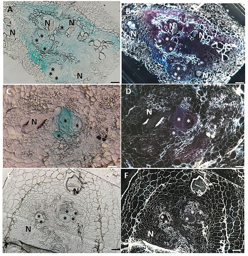
FIGURE 2 | Studying basal and wound induced LeDES GUS expression profile. Typical expression pattern of pLeDES::GUS in non-inoculated tomato root lines. (A)
Basal GUS activity in roots. (B–D) Magnified image of root cap (B), elongation zone (C), and maturation zone (D). (E,F) Promoter–GUS activity observed in lateral root
primordials in non-inoculated roots. RC, root cap; E, elongation zone; M, maturation zone. Bars: (A,E) 1 mm; (B–D,F) 250 µm. Expression analysis of pLeDES::GUS
tomato root lines in response to wounding (G–I). Histochemical GUS staining of pLeDES::GUS in control roots (G) and in roots 6 h (H) and 24 h (I) after wounding.
Bars: (G–I) 1 mm. Arrows indicate site of mechanical wounding.
primordium of the non-inoculated roots, suggesting a role for zone (Figure 2G), no LeDES promoter activity was observed
LeDES in genetic regulation of root growth and development localized at the wound site at either 6 h (Figure 2H) or 24 h
(Figures 2E,F). (Figure 2I) after wounding. It is clearly observed that a complete
loss of GUS staining characterize the specific site of mechanical
Tomato LeDES Expression Is Repressed damage as observed (arrows in Figures 2H,I) 6 and 24 h after
wounding, respectively. These results suggest wound-induced
Upon Wounding
local suppression of LeDES at the site of the mechanical damage
Mechanical damage by wounding activates defense signaling
(Figure 2).
pathways and alters hormonal levels in plants, which in turn
protects the plant against injury and pathogen attack (Savatin
et al., 2014). LeDES expression upon wounding was profiled by LeDES Mediates Me-SA and Auxin
analyzing GUS expression in pLeDES::GUS roots of wounded Phytohormone-Induced Response
and non-wounded roots 6 and 24 h after mechanical wounding. To assess whether LeDES promoter is activated through
Whereas, a typical signal was observed along the root maturation phytohormone signaling, we used the phytohormone signaling
Frontiers in Plant Science | www.frontiersin.org 7 August 2021 | Volume 12 | Article 670772Sanadhya et al. LeDES Regulates RKN Parasitism FIGURE 3 | GUS staining in transgenic tomato roots carrying pLeDES::GUS following exogenous phytohormone application. One-week-old roots were subjected to GB as a control (A,D,G,J) or to GB containing IAA [1 and 5 µM; (B,C)], IBA [1 and 10 µM; (E,F)], Me-JA [0.01 and 0.1 mM; (H,I)], or Me-SA [1 and 5 mM; (K,L)], for 16 h. GUS staining was monitored histochemically in root tips. Figures are representative of at least three independent experiments. Bars: 250 µm. molecules IAA, IBA, Me-JA, and Me-SA in an LeDES::GUS Spatiotemporal Expression Pattern of promoter bioassay (Figure 3). GUS staining revealed high LeDES Upon M. javanica Root Infection GUS expression in the root tips 16 h after treatment with Next, transcriptional activation of the GUS reporter gene driven the auxins (IAA and IBA) (Figures 3B,C,E,F) compared by the LeDES promoter in the transgenic hairy roots of tomato to the negative control (Figures 3A,D), and no activation was analyzed in a time-course analysis at 2, 3, 10, 15 and 28 of LeDES promoter by Me-JA (Figures 3H,I). Notably, dpi representing J2 (2 and 3 days), J3 and J4 (10 and 15 days) strong induction of the GUS reporter gene was evident and female (28 days) stages of nematode development. Very after Me-SA treatment, as observed in the root tip and strong staining was evident 2 and 3 dpi in the root elongation elongation zone (Figures 3K,L). Our results suggest that various zone, the preferred site for nematode penetration, along with phytohormones coregulate LeDES expression, highlighting swelling and intensive root hair growth resulting from nematode its possible role in mediating defense responses triggered by penetration compared with non inoculated roots on the left biotic stress. panel (Figures 4A–D). An intense GUS signal was observed Frontiers in Plant Science | www.frontiersin.org 8 August 2021 | Volume 12 | Article 670772
Sanadhya et al.                                                                                                                           LeDES Regulates RKN Parasitism

 FIGURE 4 | Expression-pattern analysis of pLeDES::GUS root lines during nematode infection. Non-inoculated control root demonstrated consistent GUS staining in
 the apical meristem (cell-division zone) (A,C,E,G,I). Increased GUS staining was detected in the infected swelling site located in the elongation zone on 2 dpi (B), 3 dpi
 (D), and 10 dpi, premature developing gall (F). Strong GUS staining was observed in the vasculature and vessels associated with the developed gall at 15 dpi (H). At
 28 dpi (J), GUS staining intensity decreased and became localized specifically to the cells surrounding the developed nematode. Bars: (A,C–E) 250 µm; (B,F)
 100 µm; (G–J) 500 µm.

Frontiers in Plant Science | www.frontiersin.org                                     9                                           August 2021 | Volume 12 | Article 670772
Sanadhya et al.                                                                                                                        LeDES Regulates RKN Parasitism

in the bulging root tissue at the initial nematode penetration                          deformed vascular bundles (Figure 4H). Notably, a significant
site, which is part of the developing gall (Figure 4B). High                            reduction in promoter activity was evident at 28 dpi compared
promoter activity continued to be observed in the maturing                              with the control roots (Figures 4I,J), whereas a very mild GUS
galls compared with their respective controls (Figures 4E–H) at                         signal associated with the vascular system connected to the
10 (Figure 4F) and 15 (Figure 4H) dpi, mainly confined to the                           developed female could be observed (Figure 4J).

 FIGURE 5 | Histological GUS localization within nematode feeding site. For histological GUS localization, galls were fixed and embedded in Technovit 7100 and
 3-µm-thick cross sections were analyzed using a light microscope equipped with a Nikon digital camera. All giant cells were mature and nematodes developed to the
 J4 stage. Histological analysis of roots expressing pLeDES::GUS on 15 dpi clearly shows GUS expression restricted to the vascular systems bordering the feeding
 sites, the giant cells and cells surrounding the developing female body (A,B). At 28 dpi, GUS signal is observed mostly in the mature feeding site (C,D) and (E,F). GUS
 staining is observed as blue color in whole mounts, and as a red precipitate in the dark field micrographs of the sections. N, female nematode body; *giant cells.
 Bars: 100 µm.

Frontiers in Plant Science | www.frontiersin.org                                   10                                          August 2021 | Volume 12 | Article 670772
Sanadhya et al.                                                                                                                       LeDES Regulates RKN Parasitism

Giant Cell-Specific Expression of LeDES                                                LeDES Heterologous Expression in
Accompanying Premature Stages of                                                       Arabidopsis Attenuates Nematode
M. javanica Infection                                                                  Development
To investigate the spatial expression of LeDES at nematode                             To further explore the role of LeDES in regulating plant responses
feeding sites, thin sections of galls induced by M. javanica, 15                       to nematode infection, a plant binary vector containing a
and 28 dpi, were prepared and analyzed. At 15 dpi, strong                              1,437-bp LeDES ORF under the control of the CaMV 35S
GUS staining showed a scattered signal distributed within                              promoter was introduced into A. thaliana by floral-dip method
the deformed stele harboring the feeding site and bounded                              for Agrobacterium-mediated transformation. From all of the
by the endodermis cell layer, as observed under light and                              independent kanamycin-resistant transgenic lines harboring the
dark field, respectively (Figures 5A,B). At that time point,                           35S:LeDES construct, three homozygous lines (D1, D2, and D3)
GUS signal was clearly visible in the giant cells, as well as                          were validated by RT-PCR for LeDES heterologous expression,
in the surrounding xylem and phloem parenchyma cells. As                               and used for nematode infection (Figure 6A). The transgenic
infection progressed, GUS signal was significantly quenched,                           plants did not show any phenotypic change in root or shoot
although it was predominantly retained within the developing                           growth compared to the WT Col-0 line (Figure 6B). To check
giant cells (Figures 5C,D). However, as the female matured                             for the presence of CnA and CA, indicative of active DES in
and the feeding site reached its maximum size, at 28 dpi,                              the transgenic Arabidopsis plants, root lysates of transgenic and
no GUS signal was observed in the adjacent parenchyma                                  control plants were analyzed by LC–MS (Figure 6C). The root
cells with only a very faint GUS signal in the feeding site                            homogenates of transgenic and control plants were incubated
(Figures 5E,F).                                                                        with 9-HPODE and analyzed by LC–MS for the production

 FIGURE 6 | Response of Arabidopsis plants overexpressing tomato LeDES to nematode infection. (A) RT-PCR confirmation of LeDES expression with DES-specific
 primers. Lane 1, negative control; lane 2, cloned LeDES template (positive control); Lane 3, independent transgenic line transformed with empty vector; lanes 4–6,
 independently generated transgenic lines for LeDES (B) Arabidopsis plants expressing LeDES 30 dpi; root systems of all plants show normal root with no phenotypic
 growth changes. (C) Quantification of colnelenic acid produced in transgenic Arabidopsis root homogenates as measured by LC-MS. (D) Disease development in
 Meloidogyne-infected roots of transgenic Arabidopsis plants expressing LeDES compared to control lines. All plants were inoculated with 300 sterile pre-parasitic J2s
 and the infected roots were assessed for J3/J4 and mature female development at 28 dpi through observation under the dissecting microscope following staining
 with acid fuchsin dye. General Linear Mixed Model test to infection assay data was applied. Different letters above the columns indicate significant difference (P ≤
 0.05) among different Arabidopsis lines analyzed by Tukey post-hoc range test.

Frontiers in Plant Science | www.frontiersin.org                                  11                                          August 2021 | Volume 12 | Article 670772
Sanadhya et al.                                                                                                                          LeDES Regulates RKN Parasitism

 FIGURE 7 | Effect of recombinant yeast expressing LeDES on M. javaniva J2 viability. (A) RT-PCR confirmation of LeDES expression in yeast. Lane 1, negative
 control; lane 2, transgenic yeast cells with empty vector; lane 3, transgenic yeast containing LeDES (L1) (B) Quantification of colnelenic acid production in transgenic
 yeast by LC–MS. Values are means of duplicate measurements. (C) Yeast carrying LeDES display bionematostatic activity. J2s were incubated in SD medium
 containing yeast strain and 9-HPODE or 13-HPODE for 24 h, then 300 J2s were added to the reaction for 12 h then subjected for sieving followed by microscopic
 observation. Different letters above the columns indicate significant difference (P ≤ 0.05) among the different treatments analyzed by Tukey post-hoc range test.

of CnA and CA. No CnA was observed in the control plants,                                or arrested at the earlier time points, will be starved to death
but it was detected in the D1, D2, and D3 transgenic lines                               by then, and thus barely could be visible, at 28 dai. Disease
(Figure 7C). No accumulation of CA was identified through LC–                            development, as indicated by number of developmental stages
MS in either the transgenic or control WT lines. To determine                            (J3/J4 and females) on D1, D2, and D3 and control lines (WT
the effect of LeDES overexpression on disease development in                             and vector-only) indicated that LeDES overexpression restricts
Arabidopsis transgenic lines, 10 plants each from WT, vector-                            nematode infection, and results in significantly less nematodes
only, and three transgenic lines were inoculated with 300 J2s,                           molting into J3/J4 stages, as observed in LeDES transgenic lines
and the nematode developmental stages (J3/J4 and females) were                           compared to control lines at 28 dpi. Similarly, a decrease in
monitored at 28 dpi (Figure 6D). It is commonly observed that                            the number of mature females was observed in transgenic lines
at 28 dai, analyzed infected roots, harbor mainly J3/J4, and                             overexpressing LeDES (Figure 6D). These results indicate that
mature female stages with or without egg masses. While, at                               heterologous expression of LeDES in Arabidopsis plants results
this late time point after infection, J2s which were suppressed                          in attenuation of nematode disease development in the roots.

Frontiers in Plant Science | www.frontiersin.org                                    12                                           August 2021 | Volume 12 | Article 670772
Sanadhya et al.                                                                                                   LeDES Regulates RKN Parasitism

Infection assay data were fitted with general linear mixed model          RKN M. javanica was observed, which was also evident with
in SPSS, by keeping samples as fixed factors and repeated assays          the accumulation of oxylipins at earlier stages of nematode
as random factor (Supplementary Table 1). Further, Tukey post-            parasitism (Fitoussi et al., 2021).
hoc range test was applied to check significant differences (p <              Oxylipin profiling of WT roots of tobacco plants exposed to P.
0.05) among different samples.                                            parasitica var. nicotianae (Ppn race 0) also showed preferential
                                                                          upregulation of the 9-LOX pathway and accumulation of
Functional Expression of LeDES in                                         the two DVEs CA and CnA (Fammartino et al., 2007).
Saccharomyces cerevisiae, Product                                         Similarly, NtLOX1-antisense plants were more susceptible to P.
                                                                          parasitica var. nicotianae (Rancé et al., 1998) along with reduced
Analysis, and Nematotoxic Acitivity                                       accumulation of LOX downstream pathway products, especially
To characterize the direct activity of DVEs against M. javanica           CA and CnA, highlighting the potential role of these oxylipins in
J2s, LeDES cDNA was cloned into the yeast expression vector               defense responses. Other studies, analyzing the oxylipin signature
pFL61, under the control of the constitutive PGK promoter of              in potato leaves infected with late blight disease (Weber et al.,
S. cerevisiae to obtain the pFL::LeDES construct. Then, following         1999) or in elicited potato cell suspension cultures (Stumpe et al.,
yeast transformation, pFL::LeDES was expressed in S. cerevisiae           2001) revealed the dominant induction of the DES pathway
strain INVSc1. To confirm the presence and expression of LeDES,           downstream of the 9-lipoxygenation of polyenoic fatty acids.
transgenic yeast lines were analyzed by RT-PCR, revealing                     Here, we chose to study LeDES on the basis of our previous
accumulation of DES transcripts exclusively in the positive               report which demonstrated the nematicidal activity of the DVEs
transformed line L1 (Figure 7A). Then, CnA production by                  CA and CnA (Naor et al., 2018). In the current study, we showed
the recombinant yeast was determined by incubating transgenic             that the DVE-biosynthesis pathway is activated at certain stages
yeast with 9-HPODE as a substrate. LC–MS was used to detect               of M. javanica infection in tomato roots, as reflected by elevated
the presence of CnA. Accumulation of 3.37 ± 0.060 ng/g CnA                LeDES expression. However, differences in temporal expression
was observed for the L1 recombinant yeast strain, whereas no              were detected, where the highest expression was observed at 15
accumulation was detected in the reaction containing extract              dpi. Differences in transcript accumulation in response to RKN
from yeast cells with pFL61 vector (Figure 7B).                           in the tomato host suggest differential control of RKN-induced
    Hence, recombinant yeast clones transformed with the                  DVE biosynthesis. Colneleic acid has been shown to attenuate 9-
pFL::LeDES construct and a negative control, the void                     LOX (Corey et al., 1987; Itoh and Howe, 2001; Fammartino et al.,
pFL61, were used to study the direct nematotoxicfunction                  2007), thus it can be speculated that transcription of DVE fatty
of DVEs against M. javanica J2s. Following 24 h incubation of             acids or other oxylipin (derived from 9-LOX) biosynthesis genes
recombinant yeast strains expressing either pFL::LeDES or the             may be repressed by feedback from downstream metabolites.
empty vector pFL61 with 9-HPODE or 13-HPODE M. javanica                   However, the synthesis of CA and CnA is highly specific and
J2s were added to the yeast reaction for an additional 12 h, then         strongly induced in certain instances, such as infection (Itoh
J2 viability/motility was evaluated through sieving followed by           and Howe, 2001). We therefore isolated the promoter region
microscopy observation. There was a significant difference in             (1,602 bp upstream from the start codon) to obtain insight into
percent motility of J2s following incubation with recombinant             transcriptional reprogramming of LeDES expression in response
strain pFL::LeDES and 9-HPODE (7%), compared to that with                 to RKN parasitism.
the negative control yeast carrying the empty vector pFL61 and                The modulation of LeDES expression by nematode parasitism
9-HPODE (37%) (Figure 7C). These differences in J2 motility               and signal molecules related to plant defense was monitored in
were not present when 13-HPODE was used as the substrate for              transgenic tomato hairy-root lines expressing the GUS reporter
either of the yeast strains.                                              gene driven by the LeDES promoter region. Notably, in non-
                                                                          inoculated control roots, GUS expression was not visible in the
DISCUSSION                                                                elongation zone, but was clearly detected in the apical meristem
                                                                          and maturation zone. Distinct GUS staining was also observed
Oxylipins are crucial compounds in plants, playing important              in lateral root primordia of developing roots, emphasizing
roles in developmental processes and in plant defense                     the importance of LeDES in regulating root development.
mechanisms. The involvement of oxylipins such as jasmonic                 Lateral root emergence and meristem activation are crucial
acid (JA) and its derivatives in defense responses against various        developmental mechanisms in roots which can modulate root
fungal and bacterial pathogens has been widely recognized                 architecture in response to stress, environmental, nutritional, and
and well-studied (Deboever et al., 2020); however, reports                endogenous factors (Signora et al., 2001; de Smet et al., 2006).
demonstrating the role of oxylipin-mediated signaling involved            Vellosillo et al. (2007) also showed that external application
in plant–parasitic nematode interactions are scarce (Gheysen and          of CA, can, or related oxylipins to Arabidopsis seedlings leads
Mitchum, 2019). We previously showed that several oxylipins               to loss of apical dominance and induces the development of
exhibit significant nematicidal activity against M. javanica,             lateral and adventitious roots. Thus, different oxylipins produced
emphasizing their crucial role in the plant’s defense response            in response to pathogen infection are probably endogenous
against nematode parasitism (Naor et al., 2018). In another more          regulators of lateral root emergence and consequently, might
recent study, the involvement of 9-LOX and α-dioxygenase                  be involved in reprogramming root system architecture upon
oxylipin pathways in tomato’s defense against the biotrophic              pathogen infection.

Frontiers in Plant Science | www.frontiersin.org                     13                                    August 2021 | Volume 12 | Article 670772
Sanadhya et al. LeDES Regulates RKN Parasitism
Nematode penetration inflicts mechanical damage to the galls, LeDES promoter activity was diffuse, and found throughout
roots, which lead to the release of Damage-Associated Molecular the vascular bundle, extending from the pericycle to the pith
Patterns (DAMPs) (Choi and Klessig, 2016; Holbein et al., and including the giant cells. GUS signal became constricted to
2016), which in turn initiate and perpetuate the innate immune the feeding sites as the nematodes molt into the female stage,
responses mediated by JA, salicylic acid (SA), ethylene, auxin, and diminished when feeding system was completely mature.
and reactive oxygen species (ROS) (Holbein et al., 2019). Many This specific induction of LeDES promoter in the galls and giant
reports have shown that the defense responses activated by cells in response to RKN infection indicates a putative role in
pathogens closely mimic those to wounding (Savatin et al., 2014). the defense response triggered by nematode attack. Alternatively,
Therefore, to assess the role of LeDES during the early infection it may be that auxin or Me-SA, which were shown to activate
stage, we analyzed the effect of wounding on transgenic roots LeDES, are responsible for the observed induction.
after 6 and 24 h. Expression of the GUS gene was downregulated We further cloned and expressed LeDES in the model
at the wounding site, indicating that this gene is not activated as plant A. thaliana to investigate its role in regulating plant-
part of the DAMP-triggered immunity through cell-wall damage parasitic nematode development. Heterologous expression of
and may be induced by other elicitors. the gene in A. thaliana conferred increased resistance against
Phytohormones, namely SA, ethylene, JA, abscisic acid, auxin, the RKN, evidenced by the attenuation in J3/J4 and female
brassinosteroid, gibberellic acid, and cytokinin, elicit defense stages measured in the infected roots. Similarly, Fammartino
responses by regulating the expression of defense-related genes et al. (2007) demonstrated that NtLOX1-antisense plants, which
(Santner et al., 2009; Jaillais and Chory, 2010; Pieterse et al., are compromised in their resistance to Ppn race 0, display
2012). Among them, JA and SA are key components in mounting reduced accumulation of DVEs upon inoculation. However,
defense responses in plants in response to different biotic and studies showing contradictory results have also been published.
abiotic stresses (Denancé et al., 2013). Treatment of transgenic Eschen-Lippold et al. (2007) showed that infection symptoms
roots with Me-JA showed no relevant effect on GUS signal. caused by P. infestans were unchanged in potato lines producing
However, application of Me-SA very strongly induced the signal a reduced amount of CnA after RNAi inhibition of pathogen-
in the apical meristem and elongation zone. This result fits well inducible DES. Furthermore, Fauconnier et al. (2008) later
with the notion that SA and JA act antagonistically on each other’s also showed that changes in oxylipin synthesis upon P.
pathways and biosynthesis upon sensing a pathogen (Lorenzo infestans infection do not correlate with resistance in potato.
and Solano, 2005; Pieterse et al., 2012). Plants activate mainly the These differences among the differential induction of DVEs
SA-signaling pathway to combat biotrophic pathogens, whereas in different pathosystems might be tissue-specific, in addition
JA is involved in defense against necrotrophs (Glazebrook, 2005). to its plant- or pathogen-specific features. Thus, the induced
Similarly, DES transcript in garlic leaves was induced by SA but resistance observed in the Arabidopsis–nematode pathosystem
not Me-JA application (Stumpe et al., 2008), whereas NtDES might be the result of differential substrate availability for
was not induced by SA (Fammartino et al., 2007). Considering DES in transgenic Arabidopsis plants. Nevertheless, differences
the biotrophic nature of RKNs, we can postulate a role for between Arabidopsis and tomato pathosystems might lead
SA in modulating the expression of LeDES. This suggests that to misinterpretation regards LeDES function toward RKN in
during the process of nematode infection, the LeDES branch of tomato host, thus in future, its function should be studied by
the 9-LOX pathway participates more in the systemic defense further LeDES manipulation in tomato host.
response than in the local one through production of CA and In this study, recombinant expression of LeDES in S. cerevisiae
CnA. Whether, LeDES activation is indirectly mediated through resulted in the production of measurable amounts of CnA but
potential Nematode Associated Molecular Pattern (NAMPs) not CA. Exposure of M. javanica J2s to yeast expressing LeDES
molecules, which induce SA defense-signaling pathway, that is when 9-HPODE is the substrate, resulted in reduced J2 motility.
remained to be studied. These results, along with previous ones showing that DVE is
Furthermore, treatment with IBA and IAA also induced able to attenuate motility, therefore support our findings that
promoter activity in the root’s apical meristem, thus indicating these are good candidate defense compounds at early time points
the potential role of auxins in manipulating LeDES expression of the infection process, further emphasizing their potential as
under basal and stress conditions. Auxins are proposed to be nematostatic compounds.
involved in the formation of giant cells and galls, evidenced by
its increased accumulation during the early stages of feeding-site CONCLUDING REMARKS
development (Karczmarek et al., 2004; Cabrera et al., 2014; Kyndt
et al., 2016). The antimicrobial activity of different DVEs has been well-
The infection tests performed with the transgenic hairy illustrated; however, there have been no reports of the
roots further confirmed the specific role of LeDES during the involvement of DVEs in the defense response to biotic stress
progression of nematode infection. Interestingly, during early conditions, such as nematode infection. Our study reveals the
infection stages, high GUS activity was observed in developing functional role of LeDES by showing that this tomato gene can
galls. Strong promoter activity was also evident in the middle–late be overexpressed in economically important crops to improve
infection stage (15 dpi), which then decreased (28 dpi). Hence, resistance to RKNs. Furthermore, the potential of DVEs as
the pLeDES::GUS activation pattern was consistent with our real- nematostatic and signaling molecules was confirmed. However,
time PCR results, characterized by gradual increment and then their role in RKN resistance seems to be more complex, as they
a relative reduction in expression. In sections from the 15-day have been shown to have a nematostatic effect in vitro, but to play
Frontiers in Plant Science | www.frontiersin.org 14 August 2021 | Volume 12 | Article 670772You can also read