The Inclusion Principles of Human Embryos in the WOW-Based Time-Lapse System: A Retrospective Cohort Study - Frontiers
←
→
Page content transcription
If your browser does not render page correctly, please read the page content below
ORIGINAL RESEARCH
                                                                                                                                                 published: 26 July 2021
                                                                                                                                        doi: 10.3389/fendo.2021.549216

                                            The Inclusion Principles of Human
                                            Embryos in the WOW-Based Time-
                                            Lapse System: A Retrospective
                                            Cohort Study
                                            Yuqiong Wang , Sheng Wang , Xilin Qian , Yanrong Kuai and Yang Xu *

                                            The Department of Assisted Reproduction, Peking University First Hospital, Beijing, China

                                            A time-lapse system (TLS) with a well-of-the-well (WOW) dish, which allows individual
                                            identification and the possibility of autocrine and paracrine signaling between group-
                                            cultured embryos, has been widely used in clinic. However, there is a need to re-think the
                                            inclusion principles of human embryos in WOW-based TLS, especially for grade IV (G4)
                                            embryos, which are considered to potentially have detrimental effects on surrounding
                                            embryos. Here, we carried out a single-center, large-cohort, retrospective study,
                            Edited by:      comprising 303 patients undergoing IVF (148 cases) and ICSI (155 cases), with a total
                           Richard Ivell,   of 3282 embryos, to compare embryonic development until the blastocyst stage in the
             University of Nottingham,
                      United Kingdom
                                            group culture system with or without G4 embryos. Further, LC-MS/MS was used to
                       Reviewed by:
                                            analyze the G1-G4 embryo secretome to understand the influence of G4 embryos on the
                      Stewart Russell,      group culture microenvironment. We proved that polypronuclear (PPN) embryos positively
     CReATe Fertility Centre, Canada
                                            contribute to the development of the neighboring embryos through secretion of ILIAP, ITI-
                    Avi Ben Haroush,
           Tel Aviv University, Israel      H4, and keratin. Existence of more than one G4 embryo had a negative effect on the other
                 *Correspondence:           embryos (p < 0.05). Moreover, G4 embryos were found to secrete KLKB1 and VTDB,
                          Yang Xu           which might harm the neighboring embryos. Thus, our study clarified that when embryos
           xuyang2008001@sina.com
                                            are subjected to group culture in WOW-based TLS, the PPN-derived embryos need not
                   Specialty section:       be removed, and it is important to ensure that no more than one G4 embryo is present to
         This article was submitted to      avoid negative effects on the neighboring embryos.
                         Reproduction,
               a section of the journal     Keywords: time-lapse system (TLS), well-of-the-well (WOW), embryo group culture, embryo secretome,
            Frontiers in Endocrinology      blastocyst formation
         Received: 26 October 2020
            Accepted: 08 July 2021
           Published: 26 July 2021
                                            INTRODUCTION
                             Citation:
Wang Y, Wang S, Qian X, Kuai Y and          The time-lapse system (TLS) is an effective method for continuous imaging of embryonic
Xu Y (2021) The Inclusion Principles of
                                            development in vitro; it not only provides detailed information of the developmental kinetics and
       Human Embryos in the WOW-
          Based Time-Lapse System:
                                            cleavage patterns throughout the culture period (1, 2), but also maintains a stable and optimal
       A Retrospective Cohort Study.        embryo culture environment without having to remove the culture dish from the incubator (1, 3).
        Front. Endocrinol. 12:549216.       At the same time, it greatly liberates embryologists from cumbersome clinical procedures, and is
    doi: 10.3389/fendo.2021.549216          becoming increasingly popular (3).

Frontiers in Endocrinology | www.frontiersin.org                                    1                                           July 2021 | Volume 12 | Article 549216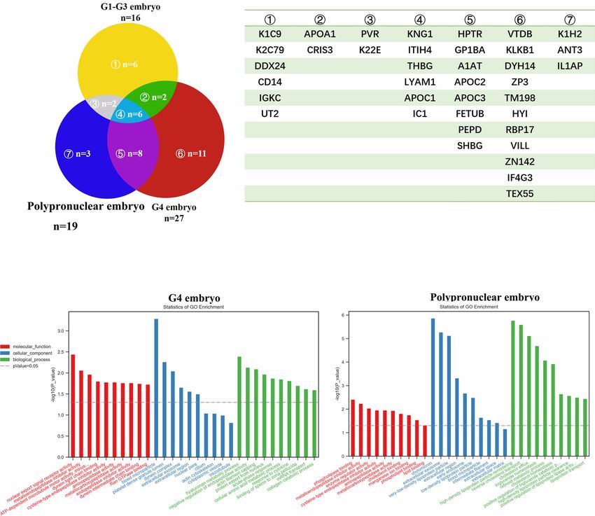
Wang et al.                                                                                                               A Retrospective Cohort

    The commonly used embryo culture dish for TLS is the so-             METHODS
called well-of-the-well (WOW) dish. This type of culture dish
uses narrow and deep microwells to designate the place of each           Study Design
embryo within a close distance from one another to facilitate            This study was a single-center, large-cohort, retrospective study.
autocrine signaling and enable paracrine communication                   Blastocyst formation rate and blastocyst quality were used to
between sibling embryos, thus combining the beneficial effects            evaluate the embryo microenvironment. Liquid chromatography-
of individual and group culture (4, 5). It was the first system to        tandem mass spectrometry (LC-MS/MS) was used to analyze the
realize individual embryo identification and the possibility of           G1–G4 embryo secretome to understand the influence of G4
autocrine and paracrine-mediated signaling between group-                embryos on the group culture microenvironment.
cultured embryos (4, 6–8). It is hypothesized that one benefit
of group embryo culture is that it allows embryos to modify their        Study Population
microenvironment in an autocrine manner to exert an effect on            We included 303 patients undergoing in-vitro fertilization (IVF)
themselves and/or on neighboring embryos (9), which may                  (148 cases) and intracytoplasmic sperm injection (ICSI) (155
provide mutual ‘‘corrective’’ factors that compensate for                cases), involving 3282 embryos in total, from the Department of
insufficiency or imbalance in the individual embryo. These                Reproductive Center, Perking University First hospital, Beijing
factors may act as growth factors or survival factors, protecting        city, China from September 1, 2018 to December 30, 2019.
the embryos from subsequent cell death (10). Potential embryo-               The patients were referred to our clinic with a history of more
derived secretory factors include members of the insulin and             than 1 year of infertility. Patients had the following primary
insulin-like growth factor and tumor necrosis factor families,           etiologies for their infertility: male factor, tubal factor,
epidermal growth factors, fibroblast growth factors, and platelet-        unexplained, endometriosis, anovulation, and polycystic
derived growth factors (11, 12).                                         ovary syndrome.
    However, some factors derived from poor-quality embryos
may have negative influences on the development of surrounding            Patients Selection
embryos. It was reported that group culture of high-quality              Inclusion criteria: Age 25~40 years old, and the number of
human embryos, rather than randomly grouped embryos                      retrieved oocytes 8-20. Exclusion criteria: Presence of organic
regardless of embryo quality, may benefit blastocyst formation,           ovarian diseases, such as ovarian cysts, ovarian benign tumors;
owing to the secretion of beneficial factors by good embryos, or          oral contraceptives, metformin and other drugs that affect
removal of detrimental factors from poor embryos (9).                    hormone levels and metabolism-related drugs within the past
Unfortunately, we lack evidence regarding what the                       three months; age > 40 years old; the number of retrieved oocytes
detrimental factors from poor embryos are and further analysis           was less than 8 or more than 20.
is necessary to determine the effect of these proteins on aspects            Ovarian stimulation was performed with gonadotropin-
such as development of neighboring embryos, clinical outcome,            releasing hormone (GnRH) analog downregulation utilizing
and even the health of the offspring (13).                               our long or microflare protocols with recombinant follicle-
    During traditional group embryo culture, grade IV (G4)               stimulating hormone and luteinizing hormone or human
embryos are abandoned because they usually consist of                    menopausal gonadotrophin, dose adjusted according to body
polypronuclear (PPN) embryos, embryos with > 50%                         mass index. During the ovarian stimulation regimen, the patients
fragmentation, and development-arrested embryos (14); even if            underwent transvaginal ultrasonographic evaluation of
these G4 embryos develop into blastocysts, they are thought to be        endometrial thickness and measurement of follicular number
unsuitable for transplantation. However, in the era of TLS, that         and size. Ovulation was induced with recombinant human
allows individual embryo identification in a WOW group culture            chorionic gonadotropin (hCG) when three or more follicles
system, we need to re-think the rule for inclusion of embryos,           were at least 18 mm in their greatest diameter.
whether it is necessary to remove the G4 embryos specifically and
determine whether G4 embryos secrete detrimental factors that            Oocyte Retrieval and In-Vitro Fertilization
harm the microenvironment of the group culture.                          The oocyte retrievals were done by transvaginal aspiration under
    Hence, we conducted a single-center, large-cohort,                   ultrasound guidance after 36 h from the hCG administration.
retrospective study to compare embryonic development in the              The follicles were flushed by G-MOPS (Vitrolife) medium. After
group culture system with or without G4 embryos to clarify the           retrieval, oocytes were rapidly isolated from follicular fluid, and
inclusion principle of human embryos in the WOW-based TLS                cultured in G-IVF medium (Vitrolife). Oocytes were
system. Based on the findings from Stokes et al. that the benefit of       inseminated 3–5 h later by classical IVF (mean concentration
group culture is usually visible after prolonged culture of human        of 200 000 motile spermatozoa/mL) or by ICSI. Spermatozoa for
embryos to the blastocyst stage, which might be because group            IVF and ICSI were prepared with the swim-up technique and
culture confers a greater advantage on development post genome           density gradient centrifugation method, respectively. For ICSI,
activation (10), the quality of blastocysts from the two groups          cumulus cells needed to be removed by hyaluronidase (Vitrolife),
was mainly compared and used as an important index to evaluate           and then the single motile spermatozoan with the best
the microenvironment of the group culture in the TLS.                    morphologic appearance was injected into each mature oocyte.

Frontiers in Endocrinology | www.frontiersin.org                     2                                    July 2021 | Volume 12 | Article 549216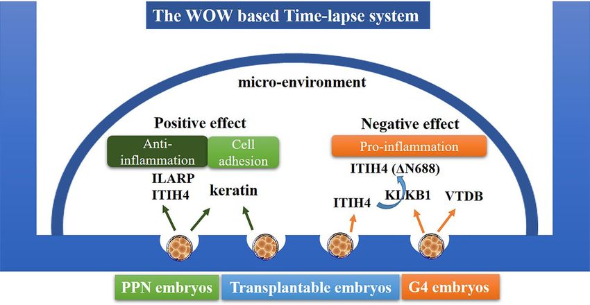
Wang et al. A Retrospective Cohort
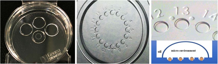
Embryo Culture cells; or C, very few cells. The trophectoderm was assessed as
After IVF or ICSI, the oocytes and/or zygotes were seeded in follows: A, many cells forming a cohesive epithelium; B, few cells
groups using 16-microwell GERI dishes (GENEA BIOMEDX) forming a loose epithelium; or C, very few large cells. The high-
(Figure 1) in a small droplet of preincubated G-1 Plus (Vitrolife) quality blastocysts were those with a score ≥ 3BB, and the
culture medium (80 ml) covered by mineral oil (3 ml) (Ovoil, criterion for cryopreservation of blastocysts was that
Vitrolife). The microwell plate was placed on the time-lapse blastocysts had a score of ≥ 3BC at days 5 or 6.
system (GERI) and fertilized oocytes were scored for pronuclei
on day 1 (usually 18-19 h post-insemination). LC-MS/MS Analysis
The culture medium first underwent enzymolysis, and was then
Day 3 Embryo Classification separated by capillary high performance liquid chromatography
Embryos were classified on day 3 into four categories as follows: and analyzed by a Q-Exactive plus mass spectrometer (Thermo
Grade I (G1): Embryos (7–9 cells) with even, regular, spherical Scientific). The conditions were: analysis duration, 60 min;
blastomeres with no or < 5% fragmentation; grade II (G2): detection mode, calibrated by standard calibration solution
Embryos (7–9 cells) with regular, spherical blastomeres, with < before use; scanning range of precursor ion, 300-1800 m/z;
20% fragmentation; grade III (G3): Embryos (≥ 4 cells) with mass spectrometry scanning mode, data-dependent acquisition
uneven shaped blastomeres, with < 50% fragmentation; and mode (DDA); the strongest 10 fragment spectrogram (MS2 scan)
grade IV (G4): Embryos (< 4 cells) with unequal, dark were collected after each full scan; fragmentation mode, high
blastomeres (develop arrest) or despite more than four cells energy collision dissociation (HCD); NEC energy, 27; and
with unequal, dark blastomeres, a fragmentation of > 50%. Next, dynamic exclusion time, 40 s. The resolution was 70000 when
the embryos derived from PPN-fertilized eggs (5) were directly the m/z of MS1 was 200, AGC target was set to 3e6, and the
marked as G4 regardless of their cells or fragmentation. maximum injection time was 50 ms; MS2 resolution was set to
The embryos were graded by SW and YRK, independently. 17500, AGC target was set to 5e5, and the maximum injection
And to avoid subjective factors, YQW only conducted the data time was 50 ms. We have shared our data on the iProX platform
analysis and did not take part in the work of embryo grading. (https://iprox.org/): project ID IPX0002563000, PXD022232.
Blastocyst Classification Gene Ontology (GO) Enrichment Analysis
On days 5-6, blastocyst formation was determined and each To give an insight into the functions of the secreted protein, we
blastocyst was graded using the system of Gardner and performed GO enrichment analysis, and the significantly
Schoolcraft. Briefly, blastocysts were given an alphanumeric changed proteins from G4 and PPN embryo culture medium
score from 1 to 6, based on their degree of expansion and compared with the G1-G3 embryos were analyzed separately
hatching status, as follows: 1, early blastocyst, the blastocoel by GSEA.
being less than half the volume of the embryo; 2, blastocyst, the
blastocoel being half or greater than half of the volume of Statistical Analysis
the embryo; 3, full blastocyst, the blastocoel completely fills the All the analyses were performed with the statistical software
embryos; 4, expanded blastocyst, the blastocoel volume is larger packages of R (http://www.R-project.org, The R Foundation)
than that of the early embryos and the zona is thinner than and EmpowerStats (http://www.empowerstats.com, X&Y
before; 5, hatching blastocyst, the trophectoderm has started to Solutions, Inc, Boston, MA). The continuous variables in our
herniate through the zona; and 6, hatched blastocyst, the study, such as blastocyst formation rate, are presented as means
blastocyst has completely escaped from the zona. and standard deviation (SD). Student’s t test or one-way analysis
The development of the inner cell mass (ICM) was graded as of variance was used to identify significant differences in
follows: A, tightly packed, many cells; B, loosely grouped, several quantitative variables. Categorical data are shown presented as
FIGURE 1 | The WOW-based 16-microwell GERI dish. The WOW-based dish uses narrow, deep microwells to designate the place of each embryo within a close
distance from each other, to facilitate autocrine signaling and enable paracrine communication between sibling embryos.
Frontiers in Endocrinology | www.frontiersin.org 3 July 2021 | Volume 12 | Article 549216Wang et al. A Retrospective Cohort
percentage (%). Chi-square tests were used to identify significant influence of PPN embryos on the development of other
differences in categorical data. Multivariate regression analysis embryos in the group culture system. The effect sizes (b) and
was used to explore the influence of G4 embryos on the 95% confidence intervals are listed in Table 2. In the IVF cycles,
development of other embryos in the group culture system. the group with PPN embryos showed a higher blastocyst
Three models were conducted: model 1, no covariates were formation rate, transplantable blastocyst formation rate, high-
adjusted; model 2, only adjusted for female’s age (y), basal quality blastocyst formation rate, and effect sizes; for example,
serum AMH (mIU/ml), BMI (kg/cm3), no. of retrieved oocytes, the effect size of 0.15 for blastocyst formation rate indicated that
fertilization rate; model 3, all covariates presented in Table 1 were the difference in every unit of PPN embryos presented in the
adjusted. To analyze the effect of G4 embryos on the co-culture group culture system was associated with a 15% increase in
system of IVF or ICSI, subgroup analyses were performed using blastocyst formation rate [0.15, 95%CI (0.02, 0.27)]. In other
stratified linear regression models. As the number of embryos in words, the PPN embryos present in the group-cultured
one GERI dish would influence the blastocyst formation rate, we microwell system contributed to the development of other
conducted subgroup analysis on embryo densities co-cultured in embryos. However, in the ICSI cycles, there were no significant
the same GERI dish. We divided that continuous variable into differences based on presence or absence of PPN embryos; this
three groups, and calculated the P for trend, which presented the may be due to the following reasons: the occurrence probability
difference in the impact of G4 embryos on the co-culture of PPN embryos in the ICSI cycles is very low (only 5 cases in this
environment under different embryo densities. A two-tailed P study), due to which the statistics are not reliable. Furthermore,
value 0.05). However, the group model (model 3) (adjusted for all covariates presented in
with PPN embryos showed a higher fertilization rate (p < 0.05), Table 1). As shown in Table 3, for each Y value (blastocyst
because PPN embryos are usually present in the IVF cycle, whose formation rate, transplantable blastocyst formation rate, high-
fertilization rate is higher than that of ICSI. quality blastocyst formation rate), the effect sizes (b) derived
from the three models were similar and had a p < 0.05, which
PPN Embryos Contribute to Improving the partly prove the stability of the above conclusion. Together, these
Development of the Neighboring Embryos results indicate that when the embryos go through group culture
in WOW-Based TLS in the WOW-based TLS, the PPN embryos need not be
To avoid interference due to the above-mentioned reason, we abandoned, as the embryos co-cultured with the PPN embryos
conducted subgroup analysis (IVF or ICSI) to explore the may benefit from it.
TABLE 1 | Baseline characteristics of selected participants.
Characteristics of the study groups Without G4 embryos (A) With G4 embryos P-value
With polypronuclear embryos (B) Without polypronuclear embryos (C)
No. of patients (n) 131 77 95
Female’s age (y) 31.11 ± 3.86 31.86 ± 3.85 31.87 ± 4.43 0.278
Basal serum AMH (mIU/ml) 4.05 ± 2.88 4.14 ± 3.00 4.06 ± 2.71 0.975
BMI (kg/cm3) 22.47 ± 3.88 21.59 ± 6.40 21.98 ± 4.78 0.442
No. of retrieved oocytes (n) 16.95 ± 7.40 18.77 ± 7.26 17.24 ± 7.11 0.210
Fertilization rate 0.67 ± 0.20 0.83 ± 0.18 0.69 ± 0.19Wang et al. A Retrospective Cohort
TABLE 2 | Subgroup analysis to explore the influence of PPN embryos on the development of other embryos in the group culture system.
Variable IVF ICSI
Group A Group B Group A Group B
effect size (95% CI) effect size (95% CI)
p value p value
Number 53 72 78 5
Blastocyst formation rate Ref 0.15 (0.02, 0.27) 0.0212 Ref 0.20 (-0.08, 0.47) 0.1669
Transplantable blastocyst formation rate Ref 0.17 (0.05, 0.29) 0.0082 Ref 0.01 (-0.07, 0.28) 0.9551
High quality blastocyst formation rate Ref 0.14 (0.03, 0.26) 0.0159 Ref 0.03 (-0.12, 0.19) 0.6832
TABLE 3 | Multivariate regression analysis to explore the influence of PPN embryos on the development of other embryos in the group culture system.
Group A Group B
Model 1 Model 2 Model 3
Blastocyst formation rate Ref 0.15 (0.02, 0.27) 0.0212 0.15 (0.02, 0.28) 0.0256 0.15 (0.02, 0.28) 0.0253
Transplantable blastocyst formation rate Ref 0.17 (0.05, 0.29) 0.0082 0.16 (0.04, 0.29) 0.0104 0.19 (0.06, 0.31) 0.0036
High quality blastocyst formation rate Ref 0.14 (0.03, 0.26) 0.0159 0.14 (0.03, 0.26) 0.0175 0.15 (0.04, 0.27) 0.0109
The data in the table: effect size (95% CI) p value.
Unadjusted model (model 1), minimum-adjusted model (model 2), fully-adjusted model (model 3).
Adjusted factors: Female’s age (y); basal serum AMH (mIU/ml); BMI (kg/cm3); no. of retrieved oocytes; fertilization rate.
Group A: control group, means that there are no grade IV embryos; group B means that there are polypronuclear embryos in the WOW-based TLS system.
G4 Embryos Might Have a Negative contained a single embryo. The G1-G2 embryo-, G3 embryo-,
Influence on the Development of the G4 embryo (except the PPN)-, and the PPN embryo-derived
Neighboring Embryos in WOW-Based TLS media were collected separately. These were then subjected to
In addition to PPN embryos, the G4 embryos also consist of LC-MS/MS analysis to determine the secretome of the different
embryos with fragmentation exceeding 50% and development- quality embryos. As shown in Figure 2A, the G4 embryo-derived
arrested embryos. Therefore, we next explored the effect of these medium consisted of 27 proteins in all and 11 proteins that were
“other G4 embryos” in the same manner mentioned above for unique to the G4 embryo (Figure 2B), which was more complex
the PPN embryos. As shown in Table 4, the results in IVF and than the other groups. Moreover, GO analysis indicated that the
ICSI cycles were similar. When there were no more than (≤) one G4 embryo-secreted proteins were related to acute phase
G4 embryo (except PPN-derived), it had no effect on other response (Figure 2C), which might explain the negative
embryos (p > 0.05). However, the existence of more than one influence of these embryos on the neighboring embryos.
G4 embryo in the group culture system negatively affected the Notably, the PPN embryo-secreted proteins were related to
other embryos (p < 0.05). lipid metabolism and synthesis (Figure 2C). And this might
The number of embryos co-cultured in one GERI dish could explain their contribution toward improving the development of
influence the blastocyst formation rate (5). To reduce the their neighboring embryos in the WOW-based TLS. And the
interference, we conducted subgroup analysis. As shown in the extracellular exosomes components were found both in the G4
Supplementary Table 1, we divided the number of embryos in and PPN embryos which suggest one possible idea: the secreted
the GERI dishes into three groups, we found that the trend of the proteins may be encapsulated in exosomes, and exert their effect
three subgroups were basically the same: more than one G4 on the co-cultured embryos. However deep research is warranted
embryo had a negative effect on the other embryos (p < 0.05). to support this hypothesis.
And we further used the three models to analyze the
independent effects of the G4 embryos on the development of
other embryos in the group culture system. The results, shown in DISCUSSION
Table 5, also confirmed the negative effect. Thus, during group
culture in WOW-based TLS, the number of G4 embryos (except The WOW-based TLS system has greatly improved the environment
PPN-derived) should be no more than (≤) 1, otherwise it might for embryo culture in vitro and facilitates clinical operation.
harm the development of other embryos. Importantly, it allows group culture during the whole process from
zygotes to blastocysts, and especially allows G4 embryos to share the
The Secretome of G4 Embryos same microenvironment with other embryos. Considering the
To determine the secretory proteins produced by the embryos increased widespread use of this system, there is a need to establish
and their potential influence on embryo development, we the inclusion principle for embryos under this system to create an
collected the culture medium in the micro-drops, which optimal microenvironment for embryo culture in vitro.
Frontiers in Endocrinology | www.frontiersin.org 5 July 2021 | Volume 12 | Article 549216Wang et al. A Retrospective Cohort
TABLE 4 | Subgroup analysis to explore the influence of grade IV embryos on the development of other embryos in the group culture system.
Groups No. Blastocyst formation rate Transplantable blastocyst formation rate High quality blastocyst formation rate
IVF A 53 Ref Ref Ref
C1 11 -0.03 (-0.24, 0.17) 0.7473 0.02 (-0.18, 0.22) 0.8259 0.01 (-0.18, 0.20) 0.9108
C2 12 -0.26 (-0.46, -0.07) 0.0099 -0.26 (-0.45, -0.06) 0.0106 -0.18 (-0.36, 0.01) 0.0424
ICSI A 78 Ref Ref Ref
C1 50 0.06 (-0.05, 0.17) 0.2785 0.05 (-0.04, 0.15) 0.2811 0.01 (-0.05, 0.07) 0.7781
C2 22 -0.07 (-0.22, 0.07) 0.0322 -0.09 (-0.22, 0.04) 0.0169 -0.03 (-0.11, 0.06) 0.05177
The data in the table: effect size (95% CI) p value.
Based on our single-center cohort study, we firstly controversial inflammatory factor that has been associated with
identified that there is no need for PPN embryos to be recurrent pregnancy loss (17). The second possible detrimental
excluded when the embryos are subjected to group culture factor identified in the G4 embryo-derived medium was
in the WOW-based TLS system. In fact, it might be desirable vitamin D-binding protein (VTDB), which was reported to
to include them because the embryos co-cultured with PPN have negative correlations with fertility ranking in bull plasm
embryos may benefit from it. Secondly, our analysis suggested (18). If these G4 embryo-derived proteins are taken up by the
that the number of G4 embryos should be no more than (≤) embryos in the microenvironment, they could have a wide
one, as higher numbers can exert a negative effect on other range of effects on embryo development. Hence, it would be
neighboring embryos. better to ensure that there is no more than one G4 embryo in
We further used the LC-MS/MS technique to identify the the WOW-based TLS.
potential autocrine/paracrine factors released by embryos that As for the PPN embryos, we found the interleukin-1
could be responsible for the observed results. Although the receptor accessory protein (IL1AP) to be specifically secreted
actual process of secretory protein production by the different by the PPN embryos. There are two forms of ILIAP:
embryos and their subsequent function remain unclear, our transmembrane form and soluble form. The soluble form of
analysis revealed some proteins that may have potential ILIAP is reported to be an inhibitor of IL-1 by directly
influence on the development of embryos. As shown in interacting with IL-1RI to abolish its capacity to transduce a
Supplementary Table 2, the inter-alpha-trypsin inhibitor signal. Thus, together with the ITI-H4, the ILIAP secreted by
heavy chain H4 (ITI-H4) was commonly shared by embryos the PPN embryos would inhibit inflammation, which might
of all grades, and might play positive roles in the maintenance contribute to the better development of embryos in the same
of Th1/Th2 balance, contributing to anti-inflammation group culture system (19). Moreover, the PPN embryos and
processes (17). In contrast, the plasma kallikrein (KLKB1) G1-G3 embryos were all found to secrete keratin proteins
was detected only in the G4 embryo-derived medium. KLKB1 (KRT2, KRT32, KRT79, KRT9), which form a complex
was reported to promote the transformation of the ITI-H4 dynamic network that controls cell architecture, cell adhesion,
longer form to its shorter form ITI-H4 (DN688), which is a cell migration, and cell differentiation (20, 21). Keratin-
TABLE 5 | Multivariate regression analysis to explore the influence of grade IV embryos on the development of other embryos in the group culture system.
Model 1 Model 2 Model 3
Y= Blastocyst formation rate
Groups
A Ref Ref Ref
C1 0.02 (-0.07, 0.11) 0.6844 0.04 (-0.05, 0.13) 0.3894 0.04 (-0.05, 0.13) 0.3764
C2 -0.15 (-0.26, -0.03) 0.0161 -0.18 (-0.30, -0.06) 0.0025 -0.18 (-0.30, -0.07) 0.0018
Y= Transplantable blastocyst formation rate
Groups
A Ref Ref Ref
C1 0.03 (-0.06, 0.12) 0.5716 0.05 (-0.04, 0.14) 0.2813 0.04 (-0.04, 0.13) 0.3159
C2 -0.15 (-0.27, -0.04) 0.0067 -0.19 (-0.30, -0.08) 0.0007 -0.20 (-0.30, -0.09) 0.0005
Y= High quality blastocyst formation rate
Groups
A Ref Ref Ref
C1 -0.02 (-0.10, 0.05) 0.5682 0.01 (-0.06, 0.08) 0.8081 0.00 (-0.07, 0.07) 0.9410
C2 -0.09 (-0.18, 0.01) 0.0665 -0.12 (-0.21, -0.03) 0.0086 -0.12 (-0.21, -0.03) 0.0082
The data in the table: effect size (95% CI) p value.
Unadjusted model (model 1), minimum-adjusted model (model 2), fully-adjusted model (model 3).
Adjusted factors:Female’s age (y); basal serum AMH (mIU/ml); BMI (kg/cm3); no. of retrieved oocytes; fertilization rate.
Group A: control group, means that there are no grade IV embryos; group C1 means that there are fewer than one grade IV embryo (except polypronuclear embryos); group C2 means that
there are more than one grade IV embryo (except polypronuclear embryo) in the WOW-based TLS system.
Frontiers in Endocrinology | www.frontiersin.org 6 July 2021 | Volume 12 | Article 549216Wang et al. A Retrospective Cohort
A B
C
FIGURE 2 | The secretome analysis of embryos with different grades. (A, B) Venn graph of the secretome of embryos with different grades, and the corresponding
protein names of each part. (C) GO analysis of the secretome of embryos with different grades.
deficient mouse embryos were reported to die from severe proteins and metabolites, the embryos can also secrete
growth retardation or restricted cytolysis in the trophoblast extracellular vehicles (EVs) to the group culture
layer (21, 22), and keratins showed a significantly low presence microenvironment (23). EVs have been proved to contain
in the low-hatched chicken embryo group, indicating that proteins and miRNAs that play an important role in embryo
keratin deficiency could be closely related to early chicken implantation. In the current study, we only analyzed the
embryo development. secretory proteins in the culture medium, without taking into
Eight proteins were found to be commonly secreted by the account the potentially huge influence of the EV miRNAs on the
PPN embryos and G4 embryos (Supplementary Table 2); neighboring embryos (24). However it is so hard to extract
however, we could not find any evidence to support the enough exosomes from the limited culture medium in the TLS.
potential detrimental function of these eight proteins. This Therefore, further studies are warranted to determine the effect
might further support our conclusion that PPN embryos could of the EVs derived from the G4 embryos on the development of
be beneficial for improving the development of the neighboring neighboring embryos.
embryos, likely through the anti-inflammatory effect of ILIAP, Nonetheless, our study also has some strengths. This is the
ITI-H4, and keratins (Figure 3), without exerting any first study to clarify the inclusion principles of human embryos
negative effects. in the WOW-based TLS system, which can guide clinical
The present study has some limitations. First, our research procedure. Second, former studies on embryo group culture
subjects comprised of only Chinese patients with group-cultured mainly used bovine or mouse embryos, and lacked
embryos in TLS. Therefore, the universality and extrapolation of observations on human embryos. Our study overcomes this
our findings to other cohorts may not be possible. Second, the gap and confirms the importance of autocrine/paracrine
microenvironment in the TLS is very complicated; apart from factors in human embryo development.
Frontiers in Endocrinology | www.frontiersin.org 7 July 2021 | Volume 12 | Article 549216Wang et al. A Retrospective Cohort
FIGURE 3 | Schematic diagram of the interaction between embryos through autocrine/paracrine proteins. In the WOW-based TLS, the PPN embryos need not be
removed, and the embryos, co-cultured with PPN embryos, may benefit through the ILIAP, ITI-H4, and keratins secreted from the PPN embryos. The G4 embryos
(except PPN-derived) would do harm to the development of other embryos through KLKB1 and VTDB secretion.
CONCLUSION AUTHOR CONTRIBUTIONS
The present research proposes the inclusion principles of human YW and YX conceived the study and designed the major
embryos in the WOW-based TLS system: (1) the PPN embryos experiments. YW and SW analyzed data. XQ and YK
need not be removed; rather, the embryos co-cultured with the contributed to materials and methods. YW and YX wrote the
PPN embryos may benefit through the ILIAP, ITI-H4, and manuscript. All authors contributed to the article and approved
keratins secreted from the PPN embryos. (2) The number of the submitted version.
G4 embryos should be no more than (≤) one to avoid their
negative effects (potentially mediated by the secreted KLKB1 and
VTDB proteins) on the development of other embryos. FUNDING
This study is supported by the following grants: Beijing
Municipal Natural Science Foundation (7194324) and
DATA AVAILABILITY STATEMENT National Natural Science Foundation of China (81901475).
The original contributions presented in the study are included in
the article/Supplementary Material. Further inquiries can be
directed to the corresponding author. ACKNOWLEDGMENTS
We acknowledge the generous support of the Department of
Assisted Reproduction, Peking University First Hospital. And we
ETHICS STATEMENT thank the Shanghai LuMing Biological Technology Co., LTD
(Shanghai, China) for providing proteomics services.
The studies involving human participants were reviewed and
approved by Ethical Review Committee of Peking University first
hospital. The patients/participants provided their written SUPPLEMENTARY MATERIAL
informed consent to participate in this study. Written
informed consent was obtained from the individual(s) for the The Supplementary Material for this article can be found online
publication of any potentially identifiable images or data at: https://www.frontiersin.org/articles/10.3389/fendo.2021.
included in this article. 549216/full#supplementary-material
REFERENCES Bovine Embryos. Biol Reprod (2010) 83:970–8. doi: 10.1095/biolreprod.
110.085522
1. Sugimura S, Akai T, Somfai T, Hirayama M, Aikawa Y, Ohtake M, et al. 2. Hoelker M, Rings F, Lund Q, Ghanem N, Phatsara C, Griese J, et al. Effect of
Time-Lapse Cinematography-Compatible Polystyrene-Based Microwell the Microenvironment and Embryo Density on Developmental
Culture System: A Novel Tool for Tracking the Development of Individual Characteristics and Gene Expression Profile of Bovine Preimplantative
Frontiers in Endocrinology | www.frontiersin.org 8 July 2021 | Volume 12 | Article 549216Wang et al. A Retrospective Cohort
Embryos Cultured In Vitro. Reproduction (2009) 137(3):415–25. doi: 10.1530/ Genome Editing. J Assist Reprod Genet (2016) 33(5):581–8. doi: 10.1007/
REP-08-0370 s10815-016-0710-8
3. Armstrong S, Bhide P, Jordan V, Pacey A, Farquhar C. Time-Lapse Systems for 16. Li M, Zhao W, Xue X, Zhang S, Shi W, Shi J. Three Pro-Nuclei (3PN)
Embryo Incubation and Assessment in Assisted Reproduction. Cochrane Incidence Factors and Clinical Outcomes: A Retrospective Study From the
Database Syst Rev (2018) 5:CD011320. doi: 10.1002/14651858.CD011320.pub3 Fresh Embryo Transfer of In Vitro Fertilization With Donor Sperm (IVF-D).
4. Ieda S, Akai T, Sakaguchi Y, Shimamura S, Sugawara A, Kaneda M, et al. A Int J Clin Exp Med (2015) 8(8):13997–4003.
Microwell Culture System That Allows Group Culture and Is Compatible 17. Li L, Choi BC, Ryoo JE, Song SJ, Pei CZ, Lee KY, et al. Opposing Roles of
With Human Single Media. J Assist Reprod Genet (2018) 35(10):1869–80. doi: Inter-a-Trypsin Inhibitor Heavy Chain 4 in Recurrent Pregnancy Loss.
10.1007/s10815-018-1252-z EBioMedicine (2018) 37:535–46. doi: 10.1016/j.ebiom.2018.10.029
5. Lehner A, Kaszas Z, Murber A, Rigo J, Urbancsek J, Fancsovits P. Embryo 18. Viana AGA, Martins AMA, Pontes AH, Fontes W, Castro MS, Ricart CAO,
Density may Affect Embryo Quality During In Vitro Culture in a Microwell et al. Proteomic Landscape of Seminal Plasma Associated With Dairy Bull
Group Culture Dish. Arch Gynecol Obstet (2017) 296(2):345–53. doi: 10.1007/ Fertility. Sci Rep (2018) 8(1):16323. doi: 10.1038/s41598-018-34152-w
s00404-017-4403-z 19. Seshagiri PB, Vani V, Madhulika P. Cytokines and Blastocyst Hatching. Am J
6. Vajta G, Korösi T, Du Y, Nakata K, Ieda S, Kuwayama M, et al. The Well-of- Reprod Immunol (2016) 75(3):208–17. doi: 10.1111/aji.12464
the-Well System: An Efficient Approach to Improve Embryo Development. 20. Gruenbaum Y, Aebi U. Intermediate Filaments: A Dynamic Network
Reprod BioMed Online (2008) 17(1):73–81. doi: 10.1016/S1472-6483(10) That Controls Cell Mechanics. F1000Prime Rep (2014) 6:54. doi:
60296-9 10.12703/P6-54
7. Kang SS, Ofuji S, Imai K, Huang W, Koyama K, Yanagawa Y, et al. The 21. Vijayaraj P, Kröger C, Reuter U, Windoffer R, Leube RE, Magin TM. Keratins
Efficacy of the Well of the Well (WOW) Culture System on Development of Regulate Protein Biosynthesis Through Localization of GLUT1 and -3
Bovine Embryos in a Small Group and the Effect of Number of Adjacent Upstream of AMP Kinase and Raptor. J Cell Biol (2009) 187(2):175–84. doi:
Embryos on Their Development. Zygote (2015) 23(3):412–5. doi: 10.1017/ 10.1083/jcb.200906094
S096719941400001X 22. Hesse M, Franz T, Tamai Y, Taketo MM, Magin TM. Targeted Deletion
8. Vajta G, Peura TT, Holm P, Pá ldi A, Greve T, Trounson AO, et al. New of Keratins 18 and 19 Leads to Trophoblast Fragility and Early
Method for Culture of Zona-Included or Zona-Free Embryos: The Well of the Embryonic Lethality. EMBO J (2000) 19(19):5060–70. doi: 10.1093/
Well (WOW) System. Mol Reprod Dev (2000) 55(3):256–64. doi: 10.1002/ emboj/19.19.5060
(SICI)1098-2795(200003)55:33.0.CO;2-7 23. Vyas P, Balakier H, Librach CL. Ultrastructural Identification of CD9 Positive
9. Tao T, Robichaud A, Mercier J, Ouellette R. Influence of Group Embryo Extracellular Vesicles Released From Human Embryos and Transported
Culture Strategies on the Blastocyst Development and Pregnancy Outcome. Through the Zona Pellucida. Syst Biol Reprod Med (2019) 65(4):273–80.
J Assist Reprod Genet (2013) 30(1):63–8. doi: 10.1007/s10815-012-9892-x doi: 10.1080/19396368.2019.1619858
10. Stokes PJ, Abeydeera LR, Leese HJ. Development of Porcine Embryos In Vivo 24. Giacomini E, Alleva E, Fornelli G, Quartucci A, Privitera L, Vanni VS, et al.
and In Vitro; Evidence for Embryo ‘Cross Talk’ In Vitro. Dev Biol (2005) 284 Embryonic Extracellular Vesicles as Informers to the Immune Cells at the
(1):62–71. doi: 10.1016/j.ydbio.2005.05.001 Maternal-Fetal Interface. Clin Exp Immunol (2019) 198(1):15–23. doi:
11. Diaz-Cueto L, Gerton GL. The Influence of Growth Factors on the 10.1111/cei.13304
Development of Preimplantation Mammalian Embryos. Arch Med Res
(2001) 32(6):619–26. doi: 10.1016/S0188-4409(01)00326-5 Conflict of Interest: The authors declare that the research was conducted in the
12. Swegen A, Grupen CG, Gibb Z, Baker MA, de Ruijter-Villani M, Smith ND, absence of any commercial or financial relationships that could be construed as a
et al. From Peptide Masses to Pregnancy Maintenance: A Comprehensive potential conflict of interest.
Proteomic Analysis of the Early Equine Embryo Secretome, Blastocoel Fluid,
and Capsule. Proteomics (2017) 17(17–18):1600433. doi: 10.1002/pmic. Publisher’s Note: All claims expressed in this article are solely those of the authors
201600433 and do not necessarily represent those of their affiliated organizations, or those of
13. Dyrlund TF, Kirkegaard K, Poulsen ET, Sanggaard KW, Hindkjær JJ, Kjems J, the publisher, the editors and the reviewers. Any product that may be evaluated in
et al. Unconditioned Commercial Embryo Culture Media Contain a Large this article, or claim that may be made by its manufacturer, is not guaranteed or
Variety of Non-Declared Proteins: A Comprehensive Proteomics Analysis. endorsed by the publisher.
Hum Reprod (2014) 29(11):2421–30. doi: 10.1093/humrep/deu220
14. Cortezzi SS, Garcia JS, Ferreira CR, Braga DPAF, Figueira RCS, Iaconelli A, Copyright © 2021 Wang, Wang, Qian, Kuai and Xu. This is an open-access article
et al. Secretome of the Preimplantation Human Embryo by Bottom-Up Label- distributed under the terms of the Creative Commons Attribution License (CC BY).
Free Proteomics. Anal Bioanal Chem (2011) 401(4):1331–9. doi: 10.1007/ The use, distribution or reproduction in other forums is permitted, provided the
s00216-011-5202-1 original author(s) and the copyright owner(s) are credited and that the original
15. Kang X, He W, Huang Y, Yu Q, Chen Y, Gao X, et al. Introducing Precise publication in this journal is cited, in accordance with accepted academic practice. No
Genetic Modifications Into Human 3PN Embryos by CRISPR/Cas-Mediated use, distribution or reproduction is permitted which does not comply with these terms.
Frontiers in Endocrinology | www.frontiersin.org 9 July 2021 | Volume 12 | Article 549216You can also read