THE EXPANSION IN LYMPHOID ORGANS OF IL-4+ BATF+ T FOLLICULAR HELPER CELLS IS LINKED TO IGG4 CLASS SWITCHING IN VIVO
←
→
Page content transcription
If your browser does not render page correctly, please read the page content below
Published Online: 5 April, 2018 | Supp Info: http://doi.org/10.26508/lsa.201800050
Downloaded from life-science-alliance.org on 30 June, 2022

Research Article

The expansion in lymphoid organs of IL-4+ BATF+
T follicular helper cells is linked to IgG4 class switching
in vivo
Takashi Maehara1,*, Hamid Mattoo1,*, Vinay S Mahajan1 , Samuel JH Murphy1 , Grace J Yuen1, Noriko Ishiguro2,
Miho Ohta2, Masafumi Moriyama2, Takako Saeki3 , Hidetaka Yamamoto4,5, Masaki Yamauchi2, Joe Daccache1,
Tamotsu Kiyoshima6, Seiji Nakamura2, John H Stone7, Shiv Pillai1

Distinct T follicular helper (TFH) subsets that influence specific                     unique TFH subsets separately and specifically drive class switching
class-switching events are assumed to exist, but the accumula-                       to different Ig isotypes is attractive, but no in vitro or in vivo data exist
tion of isotype-specific TFH subsets in secondary lymphoid organs                     to firmly establish this notion. Indeed, there have been no studies
(SLOs) and tertiary lymphoid organs has not been hitherto                            using multicolor staining approaches to examine human TFH cells
demonstrated. IL-4–expressing TFH cells are surprisingly sparse in                   in situ in secondary lymphoid organs (SLOs) or tertiary lymphoid
human SLOs. In contrast, in IgG4-related disease (IgG4-RD),                          organs (TLOs). The possibility that chronic disease states exhibiting
a disorder characterized by polarized Ig class switching, most                       polarized isotype switching could provide novel insights into spe-
TFH cells in tertiary and SLOs make IL-4. Human IL-4+ TFH cells do                   cialized TFH cells served as the rationale for undertaking this study.
not express GATA-3 but express nuclear BATF, and the tran-                               Some evidence for specialized TFH subsets, albeit indirect, comes
scriptomes of IL-4–secreting TFH cells differ from both PD1hi TFH                    from the studies of circulating human TFH cells that have described
cells that do not secrete IL-4 and IL-4–secreting non-TFH cells.                     three TFH subsets defined on the basis of chemokine receptor ex-
Unlike IgG4-RD, IL-4+ TFH cells are rarely found in tertiary lymphoid                pression patterns. The relationship between blood TFH-cell subsets and
organs in Sjögren’s syndrome, a disorder in which IgG4 is not ele-                  TFH cells in SLOs or TLOs remains unclear. In the studies of Ueno et al
vated. The proportion of CD4+IL-4+BATF+ T cells and CD4+IL-4+CXCR5+                  (Morita et al, 2011; Ueno et al, 2015) on blood TFH subsets, TFH1 cells
T cells in IgG4-RD tissues correlates tightly with tissue IgG4 plasma                secrete IFN-γ upon activation and have limited isotype-switching ac-
cell numbers and plasma IgG4 levels in patients but not with the                     tivity when examined in in vitro coculture experiments. TFH2 cells secrete
total plasma levels of other isotypes. These data describe a disease-                IL-4 after many days of in vitro stimulation and can mediate class
related TFH subpopulation in human tertiary lymphoid organs and                      switching to IgA, IgE, and essentially all IgG isotypes, including IgG4. TFH17
SLOs that is linked to IgG4 class switching.                                         cells secrete IL-17 following activation and are equally promiscuous.
                                                                                         Although all TFH cells may have the potential to secrete IL-4, one
DOI 10.26508/lsa.201800050 | Received 8 March 2018 | Revised 18 March                report has described polarized IL-4–secreting TFH cells in mice in the
2018 | Accepted 19 March 2018 | Published online 5 April 2018
                                                                                     context of an allergic disease model, and it was suggested that these
                                                                                     cells could subsequently differentiate into TH2 cells (Ballesteros-Tato
                                                                                     et al, 2016). An illuminating study using reporter mice has led to the
Introduction                                                                         view that TFH cells initially make IL-21, mature into cells that make
                                                                                     IL-21 and IL-4, and eventually make IL-4 alone (Weinstein et al, 2016).
T follicular helper (TFH) cells provide help to B cells during                       These studies also demonstrated that the use of a type 2
T-dependent immune responses, and they contribute to isotype                         response–linked murine pathogen facilitated the induction of
switching, somatic hypermutation, germinal center (GC) formation,                    IL-4–secreting “TFH4” cells. There have been no other reports estab-
and the selection of high-affinity B cells in the GC (Vinuesa et al, 2005;            lishing the existence of functionally distinct TFH subsets in human or
King et al, 2008; Crotty, 2011). However, how exactly TFH cells provide              murine SLOs or TLOs. Moreover, no cytokine-expressing subset of
specificity to class-switching events remains unclear. The idea that                  these cells in tissue sites has been linked so far to any specific disease,

1
 Ragon Institute of MGH, MIT, and Harvard, Massachusetts General Hospital, Harvard Medical School, Boston, MA, USA 2Section of Oral and Maxillofacial Oncology,
Division of Maxillofacial Diagnostic and Surgical Sciences, Faculty of Dental Science, Kyushu University, Fukuoka, Japan 3Department of Internal Medicine, Nagaoka Red
Cross Hospital, Nagaoka, Japan 4Division of Diagnostic Pathology, Kyushu University Hospital, Fukuoka, Japan 5Department of Anatomic Pathology, Kyushu University,
Fukuoka, Japan 6Laboratory of Oral Pathology, Division of Maxillofacial Diagnostic and Surgical Sciences, Faculty of Dental Science, Kyushu University, Fukuoka, Japan
7
  Division of Rheumatology, Allergy, and Immunology, Massachusetts General Hospital, Harvard Medical School, Boston, MA, USA

Correspondence: pillai@helix.mgh.harvard.edu
*Takashi Maehara and Hamid Mattoo contributed equally to this work.

© 2018 Maehara et al.                                                                 https://doi.org/10.26508/lsa.201800050   vol 1 | no 1 | e201800050       1 of 13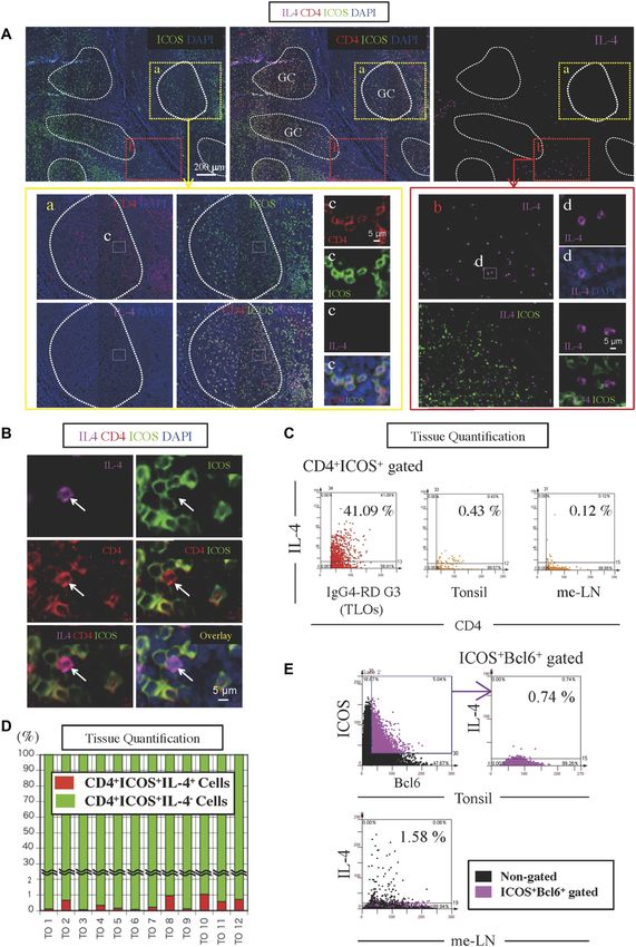
nor have TFH subsets been defined that determine specific polarized
                                                                             IL-4+ICOS− T cells are presumably TH2 cells that reside in T-cell zones
class-switching events. How the overall transcriptome of an IL-4–
                                                                             in tonsils. Quantitation revealed that CD4+ICOS+IL-4+ TFH cells
secreting TFH-cell population may differ from other TFH cell types has
                                                                             represent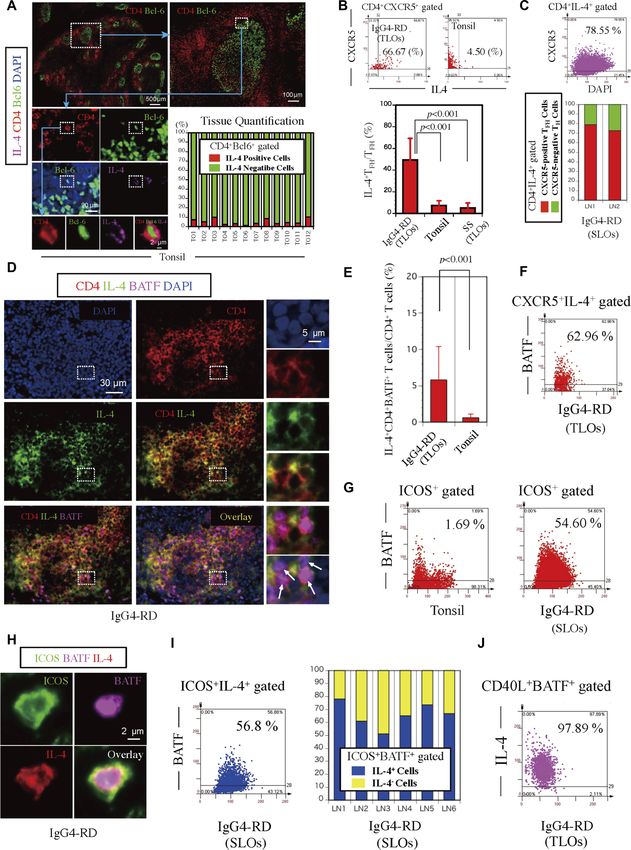
IL-4–secreting TFH cells Maehara et al. https://doi.org/10.26508/lsa.201800050 vol 1 | no 1 | e201800050 3 of 13
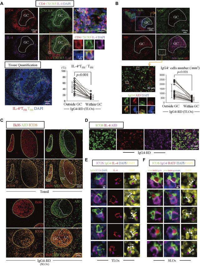
IL-4–expressing CD4+CXCR5+TFH cells were observed. Quantification of                   IL-21R, and PD1 was highest in the IL-4–secreting TFH cells. In addition,
CXCR5+BATF+IL-4+ cells revealed that approximately 60% of CXCR5+IL-4+                 they also expressed high levels of CCR4, CD200, CTLA4, and GITR and
cells in an IgG4-RD patient with TLOs expressed BATF (Fig 2F).                        low levels of CD6, CD27, CD28, SELL, IL-7R, and CD74. Although the
   We considered the possibility that the expansion of these IL-4–                    expression levels of these cell surface markers are derived from in vitro
expressing TFH cells might represent an important disease-related                     activation during cytokine capture, the CD markers specific for cells
TFH subset and contribute to the specific class-switching event in                     with an IL-4–secreting TFH phenotype may aid in the specific flow
IgG4-RD. ICOS+BATF+ TFH cells are far more abundant in IgG4-RD                        cytometric identification of ex vivo human IL-4–secreting TFH cells in
lymph nodes than in normal tonsils (Fig 2G). As shown in Fig 2H, ICOS+                future studies. As expected, CD4+ CD45RA+ cells had the highest
BATF+IL-4+ TFH cells were detected in patients with IgG4-RD, and these                level of CCR7, CD27, and CD62L. The high expression levels of
cells were abundant. Quantification of ICOS+BATF+IL-4+ TFH cells                       multiple chemokines, cytokines, and their receptors including
revealed that approximately 60% of ICOS+IL-4+ TFH cells in an IgG4-RD                 CCR2, CCR6, CXCR3, CXCR6, IL-2RA, IL-2RB, IL-10RA, CCL4, IFNG, IL-2,
lymph node patient expressed BATF (Fig 2I). Furthermore, quantifi-                     IL-3, IL-4, IL-5, IL-9, IL-10, IL-17A, IL-22, and IL-23A within the
cation revealed that the majority of ICOS+BATF+ TFH cells in IgG4-RD                  IL-4–producing CD4+CD45RA− non-TFH cells perhaps indicate the
lymph nodes expressed IL-4 as well (Fig 2I). Interestingly, quantifi-                  heterogeneity among these cells and may reflect the overlap and
cation of CD40L+BATF+IL-4+ T cells revealed that these cells repre-                   plasticity between TH1, TH2, and TH17 subsets that are seen following TCR
sented approximately 98% of CD40L+BATF+ T cells in an IgG4-RD                         stimulation in the absence of polarizing cytokines (i.e., anti-CD3 and
patient (Fig 2J). These data indicate that a large number of lesional                 anti-CD28 alone). Interestingly, the IL-4–producing TFH cells express the
IL-4+ TFH cells in IgG4-RD express BATF, CD40L, and ICOS.                             highest levels of BCL6 and BATF in this comparison but not tran-
                                                                                      scription factors related to TH2 differentiation such as GATA3, STAT5A,
                                                                                      and PRDM1 (BLIMP1), which are instead abundantly expressed in the
IL-4–secreting TFH cells are a distinct population of TFH cells
                                                                                      IL-4–producing non-TFH population, which is likely enriched for TH2 cells.
                                                                                      Furthermore, the expressions of all the markers that we studied using
Although visualization by multicolor staining permits anatomic lo-
                                                                                      immunofluorescence in IL-4–expressing TFH cells from IgG4-RD tissues
calization of TFH cells in tissues, it can provide only limited in-
                                                                                      (BCL6, ICOS, IL-4, CXCR5, BATF, GATA3, and PD1) were consistent with the
formation about a few expressed proteins in any putative TFH-cell
                                                                                      RNA sequence observations on the tonsillar IL-4–secreting TFH cells.
subset, and detailed characterization of any specific cytokine-
secreting TFH subset is currently lacking. To better understand the
biology of IL-4–secreting TFH cells found in lymphoid organs, we                      CD4+CXCR5+IL-4+ TFH cells are mainly outside GCs in IgG4-RD and
performed RNA sequence analysis of viable IL-4–secreting TFH cells                    sometimes physically associate with AID-expressing B cells
from human tonsils. Although the fraction of IL-4–producing TFH cells
is low in human tonsils, we were able to purify this subset by starting               As shown in Fig 4A, CD4+CXCR5+IL-4+ TFH cells were abundant in
with 600 million tonsil cells using an IL-4 cytokine capture strategy.                affected IgG4-RD tissues and were mainly outside GCs but were also
To promote cytokine secretion, CD19-depleted lymphocytes from                         located in the light zone within GCs. Using parallel tissue sections,
tonsils were stimulated overnight with plate-coated anti-CD3 and                      we noted that IgG4-positive B cells were abundant outside GCs in
anti-CD28 antibodies. We then compared the transcriptomes of FACS-                    the same region in which IL-4–secreting TFH cells were observed. We
sorted IL-4–producing TFH cells with those of CXCR5hiPD1hi TFH cells                  quantitated IL-4–expressing TFH cells and IgG4-expressing B cells
that did not secrete IL-4, as well as IL-4–secreting CD45RA−CXCR5−                    within and outside GCs and observed that the majority of both cell
non-TFH cells obtained from the same tonsil (Fig 3A). CD4+ CD45RA+                    types reside outside GCs (Fig 4A and B).
naive cells were also included as an additional control. Of the 26,002                   It is generally accepted that CD40L-CD40 signaling induces AID
mapped transcripts, 7,792 were differentially expressed across the                    and that specific cytokines target selected switch regions. Do the
four conditions (Fig 3B). The IL-4–producing and nonproducing TFH                     IL-4–expressing TFH cells and AID-expressing B cells make physical
cells were most similar and markedly dissimilar from IL-4–secreting                   contact with one another? We examined lymph nodes and TLOs
non-TFH cells or naive CD4+ T cells (Fig 3C). In contrast to the IL-4–                from IgG4-RD patients to determine whether ICOS+ TFH cells are in
producing non-TFH cells that express high levels of IL-4, IL-5, and                   physical contact with AID-expressing B cells or IgG4-expressing
IL-13, reflecting a TH2 signature, IL-4–producing TFH cells express only               B cells in situ. AID-expressing B cells could be visualized outside
IL-4 but much lower levels of IL-5 and IL-13 (Fig 3D). To examine                     GCs in IgG4-RD sections (Fig 4B and C); we noted that IgG4-
lineage-defining genetic regulators and functionally relevant effector                 expressing B cells outside GCs often express AID (Fig 4B). We
molecules, we focused our analysis on differentially expressed CD                     also noted the existence of IL-4+ICOS+ TFH cells in cell–cell contact
molecules, transcription factors, and cytokines (Fig 3E). The transcript              with AID-expressing B cells (Fig 4D). As has been reported earlier,
level of genes critical to TFH function such as CD40L, ICOS, CXCR5,                   much of the AID staining seen is cytosolic (Cattoretti et al, 2006).

Figure 1. CD4+ICOS+IL-4+ TFH cells are sparse in human SLOs.
(A) Immunofluorescence staining of CD4 (red), ICOS (green), IL-4 (magenta), and DAPI (blue) in tissues from normal tonsils. The yellow broken line demarcates the area
within GCs. The white broken line demarcates the area outside GC. (B) Immunofluorescence staining of CD4 (red), ICOS (green), IL-4 (magenta), and DAPI (blue) in tissues
from normal tonsils. (C) Scatter plots depict the mean fluorescence intensity per cell quantified using TissueQuest software for each fluorescent antibody used to stain
tissue from an IgG4-RD patient, normal tonsils, and normal mesenteric lymph nodes. (D) CD4+ICOS+IL-4+ (red) and CD4+ICOS+IL-4− (green) cells were quantified in tissue
from 12 tonsils. (E) Scatter plots depict the mean fluorescence intensity per cell quantified using TissueQuest software for each fluorescent antibody used to stain normal
tonsils and mesenteric lymph nodes.

IL-4–secreting TFH cells   Maehara et al.                                              https://doi.org/10.26508/lsa.201800050   vol 1 | no 1 | e201800050       4 of 13
IL-4–secreting TFH cells Maehara et al. https://doi.org/10.26508/lsa.201800050 vol 1 | no 1 | e201800050 5 of 13
Furthermore, we noted the existence of IL-4+ICOS+ and BATF+ICOS+                          (2011) reported that TFH cells from outside and within GCs secrete IL-4
TFH cells that are in cell–cell contact with IgG4-expressing B cells                      when cocultured in vitro with B cells. However, as these data did not
(Fig 4E and F), confirming visual contact with nuclear distance                            provide information at the single-cell level, the percentage of TFH
measurements. AID+IgG4+ B cells were also visualized within and                           cells capable of secreting IL-4 could not be surmised from this report.
outside GCs in TLOs in IgG4-RD.                                                           Kroenke et al used flow cytometry to quantitate intracellular cyto-
                                                                                          kines in in vitro restimulated TFH cells from tonsils. Following
CD4+CXCR5+IL-4+ TFH cells and CD4+IL-4+BATF+ T cells are enriched                         restimulation with PMA and ionomycin,
Figure 3. Transcriptomic profiling of IL-4–producing TFH cells from human tonsils. (A) Gating strategy used to sort IL-4–secreting and nonsecreting tonsillar CD45RA+CXCR5+ TFH cells following anti-CD3/anti-CD28 stimulation. CD45RA+ CXCR5− cells and CD45RA−CXCR5−IL-4+ cells were sorted as additional controls. (B) A heatmap of differentially expressed genes across all four conditions depicting the Z-scores for normalized expected read counts. (C) A correlation matrix of differentially expressed genes. (D) Expression pattern of individual ILs is depicted on a log scale. (E) Expression of CD molecules, cytokines, and transcription factors clustered into patterns using k-means. Z-scores of the expected read counts for each cluster are shown. a distinct subset. It is reasonable to consider that these IL-4– that are expanded in disease that may contribute to class switching secreting BATF-expressing TFH cells, which do not express GATA-3, to IgG4. Unlike TH2 cells, which produce IL-4, IL-5, and IL-13, the represent an activated TFH population or subset in SLOs and TLOs IL-4–producing tonsillar TFH cells produce IL-4 and IL-10 but not IL-5 IL-4–secreting TFH cells Maehara et al. https://doi.org/10.26508/lsa.201800050 vol 1 | no 1 | e201800050 7 of 13
IL-4–secreting TFH cells Maehara et al. https://doi.org/10.26508/lsa.201800050 vol 1 | no 1 | e201800050 8 of 13
or IL-13. This subset of TFH cells has a unique transcriptional profile                  and salivary glands (LSGs) of 7 patients with active SS. In addition, 2
and a distinct set of surface markers that will facilitate future                       unaffected cervical lymph nodes, 3 mesenteric lymph nodes, and
functional studies.                                                                     12 healthy human tonsils were obtained, which were histologi-
    The mechanisms and signals by which antibodies class switch to                      cally normal. All these patients with IgG4-RD had been followed
IgG4 are poorly understood. CD4+ T cells can activate IgM-positive B cells              up between 2007 and 2015 at the Department of Oral and
to switch to IgG4 and IgE in the presence of added IL-4 (Gascan et al,                  Maxillofacial Surgery of Kyushu University Hospital, a tertiary
1991). It has been argued based on in vitro studies that IL-10 may                      care center. Open SMG biopsies were obtained from patients with
contribute to IgG4 class switching indirectly by somehow facilitating                   IgG4-RD (Moriyama et al, 2014). IgG4-RD was diagnosed according
IL-4–mediated switching to IgG4 in preference to IgE (Jeannin et al,                    to the following criteria (Umehara et al, 2012): (i) persistent
1998). No molecular or cellular explanation has emerged for this in vitro               (longer than 3 mo) symmetrical swelling of more than two lac-
phenomenon. Arguments have also been made that switching to IgG4 is                     rimal and major salivary glands; (ii) high (>135 mg/dl) serum
a result of repeated division, and that switching progresses sequentially               concentrations of IgG4; and (iii) infiltration of IgG4-positive
along chromosome 14 from IgG1 to IgG3 to IgG2 to IgG4 (Tangye et al,                    plasma cells into tissue (IgG4+ cells/IgG+ cells >40%), as de-
2002; Jackson et al, 2014). We do not presume that IL-4–secreting TFH                   termined by immunostaining. All SMGs from patients with IgG4-
cells alone contribute to IgG4 class switching, although it is possible                 RD had histopathologic features of IgG4-RD. Age, sex, serum Ig,
that the same subset of IL-4–secreting TFH cells that we have char-                     and specific autoantibody levels of 17 patients with IgG4-RD
acterized might, in certain contexts, make enough IL-10 to facilitate this              (G1–G17) whose affected salivary gland biopsies were analyzed
switching event. Perhaps more efficient purification of this TFH subset                   by ex vivo in situ immunofluorescence studies are summarized in
using surface markers inferred from our transcriptomic data will fa-                    Table S1A. Age, sex, serum Ig, and specific autoantibody levels of
cilitate future functional studies on IL-4–secreting TFH cells. Tissue                  six patients with IgG4-RD (LN1–LN6) whose affected lymph node
sources of IL-10 or of other factors that work in concert with IL-4 from                biopsies were analyzed by ex vivo in situ immunofluorescence
these specific TFH cells will also need to be explored in future studies.                studies are summarized in Table S2.
    These studies are the first demonstration of an in situ expansion of                    Each patient with SS exhibited objective evidence of salivary
what may be construed to be a human TFH subset. Many diseases exist                     gland involvement based on the presence of subjective xerostomia
in which IgE dominates in the absence of IgG4 and vice versa. Our data                  and a decreased salivary flow rate, abnormal findings on parotid
indicate that only a fraction of TFH cells in SLOs and TLOs, mainly                     sialography, and focal lymphocytic infiltrates in the LSGs by his-
outside GCs, express BATF and IL-4. However, we have shown here that                    tology (Vitali et al, 2002). All patients with SS were severe cases and
these IL-4–expressing TFH cells accumulate in TLOs of patients with                     had developed abundant TLOs in each salivary gland. None of the
IgG4-RD. There is therefore a disease-specific increase in the accu-                     patients with IgG4-RD and SS had a history of treatment with
mulation of a subset of TFH cells and a tight association between the                   steroids or other immunosuppressants, infection with HIV, hepatitis
numbers of these cells and class switching, specifically to IgG4.                        B virus, hepatitis C virus, or sarcoidosis, and none had evidence of
Whether there are further subsets of BATF- and IL-4–expressing TFH                      malignant lymphoma at the time of the study. Age, sex, serum Ig,
cells that separately facilitate IgG4 or IgE class switching remains to be              and specific autoantibody levels of patients with SS whose salivary
ascertained using single-cell approaches.                                               gland biopsies were analyzed by in situ immunofluorescence are
                                                                                        summarized in Table S1B.
                                                                                           Normal human mesenteric lymph node and tonsil patients were
                                                                                        obtained from Massachusetts General Hospital. Normal human
Materials and Methods                                                                   cervical lymph nodes were obtained from the Department of Oral
                                                                                        and Maxillofacial Surgery of Kyushu University Hospital.
Study population                                                                           The study protocol was approved by the Ethics Committee of
                                                                                        Kyushu University, Japan, and the Institutional Review Board at
SMGs were obtained from 17 Japanese patients with IgG4-RD, from                         Massachusetts General Hospital. All patients provided written in-
affected lymph nodes of 6 patients with IgG4-RD, and from lacrimal                      formed consent before participating in the study.

Figure 4. CD4+CXCR5+IL-4+ TFH cells are abundant around GCs in IgG4-RD and sometimes physically contact AID-expressing B cells.
(A) CD4+CXCR5+IL-4+ T cells were enriched around TLOs in IgG4-RD SMGs. Immunofluorescence staining of CD4 (red), IL-4 (magenta), CXCR5 (green), and DAPI (blue) in IgG4-
RD SMGs (patient G3). Quantification of CD4+CXCR5+IL-4+ TFH cells and CD4+CXCR5+ TFH cells comparing those outside and within GCs from each of five different areas in TLOs
of 17 patients with IgG4-RD (G1–G17). The P-value is based on the Mann–Whitney U test. (B) IgG4+B cells were enriched outside GC, especially some these cells express AID.
Immunofluorescence staining of CD4 (red), CD19 (blue), and Bcl6 (green) in IgG4-RD SMGs (patient G3). Immunofluorescence staining of AID (red), Bcl6 (magenta), IgG4
(green), and DAPI (blue) in IgG4-RD SMGs (patient G3). The numbers of IgG4+ cells (per square micrometer) outside and within GCs were quantified from each of five
different areas in TLOs of 17 patients with IgG4-RD (G1–G17). The P-value is based on the Mann–Whitney U test. (C) AID-expressing B cells outside GCs in IgG4-RD lymph
nodes were abundant. Immunofluorescence staining of Bcl6 (red), AID (green), and ICOS (orange) in a normal tonsil and IgG4-RD lymph node. (D) Immunofluorescence
staining of ICOS (green), IL-4 (red), and AID (magenta) in IgG4-RD SMGs. (E) Immunofluorescence staining of IgG4 (green), ICOS (magenta), IL-4 (red), and DAPI (blue) in TLOs
with an IgG4-RD patient (G3). Magenta arrows indicate ICOS+IL-4+ TFH cells. Green arrows indicate IgG4+ B cells. A number of IL-4–expressing TFH cells and IgG4+ B cells
formed close and extensive intercellular plasma membrane contacts. The distance was measured between the edge of each IgG4+ B cell nucleus to the edge of the closest
IL-4–expressing TFH-cell nucleus, and an internuclear distance of less than 0.4 μm was indicative of a T-B conjugate. (F) Immunofluorescence staining of ICOS (green), IgG4
(magenta), BATF (red), and DAPI (blue) in the lymph nodes of an IgG4-RD patient. Green arrows indicate ICOS+BATF+ TFH cells. Magenta arrows indicate IgG4+ B cells. A
number of BATF+ICOS+ TFH cells and IgG4+ B cells formed close and extensive intercellular plasma membrane contacts. The distance was measured between the edge of
each IgG4+ B-cell nucleus to the edge of the closest BATF+ICOS+ TFH-cell nucleus. All internuclear distances in this figure were
IL-4–secreting TFH cells Maehara et al. https://doi.org/10.26508/lsa.201800050 vol 1 | no 1 | e201800050 10 of 13
Multicolor immunofluorescence staining The number of TLOs with GCs and the size of GCs in each TLO
were evaluated in 4 mm2 sections from five different areas of 17
Tissue samples were fixed in formalin, embedded in paraffin, and patients with IgG4-RD (G1–G17) and 7 patients with SS (SS1–SS7)
sectioned. These specimens were incubated with the following (Table S3).
antibodies: anti-AID (clone: ZA001; Invitrogen), anti-IgG4 (clone:
ab109493; Abcam), anti-ICOS (clone: 89601; Cell Signaling Technol- IL-4 capture assay and cell sorting
ogy), anti-IL-4 (clone: MAB304; R&D Systems), GATA3 (clone: CM405A;
Biocare), CXCR5 (clone: MAB190; R&D Systems), Bcl-6 (clone: CM410A, 600 million cells from human tonsils were resuspended in Miltenyi
C; Biocare), BATF (clone: 10538; Cell Signaling Technology), CD4 (clone: magnetic-activated cell sorting buffer and stained with biotinylated
CM153A; Biocare), and CD19 (clone: CM310 A,B; Biocare), followed by anti-human CD19 (clone: HIB19; BioLegend) on ice for 25 min. Cells
incubation with secondary antibodies using a SuperPicTure Polymer were washed once in magnetic-activated cell sorting buffer and
Detection Kit (Invitrogen) and an Opal 3-Plex Kit (Fluorescein, Cya- incubated with anti-biotin microbeads (Miltenyi Biotec) for 25 min
nine3, and Cyanine5). The samples were mounted with ProLong Gold on ice. Cells were then loaded on two separate Miltenyi LS columns
Antifade mountant containing DAPI (Invitrogen). (at 300 million cells per column), and the flow-through was col-
lected as the B cell–depleted fraction. These cells were spun down
Microscopy and quantitative image analysis and stimulated overnight with plate-coated anti-CD3 (5 μg/ml) and
anti-CD28 (5 µg/ml) antibodies. The stimulated cells were then
Images of the salivary gland specimens were acquired using the enriched for IL-4–secreting cells using the vendor’s protocol (IL-4
TissueFAXS platform (TissueGnostics). For quantitative analysis, the Secretion Assay Kit; Miltenyi Biotec, #130-054-101). The enriched IL-4+
entire area of the tissue involved by the lymphoplasmacytic in- cell fraction was surface stained with antibodies against CD4
filtrate was acquired as digital grayscale images in four channels (BioLegend, #317420), CD45RA (clone: H100; BioLegend, #304122),
with filter settings for FITC, Cy3, and Cy5 in addition to DAPI. Cells of CXCR5 (clone: J252D4; BioLegend, #356920), and PD1 (clone: EH12.2H7;
a given phenotype were identified and quantitated using Tissue- BioLegend, #329924). The following populations were viably sorted
Quest software (TissueGnostics), with cutoff values determined using FACSAria2 (Becton Dickinson) directly into Qiagen RLT-plus
relative to the positive controls. This microscopy-based multicolor buffer (with 1% 2-ME): (i) IL-4–secreting CD4+CXCR5+ T FH cells, (ii)
tissue cytometry software permits multicolor analysis of single cells IL-4–secreting CD4 + CD45RA-CXCR5 − T H2 cells, (iii) IL-4–negative
within tissue sections similar to flow cytometry. The principle of the CD4+CXCR5+PD1+ TFH cells, and (iv) IL-4–negative CD45RA+ cells.
method and the algorithms used have been described in detail
elsewhere (Ecker & Steiner, 2004). Trancriptomic analyses
Evaluation of TLOs with IgG4-RD and SS Total RNA was isolated from the FACS-sorted cells using the RNeasy
plus Micro Kit (Qiagen). RNA-sequencing libraries were prepared
TLOs with GCs (Ruddle, 2014) were identified using multicolor im- as previously described (Picelli et al, 2013). Briefly, whole tran-
munofluorescence approaches (CD4, CD19, Bcl6, and DAPI). In this scriptome amplification and tagmentation-based library prepara-
study, SMG and LSG tissue sections from 25 patients with IgG4-RD tion were performed using the SMART-Seq2 protocol, followed by
and 15 patients with severe SS were evaluated. Distinct Bcl-6+ GCs 35-bp paired-end sequencing on a NextSeq 500 instrument (Illu-
were observed in affected IgG4-RD tissues. Bcl-6+ GC B cells in TLOs mina). 5–10 million reads were obtained from each sample and
were within B-cell follicles, delineated using antibodies to CD19. aligned to the University of California, Santa Cruz hg38 transcriptome.
17 of 25 patients (68%) with IgG4-RD had TLOs in affected salivary Gene expression was calculated using RSEM as previously described
glands. Age, sex, serum Ig, and specific autoantibody levels of 17 (Li & Dewey, 2011). The EBSeq package was used to identify differ-
patients with IgG4-RD (G1–G17) whose affected salivary gland entially expressed genes with a posterior probability of differential
biopsies were analyzed by in situ immunofluorescence are expression >0.95 (Leng et al, 2013). Our analysis focused on cytokines,
summarized in Table S1A. 7 of 15 patients (47%) with severe SS transcription factors, and CD molecules. Cytokines and transcription
had TLOs in affected salivary glands. Age, sex, serum Ig, and factors were obtained using the gene ontology terms “GO:0003700”
specific autoantibody levels of patients with SS whose salivary and “GO:0005125,” respectively. Transcription factors pertaining to
gland biopsies were analyzed in this study are summarized in immune cells were filtered using a literature-based gene prioritization
Table S1B. approach with the query terms “immune response,” “immunity,”
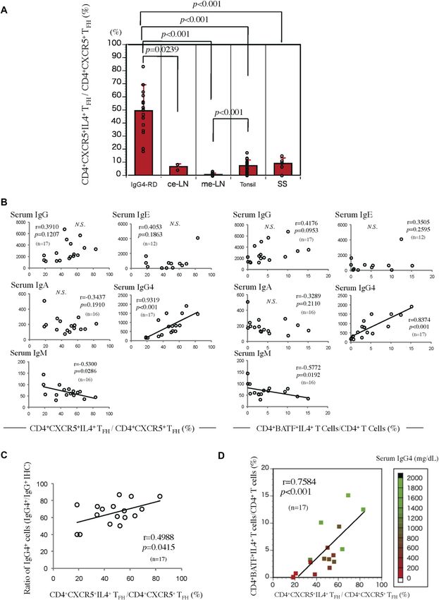
Figure 5. CD4+CXCR5+IL-4+ TFH cells and CD4+IL-4+BATF+ T cells are enriched in IgG4-RD, and their proportions are tightly linked to serum IgG4 levels and IgG4-positive
plasma cells.
(A) Quantification of CD4+CXCR5+IL-4+ and CD4+CXCR5+ TFH cells in 17 IgG4- RD SMGs, seven SS LSGs, 12 tonsils, two cervical lymph nodes, and three mesenteric lymph nodes.
The P-value is based on the Mann–Whitney U test. (B) Correlations of the proportion of CD4+CXCR5+IL-4+ TFH cells and CD4+BATF+IL-4+ T cells in SMGs from patients with
IgG4-RD and their serum IgG, IgA, IgE, IgM, or IgG4 levels. The r and P-value were determined using Spearman’s rank correlations. (C) Correlations of the proportions of CD4+
CXCR5+IL-4+ TFH cells in SMGs from patients with IgG4-RD and their ratio of IgG4+/IgG+ cells (n = 17). The r and P-value were determined using Spearman’s rank correlations.
(D) The frequency of CD4+CXCR5+IL-4+ TFH cells in SMGs from patients with IgG4-RD (n = 17) correlated with the frequency of CD4+BATF+IL-4+ T cells and serum IgG4
concentrations. The r and P-value were determined using Spearman’s rank correlations.
IL-4–secreting TFH cells Maehara et al. https://doi.org/10.26508/lsa.201800050 vol 1 | no 1 | e201800050 11 of 13“T cells,” and “lymphocytes” (Fontaine et al, 2011). The data dis- References
cussed in this publication have been deposited in National Center
for Biotechnology Information’s Gene Expression Omnibus and are Akiyama M, Suzuki K, Yamaoka K, Yasuoka H, Takeshita M, Kaneko Y, Kondo H,
accessible through GEO Series accession number GSE111968. Kassai Y, Miyazaki T, Morita R, et al (2015) Number of circulating
follicular helper 2 T cells correlates with IgG4 and interleukin-4 levels
Statistical analyses and plasmablast numbers in IgG4-related disease. Arthritis
Rheumatol 67: 2476–2481. doi:10.1002/art.39209
Differences between groups were determined using χ2 tests, Akiyama M, Yasuoka H, Yamaoka K, Suzuki K, Kaneko Y, Kondo H, Kassai Y,
Koga K, Miyazaki T, Morita R, et al (2016) Enhanced IgG4 production by
t tests, Mann–Whitney U tests, and Spearman’s rank correlations.
follicular helper 2 T cells and the involvement of follicular helper 1
All statistical analyses were performed using JMP Pro software, version T cells in the pathogenesis of IgG4- related disease. Arthritis Res Ther
11 (SAS Institute) for Mac. P-values < 0.05 were considered statistically 18: 167. doi:10.1186/s13075-016-1064-4
significant. Nonsignificant differences were not specified. In all figures, Ballesteros-Tato A, Randall TD, Lund FE, Spolski R, Leonard WJ, Leon B (2016)
bar charts and error bars represent means ± SEM. T follicular helper cell plasticity shapes pathogenic T helper 2 cell-
mediated immunity to inhaled house dust mite. Immunity 44: 259–273.
doi:10.1016/j.immuni.2015.11.017
Supplementary Information Bentebibel SE, Schmitt N, Banchereau J, Ueno H (2011) Human tonsil B-cell
lymphoma 6 (BCL6)-expressing CD4+ T-cell subset specialized for
B-cell help outside germinal centers. Proc Natl Acad Sci USA 108:
Supplementary Information is available at https://doi.org/10.26508/lsa. E488–E497. doi:10.1073/pnas.1100898108
201800050.
Cattoretti G, Buttner M, Shaknovich R, Kremmer E, Alobeid B, Niedobitek G
(2006) Nuclear and cytoplasmic AID in extrafollicular and
germinal center B cells. Blood 107: 3967–3975. doi:10.1182/
Acknowledgements blood-2005-10-4170
Crotty S (2011) Follicular helper CD4 T cells (TFH). Annu Rev Immunol 29:
This work was supported by awards AI110495 (to S Pillai) and AI113163 (to VS 621–663. doi:10.1146/annurev-immunol-031210-101400
Mahajan) from the National Institute of Health and supported by the Jap- Ecker RC, Steiner GE (2004) Microscopy-based multicolor tissue cytometry at
anese Society for the Promotion of Science Postdoctoral Fellowships for the single-cell level. Cytometry A 59: 182–190. doi:10.1002/cyto.a.20052
Research Abroad to T Maehara and Mochida Memorial Medical and Phar-
Fontaine JF, Priller F, Barbosa-Silva A, Andrade-Navarro MA (2011) Genie:
maceutical Foundation to T Maehara. The authors thank Eric Safai for help
Literature-based gene prioritization at multi genomic scale. Nucleic
with the collection and processing of samples from Massachusetts General
Acids Res 39:W455–W461. doi:10.1093/nar/gkr246
Hospital and Thomas Diefenbach of the Imaging Core at the Ragon Institute
for help and advice. Gascan H, Gauchat JF, Roncarolo MG, Yssel H, Spits H, de Vries JE (1991)
Human B cell clones can be induced to proliferate and to switch to
IgE and IgG4 synthesis by interleukin 4 and a signal provided by
Author Contributions activated CD4+ T cell clones. J Exp Med 173: 747–750. doi:10.1084/
jem.173.3.747
T Maehara: conceptualization, formal analysis, investigation, and Jackson KJ, Wang Y, Collins AM (2014) Human immunoglobulin classes and
writing—original draft, review, and editing. subclasses show variability in VDJ gene mutation levels. Immunol Cell
H Mattoo: conceptualization, investigation, and writing—original Biol 92: 729–733. doi:10.1038/icb.2014.44
draft, review, and editing. Jeannin P, Lecoanet S, Delneste Y, Gauchat JF, Bonnefoy JY (1998) IgE versus
VS Mahajan: formal analysis, investigation, and writing—original IgG4 production can be differentially regulated by IL-10. J Immunol
160: 3555–3561.
draft, review, and editing.
SJH Murphy: investigation. Kamisawa T, Zen Y, Pillai S, Stone JH (2015) IgG4-related disease. Lancet 385:
1460–1471. doi:10.1016/s0140-6736(14)60720-0
GJ Yuen: investigation and writing—review and editing.
N Ishiguro: resources. King C, Tangye SG, Mackay CR (2008) T follicular helper (TFH) cells in normal
and dysregulated immune responses. Annu Rev Immunol 26: 741–766.
M Ohta: resources.
doi:10.1146/annurev.immunol.26.021607.090344
M Moriyama: resources.
Kroenke MA, Eto D, Locci M, Cho M, Davidson T, Haddad EK, Crotty S (2012) Bcl6
T Saeki: resources.
and Maf cooperate to instruct human follicular helper CD4 T cell
H Yamamoto: resources. differentiation. J Immunol 188: 3734–3744. doi:10.4049/
M Yamauchi: resources. jimmunol.1103246
J Daccache: investigation. Leng N, Dawson JA, Thomson JA, Ruotti V, Rissman AI, Smits BM, Haag JD,
T Kiyoshima: resources. Gould MN, Stewart RM, Kendziorski C (2013) EBSeq: An empirical Bayes
S Nakamura: resources. hierarchical model for inference in RNA-seq experiments.
Bioinformatics 29: 1035–1043. doi:10.1093/bioinformatics/btt087
JH Stone: resources.
S Pillai: conceptualization, formal analysis, supervision, funding Li B, Dewey CN (2011) RSEM: Accurate transcript quantification from RNA-Seq
data with or without a reference genome. BMC Bioinformatics 12: 323.
acquisition, and writing—original draft, review, and editing.
doi:10.1186/1471-2105-12-323
Ma CS, Suryani S, Avery DT, Chan A, Nanan R, Santner-Nanan B, Deenick EK,
Conflict of Interest Statement Tangye SG (2009) Early commitment of naive human CD4(+) T cells to
the T follicular helper (T(FH)) cell lineage is induced by IL-12. Immunol
The authors declare that they have no conflict of interest. Cell Biol 87: 590–600. doi:10.1038/icb.2009.64
IL-4–secreting TFH cells Maehara et al. https://doi.org/10.26508/lsa.201800050 vol 1 | no 1 | e201800050 12 of 13Maehara T, Mattoo H, Ohta M, Mahajan VS, Moriyama M, Yamauchi M, Drijvers Sahoo A, Alekseev A, Tanaka K, Obertas L, Lerman B, Haymaker C, Clise-Dwyer
J, Nakamura S, Stone JH, Pillai SS (2017) Lesional CD4+ IFN-gamma+ K, McMurray JS, Nurieva R (2015) Batf is important for IL-4 expression in
cytotoxic T lymphocytes in IgG4-related dacryoadenitis and T follicular helper cells. Nature Commun 6: 7997. doi:10.1038/
sialoadenitis. Ann Rheum Dis 76: 377–385. doi:10.1136/annrheumdis- ncomms8997
2016-209139
Tangye SG, Ferguson A, Avery DT, Ma CS, Hodgkin PD (2002) Isotype switching
Mahajan VS, Mattoo H, Deshpande V, Pillai SS, Stone JH (2014) IgG4- related by human B cells is division-associated and regulated by cytokines.
disease. Annu Rev Pathol 9: 315–347. doi:10.1146/annurev-pathol- J Immunol 169: 4298–4306. doi:10.4049/jimmunol.169.8.4298
012513-104708
Ueno H, Banchereau J, Vinuesa CG (2015) Pathophysiology of T follicular helper
Mattoo H, Mahajan VS, Della-Torre E, Sekigami Y, Carruthers M, Wallace ZS, cells in humans and mice. Nat Immunol 16: 142–152. doi:10.1038/ni.3054
Deshpande V, Stone JH, Pillai S (2014) De novo oligoclonal expansions
Umehara H, Okazaki K, Masaki Y, Kawano M, Yamamoto M, Saeki T, Matsui S,
of circulating plasmablasts in active and relapsing IgG4-related
Sumida T, Mimori T, Tanaka Y, et al; Research Program for Intractable
disease. J Allergy Clin Immunol 134: 679–687. doi:10.1016/j.jaci.
Disease by Ministry of Health, and G.t. Welfare Japan (2012) A novel
2014.03.034
clinical entity, IgG4-related disease (IgG4RD): General concept and
Mattoo H, Mahajan VS, Maehara T, Deshpande V, Della-Torre E, Wallace ZS, details. Mod Rheumatol 22:1–14. doi:10.3109/s10165-011-0508-6
Kulikova M, Drijvers JM, Daccache J, Carruthers MN, et al (2016) Clonal
Vinuesa CG, Tangye SG, Moser B, Mackay CR (2005) Follicular B helper T cells
expansion of CD4 cytotoxic T lymphocytes in patients with IgG-related
in antibody responses and autoimmunity. Nat Rev Immunol 5:
disease. J Allergy Clin Immunol 138: 825–838. doi:10.1016/j.jaci.2015.12.1330
853–865. doi:10.1038/nri1714
Morita R, Schmitt N, Bentebibel SE, Ranganathan R, Bourdery L, Zurawski G,
Foucat E, Dullaers M, Oh S, Sabzghabaei N, et al (2011) Human blood Vitali C, Bombardieri S, Jonsson R, Moutsopoulos HM, Alexander EL, Carsons
CXCR5(+)CD4(+) T cells are counterparts of T follicular cells and SE, Daniels TE, Fox PC, Fox RI, Kassan SS, et al (2002) Classification
contain specific subsets that differentially support antibody criteria for Sjogren’s syndrome: A revised version of the European
secretion. Immunity 34: 108–121. doi:10.1016/j.immuni.2010.12.012 criteria proposed by the American-European Consensus Group. Ann
Rheum Dis 61: 554–558. doi:10.1136/ard.61.6.554
Moriyama M, Furukawa S, Kawano S, Goto Y, Kiyoshima T, Tanaka A, Maehara
T, Hayashida JN, Ohta M, Nakamura S (2014) The diagnostic utility of Weinstein JS, Herman EI, Lainez B, Licona-Limon P, Esplugues E, Flavell R, Craft J
biopsies from the submandibular and labial salivary glands in IgG4- (2016) TFH cells progressively differentiate to regulate the germinal
related dacryoadenitis and sialoadenitis, so-called Mikulicz’s disease. center response. Nat Immunol 17: 1197–1205. doi:10.1038/ni.3554
Int J Oral Maxillofac Surg 43: 1276–1281. doi:10.1016/j.ijom.2014.06.014
Picelli S, Bjorklund AK, Faridani OR, Sagasser S, Winberg G, Sandberg R (2013)
Smart-seq2 for sensitive full-length transcriptome profiling in single License: This article is available under a Creative
cells. Nat Methods 10: 1096–1098. doi:10.1038/nmeth.2639 Commons License (Attribution 4.0 International, as
Ruddle NH (2014) Lymphatic vessels and tertiary lymphoid organs. J Clin described at https://creativecommons.org/
Invest 124: 953–959. doi:10.1172/jci71611 licenses/by/4.0/).
IL-4–secreting TFH cells Maehara et al. https://doi.org/10.26508/lsa.201800050 vol 1 | no 1 | e201800050 13 of 13You can also read