The box C/D snoRNP assembly factor Bcd1 interacts with the histone chaperone Rtt106 and controls its transcription dependent activity - Infoscience
←
→
Page content transcription
If your browser does not render page correctly, please read the page content below
ARTICLE
                  https://doi.org/10.1038/s41467-021-22077-4                 OPEN

                  The box C/D snoRNP assembly factor Bcd1
                  interacts with the histone chaperone Rtt106
                  and controls its transcription dependent activity
                  Benoît Bragantini 1,6,8, Christophe Charron1,8, Maxime Bourguet2,8, Arnaud Paul1,8, Decebal Tiotiu1,
                  Benjamin Rothé1,7, Hélène Marty1, Guillaume Terral2, Steve Hessmann2, Laurence Decourty3,
                  Marie-Eve Chagot1, Jean-Marc Strub2, Séverine Massenet1, Edouard Bertrand 4, Marc Quinternet5,
                  Cosmin Saveanu 3, Sarah Cianférani2, Stéphane Labialle 1,9 ✉, Xavier Manival 1,9 ✉ &
1234567890():,;

                  Bruno Charpentier 1,9 ✉

                  Biogenesis of eukaryotic box C/D small nucleolar ribonucleoproteins initiates co-transcriptionally
                  and requires the action of the assembly machinery including the Hsp90/R2TP complex, the
                  Rsa1p:Hit1p heterodimer and the Bcd1 protein. We present genetic interactions between the
                  Rsa1p-encoding gene and genes involved in chromatin organization including RTT106 that codes
                  for the H3-H4 histone chaperone Rtt106p controlling H3K56ac deposition. We show that Bcd1p
                  binds Rtt106p and controls its transcription-dependent recruitment by reducing its association
                  with RNA polymerase II, modulating H3K56ac levels at gene body. We reveal the 3D structures
                  of the free and Rtt106p-bound forms of Bcd1p using nuclear magnetic resonance and X-ray
                  crystallography. The interaction is also studied by a combination of biophysical and proteomic
                  techniques. Bcd1p interacts with a region that is distinct from the interaction interface between
                  the histone chaperone and histone H3. Our results are evidence for a protein interaction interface
                  for Rtt106p that controls its transcription-associated activity.

                  1 Université de Lorraine, CNRS, IMoPA, Nancy, France. 2 Laboratoire de Spectrométrie de Masse BioOrganique, Université de Strasbourg, CNRS IPHC

                  UMR 7178, Strasbourg, France. 3 Génétique des Interactions Macromoléculaires, Département de Génomes et Génétique, Institut Pasteur
                  (UMR3525-CNRS), Paris, France. 4 IGH, CNRS, Université de Montpellier, Montpellier, France. 5 Université de Lorraine, CNRS, INSERM, IBSLor, Nancy, France.
                  6
                    Present address: Department of Biochemistry and Molecular Biology, Mayo Clinic, Rochester, MN, USA. 7Present address: Ecole polytechnique fédérale
                  de Lausanne (EPFL) SV ISREC, Station 19, Lausanne, Switzerland. 8These authors contributed equally: Benoît Bragantini, Christophe Charron, Maxime Bourguet,
                  Arnaud Paul. 9These authors jointly supervised this work: Stéphane Labialle, Xavier Manival, Bruno Charpentier. ✉email: stephane.labialle@univ-lorraine.fr;
                  xavier.manival@univ-lorraine.fr; bruno.charpentier@univ-lorraine.fr

                  NATURE COMMUNICATIONS | (2021)12:1859 | https://doi.org/10.1038/s41467-021-22077-4 | www.nature.com/naturecommunications                                 1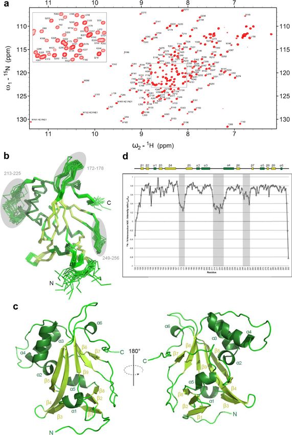
ARTICLE                                                                 NATURE COMMUNICATIONS | https://doi.org/10.1038/s41467-021-22077-4

S
       mall nucleolar RNAs (snoRNAs) form a large abundant                   that Bcd1p binds with the PH1 (Pleckstrin-Homology 1) domain
       family of noncoding RNAs predominantly localized in the               of Rtt106p. These data support a model in which the binding of
       nucleolus1. Based on conserved sequence motifs, snoRNAs               Bcd1p to Rtt106p can inhibit the transcription-related activities of
fall into one of two classes—box C/D snoRNAs or box H/ACA                    this histone chaperone.
snoRNAs. Each snoRNA associates with a set of class-specific and
well-characterized core proteins to form ribonucleoprotein (RNP)
complexes referred to as small nucleolar ribonucleoproteins, i.e.,           Results
box C/D snoRNPs and box H/ACA snoRNPs. These particles                       Genetic Interaction Mapping (GIM) screens of the C/D
primarily catalyze post-transcriptional modifications in ribosomal            snoRNP assembly factor gene RSA1. To identify potential
RNAs2 (rRNAs). In these reactions, the snoRNA functions as a                 genetic interactions with the snoRNP assembly machinery, we
guide by base-pairing with a target sequence and selecting                   performed a high-throughput genetic screen. We used deletion of
the precise nucleotide that will be modified by the catalytic                 RSA1 (rsa1Δ::prMFα2NatR) as the query mutation and combined
activity of the snoRNP3. A few other snoRNPs, e.g., U3, are                  it in a single pool by mating it with individual mutants of the
involved in endo-ribonucleolytic processing of the original pre-             yeast systematic deletion library of nonessential genes31 and with
rRNA transcript4,5.                                                          a collection of DAmP (decreased abundance by mRNA pertur-
   Eukaryotic snoRNP biogenesis is a complex process that                    bation) mutants for essential genes32,33. Double mutants were
involves coordinated assembly, RNA processing, and localization              selected from the pool after sporulation and grown for ~18
factors6. In yeast, box C/D snoRNPs are formed by assembling                 generations in rich liquid medium. In parallel, a similar screen
core proteins Snu13, Nop56, Nop58, and 2’-O-methyl transferase               was performed with other mutant strains for use as a control
Nop1 on box C/D snoRNAs. This process requires assembly                      population. The relative growth rates of double mutant strains
machinery including the protein heterodimer Rsa1:Hit17 and the               were estimated by measuring the relative abundance of cells in the
R2TP chaperone complex, which is composed of proteins Pih1,                  query versus reference populations using DNA barcodes and
Tah1, and AAA+ ATPases Rvb1 and Rvb2 (Rvb1/2)8,9. In addi-                   microarrays. Normalized results are expressed as log2(Q/R)
tion, the Bcd1 protein is essential for both cell viability and box C/       where Q represents the signal intensities of the tag marking a
D snoRNA steady-state stability10,11, and was shown to control               given mutant when combined with the query mutation, and R is
the loading of Nop58p to pre-snoRNPs12. A conserved motif in                 the signal from the same mutant when introduced in the refer-
the N-terminal region of Bcd1p was proposed as a determinant                 ence population. In agreement with previous GIM screens, we
for binding with Snu13p and snoRNA13. This tripartite R2TP/                  obtained a relatively large number of both negative and positive
Rsa1p:Hit1p/Bcd1p machinery is conserved in metazoans and                    log2(Q/R) values, representing aggravating and buffering (or
plants where it is also used for snoRNP biogenesis8,14–18. The               alleviating) effects, respectively (Source data file). In agreement
human ortholog of R2TP associates with an additional prefoldin-              with the contribution of Rsa1p to snoRNP biogenesis and vali-
like (PFDL) module to form a larger chaperone complex called                 dating the approach, we observed strong genetic links between
the PAQosome19. This chaperone is involved in the folding of                 RSA1 and RVB1, encoding one of the R2TP components, and
critical macrocomplexes, e.g., RNPs involved in fundamental                  NOP56, encoding one of the box C/D core proteins (Fig. 1).
cellular processes, and RNA polymerases.                                     However, whereas a negative value (log2(Q/R) = −1.8) was
   Here we show that the snoRNP assembly factor gene RSA1                    obtained for the rvb1-DAMP mutation pointing to a synthetic
genetically interacts with genes involved in chromatin structure             growth defect, combining rsa1Δ mutation with the nop56-DAMP
and nucleosome assembly including histone chaperones such as                 mutation had an epistatic effect with a positive value (log2(Q/R)
Rtt106p (Regulator of Ty transposition), as well as with genes               = +3.0).
involved in transcription and RNA processing. We also demon-                    We used g:Profiler [https://biit.cs.ut.ee/gprofiler/gost] to check
strate that the box C/D snoRNP assembly factor Bcd1p binds                   if specific annotations were enriched among our results. The list
Rtt106p directly. This chaperone cooperates with the H3-H4                   of the top 50 mutants showing an epistatic or suppressor effect in
histone chaperone CAF-1 (chromatin assembly factor 1) and with               combination with rsa1Δ was enriched in two broad gene ontology
the heterodimeric chaperone FACT (facilitates chromatin tran-                (GO) categories: mRNA metabolic process (GO:0016071) and
scriptions, composed of the Spt16p:Pob3p heterodimer) to facil-              chromatin organization (GO:0006325). For a more detailed view
itate DNA replication-coupled (RC) nucleosome assembly20,21.                 of which specific processes among RNA metabolism and
During this process, Rtt106p participates in the deposition of               chromatin organization were affected by RSA1 deletion, we used
newly synthesized H3K56ac-carrying H3:H4 complex on repli-                   a curated list of 518 yeast complexes34 and searched for gene set
cating DNA22–26. In addition, Rtt106p is involved in replication-            enrichment in the GIM screen data using GAGE35. Among the
independent pathways including heterochromatin silencing20,27,               top results, we found genes, e.g., SPT21, encoding proteins
HIR-dependent control of histone gene expression28, regulation               implicated in transcriptional silencing and required for normal
of transcription-dependent histone H3 deposition during RNA                  transcription at other loci including HTA2-HTB2 and HHF2-
polymerase elongation29, and maintenance of promoter fidelity in              HHT2 that encode histone proteins. Deletion of genes involved in
cooperation with HIR (histone regulatory) and Asf1 (anti-silen-              termination and processing of the 3’ end of pre-snoRNAs rescued
cing factor 1) proteins30.                                                   growth of rsa1Δ cells, including the exosome components MTR3,
   Our data reveal a Bcd1p-dependent association of Rtt106p on               RRP42, RRP46, and CSL4, as well as the LEO1 and RTF1 subunits
box C/D pre-snoRNAs, with no discernible effect on snoRNP                    of the PAF complex, which is involved in transcription elongation
assembly. On the contrary, we observe that the association of                (Fig. 1). It is conceivable that impairing these RNA processing
Rtt106p with transcriptionally active loci correlates with its               events compensates for the imperfect box C/D snoRNP assembly
association with RNA polymerase II, and that these associations              resulting from the rsa1Δ disruption. Assembly of snoRNP starts
are negatively modulated by Bcd1p. In coherence with these                   early on nascent snoRNAs and this process has been proposed to
observations, we show that H3K56ac levels are increased at sev-              be coupled to 3’ processing36,37. One possibility is that RNP
eral gene bodies upon Bcd1p depletion. We characterize this                  assembly is delayed in the rsa1Δ mutant and becomes kinetically
important Bcd1p:Rtt106p interaction for the transcription-                   uncoupled from 3’ processing. Slower transcription termination
dependent Rtt106p function at the molecular and atomic levels.               and 3’ processing would then compensate the slower RNP
A combination of ITC, MS approaches, NMR, and X-ray show                     assembly.

2                NATURE COMMUNICATIONS | (2021)12:1859 | https://doi.org/10.1038/s41467-021-22077-4 | www.nature.com/naturecommunications
NATURE COMMUNICATIONS | https://doi.org/10.1038/s41467-021-22077-4                                                                           ARTICLE

Fig. 1 Data from the GIM screen. Two independent screens were performed, one with a mutant strain in which the RSA1 gene was deleted (rsa1Δ::NatR)
and the other with a mutant used as reference strain. Two independent experiments were performed for each of these screens. The query strain and the
reference strain were mated with a pool of strains containing all the viable strains from the haploid gene deletion collection. After selection of heterozygous
diploids, sporulation and selection of the haploid, double mutants were grown for ~18 generations in rich liquid medium (YPD). Microarrays were used to
measure the relative abundance of double mutants with query versus the reference populations. Normalized results are expressed as log2(Q/R) (see
Methods section). Negative values indicate a synthetic growth defect. Positive values reveal either epistatic (buffering) or suppressive (alleviating)
interactions between RSA1 and the selected genes. The genetic interactions with RSA1 are indicated by green arrows for the positive log2(Q/R) values and
by red arrows for the negative values. The exact values are given in parentheses. HC heterochromatin.

   In addition, deletion of RSA1 had multiple epistatic effects                  Retention of 3xHA-Bcd1p on beads occurred with the co-
when combined with deletion of genes encoding components of                      purification of Rtt106p-TAP. To determine whether this interac-
the chromatin remodeling complexes HIR (HIR2, HIR3, HPC2),                       tion is direct, we performed co-purification assays in Escherichia
INO80 (IES1, IES5, NHP10), and CAF-1 (CAC2 and CAC3), as                         coli upon ectopic co-expression of different combinations of
well as genes HDA1 and HDA2 encoding members of the HDA1                         truncated or full-length His6-tagged and untagged proteins
histone deacetylase complex and genes encoding histone variants                  (Fig. 2c). The co-expressed proteins in the bacterial extract were
(HTZ1 and HTA1; Fig. 1). Interestingly, rsa1Δ had a positive log2                co-purified by immobilized metal ion affinity chromatography
(Q/R) value (+1.5) in combination with the deletion of RTT106,                   (IMAC), fractionated by SDS-PAGE and analyzed by mass
which is connected to the HIR and CAF-1 complexes27,28.                          spectrometry (MS) (Supplementary Table 1). This confirmed the
We focused on the Rtt106 protein as it is linked to several                      formation of complexes Bcd1pFL:Rtt106pFL and Bcd1pFL:Rtt106p-
fundamental functions such as DNA replication, stability, and                    M. Recombinant Bcd1pFL and the fragment Rtt106p-M were
transcription, but not yet to snoRNP biogenesis and function,                    also independently purified and isothermal titration calorimetry
except for the observation based on modified chromatin                            (ITC) revealed direct and endothermic in vitro binding with a
immunopurification38 (mChIP) suggesting that it can associate                     1.17 ± 0.17 μM dissociation constant (Kd; Fig. 2d, left panel).
with complexes containing Rvb1/2 proteins in the vicinity of                        We completed these data by native MS analysis of the co-
chromatin.                                                                       expressed complex Bcd1pFL:Rtt106p-M and its individual sub-
                                                                                 units (Fig. 2e). Bcd1pFL was mainly detected as a monomer in
Rtt106p binds Bcd1p. To investigate interactions between                         interaction with two zinc ions (42,752 ± 1 Da, Fig. 2e upper
Rtt106p and the proteins known to be involved in snoRNP bio-                     spectrum), as already reported41. For Rtt106p-M, monomeric
genesis, we performed systematic pairwise yeast two-hybrid                       species were also mainly detected (29,695 ± 1 Da, Fig. 2e middle
(Y2H) assays. This revealed a strong in vivo interaction between                 spectrum). Finally, native MS revealed the formation of a 1:1
Rtt106p and Bcd1p, which resisted high concentrations of 3-AT                    Bcd1pFL:Rtt106p-M heterodimer with a mass of 72,451 ± 1 Da.
(30–40 mM; Fig. 2a). The Y2H interaction derived from the                        These data demonstrated that Bcd1pFL binds the 65–320 (M)
middle domain of Rtt106p encompassing the tandem PH1 and                         domain of Rtt106p with 1:1 stoichiometry.
PH2 domains. Indeed, full-length Bcd1p (Bcd1pFL) interacted                         We then monitored the Bcd1pFL:Rtt106p-M heterodimer
with the fragment of Rtt106p spanning amino acids 65–320                         formation by ion mobility coupled with mass spectrometry
(Rtt106p65-320 also named Rtt106p-M, Fig. 2a). This interaction                  (IM-MS). This approach makes it possible to determine collision
was specific as it was not detected with the homologous PH                        cross section (TWCCSN2) by measuring the drift time of ions
domains from histone chaperones Pob3p and Spt16p (Fig. 2a)                       through a gas-filled IM cell. The arrival drift time (ADT) is
that are members of the FACT chromatin remodeling                                closely linked to the charge state and to the shape of the ions in
complex22,39,40.                                                                 the gas phase42. IM-MS data and TWCCSN2 measured for each
   To validate the Bcd1p:Rtt106p interaction, we used pull-down                  individual subunit Bcd1pFL and Rtt106p-M and for the Bcd1pFL:
assays in yeast cells engineered to express the tagged proteins                  Rtt106p-M heterodimer (Supplementary Fig. 1) revealed that a
3xHA-Bcd1p and Rtt106-TAP from their genome (Fig. 2b).                           significant conformational change occurs in one or both proteins

NATURE COMMUNICATIONS | (2021)12:1859 | https://doi.org/10.1038/s41467-021-22077-4 | www.nature.com/naturecommunications                                     3
ARTICLE                                                               NATURE COMMUNICATIONS | https://doi.org/10.1038/s41467-021-22077-4

upon heterodimer formation. The IM-MS measured TWCCSN2                     Rtt106p binds pre-snoRNPs but does not appear to contribute
(44.4 ± 1.4 nm2) was larger than the value expected from mass              to or interfere with snoRNP biogenesis. To investigate if
and 15% smaller than the one predicted from a juxtaposition of             Rtt106p contributes to snoRNP biogenesis, we used RNA
the two subunits. We concluded that the heterodimer is not                 immunoprecipitation (RIP) assays. We observed that Rtt106p-
formed merely by simple juxtaposition of the two partners.                 TAP was highly enriched on box C/D snoRNAs U3, U14 and the

4              NATURE COMMUNICATIONS | (2021)12:1859 | https://doi.org/10.1038/s41467-021-22077-4 | www.nature.com/naturecommunications

NATURE COMMUNICATIONS | https://doi.org/10.1038/s41467-021-22077-4 ARTICLE
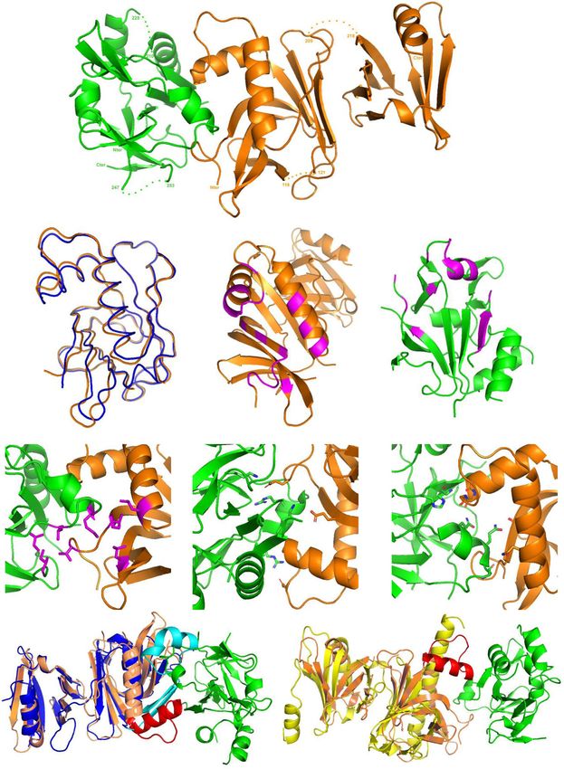
Fig. 2 Bcd1p and histone chaperone Rtt106p form a stable heterodimer. a Schematic representation of the Rtt106p and Bcd1p domain organization. DD
dimeric domain, PH Plekstrin homology, PH1 + PH2 = MD middle domain, AR acidic region, ZHD zinc finger HIT domain, RBD Rtt106 binding domain, DR
disorder region. Y2H assay: full-length Bcd1p (Bcd1pFL) fused to the Gal4 binding domain (BD) interacts with the full-length Rtt106p (Rtt106pFL) or
fragment spanning amino acids 65–320 (Rtt106p-M) fused to the Gal4 activation domain (AD) as evidenced by growth on a His deprived medium
supplemented with increased concentrations of 3-amino-1, 2, 4-triazol (3-AT). No interaction was observed with homologous fragments of FACT
chaperone components Spt16p and Pob3p (Spt16p-M and Pob3p-M). b Interaction of Bcd1pFL with Rtt106pFL in yeast. Co-immunoprecipitation (co-IP) was
performed on GAL1::3HA-BCD1 × RTT106-TAP cells expressing 3xHA-tagged Bcd1pFL and TAP-tagged Rtt106pFL. Cells expressing the nontagged Bcd1p were
used as negative control. Extracts were incubated with anti-HA beads. The co-immunoselected proteins were analyzed by SDS-PAGE and western blotting.
10% of total proteins used per assay were loaded in the input lane. Tagged proteins were detected with PAP antibodies for Rtt106p and anti-HA antibodies
for Bcd1p. The Dps1 protein used to control protein loading was detected using specific anti-Dps1p antibodies. The two panels correspond to a cropping of
two sections of the same membrane. The full-length membrane is presented in the Source data file. The experiment was independently repeated three
times with similar results. c Interaction of recombinant Bcd1p and Rtt106p in E. coli. His6-tagged full-length or M domain of Rtt106p were co-expressed with
Bcd1pFL. His6-tagged Bcd1pFL was co-expressed with Rtt106pFL or Rtt106p-M. Complexes were selected from crude extract by immobilized metal ion
affinity chromatography (IMAC), fractionated by SDS-PAGE and revealed by Coomassie blue staining. The results correspond to co-expression with high
salt (400 mM) buffer. Molecular weight markers (MW) in kilo Dalton (kDa) were loaded on the left. The experiment was repeated twice with similar
results. The identity of the proteins in bands 1 A, 1B, 1 C, 1D, 1E, 1 F, 1 G, and 1H was confirmed by in-gel digestion of gel slices and mass spectrometry (MS)
analysis of the peptide extract (Supplementary Table 1). d Bcd1p and Rtt106p interacting domains. ITC data for the interaction of Bcd1pFL with Rtt106p-M
(on the left) and Bcd1p120-303 with Rtt106p65-301 (on the right) recorded at 293 K in buffer containing 10 mM NaPi at pH 7.5, 150 mM NaCl and 0.5 mM
TCEP. The calculated affinities Kd, and thermodynamic parameters as variations in enthalpy (ΔH) and entropy (ΔS) are indicated. e Nondenaturing
MS characterization of the complex formed upon incubation of recombinant Bcd1pFL with fragment Rtt106p-M. NanoESI mass spectra performed
under nondenaturing conditions confirmed the presence of a 1:1 binding stoichiometry of Bcd1pFL:Rtt106p-M complex (Da = Dalton). Source data for panel
b are provided as a Source Data file.
precursor form of U14 (Fig. 3a). Moreover, using GAL1::3HA- transcription-related recruitment of Rtt106p, we solved the pat-
BCD1 cells grown in galactose-containing (YPG) medium for tern of association at the U3 actively transcribed loci (Fig. 3c).
expression of Bcd1p and in glucose-containing (YPD) medium Rtt106p enrichment increased in the body of the gene compared
for depletion of Bcd1p12, we showed that the association of to the flanking intergenic regions, and the association was sen-
Rtt106p with these snoRNAs is Bcd1p dependent. Using the sitive to the level of expression of Bcd1p only in the transcribed
rsa1Δ mutant cells, we showed that binding of Rtt106p with region. To test the possibility that association to chromatin relied
RNPs containing Bcd1p is also Rsa1p-dependent, which could be on RNA, we performed ChIP analyses in the presence of RNase
explained by the fact that a detectable association of Bcd1p with treatment (Supplementary Fig. 2e). Interestingly, we observed a
snoRNA species is Rsa1p-dependent (Supplementary Fig. 2a). slight reduction of ChIP signals at both transcribed and untran-
Although these data revealed the presence of Rtt106p on box C/D scribed loci, suggesting that Rtt106p associated moderately via
pre-snoRNPs, the hypothesis of a role for Rtt106p in facilitating undetermined chromatin-associated RNA species. Importantly,
or modulating the function of Bcd1p in snoRNP biogenesis the effect of RNase treatment was similar in cells expressing or
seemed unlikely. Indeed, the steady-state levels of box C/D pre- not Bcd1p, excluding the possibility that it occurs in a Bcd1p-
snoRNAs and their mature forms did not change in absence of dependent way. Therefore, the data suggest that Rtt106p interacts
Rtt106p expression, at least in yeast cells grown in standard with chromatin by several, independent ways with at least one
culture conditions (Supplementary Fig. 2b). Accordingly, involving RNAs present at the vicinity of chromatin (including at
enrichments of Bcd1p to box C/D snoRNAs (Supplementary untranscribed loci), and a second one that is independent from
Fig. 2c) and to snoRNA gene loci did not change appreciably in RNA but dependent on Bcd1p expression level.
the absence of Rtt106p (Supplementary Fig. 2d).
Bcd1p controls the association of Rtt106p with RNA poly-
Bcd1p regulates the presence of Rtt106p at transcriptionally merase II and H3K56ac levels at gene bodies. As it has been
active loci. To investigate a potential regulatory function of proposed that Rtt106p acts in the wake of the transcriptional
Bcd1p in Rtt106p activity, we checked whether the recruitment of machinery to promote new histone deposition29, we first tested
Rtt106p to DNA loci could be Bcd1p dependent. Using ChIP, we whether an interaction between Rtt106p and RNA polymerase II
analyzed the presence of FLAG-tagged Rtt106p to the HIR- was detectable. Using co-IP, we indeed observed that Rtt106p was
dependent histone genes HTA1-HTB1 and to the HMR locus associated with Rpb1p, the largest RNA polymerase II subunit
where Rtt106p has been shown to be enriched26,30,43,44. Enrich- (Fig. 4a). Interestingly and in agreement with an effect of Bcd1p
ment in GAL1::3HA-BCD1 cells at box C/D snoRNA-encoding on transcription-related Rtt106p function, this association
genes SNR17A (U3), SNR128 (U14), SNR52 (snR52), and SNR32 increased in the absence of BCD1 gene expression. Taken toge-
encoding box H/ACA snoRNA snR32, as well as at the protein ther, these data suggest that direct binding of Bcd1p with Rtt106p
encoding gene ALG9 was tested for the purpose of comparison precludes association of the histone chaperone with the RNA
(Fig. 3b). Enrichment of Rtt106p at either C/D box or H/ACA polymerase II, and therefore reduces its presence at tran-
box snoRNA gene loci was equivalent to enrichment at HTA1- scriptionally active loci. In agreement with this proposal, the
HTB1 locus. Therefore, as described for coding genes that are presence of histone H3 acetylated at lysine 56 (H3K56ac) was
actively transcribed29,45, Rtt106p likely acts as a histone chaper- found to be higher in these transcribed regions upon Bcd1p
one at snoRNA gene loci. Disruption of the RSA1 gene has no depletion (Fig. 4b). We concluded that Bcd1p negatively regulates
effect on the presence of Rtt106p to these loci. However, we the histone chaperone activity of Rtt106p during transcription.
observed that this enrichment increased significantly in the
absence of BCD1 gene expression, i.e., in YPD medium, at actively
transcribed loci, but not at a chromosome V region devoid of Identification of the Rtt106p binding domain in Bcd1p.
transcription (Fig. 3b). To confirm that Bcd1p affects the Sequence analysis of Bcd1p did not enable us to predict the
NATURE COMMUNICATIONS | (2021)12:1859 | https://doi.org/10.1038/s41467-021-22077-4 | www.nature.com/naturecommunications 5ARTICLE NATURE COMMUNICATIONS | https://doi.org/10.1038/s41467-021-22077-4 Fig. 3 Interaction of Rtt106p with selected transcripts and loci. a RNA immunoprecipitation (RIP) assays were performed on extracts prepared from RTT106-TAP; GAL1:3HA-BCD1 cells disrupted (rsa1Δ) or not of the RSA1 ORF. The cells were cultivated in the presence of galactose (YPG) or shifted to a glucose-containing medium (YPD) for 6 h before preparation of the extract. IP was performed using IgG-sepharose beads. RNAs retained on beads were quantified by RT-qPCR as described in the Methods section. The snoRNAs and pre-snoRNAs analyzed are indicated. b Chromatin immunoprecipitation (ChIP) assays were performed on yeast GAL1:3HA-BCD1 cells transformed with recombinant plasmid FLAG-RTT106; with (rsa1Δ) or without the disruption of the RSA1 ORF. Cells were cultivated in YPG or shifted to YPD medium before ChIP assays. Cells expressing nontagged Rtt106p (GAL1:3HA-BCD1 and GAL1:3HA-BCD1; rsa1Δ) were used as controls and IP was performed using anti-FLAG antibody. The loci analyzed by qPCR analysis are indicated. c Chip assays performed as in panel b on GAL1:3HA-BCD1 cells transformed with recombinant plasmid FLAG-RTT106. Primers were used for qPCR analysis on both side and across the U3 encoding gene (SNR17A). Data reported in the panels a and c are mean values plus standard error of the mean of three biological replicates (n = 3). Data reported in panel b are mean values plus standard error of the mean of three (for GAL1::3HA-BCD1, rsa1Δ + p413 (−) and GAL1::3HA-BCD1, rsa1Δ + p413::FLAG-RTT106; n = 3) or four (for GAL1::3HA-BCD1 + p413 (−) and GAL1::3HA-BCD1 + p413::FLAG-RTT106; n = 4) biological replicates. Two-tailed t-tests: *P < 0.05, **P < 0.01, ***P < 0.001. In panel b, for FLAG-RTT106 YPG versus YPD: * = 0.012 for HTA/B1, *** = 0.0006 for SNR128 (U14), * = 0.045 for SNR52, ** = 0.002 for SNR32, * = 0.032 for HMR a1. Note that SNR17A (U3) is close to significance: P = 0,056. For FLAG-RTT106 YPG versus rsa1Δ; FLAG-RTT106 YPG: * = 0.02 for SNR128 (U14) and * = 0.017 for SNR32. In panel c, for FLAG-RTT106 YPG versus YPD: ** = 0.006, * = 0.031, and ** = 0.001 from the left to the right. Source data for all these panels are provided as Source Data files. existence of known domain(s) except the zf-HIT domain (ZHD) fragments of Bcd1p that maintain binding with His6Rtt106p-M, in the N-terminal part of the protein41 (Fig. 2a). To delineate the with most fragments spanning amino acids 113–366 (Supple- structural domain that engages Rtt106p, we performed partial mentary Fig. 3). Guided by these data, we tested the potential proteolysis followed by native MS analysis of the co-purified interaction of Rtt106p-M with various fragments of Bcd1p by ITC Bcd1p:His6Rtt106p-M complex. We were able to identify several (Fig. 2d right panel and Supplementary Fig. 4). In these studies, 6 NATURE COMMUNICATIONS | (2021)12:1859 | https://doi.org/10.1038/s41467-021-22077-4 | www.nature.com/naturecommunications
NATURE COMMUNICATIONS | https://doi.org/10.1038/s41467-021-22077-4                                                                        ARTICLE

Fig. 4 Bcd1p controls transcription-dependent activity of Rtt106p. a Interaction of Bcd1p with the RNA polymerase II large subunit Rpb1p in yeast. Co-
immunoprecipitation (co-IP) was performed on GAL1::3HA-BCD1 × RPB1-TAP cells transformed with p416GDP::FLAG-Rtt106p expressing 3xHA-tagged
Bcd1p, TAP-tagged Rpb1p and FLAG-Rtt106p. Cells were transformed with empty vector p416GDP as a control. Cells were grown in YPG (+Bcd1p) or YPD
(−Bcd1p). Procedure was as described in Fig. 2b but with the IP performed with anti-TAP antibodies. The image corresponds to a cropping of different
sections of the same membrane. The full-length membrane incubated with different antibodies is presented in the Source data file. Histogram on the right
presents quantification of Western blots obtained from six independent co-IP experiments. Quantification was performed using Fusion Solo (Vilber
Lourmat) and Fusion-Capt Advance Solo 4 software. The FLAG signal (Rtt106p) was normalized to the TAP signal (Rpb1p). The data represent mean
values plus standard error to the mean of four biological replicates. b Depletion of Bcd1p affects H3K56ac levels at several RNA polymerase II-dependent
genes. ChIP H3K56ac enrichment (IP/INPUT) for parental BY4741 strain and the GAL1::3HA-BCD1 strain grown in YPG (+Bcd1p) or YPD (−Bcd1p) is
presented. Signal specificity was controlled with IgG antibodies. The histogram represents mean values plus standard error to the mean of three biological
replicates. Two-tailed t-tests: *P < 0.05, **P < 0.01, ***P < 0.001. In panel a, *** = 0.0001; in panel b, * = 0.011 for SNR17A (U3), ** = 0.002 for HTA/B1,
 * = 0.039 for ALG9. Source data for these panels are provided as Source Data files.

the shortest fragment of Bcd1p that conserved the interaction                   This observation prompted us to search for additional structural
with Rtt106p without decreasing the affinity encompassed amino                   information on this domain in an unbound state. We solved the
acids 120–303 (Fig. 2a). We measured a Kd of 0.94 ± 0.06 μM for                 three-dimensional (3D) structure of Bcd1p120–303 using multi-
Bcd1p120–303 vs Rtt106p65–301, equivalent to the Kd measured with               dimensional NMR spectroscopy. This stable Bcd1p subfragment
the full-length Bcd1p for Rtt106p65-320 (Fig. 2d left panel). We                provided well-resolved NMR spectra (Fig. 5a), which enabled the
therefore concluded that the central region of Bcd1p is necessary               unambiguous assignment of more than 90% of the 1H, 13C, and
and sufficient for interaction with the tandem PH domains of                     15N resonances. NMR data provided a well-defined ensemble of
Rtt106p. Then, we confirmed the direct effect of Bcd1p on                        20 water-refined structures with respective backbone and heavy
Rtt106p-chromatin association by generating the strain                          atom RMSD values of 0.74 ± 0.16 Å and 1.36 ± 0.20 Å for residues
BCD11–115 harboring an endogenous BCD1 gene deleted from the                    123–301 (Fig. 5b; statistics are detailed in Table 1). The 120–303
coding region necessary for Bcd1p:Rtt106p binding (Supple-                      domain is composed of nine β-strands and six α-helices, with
mentary Fig. 2 f). Compared to WT cells, Rtt106p was better                     strands β4–7 and β9 forming a central twisted β-sheet (Fig. 5c
associated with chromatin in BCD11–115 cells even if the effect was             and Supplementary Fig. 7). The majority of the helices (α2, α3,
milder compared to the effect obtained by inducing transient                    α4, and α5) are packed on one side of the central sheet, while on
Bcd1p depletion (Fig. 3b, c).                                                   the opposite side, only the small helix α6, the β4–β5 (172–178)
                                                                                and β6–β7 (249–256) loops, and the C-terminal region are pre-
NMR solution structure of Bcd1p120–303. Sequence comparison                     sent. The N-terminal region Bcd1p120–149, whose deletion pre-
between the yeast Bcd1 protein and its human homologous                         vents binding with Rtt106p (Supplementary Fig. 4 and
ZNHIT6 showed that the central Bcd1p120–303 domain resembles                    Supplementary Fig. 5), comprises helix α1 and strands β1 and β2.
the C-terminal region of ZNHIT6 (Supplementary Fig. 6).                         Since deletion of this 120–149 region in Bcd1p results in soluble

NATURE COMMUNICATIONS | (2021)12:1859 | https://doi.org/10.1038/s41467-021-22077-4 | www.nature.com/naturecommunications                                  7
ARTICLE NATURE COMMUNICATIONS | https://doi.org/10.1038/s41467-021-22077-4 Fig. 5 Solution 3D structure of Bcd1p120-303. a 1H-15N-HSQC spectra of Bcd1p120-303. The assigned peaks are labeled (ω = frequency). The box shows a zoom of the center of the spectra. b Ribbon representation of the 20 best NMR solutions for the 3D structure of Bcd1p120-303. Flexible loops are circled in gray and labelled. Secondary structure elements are α-helices (in dark green) and β-strands (in light green). N and C are the N-terminal and C-terminal extremities of the protein, respectively. c Two opposite views 180° apart in a cartoon representation of Bcd1p120-303 with secondary structures labeled and numbered. The color code is the same as in panel b. d NMR heteronuclear nOe. Residue numbers are indicated at the bottom. Secondary structure elements are represented at the top in the same colors as in panel b. The flexible internal regions are highlighted in gray, and reported in panel b. 8 NATURE COMMUNICATIONS | (2021)12:1859 | https://doi.org/10.1038/s41467-021-22077-4 | www.nature.com/naturecommunications
NATURE COMMUNICATIONS | https://doi.org/10.1038/s41467-021-22077-4                                                                                ARTICLE

 Table 1 NMR and refinement statistics for protein                                             The loop that connects the β-strands S4 and S5 (residues
 structures.                                                                                  119–120) in the PH1 domain and the peptide link between the
                                                                                              two PH domains (residues 206–217) were not present in the final
                                                                                              crystal structure because of the lack of density and are possibly
                                                                       Bcd1p120–303
                                                                                              disordered in their native state. The overall fold of Rtt106p65-301
 NMR distance and dihedral constraints                                                        bound to Bcd1p120-303 resembles that of Rtt106p65-301 in free
   Distance constraints                                                                       state22 (entry PDB code 3TW1 [https://www.rcsb.org/structure/
         Total NOE                                                     4186
                                                                                              3TW1]), with a 0.95 Å RMSD for 202 Cα atoms. However, several
         Intraresidue                                                  1115
         Inter-residue                                                 3071
                                                                                              local conformational changes, with structural deviations that can
           Sequential (|i – j| = 1)                                    1087                   reach 4 Å, occurred in the PH1 domain upon binding of
           Medium range (|i – j| ≤ 4)                                  694                    Bcd1p120–303 (Fig. 6b and Supplementary Fig. 8). For instance,
           Long range (|i – j| ≥ 5)                                    1290                   some structural modifications occurred in the N-terminal part
      Total dihedral angle restraints                                  280                    of the PH1 domain and were identified in the C-terminal part of
          ϕ                                                            138                    β-strand S1, in the N-terminal part of β-strand S2 and in the
          ψ                                                            142                    loops connecting β-strands S2 to S3, and S3 to S4. Two major
     Total RDCs                                                        111                    conformational changes were observed in the C-terminal part of
 Structure statistics                                                                         the PH1 domain that involve α-helices H1 and H2 as well as the
   Violations (mean and s.d.)                                                                 loop connecting these two α-helices. We concluded that the
         Distance constraints (Å)                                      0.039 ± 0.036          conformational changes induced by complex formation mostly
         Dihedral angle constraints (°)                                0.889 ± 0.699
                                                                                              occurred in Rtt106p.
         Max. distance constraint violation (Å)                        0.24 ± 0.04
         Max. dihedral angle violation (°)                             2.79 ± 0.48
   Deviations from idealized geometry                                                         Bcd1p120–303 interacts with the domain PH1 of Rtt106p. The
         Bond lengths (Å)                                              0.0114 ± 0.0003        crystal structure of the Bcd1p120–303:Rtt106p65-301 complex
         Bond angles (°)                                               1.2036 ± 0.0305        revealed that Rtt106p interacts with Bcd1p only via its PH1
         Impropers (°)                                                 1.3914 ± 0.0716        domain (Fig. 6a). Most of the residues from Rtt106p that are
   Average pairwise r.m.s. deviationa (Å)
                                                                                              buried upon interaction with Bcd1p are located in two distinct
         Backbone                                                      0.74 ± 0.16
         Heavy                                                         1.36 ± 0.20
                                                                                              parts (Fig. 6c), including residues of the first S1 and S2 β-strands,
                                                                                              solvent-exposed residues from the contiguous α-helices H1 and
 aPairwise   r.m.s. deviation (residues 123–301) was calculated among 20 refined structures.   H2, and residues from the loop connecting these two α-helices.
                                                                                              For Bcd1p, the interaction interface mainly involved the solvent-
                                                                                              exposed faces of helix α1 and of strand β6 located on one edge of
                                                                                              the central β-strand (Fig. 6d). This observation reinforces the
and folded subfragments (Supplementary Fig. 4 and Supple-                                     major role we already highlighted for the region 120–149 of Bcd1p
mentary Fig. 5), it could be considered as an independent module,                             in holding its helix α1 (Supplementary Fig. 4 and Supplementary
packed on the edge of Bcd1p150–303, mainly via a β-sheet invol-                               Fig. 5). In addition, the N-terminal parts of strands β8 and β10 of
ving strands β1, β2, and β8. With the exception of the N- and                                 Bcd1p as well as the loop upstream from strand β9 were partially
C-terminal tails of Bcd1p120–303, the analysis of the 1H–15N                                  buried upon binding to Rtt106p65–301. A hydrophobic cluster
heteronuclear nOe ratios revealed three flexible regions (172–178,                             formed at the heterodimer interface with side-chain interactions of
213–225, and 249–256) related to large loops in the structure                                 four hydrophobic residues from each protein (Fig. 6e). Moreover,
(Fig. 5b, d).                                                                                 an ionic network involved positively charged residues from Bcd1p
                                                                                              and negatively charged residues from Rtt106p (Fig. 6f). This
                                                                                              observation is in accordance with the electrostatic potential
                                                                                              mapped on the molecular surface of Bcd1p120–303 (Supplementary
General view of the crystal structure of the Bcd1p120-303:
                                                                                              Fig. 9). Finally, many hydrogen bonds involving main chains
Rtt106p65-301 heterocomplex. To understand the mode of
                                                                                              atoms were also observed (Fig. 6g).
interaction of Bcd1p120-303 with Rtt106p65-301, we characterized
the 3D structure of the complex using X-ray crystallography
(Fig. 6). The final structure was refined against the dataset col-                              Structural MS characterization in solution of Bcd1pFL:
lected from a native crystal to an Rfactor of 21.0% and an Rfree of                           Rtt106p-M. We used alternative approaches to characterize
29.7%, including all reflections between 20 and 2.79 Å resolution                              complexes containing the full-length Bcd1 protein (Bcd1pFL)
(Table 2). The asymmetric unit contains one Bcd1p120-303:                                     based on mass spectrometry. First, we used hydrogen deuterium
Rtt106p65-301 heterodimer.                                                                    exchange coupled with mass spectrometry (HDX-MS) to char-
   The three loops in bound Bcd1p encompassing residues 173–177,                              acterize the conformational dynamics of Bcd1FL and Rtt106p-M
211–224, and 248–252, respectively, were not built due to the lack                            proteins upon the complex formation. HDX enables identification
of density explained by their high flexibility in their native free state                      of regions that either are protected from the solvent upon com-
(Figs. 5d and 6a). Comparison of the crystal structure of                                     plex formation or undergo conformational changes resulting in
Bcd1p120–303 bound to Rtt106p65-301 with the NMR structure of                                 differences in solvent accessibility47. We consequently compared
Bcd1p120–303 in a free state yielded a root-mean-square devia-                                the deuterium incorporations of the Bcd1pFL:Rtt106p-M complex
tion (RMSD) of Cα positions of 1.13 Å. Indeed, the two 3D                                     to those of the isolated partners (Fig. 7). Concerning the impact of
structures closely resembled one another and 3D superimposition                               Rtt106p-M binding on Bcd1pFL, the N-terminal region of
revealed that no significant conformational modifications of                                    Bcd1p1–125 was not much affected upon Rtt106p-M binding
Bcd1p120–303 occurred upon binding to Rtt106p65–301.                                          (Supplementary Fig. 10a and Supplementary Fig. 11). Moreover,
   The overall structure of bound Rtt106p65–301 displayed the                                 Bcd1p showed significant protection upon the complex formation
tandem PH domain architecture as previously described22,26,46                                 for regions 125–202; 218–234, and 242–281, encompassing
with a large intramolecular interface between the first (PH1) and                              respectively, strands β1 to β5 and helices α1–α2; helix α4; helix α5
second (PH2) domains (Fig. 6a and Supplementary Fig. 7).                                      and strands β7 to β8 (Supplementary Fig. 7a, Supplementary

NATURE COMMUNICATIONS | (2021)12:1859 | https://doi.org/10.1038/s41467-021-22077-4 | www.nature.com/naturecommunications                                        9
ARTICLE NATURE COMMUNICATIONS | https://doi.org/10.1038/s41467-021-22077-4 Fig. 6 Bcd1p120-303 interacts with the PH1 domain of Rtt106p65-301. a Ribbon representation of the crystal structure of the complex between Rtt106p65-301 and Bcd1p120-303. Rtt106p is in orange and Bcd1p in green. b Superimposition of the PH1 domain of Rtt106p bound to Bcd1p (in orange) and in a free state (in blue, entry PDB code 3TW122 [https://www.rcsb.org/structure/3TW1]). H = Helix, S = Strand, N = N-terminal extremity, C = C-terminal extremity. c Residues located at the molecular surface of Rtt106p that are buried upon Bcd1p binding are in magenta. d Residues located at the molecular surface of Bcd1p that are buried in the Rtt106p:Bcd1p interface are in magenta. e–g Hydrophobic contacts, salt-bridges, and hydrogen bonds at the Rtt106p:Bcd1p interface: e Hydrophobic cluster (magenta) at the interface of the heterodimer. f Charged residues located at the interface form ionic interactions between Rtt106p and Bcd1p. g Network of hydrogen bonds between Rtt106p and Bcd1p. h, i Comparison of the 3D structures of Pop3p (in blue, entry PDB code 4PQ0 [https://www.rcsb.org/structure/4PQ0]) and Spt16p (in yellow, entry PDB code 4IOY [https://www.rcsb.org/structure/4IOY]). The sequence spanning amino acids 162–182 in Rtt106p, which differs strongly from Pob3p and Spt16p, is in red. Fig. 10a and Supplementary Fig. 11). While helix α1 was shown to the C-terminal part of S2 and S3 (Supplementary Fig. 7b) showed be involved in the interaction (Fig. 6d), the β sheet composed of protection upon Bcd1pFL binding. Furthermore, protection strands β1, β2, and β8 showed strong protection upon Rtt106p-M was also identified for regions 126–137; 148–175, and binding (Fig. 7, Supplementary Fig. 10a and Supplementary 188–233 spanning respectively β-strands S5, S7 and α-helix H1; Fig. 11), highlighting a higher stability of this region. Interest- the C-terminal part of α-helix H2 and β-strands S8 and S9. ingly, region 208–217 (not built in the crystal structure), spanning Interestingly, strands S7, S8, and S9 are at the interface of the C-terminal part of helix α3 and part of the loop α3-α4 showed domains PH1 and PH2. The variations observed in these regions higher deuterium uptake in presence of Rtt106p-M, highlighting a are probably due to the relative intramolecular flexibility between higher flexibility of this region upon the complex formation. The these two domains while strands S1, S2 and helices H1 and H2 same analysis was performed on Rtt106p-M and highlighted were shown to be involved in the interaction with Bcd1p (Fig. 6c). several regions that were affected upon the complex formation Next, we performed chemical cross-linking experiments (Fig. 7, Supplementary Fig. 10b and Supplementary Fig. 12). followed by mass spectrometry (XL-MS) to identify potential Firstly, regions 75–80 and 94–106 encompassing β-strands S1, intercross-linked peptides and assess amino-acid residues of 10 NATURE COMMUNICATIONS | (2021)12:1859 | https://doi.org/10.1038/s41467-021-22077-4 | www.nature.com/naturecommunications
NATURE COMMUNICATIONS | https://doi.org/10.1038/s41467-021-22077-4                                                                                      ARTICLE

 Table 2 X-ray data collection and refinement statistics.

                                                                  Bcd1p120–303:Rtt106p65–301
 Data collection
 Space group                                                      P21
 Cell dimensions
        a, b, c (Å)                                               56.71, 66.68, 65.12
         α, β, γ (°)                                              90.00, 104.72, 90.00
 Resolution (Å)a                                                  50–2.79 (2.79–2.95)
 Rsym                                                             4.7 (23.3)
 I / σI                                                           27.4 (6.7)
 Completeness (%)                                                 99.1 (96.7)
 Redundancy                                                       6.7 (6.4)
 Refinement
 Resolution (Å)                                                   20–2.79
 No. reflections                                                   10,224
 Rwork / Rfree                                                    0.210/0.297
 No. atoms                                                                                     Fig. 7 Summary of the XL-MS and HDX-MS experiments. Relative
     Protein                                                      3007
                                                                                               fractional uptake (RFU) differences export on the crystal structure of the
     Ligand/ion                                                   —
                                                                                               complex between Rtt106p-M and Bcd1pFL determined by hydrogen
     Water                                                        —
 B-factors                                                                                     deuterium exchange mass spectrometry (HDX-MS). Export is realized for 2
     Protein                                                      58.0                         min deuteration experiments. Differences are color scaled on Bcd1pFL from
     Ligand/ion                                                   —                            blue (deprotection) to red (protection) upon Rtt106p-M binding (−10% to
     Water                                                        —                            10% RFU difference range). Differences are color scaled on Rtt106p-M
 R.m.s. deviations                                                                             from green (deprotection) to magenta (protection) upon Bcd1pFL binding
     Bond lengths (Å)                                             0.014                        (−10% to 10% RFU difference range). The Cα-Cα distances of intercross-
     Bond angles (°)                                              1.733                        linked peptides are represented with black dotted lines. Orange residues
 aValues
                                                                                               and secondary structures represent the most affected regions on Bcd1pFL
           in parentheses are for the highest-resolution shell.
                                                                                               while the purple ones represent the most affected regions on Rtt106p-M.

                                                                                               distinct interaction regions on Rtt106p. We also performed ITC
proximity. The resulting peptide fragments were analyzed to look                               measurements with an acetylated peptide (H3K56ac) known to
for intermolecular cross-links present as doublets peaks of heavy                              interact with the PH2 domain22. Our data showed similar
BS3-d4 and light BS3-d0 modified peptides. Interestingly, a total                               interaction modes between H3K56ac and the free form of
11 intramolecular cross-linked peptides of Bcd1pFL and one                                     Rtt106p65–301 or the form bound to Bcd1p120–303 (Supplementary
intermolecular cross-linked peptide involving K151 of Bcd1pFL                                  Fig. 13b). Affinities and thermodynamic values in both experi-
and S177 of Rtt106p-M were identified and validated (Fig. 7,                                    ments were of the same order of magnitude. We concluded that
Table 3 and Supplementary Table 2). Of note, no intra XL                                       Rtt106p65–301 can simultaneously interact with Bcd1p120–303 and
peptides were manually validated for Rtt106p-M, mostly because                                 the H3K56ac peptide.
only the heavy BS3-d4 cross-link peptide was detected in the
experiments. K151 of Bcd1pFL and S177 of Rtt106p-M residues are
located at the dimer interface of the complex: while Bcd1p K151                                Discussion
residue is part of the strand β3, S177 residue of Rtt106p is part of                           Here, we report on a link between the machinery involved in box
the loop between helices H1 and H2, and neighbor residues                                      C/D snoRNP assembly and the machinery for chromatin
directly involved in the ionic and hydrogen bond networks                                      assembly and remodeling. Genetic interaction mapping (GIM)
(Fig. 7, Supplementary Fig. 7 and Fig. 6f, g). Thus, HDX and                                   analysis performed with the query mutation strain disrupted for
chemical XL-MS results showed remarkable agreement among                                       the snoRNP assembly factor Rsa1p revealed an epistatic effect
themselves and with the previous structural characterization of                                with several genes involved in chromatin remodeling, including
the complex. Besides, HDX allowed to highlight conformational                                  histone chaperones (Fig. 1). We observed that one of these his-
dynamics of the proteins and particularly for Bcd1p, showing a                                 tone chaperones—Rtt106p—directly binds Bcd1p (Fig. 2),
strong impact upon the complex formation for additive region,                                  another RNP assembly factor that is crucial for box C/D snoRNP
especially the β1–β2–β8 sheet, which showed a strong protection.                               assembly and cell growth. The selective interaction of Rtt106p
                                                                                               with Bcd1p relies on a structural motif absent in other structurally
                                                                                               related histone chaperones Pob3p and Spt16p22,26,39,40 (Fig. 6h,
3D model of a potential Bcd1p:Rtt106p:(H3:H4)2 complex.                                        i). No clear homolog of Rtt106p has been identified in human, but
Deposition of newly synthesized H3:H4 complex on replicating                                   the Rtt106p binding domain (RBD, Fig. 2a) in Bcd1p is conserved
DNA relies on the interaction of Rtt106p with histone H3:H4                                    in human and mouse13 (Supplementary Fig. 6a), and in other
tetramers and requires acetylation of H3 lysine 5622. To                                       yeasts including pathogenic Candida species (Supplementary
explore whether Bcd1p could interfere in the formation of the                                  Fig. 6b). Our structural studies revealed the 3D structure of this
Rtt106p:(H3:H4)2 complex, we built a 3D model of the hetero-                                   interaction domain spanning amino acids 120–303 (Fig. 5b and
dimer Rtt106p65–301:Bcd1p120–303 bound to the histone tetramer                                 Fig. 6a). Structural MS analysis and our X-ray structure of
(H3:H4)2, on the basis of the 3D model established for Rtt106p:                                Bcd1p120-303:Rtt106p65-301 indicated that the region spanning
(H3:H4)222 and the present crystal structure of Rtt106p65–301:                                 amino acids 120–149 drives the interaction with the PH1 domain
Bcd1p120–303 (Supplementary Fig. 13a). In this model, the binding                              of Rtt106p. Using the Dali server ([http://ekhidna.biocenter.
of Bcd1p120–303 appears to be compatible with the structure of                                 helsinki.fi/dali_server]), the wheel domain of the protein Cns1
Rtt106p:(H3:H4)2 complex as histones H3:H4 and Bcd1p have                                      was retrieved as the best 3D homolog of Bcd1p120–303. This result

NATURE COMMUNICATIONS | (2021)12:1859 | https://doi.org/10.1038/s41467-021-22077-4 | www.nature.com/naturecommunications                                              11
ARTICLE NATURE COMMUNICATIONS | https://doi.org/10.1038/s41467-021-22077-4
Table 3 Cross-linked sites observed with different protein:BS3 (d0/d4) ratios for the complex Bcd1pFL:Rtt106p-M.
Cross-linked proteins Linkage sites Ratio 1:50 Ratio 1:100 Distancea Cα–Cα (Å)
Bcd1pFL–Bcd1pFL K16–K62 √ N.C.
K16–K88 √ √ N.C.
K16–S106 √ N.C.
K16–K109 N.C.
K16–K151 √ N.C.
K16–K258 √ √ N.C.
K62–K80 √ N.C.
K80–K151 √ N.C.
K87–K151 √ N.C.
K140–K151 √ 15.4
K188–K151 √ √ 10.1
Rtt106p-M–Bcd1pFL S177–K151 √ √ 19.4
Identified cross-linked sites are represented for all tested ratios (technical duplicates).
aCα–Cα distances are indicated according to the crystal structure (N.C. for “not crystallized” mention is indicated for cross-links involving at least one site that has not been crystallized).
confirms recent predictions about the presence of a wheel domain Bcd1p and the Snu13p-interacting protein Rsa151 (Fig. 3a).
in Bcd1p13. When aligned, these two homologs showed structural Hence, Rtt106p is likely recruited to snoRNAs by binding Bcd1p,
similarities, especially in the organization of the β-strands, despite which is either present in a pre-existing pre-snoRNA:Snu13p:
low amino-acid sequence conservation (Supplementary Fig. 14). Rsa1p:Bcd1p RNPs or in a protein-only complex before its
Interestingly, region 150–303 in Bcd1p, which encompasses the loading to RNA. However, in both scenarios, binding could occur
core β-sheet formed with strands β3–β7 and β9, preferentially during preparation of the cell extract thereby explaining why no
aligns with the wheel domain of Cns1p, especially with strands β2 contribution of Rtt106p to the biogenesis of snoRNPs was
to β6. Cns1p has been described as a co-chaperone of Hsp90, observed. Nonetheless, it is possible that under specific growth
assisting the folding of the elongation factor eEF248. One can thus conditions, association of Rtt106p with pre-snoRNP contributes
assume that Bcd1p adopts similar functions via its region to or regulates snoRNP biogenesis.
150–303, which is mainly free of interactions with Rtt106p. Can Bcd1p regulate Rtt106p activity? Rtt106p contributes to
In agreement with data resulting from the GIM analysis, it has the formation of nucleosome on replicating DNA22–26 but is also
already been suggested that Rsa1p is connected with chromatin linked to transcription activity29. During the replication-
dynamics. Rsa1p and its binding partner Hit1p were shown to dependent process, new (H3:H4) dimers present in a nuclear
contribute to condensin Brn1p accessibility to rDNA and to delay heterotrimeric complex Asf1p:(H3:H4) are transferred to CAF-1
rDNA compaction during the cell cycle, thereby enabling the and Rtt106p for formation and deposition of (H3:H4)2 tetramers
coordination of nucleolar segregation with mitotic exit49. The onto newly synthesized DNA52. Less information is available on
mechanism of action of Rsa1p:Hit1p in the control of rDNA the mode of action of Rtt106p for nucleosome disassembly
compaction is not known, but the role of snoRNP assembly factors associated with transcription initiation and nucleosome reas-
associated with chromatin regulation nevertheless appears to be sembly in the wake of RNA polymerase compared to the docu-
conserved. Indeed, mitotic exit in Schwan cells involves ZNHIT3, mented activities of FACT, Rtt109p, and Asf1p in such
the counterpart of Hit1p in human50. Since rsa1Δ leads to chro- processes45,53. Rtt106p interacts functionally and genetically with
matin hypercondensation49, it is possible that mutations that various regulators of RNA polymerase II transcription43,54–56 and
affect actors of DNA condensation compensate for the negative is physically associated with transcribed chromatin regions29. Our
effect of rsa1Δ and increase the fitness of mutant cells, as selected ChIP analysis confirmed enrichment of Rtt106p in the body of
in the GIM approach. We thus favor the hypothesis of a sup- active genes including the U3 snoRNA locus (SNR17A, Fig. 3b, c).
pressive effect when rsa1Δ is combined with the disruption of Remarkably, a co-immunoprecipitation assay revealed a robust
RTT106 (rtt106Δ) or with the disruption of genes encoding association between Rtt106p and Rpb1p, the largest subunit of
components of the chromatin remodeling complexes HIR, INO80, RNA polymerase II (Fig. 4a). This observation suggests that
and CAF-1. enrichment of Rtt106p to these loci is RNA polymerase II-
At first glance, the characterization of a Bcd1p:Rtt106p com- dependent and thus identifies histone chaperone Rtt106p as an
plex suggested that Rtt106p could have a function in snoRNP actor involved in chromatin control during transcription, as
assembly, and/or that Bcd1p regulates the activity of Rtt106p in primarily proposed29. Whether Rtt106p provides H3:H4 for
the control of chromatin structure, and/or that the Bcd1p:Rtt106p nucleosome assembly by traveling with elongating RNA poly-
complex contributes to another cellular mechanism. The present merase II complexes or afterwards is still an open question. The
work did not reveal any strong involvement of Rtt106p in the identification of the Rtt106p:Rpb1p interaction favors the first
function of Bcd1p in snoRNP biogenesis under standard hypothesis.
laboratory culture conditions. Indeed, under these conditions, the Most importantly, the present data reveal that Bcd1p reduces
disruption of RTT106 (Supplementary Fig. 2b) had no major the association between Rtt106p and the RNA polymerase II as
effect on the steady-state levels of snoRNAs. We previously well as the enrichment of H3K56ac marks to several tran-
identified the N-terminal region as necessary for Bcd1p function scriptionally active genes (Fig. 4). Therefore, the data strongly
and it did not correspond to the region involved in Rtt106p suggest that, by direct interaction with Bcd1p, Rtt106p dissociates
interaction12. Nonetheless, we show that the histone chaperone from the RNA polymerase machinery that allows its recruitment
Rtt106p is present on box C/D snoRNAs including pre-snoRNAs and function at actively transcribed genes.
(Fig. 3a) and remarkably also at snoRNA gene loci (Fig. 3b). The We propose that the control of Rtt106p activity by Bcd1p is not
specificity of the association of Rtt106p with pre-snoRNAs was performed on chromatin, at the site of transcription. Originally, it
confirmed by the observation that it requires the presence of was suggested that Rtt106p must interact with DNA to deliver
12 NATURE COMMUNICATIONS | (2021)12:1859 | https://doi.org/10.1038/s41467-021-22077-4 | www.nature.com/naturecommunicationsNATURE COMMUNICATIONS | https://doi.org/10.1038/s41467-021-22077-4 ARTICLE
histones H3:H4 during replication30 and a conserved positively targeted gene. GAL1 promoter or TAP tag was inserted by homologous
charged surface of Rtt106p was shown to be responsible for recombination. Correct insertion at the locus was checked by PCR and sequencing.
dsDNA binding30,46. First, in the crystal Bcd1p:Rtt106p complex
(Fig. 6), the binding site for dsDNA at the surface of Rtt106p is Yeast two-hybrid assays. pGBKT7 plasmids expressing the bait protein fused to
still exposed to the solvent and a second positive patch continuing the DNA binding domain of Gal4 and pACTII expressing the prey protein fused to
the transcription activation domain of Gal4 were used to transform haploid yeast
the one of Rtt106p is present on the surface of Bcd1p (Supple- cells Y187 and Y190, respectively. The transformed cells were selected on single
mentary Fig. 15). In addition, the 3D model of the heterodimer selective medium lacking tryptophan (Trp–) for pGBKT7 and leucine (Leu–) for
Rtt106p65–301:Bcd1p120–303 bound to the histone tetramer (H3: pACTII. After mating, the diploid cells containing both plasmids were selected on
H4)2 (Supplementary Fig. 13a) also predicts absence of direct double selective medium Leu–Trp– and then plated on triple selective medium Leu–
Trp–His– in order to reveal expression of the reporter HIS3 gene. Increase con-
interference of Bcd1p in Rtt106p activity for the nucleosome centration of 3-amino-1, 2, 4-triazol (3-AT) (Sigma), a competitive inhibitor of the
assembly. Second, we observed the same positive effect of Bcd1p product of HIS3, was used to evaluate the strength of the interaction between the
depletion on Rtt106p enrichment at transcribed loci where Bcd1p bait and the prey proteins. Growth was assessed after three days of incubation
was not enriched (Fig. 3b and Supplementary Fig. 2e). Indeed, the at 30 °C.
enrichment of Bcd1p to snoRNA loci observed in the ChIP
experiment corresponds to the presence of Bcd1p on nascent pre- Genetic interactions mapping (GIM). The method is described in detail
snoRNAs, as evidenced by the loss of enrichment upon RNase elsewhere58,59. Here we describe the main principles of the different steps of the
method. The query strain rsa1Δ::prMFα2NatR was generated by changing the
treatment12. Interestingly, we found that the association of KanMX4 marker to the prMFα2NatR marker in the knockout mutant Matα strain
Rtt106p at chromatin is partially sensitive to RNase treatment at BY4742 rsa1Δ::KanMX4. The query strain was transformed with plasmid pG1D1
both transcribed and nontranscribed loci (Supplementary bearing hygromycin resistance and mated in mass with the pooled MATa yeast
Fig. 2e); the underlaying mechanism is unknown but we deter- deletion library (KanR) comprising 4885 S. cerevisiae mutants from the systematic
collection of haploid deletion strains and 977 barcoded haploid DAmP (decreased
mined that it was independent from the effect generated by abundance by mRNA perturbation) strains in which the function of essential genes
Bcd1p. It remains to be determined how the interaction of Bcd1p was perturbed by the introduction of a drug resistance cassette downstream from
precludes the transcription-dependent recruitment of Rtt106p. the stop codon, leading to an extended 3’ UTR32. The diploids were selected for
We propose that Bcd1p could deplete Rtt106p from active loci by hygromycin and kanamycin resistance. After sporulation, the Matα double mutant
haploids cells were directly selected for combined nourseothricin and kanamycin
forming delocalized Bcd1p:Rtt106p complexes. Concerning the resistance in standard rich liquid medium (YPD) and grown at constant turbidity
snoRNA loci, an attractive possibility would be that the interac- for 18 generations. The relative measured fitness of the double mutants log2(Q/R)
tion of Rtt106p with the nascent pre-snoRNPs containing Bcd1p was estimated from the values of the intensity of hybridization signal on custom
could reduce the activity of Rtt106p during transcription at these glass slide oligonucleotide microarrays (custom Agilent, GEO GPL18088) for the
query double mutant population (Q) compared with a reference double mutant
loci. However, it is likely the free RNA-unbound form of the population obtained in parallel (R). Hybridizations were performed with fragments
Rtt106p:Bcd1p complex that has a regulatory function and not obtained by amplification of the tags by PCR with Cy3 and Cy5 5’-end-labeled
the one associated with nascent pre-snoRNPs. Indeed, the mod- oligonucleotides.
ulation of Rtt106p association across the SNR17A gene encoding
U3 is Rsa1p-independent and consequently does not rely on the Protein co-immunoprecipitation (co-IP) and RNA immunoprecipitation (RIP)
formation of the pre-snoRNP (Fig. 3b). assays. Yeast cells were grown at 30 °C in YPD to A600 ~0.8–1. After cen-
In conclusion, the present data reinforce the proposal that, in trifugation, cell pellets were resuspended in breaking buffer (HEPES-KOH 20 mM
(pH 7.9); NaCl 150 mM; MgCl2 3 mM; DTT 0.5 mM, TRITON-X-100 0.1%; Gly-
addition to its activity during replication, the chaperone Rtt106p cerol 10%; Antiproteases 1× (Thermo Scientific) and lysed by bead-beating. The
contributes to chromatin structure during transcription lysate was then clarified twice by centrifugation at 6,000 × g for 5 min. The yeast
elongation29. We identified a new interaction interface in its PH1 cell extract was incubated, depending on the tagged protein, either with protein G
domain that binds Bcd1p, an essential factor for C/D box magnetic beads (Invitrogen) coupled with anti-HA 3F10 antibodies (Roche) at
dilution 1/40, or with IgG-Sepharose beads (GE Healthcare), or with ANTI FLAG-
assembly, and therefore for functional ribosome formation and M2 agarose beads (Sigma) for 2 h at 4 °C. The beads were washed three times in
cell proliferation10–13. The interaction between Rtt106p and breaking buffer. Proteins retained on beads were extracted by boiling in 1×
Bcd1p we evidenced here may represent an important connection Laemmli buffer (2% SDS, 10% glycerol, 5% 2-mercaptoethanol, 0.01% bromo-
point to coordinate RNA polymerase II transcription activity and phenol blue and 60 mM Tris-HCl, pH 6.8), fractionated on 12.5% SDS-PAGE and
ribosome formation. analyzed by Western blotting according to standard procedures using rabbit
commercial Peroxidase Anti-Peroxidase (PAP) at dilution 1/2000 or monoclonal
anti-HA antibodies (Roche) at dilution 1/250 and ECL Prime Western blotting
system (GE healthcare). Anti-Dps1p (provided by C. Allmang and G. Eriani,
Methods IBMC, Strasbourg, France) was used at 1/5000 dilution to reveal loading controls.
Reagents. Ammonium acetate (NH4Ac), 4-(2-Hydroxyethyl)piperazine-1-etha- For RIP experiments, RNAs retained on beads were extracted with phenol-
nesulfonic acid (HEPES), tris(2-carboxyethyl)phosphine (TCEP), dibasic potas- chloroform-isoamyl alcohol and analyzed by RT-qPCR. cDNAs were generated
sium phosphate (K2HPO4), monobasic potassium phosphate (KH2PO4), guanidine using M-MLV Reverse Transcriptase (Invitrogen) and random hexamers or oli-
hydrochloride (GuHCl), and hydrochloric acid (HCl) were purchased from Sigma gonucleotide primers specific of pre-snoRNAs. Quantitative PCR using iTaq
(St. Louis, MO, USA). Deuterium oxide (D2O) and deuterium chloride were Universal SYBRGreen premix (Biorad) were performed on the STEPONE appa-
purchased from Euriso-top (Saarbrücken, Germany). Bis(sulfosuccinimidyl) ratus using a relative quantification and a standard curve method.
suberate d0/d4 (BS3-d0/d4) and Zeba column were purchased from Thermo Sci-
entific (Rockford, IL, USA). Vivaspin cutoff 5 kDa was purchased from Sartorius
(Goettingen, Germany), Glu-fibrinogen peptide (GFP) from ERA (Golden, CO, Chromatin immunoprecipitation (ChIP). Yeast cells grown as described above to
USA), trypsin from Promega (Madison, WI, USA), and pepsin-immobilized car- the mi-log phase were fixed with 1% formaldehyde (10 min at room temperature),
tridge from Applied Biosystems (Forster city, CA, USA). Protein samples were quenched with 0.125 M glycine, and lysed by bead-beating in breaking buffer (see
home produced. Oligonucleotides are listed in Supplementary Table 3. previous paragraph) and presence of 400 µL glass beads (Sigma). Chromatin was
sonicated to an average length of 200–500 bp and incubated on a wheel at 4 °C
during 60 min in solubilization buffer (50 mM HEPES-KOH (pH 7.5), 500 mM
Plasmids and strains. Plasmids are listed in Supplementary Table 4. The frag- NaCl, 1 mM EDTA, 1% Triton X-100, 0.1% Na-deoxycholate, 0.1% SDS, Anti-
ments of proteins Bcd1 and Rtt106 used for biochemical and structural assays were proteases 1× (Thermo Scientific)). FLAG-M2 Agarose FLAG-beads or protein G
overexpressed in E. coli BL21 pRARE2 grown in LB rich media and transformed magnetic beads (Invitrogen) coupled with H3K56ac antibody (active motif) at
with recombinant pnEA-3CH plasmid leading to expression of a 6xHis-tag at the dilution 1/50 for IP and 1/1000 for blotting were pre-cleared in breaking buffer
N-terminal extremity of the protein followed by the PreScission protease cleavage containing 20 μg/mL BSA for 2 h. Protein-DNA complexes were captured on beads
site57. In addition, for the co-expression assays, cells were transformed by the for 2 h at 4 °C, washed twice with low salt buffer (50 mM HEPES-KOH (pH 7.5),
recombinant plasmid pnCS57. 50 mM NaCl, 1 mM EDTA, 0.1% Triton X-100, 0.01% Na-deoxycholate, 0.05%
All yeast strains were generated in the S. cerevisiae BY4741 background unless SDS), followed by washes with LiCl buffer (250 mM LiCl, 10 mM Tris-HCl (pH 8),
otherwise indicated (Supplementary Table 5). Genes were deleted by one-step 1 mM EDTA, 0.01% Igepal, 0.05% Na-deoxycholate) and IPP150 buffer (10 mM
integration of KO cassettes, followed by PCR verification of the 5’ and 3’ ends of Tris-HCl (pH 8), 15 mM NaCl, 0.01% Igepal). After digestion with proteinase K
NATURE COMMUNICATIONS | (2021)12:1859 | https://doi.org/10.1038/s41467-021-22077-4 | www.nature.com/naturecommunications 13You can also read