Taxonomic Revision of the pulcherrima Clade of Metschnikowia (Fungi): Merger of Species - MDPI
←
→
Page content transcription
If your browser does not render page correctly, please read the page content below
Article
Taxonomic Revision of the pulcherrima Clade of Metschnikowia
(Fungi): Merger of Species
Matthias Sipiczki

                                          Department of Genetics and Applied Microbiology, University of Debrecen, 4032 Debrecen, Hungary;
                                          gecela@post.sk

                                          Abstract: The type strains of 10 small-spored species of the ascomycetous yeast genus Metschnikowia
                                          usually form a compact group on the phylogenetic trees inferred from barcode sequences. Based on
                                          the name of the species, which was described first (Metschnikowia pulcherrima), the group is frequently
                                          referred to as the pulcherrima clade. All strains produce the iron-chelate pigment pulcherrimin and
                                          have antagonistic effects on many microorganisms. Recent results of molecular phylogenetic, genetic,
                                          and genomic research raised doubts about the taxonomic division of the clade. Those data—combined
                                          with results obtained in this study by comprehensive analysis of primary and secondary barcode
                                          sequences, physiological tests, and hybridisation experiments—demonstrate that the species cannot
                                          be distinguished from each other by the criteria of any of the phenotypic, phylogenetic, and biological
                                          species concepts. Therefore, I propose that the species of the pulcherrima clade be merged into one
                                          species under the oldest species name, M. pulcherrima.

                                          Keywords: yeast; taxonomy; barcode; rDNA; reticulation; chimeric genome; hybridisation; pulcherrimin


                                   1. Introduction
Citation: Sipiczki, M. Taxonomic                Metschnikowia (Saccharomycetales) is a large genus of budding yeasts currently com-
Revision of the pulcherrima Clade of      prising 71 species and a large number of related strains (Mycobank and NCBI Taxonomy
Metschnikowia (Fungi): Merger of          Database, 31 October 2020). A group of species, the so-called M. pulcherrima clade (or
Species. Taxonomy 2022, 2, 107–123.       pulcherrima clade), produces pulcherrimin, a chelate complex of pulcherriminic acid and
https://doi.org/10.3390/                  ferric ions [1]. The complex turns the colony faintly violet to maroon-red depending on
taxonomy2010009                           the concentration of ferric ions in the medium. As pulcherriminic acid can be secreted into
Academic Editor: Li-Wei Zhou              the environment, pigmented halos can frequently be seen around the pigmented colonies.
                                          In contrast to its secreted precursor, pulcherrimin is not soluble in water, so its produc-
Received: 11 January 2022                 tion immobilises the iron in the medium [2]. The process makes the iron inaccessible to
Accepted: 25 February 2022
                                          microorganisms. Numerous pulcherrima-clade strains have been found to have antagonistic
Published: 1 March 2022
                                          activity against bacteria and fungi (e.g., [2–7]). The strength of their antagonistic effect
Publisher’s Note: MDPI stays neutral      correlates with the intensity of the extracellular production of pulcherrimin, and supple-
with regard to jurisdictional claims in   mentation of the medium with ferric salts reduces the size of both the pigmented halo and
published maps and institutional affil-   the inhibition zone around the Metschnikowia colony. From these results, it was inferred
iations.                                  that the pulcherrimin-producing strains antagonise fungal and bacterial growth by iron
                                          depletion [2]. Although iron sequestration seems to be the major element in the antimi-
                                          crobial antagonism, other factors—such as the secretion of cell-wall degrading enzymes
                                          (e.g., [8]) and the release of volatile compounds [9]—may also contribute to the antagonistic
Copyright:     © 2022 by the author.
                                          effect. Due to their antimicrobial activities, these strains can be used as biological agents to
Licensee MDPI, Basel, Switzerland.
                                          protect agricultural commodities and food products against pathogenic and destructive
This article is an open access article
                                          microorganisms (for a review, see [10]).
distributed under the terms and
                                                Pulcherrima-clade yeasts occur in a broad spectrum of natural substrates, fermenting
conditions of the Creative Commons
Attribution (CC BY) license (https://
                                          beverages, and also in processed food products. However, their taxonomic position within
creativecommons.org/licenses/by/
                                          the clade cannot be easily determined because of the frequent occurrence of ambiguous
4.0/).
                                          (di- or polymorphic) nucleotides in the D1/D2 and ITS barcode sequences routinely used

Taxonomy 2022, 2, 107–123. https://doi.org/10.3390/taxonomy2010009                                       https://www.mdpi.com/journal/taxonomy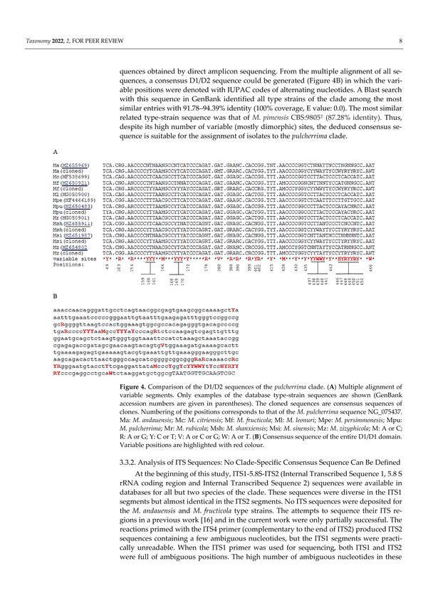
Taxonomy 2022, 2                                                                                             108

                   for taxonomic identification of yeast strains. The situation is further complicated by
                   the presence of such nucleotides in the database sequences of the type strains of certain
                   species. The clade consists of 10 species (M. andauensis, M. citriensis, M. fructicola, M.
                   leonuri, M. persimmonesis, M. pulcherrima, M. rubicola, M. shanxiensis, M. sinensis, and M.
                   ziziphicola) [3,5,6,11–14]. However, the taxon name M. citriensis is listed in MycoBank as
                   invalid. All but one species were delimited on the basis of differences between the above-
                   mentioned barcode sequences of their ex-type strains (living strains of types permanently
                   preserved in a metabolically inactive state). Cloning and sequencing of the D1/D2 and ITS
                   segments of individual rDNA repeats revealed that the nucleotide ambiguities are caused
                   by intragenomic rDNA repeat heterogeneity [15,16]. A recent study found that secondary
                   barcode sequences are also heterogeneous in the ex-type strains for which such sequences
                   are available [17]. Thus, the type strains of the species of the clade are not separated by
                   clear barcode gaps and therefore they cannot represent distinct species in terms of the
                   phylogenetic species concept. This concept is based on the premise that a species represents
                   a distinct entity containing its own synapomorphic characteristics (e.g., specific barcode
                   sequences) not shared with neighbouring taxa [18]. From a recent analysis of a larger group
                   of orthologous genes of seven whole-genome sequences, including those of three ex-type
                   strains, it was inferred that the failure to differentiate the species by clear barcode gaps
                   might be attributed to their admixed chimeric genomes consisting of sequences of diverse
                   phylogenetic histories [17].
                         Delimitation of fungal species by the application of the phylogenetic species concept
                   is based on the analysis of DNA barcodes. However, at least 30 other species concepts
                   have been described in the literature [19]. Those preferably applied to fungi have been
                   critically reviewed in numerous recent works (e.g., [20–22]). One of these concepts is the
                   biological concept which defines a species as a group of interbreeding natural populations
                   that are reproductively isolated from other such groups [23]. In yeasts, it was successfully
                   applied, e.g., to the delimitation of Saccharomyces species separated by postzygotic sterility
                   barriers [24]. All Saccharomyces strains can form viable hybrids, but only the hybrids
                   of conspecific strains can produce functional (mating-competent) gametes. Interspecies
                   hybrids either do not form viable gametes (ascospores) or their gametes (spores and spore
                   clones) are sterile due to the second sterility barrier underlain by MAT heterozygosity (for
                   a review, see [25]). Formation of viable hybrids was also reported for certain combinations
                   of auxotrophic mutants of Metschnikowia ex-type strains, and mating-competent segregants
                   were detected in a M. andauensis × M. fructicola hybrid [16,17] which implies that the
                   reproductive (biological) isolation of the species of the clade may be incomplete.
                         Since both the barcoding results and the hybridisation tests raised doubts as to whether
                   the taxonomic division of the clade is sufficiently justified, I set out to perform barcode
                   analyses with more sequences and hybridisation tests with more strains. Certain phenotypic
                   traits not tested or found variable in previous studies were also re-examined. The results
                   show that the species of the clade cannot be distinguished from each other by any of the
                   phenotypic, phylogenetic, and biological species concepts. Besides, their non-homogenised
                   rDNA repeats seem to evolve by reticulation, a complex process that involves horizontal
                   exchange and recombination of genetic elements, e.g., [15,17]. Therefore, I propose their
                   unification into one species.

                   2. Materials and Methods
                   2.1. Strains and Culture Conditions
                        The Metschnikowia strains used in this study are listed in Table S1. The composition of
                   the growth media YEA (yeast-extract agar), YEL (yeast-extract liquid), and SMA (synthetic
                   minimal agar) were described previously [26]. Assimilation tests were performed on YNB
                   plates supplemented with the test carbon sources. Sporulation was examined in cultures
                   grown on vegetable juice medium prepared from diluted BIO Gemüsesaft (Josef Pölz,
                   Bio-Produkte, 84,518 Garching an der Alz, Germany) and supplemented with 2% agar [16].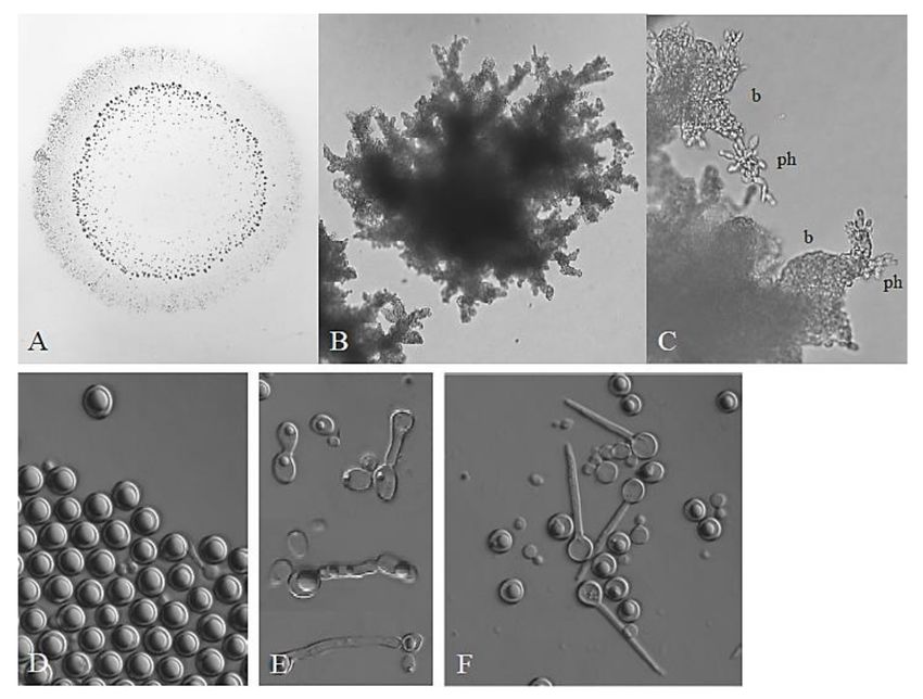
Taxonomy 2022, 2                                                                                                  109

                   2.2. Phenotypic Examination
                         Pulcherrimin production was examined on YEA plates supplemented with various
                   amounts of FeCl3 . Colonies producing pulcherrimin turn red on this medium. The higher
                   the concentration of FeCl3 , the more intense the pigmentation. If pulcherrimin is also
                   produced extracellularly, a coloured halo can be seen around the pigmented colony. Segre-
                   gants producing no or less pulcherrimin form white or pale red sectors in the colonies. To
                   test the segregants for stability, samples were taken from the sectors by sterile loops and
                   inoculated onto a fresh medium supplemented with 0.02 mg/mL FeCl3 . Sectors differing
                   in pigmentation intensity in the cultures of the segregants indicated instability.
                         To test the strain for the ability to penetrate the agar medium, 10 µL of dense cell
                   suspension (107 cells/mL) was dropped onto YEA. After two weeks of incubation at
                   room temperature, the colony was washed off from the plate with water to visualise the
                   intrusions formed beneath the colony. The cell morphology in the intrusions was examined
                   microscopically.
                         For the examination of assimilation of carbon sources, 5 µL of cell suspension (106 cells/mL)
                   was dropped onto the YNB plate supplemented with a carbon source. The plates were
                   incubated at room temperature for two weeks.
                         Formation of asci and chlamydospores was tested on vegetable juice medium. A 10 µL
                   measure of dense suspension of cells (107 cells/mL) was dropped onto the medium and
                   incubated at 14 ◦ C. After 4 weeks of incubation, samples of the culture were examined mi-
                   croscopically. For germination tests, ascospores were released from the asci by Zymolyase
                   20T treatment, (1.0 mg/mL, 25 ◦ C, 3 h), spread on thin YEA films on glass slides, covered
                   with cover slips and examined microscopically at regular intervals for 7 days.

                   2.3. Barcode Amplification and Sequencing
                        DNA was isolated from overnight cultures grown in YEL broth as described previ-
                   ously [27]. The D1/D2 domains and the ITS1-5.8S-ITS2 segments of the rDNA repeats were
                   amplified with the primer pairs NL1-NL4 [28] and ITS1-ITS4 [29], respectively. The ACT1
                   sequences were amplified as described in [13]. The primer pairs used for the amplification
                   of barcode segments of the RPB2 were RPB2F5 (aggagcctgacgacaga), RPB2R5 (gcgtgaaggt-
                   gtgtgtgga, PBR2FreF (gaggaggcccagctgaaggt), and PBR2FreR (ggtaagctttttgaataagag). The
                   amplified fragments were sequenced in both directions using the same primer pairs. The
                   sequences were deposited in GenBank under accession numbers listed in Tables S2 and S3.

                   2.4. Sequence Analysis
                        The accession numbers of the rDNA barcode sequences (‘primary barcodes’) used
                   in this study are listed in Table S2. The accession numbers of the ACT1, TEF1, and RPB2
                   barcode sequences (‘secondary barcodes’) are listed in Table S3.
                        Multiple alignments were produced with the Clustal W 1.7 [30] or the MUSCLE
                   3.8.31 [31] algorithms. Maximum-likelihood phylogenetic trees were generated from the
                   alignments with the PhyML 3.0 algorithm [32] using the best model suggested by the
                   Akaike information criterion (AIC) in jMedelTest version 1.0.2 [33].

                   2.5. Hybridisation and Segregation
                         Mutants of different (complementary) auxotrophic markers were hybridised by the
                   replica-plate method described previously [16]. Briefly, the mutants were streaked on YEA
                   plates, incubated for 3 days, then replica-plated onto fresh YEA plates perpendicularly to
                   each other to produce grids of prints in which the mutants intersected each other. After
                   7 days of incubation, the grids were replica-plated onto SMA plates. After two weeks of
                   incubation, individual prototrophic (hybrid) colonies were isolated from the intersections
                   and maintained on SMA plates.
                         The hybrids were tested for segregation by cultivating their cultures on YEA plates
                   where both the protrophic hybrid cells and the auxotrophic segregants could propagate. Af-
                   ter three days, samples were taken from the cultures and spread on YEA plates to obtain at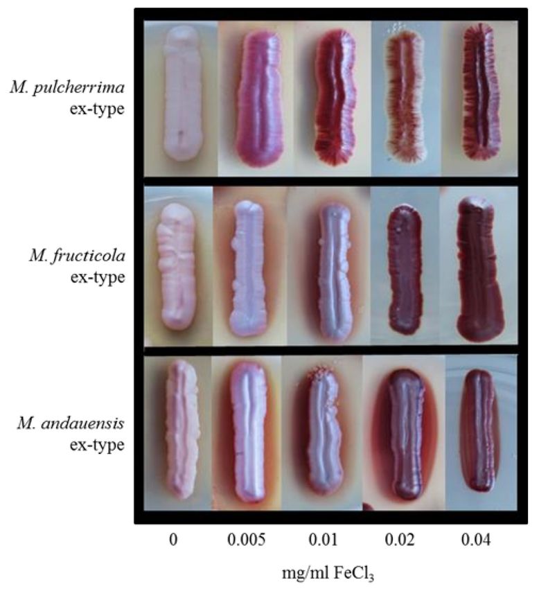
Taxonomy 2022, 2                                                                                               110

                   least 1000 colonies. The colonies were replica-plated on SMA plates to identify auxotrophic
                   segregants. The segregants were tested for auxotrophies by inoculating them onto SMA
                   plates supplemented with compounds for which the parental strains were auxotrophic.

                   3. Results
                   3.1. Phenotypic Examination: Lack of Taxonomically Relevant Differences
                         In the taxonomic descriptions, the type strains of the clade had highly similar pheno-
                   typic properties [5,6,11,34]. Since different laboratories used different test methods and/or
                   certain strains were not tested for the utilisation of complete sets of carbon sources, the as-
                   similation tests were repeated in this study. All ex-type strains assimilated the same subset
                   of carbon sources, albeit there were slight differences in the efficiency of the assimilation of
                   glucitol, D-gluconate, mannitol, methyl-alpha-D-glucoside, salicin, L-sorbose, trehalose,
                   and D-xylose. The differences were too small for reliable and reproducible differentiation
                   of strains. When grown on YEA plates, all ex-type cultures formed pseudohyphae capable
                   of invasive growth in the medium associated with the formation of blastoconidia along
                   the pseudohyphae (Figure 1A–C). All cultures grown on the vegetable medium contained
                   chlamydospores (5–60%) (Figure 1D) and asci (1–14%) (Figure 1F). The asci contained
                   two, not always clearly discernible ascopores. Ascospores were released from asci of the
                   ex-type strains of M. andauensis, M, fructicola, M. pulcherrima, M. shanxiensis, M sinensis, and
                   M. ziziphicola by Zymolyase treatment and tested for germination. No germination was
                   observed among the 24–84 ascospores examined for each strain. On the vegetable medium,
                   cell pairs connected with protuberances reminiscent of conjugation tubes formed by cells
                   of opposite mating types in other yeast species were also observed at low frequency in all
                   ex-type cultures (Figure 1E).

                   Figure 1. Morphology. (A) Invasive growth (penetration the medium) made visible by washing
                   off the colony from the surface of the medium. Penetrating structures are visible as dark spots.
                   (B) Penetrating pseudomycelium. (C) Pseudohyphae growing in the medium. Ph: pseudohypha;
                   b: blastoconidia. (D) Chlamidospores formed on vegetable agar. (E) Conjugation-like connections
                   between cells. (F) Asci.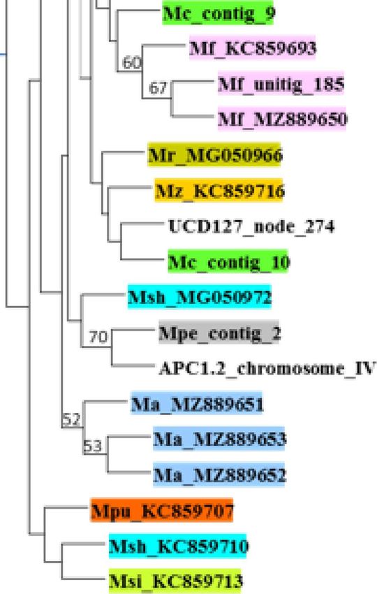
Taxonomy 2022, 2                                                                                             111

                   3.2. Pulcherrimin Production: A Variable and Unstable Trait
                        The formation of the maroon-red complex pulcherrimin is a characteristic feature of
                   the clade [10,34]. Pulcherrimin accumulation in the cells pigments the colonies on most
                   laboratory media. To find out whether the intensity of pulcherrimin synthesis can be used
                   in the differentiation of the species, in this study the ex-type cultures of M. andauensis, M.
                   fructicola, M. pulcherrima, M. shanxiensis, M. sinensis, and M. ziziphicola were inoculated
                   on the yeast extract medium YEA supplemented with various amounts (0, 0.005, 0.01,
                   0.02, 0.04 mg/mL) of FeCl3 . As expected, all formed pigmented colonies after 3–4 days
                   of incubation on the media supplemented with iron, and the intensity of pigmentation
                   increased with the concentration of the salt (examples are shown in Figure 2).

                   Figure 2. Effect of FeClš concentration on pigmentation. The cultures shown are ex-types of M.
                   pulcherrima CBS5833T , M. fructicola CBS 8853T , and M. andauensis CBS 10809T , respectively.

                        M. pulcherrima and M. sinensis formed darker colonies than the other strains at the
                   lowest FeCl3 concentration, but at 0.04 Mg/mL all cultures were equally pigmented. How-
                   ever, the weaker colony pigmentation does not necessarily imply less efficient pulcherrimin
                   synthesis. It can also be due to the formation of some of the pigment in the medium around
                   the colonies (generating a pigmented halo). Upon prolonged incubation, less pigmented
                   sectors appeared at the edges of all cultures (Figure 3). When isolated and inoculated onto
                   fresh medium, these sectors (segregants) formed pale or pink cultures. As the pigment is
                   the chelate of ferric irons and pulcherriminic acid [1], the reduced pigmentation implies
                   that pulcherriminic acid was synthesised at a reduced rate in these segregants. The dark
                   sectors formed dark colonies when inoculated onto fresh medium. However, neither of the
                   segregants was stable; their colonies occasionally contained segments or spots differing
                   from the rest of the colony in pigmentation intensity (Figure 3). Because of the instability
                   of colony pigmentation and the pigmented halos around the colonies, the intensity of
                   pulcherrimin production cannot be used as a distinctive taxonomic trait either.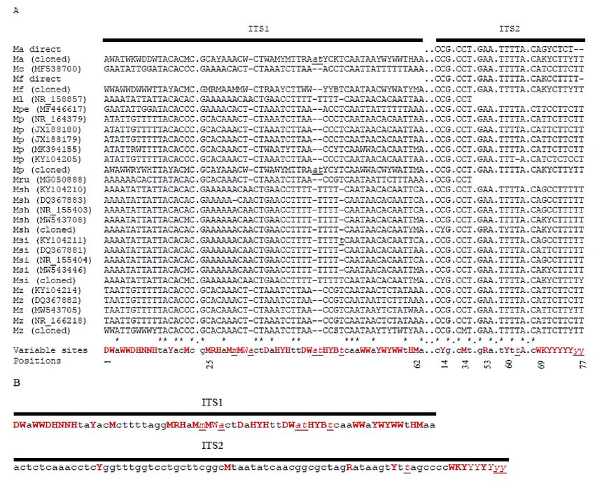
Taxonomy 2022, 2 112
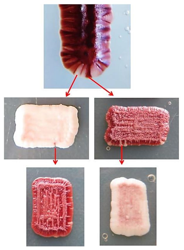
Figure 3. Segregation of pulcherrimin production (pigmentation intensity).
3.3. Analysis of Primary Barcodes
3.3.1. Analysis of D1/D2 Sequences: A Clade-Specific Consensus Sequence Can Be Defined
The sequences containing D1/D2 LSU domains of the type strains of the pulcherrima
clade were downloaded from the databases and compared with each-other by generating
multiple alignments. For the M. andauensis, M. fructicola, and M. pulcherrima type strains,
multiple slightly different sequences were deposited in the databases by different laborato-
ries and certain sequences contained ambiguous nucleotides. The presence of ambiguous,
mostly dimorphic (SND), rarely polymorphic (SNP) sites, and the intra-strain differences
hinted at rDNA heterogeneity (rDNA repeat diversity) in the genomes of the strains. For the
rest of the type strains, single unambiguous sequences were deposited. Since the database
entries do not provide information about the ‘pre-submission processing’ of the nascent
Sanger sequences, the D1/D2 domains of six type strains were resequenced in this study.
The accession numbers of the new sequences of M. pulcherrima CBS 5833T , M. fructicola CBS
8853T , M. sinensis CBS 10357T , M. ziziphicola CBS 10357T , M. shanxiensis CBS 10359T , and
M. andauensis CBS 10809T are listed in Table S2. All newly generated sequences contained
ambiguous nucleotides and differed from all amplicon sequences previously deposited
in databases for these strains. As a control, the D1/D2 domain of the ex-type strain (CBS
9803T ) of the closely related species M. chrysoperlae was also sequenced; its sequence had
no ambiguous positions and was identical with that available in the GenBank database.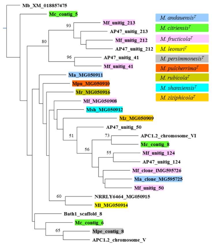
Taxonomy 2022, 2                                                                                                      113

                         In previous studies, individual D1/D2 domain sequences were cloned from six ex-type
                   strains and the clones representing individual rDNA repeats showed high intrastrain diver-
                   sity and formed a continuous cloud in the phylogenetic analyses [10,15,17]. In this study,
                   all cloned sequences were aligned with all non-cloned amplicon sequences (including those
                   produced here) (Figure 4A). The cloned sequences differed from one another in positions
                   corresponding to the positions of ambiguous nucleotides in the sequences obtained by
                   direct amplicon sequencing. From the multiple alignment of all sequences, a consensus
                   D1/D2 sequence could be generated (Figure 4B) in which the variable positions were
                   denoted with IUPAC codes of alternating nucleotides. A Blast search with this sequence
                   in GenBank identified all type strains of the clade among the most similar entries with
                   91.78–94.39% identity (100% coverage, E value: 0.0). The most similar related type-strain
                   sequence was that of M. pimensisCBS:9805T (87.28% identity). Thus, despite its high number
                   of variable (mostly dimorphic) sites, the deduced consensus sequence is suitable for the
                   assignment of isolates to the pulcherrima clade.

                   Figure 4. Comparison of the D1/D2 sequences of the pulcherrima clade. (A) Multiple alignment
                   of variable segments. Only examples of the database type-strain sequences are shown (GenBank
                   accession numbers are given in parentheses). The cloned sequences are consensus sequences of
                   clones. Numbering of the positions corresponds to that of the M. pulcherrima sequence NG_075437.
                   Ma: M. andauensis; Mc: M. citriensis; Mf: M. fructicola; Ml: M. leonuri; Mpe: M. persimmonesis; Mpu: M.
                   pulcherrima; Mr: M. rubicola; Msh: M. shanxiensis; Msi: M. sinensis; Mz: M. zizyphicola; M: A or C; R:
                   A or G; Y: C or T; V: A or C or G; W: A or T. (B) Consensus sequence of the entire D1/D1 domain.
                   Variable positions are highlighted with red colour.
Taxonomy 2022, 2                                                                                            114

                   3.3.2. Analysis of ITS Sequences: No Clade-Specific Consensus Sequence Can Be Defined
                        At the beginning of this study, ITS1-5.8S-ITS2 (Internal Transcribed Sequence 1, 5.8
                   S rRNA coding region and Internal Transcribed Sequence 2) sequences were available in
                   databases for all but two species of the clade. These sequences were diverse in the ITS1
                   segments but almost identical in the ITS2 segments. No ITS sequences were deposited
                   for the M. andauensis and M. fructicola type strains. The attempts to sequence their ITS
                   regions in a previous work [16] and in the current work were only partially successful. The
                   reactions primed with the ITS4 primer (complementary to the end of ITS2) produced ITS2
                   sequences containing a few ambiguous nucleotides, but the ITS1 segments were practically
                   unreadable. When the ITS1 primer was used for sequencing, both ITS1 and ITS2 were full
                   of ambiguous positions. The high number of ambiguous nucleotides in these sequences
                   was puzzling because the sequences of individual repeats cloned in a previous study
                   from these strains differed by much lower numbers of substitutions [16]. Obviously, these
                   substitutions cannot account for all the ambiguous nucleotides of the amplicon sequences.
                   Rather, they could be attributed to the presence of indels in the ITS segments of certain
                   rDNA repeats. When the cloned sequences were realigned in this study, single-nucleotide
                   indels were detected in some of them. Obviously, the shifts caused by these indels during
                   the sequencing process resulted in mass ambiguities in the final amplicon sequences.
                        Figure 5A shows the alignment of the database ITS1 and ITS2 sequences of the type
                   strains, the consensus sequences of the individual repeats cloned from six ex-type strains
                   in a previous study, and the ITS2 segments of the M. andauensis and M. fructicola ex-type
                   strains sequenced in this study. From the alignment, a consensus ITS1 and a consensus
                   ITS2 sequence were generated for the entire clade (Figure 5B). The former contained a very
                   high number of di- and polymorphic sites. Both turned out to be unsuitable for taxonomic
                   identification by Blast searches in sequence databases. ITS1 is not recognised as nucleotide
                   sequence by the NCBI Blast algorithm (due to the high proportion of symbols different from
                   A, G, T, and C, Blast recognises it as an amino-acid sequence) and ITS2 cannot distinguish
                   the pulcherrima-clade type strains from the type strains of closely related species.

                   3.4. Analysis of Secondary Barcodes
                   3.4.1. Analysis of ACT1 (Actin) Sequences: Insufficient Variability
                        To explore the diversity of the ACT1 barcodes in the clade, all ACT1 sequences available
                   in databases (Table S3) were downloaded and examined for the presence of ambiguous
                   nucleotides and for interstrain differences. For the M. andauensis type strain, two different
                   sequences are available in GenBank; they have ambiguous nucleotides in 8 and 10 positions,
                   respectively. The database sequences of a group of related strains isolated in the same
                   Austrian wine-growing region [13] and the M. shanxiensis, M. sinensis, and M. ziziphicola
                   type strains also had a few ambiguous nucleotides, mostly in corresponding positions.
                   However, when re-sequenced in this study, the latter three strains had clean sequences
                   which only differed from each other by one nucleotide. The ACT1 genes had identical
                   sequences in all sequenced genomes, including those of the type strains and only differing
                   from those of all but one type (M. andauensis) strains by 1 to 2 nucleotides. The very low
                   interstrain diversity precludes the application of ACT1 to taxonomic differentiation of the
                   type strains of the pulcherrima clade.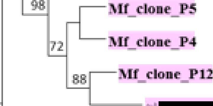
Taxonomy 2022, 2                                                                                                         115

                   Figure 5. Comparison of the ITS sequences of the pulcherrima clade. (A) Multiple alignment of variable
                   segments. Only examples of the database type-strain sequences are shown (GenBank accession
                   numbers are given in parenthesis). Direct: sequence obtained in this study by direct sequencing
                   of the PCR amplicon. The cloned sequences are consensus sequences of clones. Numbering of the
                   positions corresponds to that of the M. pulcherrima sequence NR_164379. Ma: M. andauensis; Mc:
                   M. citriensis; Mf: M. fructicola; Ml: M. leonuri; Mpe: M. persimmonesis; Mpu: M. pulcherrima; Mr: M.
                   rubicola; Msh: M. shanxiensis; Msi: M. sinensis; Mz: M. zizyphicola; M: A or C; R: A or G; Y: C or T; V: A
                   or C or G; W: A or T. Nucleotide symbols are lower-case and underlined at positions in which indels
                   or insertions occur. (B) Consensus sequences of the entire ITS1 and ITS2 segments. Variable positions
                   are highlighted with red colour.

                        When all type-strain and non-type-strain sequences were aligned, nucleotide differ-
                   ences were detected at 15 sites, but these differences were mainly due to the high number
                   of ambiguous positions of the M. andauensis sequences. All these sites were dimorphic;
                   no transversion was detected and the C/T transitions were more frequent than the A/G
                   transitions. All but two dimorphic sites were third nucleotides of triplets encoding amino
                   acids. It can be assumed that the strains having ambiguous positions have two (or more)
                   different ACT1 genes. This assumption seems to be corroborated by the genome sequences
                   of the M. fructicola type strain and the strain AF47 which both have two ACT1 genes,
                   although one is incomplete in both cases. The consensus sequence derived from the multi-
                   ple alignment identified all type strains of the clade in the GenBank Blast search (96.61%
Taxonomy 2022, 2 116
to 98.57% sequence identity), but did not distinguish them from M. picachoensis (97.32%
sequence identity) and M. rancensis (96.96% sequence identity).
3.4.2. Analysis of TEF1 (Translation Elongation Factor 1-α) Sequences: Intragenomic
Diversity Obscures Species Boundaries
The TEF1 barcode sequences turned out to be more diverse. Although listed in
Table S3, the TEF1 sequences of certain sequenced genomes could not be used in the
analysis because they were incomplete and did not overlap all regions regularly used
as barcode sequences. Large parts of the TEF1 ORFs were missing in unitig 43 of the
strain M. fructicola CBS8853T , unitig 43 of AP47, the NODES 547 and 1051 of UCD127, and
scaffold 137 of Bath1 (the deposited M. fructicola CBS8853T genome sequence is organised
in 93 unitigs, high-confidence contigs; the deposited Bath1 genome sequence is organised
in 524 scaffolds, ordered sets of contigs). In the alignment of 890 nt-long segments of the
rest of the TEF1 sequences, C/T, A/G, and G/C dimorphism were detected at 22, 2, and
2 sites, respectively. All dimorphic sites were third nucleotides of triplets, and none of the
substitutions caused amino-acid replacements in the proteins. Hence, these differences in
the gene sequences are unlikely to affect the fitness of the organisms. Remarkably, the M.
fructicola CBS 8853T , M. citriensis FL01T , and AP47 genomes had 3 to 6 TEF1 genes which
differed in sequence (intrastrain diversity). The phylogenetic analysis (Figure 6) grouped
the sequences into clusters that did not correspond to their origin. The genes showing
higher interstrain than intrastrain (intragenomic) similarity are most probably not paralogs
of the other genes of the genome, but their orthologs of different evolutionary histories.
3.4.3. Analysis of RPB2 (Second Largest Subunit of RNA-Polymerase II) Sequences:
Genomes Contain Genes of Different Phylogenetic Relationships
The RPB2 barcode sequences of the pulcherrima clade available in databases are diverse
in length and their locations within the genes. The sequences analysed in this work are
listed in Table S3. Their multiple alignment identified a 398-long segment (segment I: 1183–
1580 in Figure 7A) which was present in most of them and also in most genes extracted
from the whole-genome sequences. The genes of contig 43 of the genome Bath1 and NODE
247 of the genome UCD 127 lacked it, most probably due to incompleteness of the genome
sequences. As no M. andauensis RPB2 sequences were available at the beginning of this
study, this region was amplified and sequenced in this study from an ex-type culture and
from its two auxotrophic mutants generated for hybridisation purposes (see below).
All but one of these sequences contained ambiguous nucleotides in positions corre-
sponding to third nucleotides of triplets. The exception was the M. rubicola type strain.
All these sites were dimorphic (SND), in which either C/T (12) or A/G (6) transitions
occurred. No transversion and no polymorphic sites (SNP) were detected. The intrastrain
variability hints at the presence of two or more gene copies in the genomes. To test this
assumption, individual fragments were cloned from the ex-type cultures of M. andauensis
and M. fructicola (accession numbers are listed in Table S3). The cloned sequences contained
no ambiguous nucleotides. On the phylogenetic tree (Figure 7B), both the clones and
the mutants grouped together with the corresponding type strains, but the M. fructicola
sequences shared a branch with their M. leonuri counterpart and with one of the genes of
the M. citriensis genome.
Because of the close relationship of its sequences to sequences of other type strains, a
longer upstream region (segment II: 460-1264 in Figure 7A) was also sequenced for the M.
fructicola ex-type strain, both directly and after cloning. As expected, the direct amplicon
sequencing resulted in a sequence containing numerous ambiguous nucleotides, but the
clones were clean (for accession numbers, see Table S3). When compared with each other
by aligning all new sequences, five types of cloned sequences could be distinguished that
differed from each other at positions corresponding to the ambiguous sites of the amplicon
sequence. Again, all variability was due to SNDs (7 C/T, 7 A/G, 3 A/C, 2 G/C, and 2 A/T)
in third positions of triplets, causing no amino-acid substitutions. On the PhyML treeTaxonomy 2022, 2 117
(Figure 7C), they formed two well-separated clusters. One of the clusters shared a branch
with the sequence located in unitig 185 of the genome sequence of the ex-type strain.
Figure 6. The neighbour joining tree of TEF1 barcode sequences. Sequences from type materials are
marked with colours (see colour key at the top right). Boostrap values higher than 50 are shown
at nodes.
3.5. Hybridisation and Segregation
As shown above, the tested ex-type strains sporulated poorly and no spore germination
was observed. There can be several reasons for the low sporulation efficiency and the
inability of the spores to germinate. One is the complexity of genome structures. The
results of the analysis of the secondary barcode genes in this study (see above) and 35 non-
barcode genes described in a recent comparative analysis [17] pointed to segmental mosaic
genome structures in the strains of the clade. Segmental genomes are usually poor at
meiosis because of poor chromosome paring in Meiosis I, and the abruption of meiosis
results in low gamete viability (e.g., [35,36]).Taxonomy 2022, 2 118
RPB2 (cbromo ome V of APCl.2):
1 a• a• I • • • • • .. 3702
Examined segments:
460 1264
II 1183 1580
)fl>_K 85�681
:\Jb_X;\I_0188M993
B la KC�%78 C Ir (01
lll!bl _$CtITOIO _ 3�
Batbl_scattold_43
.tN1_unltlg_l3S
�:,ic:;05�
Mf_ll.lllll&_18S
I II
l\U_cloo8
Ml_eto1e_P7
Batbl_scattold_.l26
Figure 7. Neighbour joining trees of RPB2 segments. (A) Segments used for analysis. (B) Tree inferred
from the sequences of segment I. Outgroup: M. bicuspidata Mb_KC859681. (C) Tree inferred from
the sequences of segment II. Outgroup: M. bicuspidata Mb_XM_018855993. On both trees: sequences
from type materials are marked with colours (for the colour key, see Figure 6) and boostrap values
higher than 50 are shown at nodes.
Mosaic (chimeric) genomes can evolve from hybrids by gradual loss of segments of
the subgenomes during propagation of the hybrid cells (postzygotic genome evolution).
Similar processes can be assumed to take place in the pulcherrima clade. Recently, the
ex-type strains of M. andauensis and M. fructicola were found to be able to form hybrids with
the M. pulcherrima ex-type strain (e.g., [16,17]). In this study, prototrophic hybrids were
also obtained between auxotrophic mutants of M. pulcherrima CBS 5833T and auxotrophic
mutants of the rest of the ex-type strains (all mutants are listed in Table S1) but not in all
possible mutant combinations. The M. sinensis and M. ziziphicola mutants only formed
hybrids with the M. pulcherrima mutant of a mating type. Apparently, the mutants of
these species had alpha mating type. Consistently, no M. sinensis × M. ziziphicola hybrids
were obtained. The microscopic examination of six M. pulcherrima × M. andauensis, five M.
pulcherrima × M. fructicola, two M. pulcherrima × M. shanxiensis, two M. pulcherrima × M.
sinensis, and six M. pulcherrima × M. ziziphicola hybrids, numerous ‘intrastrain’ M. pul-
cherrima and M. andauensis hybrids and the parental mutants cultured on vegetable agar
revealed no increase in sporulation efficiency in the hybrids.
Two 1591 M. pulcherrima his− × 1622 M. ziziphicola ade− and two 1591 M. pulcherrima
his × 1605 M. shanxiensis asp− hybrids were tested for segregation. After cultivating
−
their cultures for two days under non-selective conditions, cells were spread on a complete
medium to obtain detached colonies. Colonies with auxotrophic markers of the parental
mutants (4 and 6 among 989 and 1201 colonies, respectively) segregated from both M.
pulcherrima × M. ziziphicola hybrids. Two his− and two ade− segregants were then testedTaxonomy 2022, 2 119
for hybridisation with parental mutants of complementary auxotrophies. Prototrophs were
formed in all backcrosses. The M. pulcherrima × M. shanxiensis hybrids did not segregate.
4. Discussion
The experimental results and the analysis of barcode sequences presented in this study
reinforce doubts about the validity of the taxonomic division of the pulcherrima clade
of Metschnikowia.
When the utilisation of carbon sources was examined, all tested ex-type cultures
assimilated the same set of compounds and only slightly differed in growth intensity.
The morphological examinations also found the ex-type strains very similar. All formed
chlamydospores and asci and could penetrate the solid medium by means of pseudohyphal
growth. All produced the pigment pulcherrimin, albeit with somewhat different intensities.
However, the differences in the production of the pigment turned out to be unstable. The
cultures segregated into clones of different pigmentation intensities and the segregants
were not stable either. Because of the absence of characteristic differences, phenotypic
species boundaries cannot be defined in the clade.
A similarity search in databases identified multiple D1/D2 and ITS sequences for most
type strains that differed at certain positions or contained ambiguous nucleotides. In this
study, the rDNA segments of the ex-type strains of six species were re-sequenced, including
those originally delimited on the basis of single cloned sequences. All newly generated
amplicon sequences contained ambiguous positions. In other cases, the sequences used
for species delimitation differed from those found in the whole-genome sequences of the
ex-type strains of the species. Thus, all known species of the clade have non-homogenised
heterogeneous rDNA repeats. This conclusion is consistent with previous studies that
revealed enormous internal diversity among individual D1/D2 and ITS segments cloned
from the genomes of these strains (e.g., [17]). On the phylogenetic trees and networks,
the D1/D2 and ITS sequences of the strains had intermixed positions (forming no distinct
clusters separated by clear barcode gaps), indicating conspecificity [17]. Additionally, the
secondary barcode analysis did not identify clear barcode gaps between the type strains.
Most strains harboured more than one copy of the barcode genes that usually differed in
sequence. Like the rDNA repeats, these copies frequently did not group with each other on
the trees but with their counterparts from other strains. A recent analysis of the genome
sequences of three ex-type strains and three strains of uncertain position in the clade found
that other protein-encoding genes also have copies that differ more from each other than
from their counterparts in other genomes [17]. Thus, neither phylogenetic boundaries can
be clearly defined in the pulcherrima clade.
The intragenomic (intrastrain) barcode diversity implies that segmental chimeric
genomes are common in the clade. Chimeric genomes consisting of sequences of dif-
ferent phylogenetic histories can evolve from hybrids of organisms (strains) of different
genomes by postzygotic evolution of the hybrid genomes. For example, in the hybrids of
the Saccharomyces species, the hybrid genomes are usually unstable and convert to chimeric
genomes during meiotic and mitotic divisions of the cells by gradually losing large parts
of the subgenomes (for a review, see [25]). The chimeric composition of the genomes
of the pulcherrima-clade strains can be attributed to a similar process. Like the cultures
of the Saccharomyces species, the ex-type cultures of the tested pulcherrima-clade species
also produced viable hybrids and the spores (gametes) of the hybrids also were unable
to germinate. However, the analogy between the two groups of strains is only partial
because, unlike the non-chimeric (“pure”) genomes of the natural Saccharomyces species,
the pulcherrima-clade type strains seem to have composite genomes. This is probably the
reason why their ascospores do not germinate, whereas the ascospores of the natural Sac-
charomyces species are viable. However, despite the fact that the spores appear unviable, the
hybrids investigated in this and a previous study [16] segregated, and the segregants were
mating-competent. As the culturing conditions were not favourable for sporulation, these
segregants must have arisen by mitotic segregation. This assumption is consistent withTaxonomy 2022, 2 120
the observations of Pitt and Miller [37] who found that the life cycle of the M. pulcherrima
type strain is parasexual. Due to the hybridisation–segregation cycle, the pulcherrima-clade
strains can evolve in interactions including horizontal exchange of genetic material and
reticulation of repeat sequences. Thus, the species of the clade do not meet the criteria of
the biological species concept either.
In conclusion, the type strains of the species of the pulcherrima clade cannot be dis-
tinguished from each other by the criteria of any of the phenotypic, phylogenetic, and
biological species concepts. The lack of reliable phenotypic differences and barcode gaps
hampers the taxonomic identification of new isolates. The high numbers of misidentified
records and entries labelled as “Metschnikowia sp.” or “Metschnikowia aff.” among the pul-
cherrima-clade strains in sequence databases can be attributed to this problem. Therefore, I
propose that the species of the pulcherrima clade be merged into one species.
As the unified taxon the name M. pulcherrima has to be chosen because according to
the International Code of Nomenclature for Algae, Fungi, and Plants [38] the oldest epithet has
nomenclatural priority when one of the competing legitimate names is to be chosen. In
this case, the oldest name is M. pulcherrima. The other species names have to be relegated
to synonyms of M. pulcherrima. However, in contrast to other mergers of fungal species
which were usually based on high barcode similarity or sequence identity, here diverse
sequences coexist even within strains. Which barcode sequences can then be assigned to
the merged species? The pragmatic solution is to retain all amplicon sequences deposited
in the databases for the ex-type strains of CBS 5833. These sequences can be considered
various (slightly different) consensus sequences of all repeats and copies of the barcode
genes in the CBS 5833T genome. The problem with this option is that even closely related
strains can differ significantly in the D1/2 and ITS sequences if they differ in the proportion
of the different rDNA repeats (that determine the ‘consensus’ Sanger sequence) in their
genomes. The unorthodox alternative could be the use of consensus barcode sequences
inferred from all known gene and repeat sequences. I compiled one consensus sequence
for each of the D1/D2 domain, the ITS1, and the ITS2 segments. The D1/D2 sequence,
despite its numerous dimorphic positions, is suitable for the taxonomic identification
of pulcherrima-clade strains. However, the ITS1 consensus sequence contains too many
ambiguous positions to be recognisable as DNA sequence for the GenBank blast algorithm.
The ITS2 consensus sequence is not specific enough for the differentiation of the clade
from other species of the genus. The usage of such consensus sequences in taxonomic
identification is not only impossible because of these problems, but also because databases
do not accept consensus sequences. Thus, the typological, type-centric approach to species
delimitation and strain identification has limits in this group of yeasts. In their case, Mayr’s
population approach (“population thinking”) is more appropriate [39,40]. Population
thinking rejects the use of a central representative type, and emphasizes the variation
among individuals. According to Mayr, a species is a genetic unit consisting of a large
intercommunicating gene pool.
The proposed unification of the clade in M. pulcherrima requires emendation of its
original taxonomic description. I propose the following new description.
Metschnikowia pulcherrima J.I. Pitt & M.W. Miller (1968) emend M. Sipiczki
The description is as before (Pitt and Miller, 1968) with the following addition:
Pseudohyphae capable of invasive growth are formed on solid media. During invasive
growth, blastoconidia are formed on the pseudohyphae. The pigment pulcherrimin can
be produced both intracellularly (pigmented colonies) and extracellularly (pigmented
zones around colonies), but certain strains only produce it intracellularly. The intensity of
pigmentation is variable and unstable. Colonies can segregate into more and less pigmented
sectors. Reduction in pigment formation by segregation is reversible. Strains can have
antimicrobial activity which correlates with pigmentation intensity. Hybridisation can take
place between sexually competent strains and the hybrids can segregate to form mating-
competent segregants. Genomes are variable in size and can have chimeric structures
associated with intragenomic barcode diversity.Taxonomy 2022, 2 121
MycoBank: MB#334124
New synonyms:
Metschnikowia andauensis O. Molnar & H. Prillinger, Systematic and Applied Microbi-
ology 28 (8): 723 (2005) [MB#356956]
Metschnikowia fructicola C.P. Kurtzman & S. Droby, Systematic and Applied Microbiol-
ogy 24 (3): 396 (2002) [MB#375792]
Metschnikowia leonuri C.P. Kurtzman, C.J. Robnett & E. Basehoar, Antonie van Leeuwen-
hoek 111 (11): 2026 (2018) [MB#823166]
Metschnikowia persimmonesis Y.M. Kang, J.E. Choi, R. Komakech, J.H. Park, D.W. Kim,
K.M. Cho, S.M. Kang, S.H. Choi, K.C. Song, C.M. Ryu, K.C. Lee & J.S. Lee, AMB Express
Journal 7 (199): 1 (2017) [MB#823579]
Metschnikowia rubicola C.P. Kurtzman, C.J. Robnett & E. Basehoar, Antonie van Leeuwen-
hoek 111 (11): 2014 (2018) [MB#823165]
Metschnikowia shanxiensis M.L. Xue & L.Q. Zhang, International Journal of Systematic
and Evolutionary Microbiology 56: 2249 (2006) [MB#501113]
Metschnikowia sinensis M.L. Xue & L.Q. Zhang, International Journal of Systematic and
Evolutionary Microbiology 56: 2248 (2006) [MB#529862]
Metschnikowia ziziphicola M.L. Xue & L.Q. Zhang, International Journal of Systematic
and Evolutionary Microbiology 56:2248 (2006) [MB#529862]
Metschnikowia citriensis Y. Liu, S.X. Yao, L.L. Deng, J. Ming & K.F. Zeng, Biological
Control 125: 17 (2018) [MB#838084] is not listed among the new synonyms because this
name is invalid
Supplementary Materials: The following are available online at https://www.mdpi.com/article/
10.3390/taxonomy2010009/s1, Table S1: List of strains; Table S2: Accession numbers of rDNA
(primary) barcode sequences; Table S3: Accession numbers of secondary barcode sequences.
Funding: This research was funded by the National Research, Development, and Innovation Office
of Hungary (grant nos. 2020-1.1.2-PIACI-KFI-2020-00130 and 2019-2.1.11-TÉT-2019-00001).
Institutional Review Board Statement: Not applicable.
Informed Consent Statement: Not applicable.
Data Availability Statement: All sequences produced in this study are available in GenBank under
accession numbers listed in Tables S2 and S3.
Acknowledgments: The author thanks Anita Kovacs and Anita Olah for excellent technical assistance.
Conflicts of Interest: The author declares no conflict of interest.
References
1. Cook, A.H.; Slater, C.A. The structure of pulcherrimin. J. Chem. Soc. 1956, 4133–4135. [CrossRef]
2. Sipiczki, M. Metschnikowia strains isolated from botrytized grapes antagonize fungal and bacterial growth by iron depletion. Appl.
Environ. Microbiol. 2006, 72, 6716–6724. [CrossRef] [PubMed]
3. Kurtzman, C.P.; Droby, S. Metschnikowia fructicola, a new ascosporic yeast with potential for biocontrol of postharvest fruit rots.
Syst. Appl. Microbiol. 2001, 24, 395–399. [CrossRef] [PubMed]
4. Türkel, S.; Korukluoglu, M.; Yavuz, M. Biocontrol activity of the local strain of Metschnikowia pulcherrima on different postharvest
pathogens. Biotechnol. Res. Int. 2014, 2014, 1–6. [CrossRef]
5. Kang, Y.M.; Choi, J.E.; Komakech, R.; Park, J.H.; Kim, D.W.; Cho, K.M.; Kang, S.M.; Choi, S.H.; Song, K.C.; Ryu, C.M.; et al.
Characterization of a novel yeast species Metschnikowia persimmonesis KCTC 12991BP (KIOM G15050 type strain) isolated from a
medicinal plant, Korean persimmon calyx (Diospyros kaki Thumb). AMB Express 2017, 7, 199. [CrossRef]
6. Liu, Y.; Yao, S.; Deng, L.; Ming, J.; Zeng, K. Metschnikowia citriensis sp. nov., a novel yeast species isolated from leaves with
potential for biocontrol of postharvest fruit rot. Biol. Control 2018, 125, 15–19. [CrossRef]
7. Gore-Lloyd, D.; Sumann, I.; Brachmann, A.O.; Schneeberger, K.; Ortiz-Merino, R.A.; Moreno-Beltrán, M.; Schläfli, M.; Kirner, P.;
Santos Kron, A.; Rueda-Mejia, M.P.; et al. Snf2 controls pulcherriminic acid biosynthesis and antifungal activity of the biocontrol
yeast Metschnikowia pulcherrima. Mol. Microbiol. 2019, 112, 317–332. [CrossRef]
8. Saravanakumar, D.; Spadaro, D.; Garibaldi, A.; Gullino, M.L. Detection of enzymatic activity and partial sequence of a chitinase
gene in Metschnikowia pulcherrima strain MACH1 used as postharvest biocontrol agent. Eur. J. Plant Pathol. 2009, 123, 183–193.
[CrossRef]Taxonomy 2022, 2 122
9. Oro, L.; Feliziani, E.; Ciani, M.; Romanazzi, G.; Comitini, F. Volatile organic compounds from Wickerhamomyces anomalus,
Metschnikowia pulcherrima and Saccharomyces cerevisiae inhibit growth of decay causing fungi and control postharvest diseases of
strawberries. Int. J. Food Microbiol. 2018, 265, 18–22. [CrossRef] [PubMed]
10. Sipiczki, M. Metschnikowia pulcherrima and related pulcherrimin-producing yeasts: Fuzzy species boundaries and complex
antimicrobial antagonism. Microorganisms 2020, 8, 1029. [CrossRef]
11. Kurtzman, C.P.; Robnett, C.J.; Basehoar, E.; Ward, T.J. Four new species of Metschnikowia and the transfer of seven Candida species
to Metschnikowia and Clavispora as new combinations. Antonie Van Leeuwenhoek 2018, 111, 2017–2035. [CrossRef]
12. Pitt, J.I.; Miller, M.W. Sporulation in Candida pulcherrima, Candida reukaufii and Chlamydozyma species; their relationship with
Metschnikowia. Mycologia 1968, 60, 663–685. [CrossRef]
13. Molnar, O.; Prillinger, H. Analysis of yeast isolates related to Metschnikowia pulcherrima using the partialsequences of the large
subunit rDNA and the actin gene; description of Metschnikowia andauensis sp. nov. Syst. Appl. Microbiol. 2005, 28, 717–726.
[CrossRef] [PubMed]
14. Xue, M.L.; Zhang, L.Q.; Wang, Q.M.; Zhang, J.S.; Bai, F.Y. Metschnikowia sinensis sp. nov., Metschnikowia zizyphicola sp. nov. and
Metschnikowia shanxiensis sp. nov., novel yeast species from jujube fruit. Int. J. Syst. Evol. Microbiol. 2006, 56, 2245–2250. [CrossRef]
15. Sipiczki, M.; Pfliegler, W.P.; Holb, I.J. Metschnikowia species share a pool of diverse rRNA genes differing in regions that
determine hairpin-loop structures and evolve by reticulation. PLoS ONE 2013, 8, e67384. [CrossRef]
16. Sipiczki, M.; Horvath, E.; Pfliegler, W.P. Birth-and-death evolution and reticulation of ITS segments of Metschnikowia andauensis
and Metschnikowia fructicola rDNA repeats. Front. Microbiol. 2018, 9, 1193. [CrossRef]
17. Sipiczki, M. When barcoding fails: Genome chimerisation (admixing) and reticulation obscure phylogenetic and taxonomic
relationships. Mol. Ecol. Resour. 2022. [CrossRef]
18. Wheeler, Q.D.; Platnick, N.I. The Phylogenetic species concept (sensu Wheeler and Platnick). In Species Concepts and Phylogenetic
Theory: A Debate; Wheeler, Q.D., Meier, R., Eds.; Columbia University Press: New York, NY, USA, 2000; pp. 55–69.
19. Zachos, F.E. Species Concepts in Biology: Historical Development, Theoretical Foundations and Practical Relevance; Springer Nature:
Cham, Switzerland, 2016.
20. Lücking, R.; Aime, M.C.; Robbertse, B.; Miller, A.N.; Ariyawansa, H.A.; Aoki, T.; Cardinali, G.; Crous, P.W.; Druzhinina, I.S.;
Geiser, D.M.; et al. Unambiguous identification of fungi: Where do we stand and how accurate and precise is fungal DNA
barcoding? IMA Fungus 2020, 11, 14. [CrossRef] [PubMed]
21. Boekhout, T.; Aime, M.C.; Begerow, D.; Gabaldón, T.; Heitman, J.; Kemler, M.; Khayhan, K.; Lachance, M.-A.; Louis, E.J.;
Sun, S.; et al. The evolving species concepts used for yeasts: From phenotypes and genomes to speciation networks. Fungal Divers.
2021, 109, 27–55. [CrossRef] [PubMed]
22. Yurkov, A.; Alves, A.; Bai, F.Y.; Boundy-Mills, K.; Buzzini, P.; Čadež, N.; Cardinali, G.; Casaregola, S.; Chaturvedi, V.;
Collin, V.; et al. Nomenclatural issues concerning cultured yeasts and other fungi: Why it is important to avoid unneeded
name changes. IMA Fungus 2021, 12, 18. [CrossRef]
23. Mayr, E. Speciation phenomena in birds. Am. Nat. 1940, 74, 249–278. [CrossRef]
24. Naumov, G. Genetic identification of biological species in the Saccharomyces sensu stricto complex. J. Ind. Microbiol. 1996, 17,
295–302. [CrossRef]
25. Sipiczki, M. Interspecies hybridisation and genome chimerisation in Saccharomyces: Combining of gene pools of species and its
biotechnological perspectives. Front. Microbiol. 2018, 9, 3071. [CrossRef]
26. Sipiczki, M. Pichia bruneiensis sp. nov., a biofilm-producing dimorphic yeast species isolated from flowers in Borneo. Int. J. Syst.
Evol. Microbiol. 2012, 62, 3099–3104. [CrossRef]
27. Sipiczki, M. Candida zemplinina sp. nov., an osmotolerant and psychrotolerant yeast that ferments sweet botrytized wines. Int. J.
Syst. Evol. Microbiol. 2003, 53, 2079–2083. [CrossRef]
28. O’Donnell, K. Fusarium and its near relatives. In The Fungal Holomorph: Mitotic, Meiotic and Pleomorphic Speciation in Fungal
Systematics; Reynolds, D.R., Taylor, J.W., Eds.; CAB International: Wallingford, UK, 1993; pp. 225–233.
29. White, T.J.; Bruns, T.; Lee, S.; Taylor, J. Amplification and sequencing of fungal ribosomal RNA genes for phylogenetics. In PCR
Protocols. A Guide to Methods and Applications; Innis, M.A., Gelfand, D.H., Sninsky, J.J., White, T.J., Eds.; Academic Press: San
Diego, CA, USA, 1990; pp. 315–322.
30. Thompson, J.D.; Higgions, D.G.; Gibson, T.J. CLUSTAL W: Improving the sensitivity of progressive multiple sequence alignment
through sequence weighting, position-specific gap penalties and weight matrix choice. Nucleic Acids Res. 1994, 22, 4673–4680.
[CrossRef]
31. Edgar, R.C. MUSCLE: Multiple sequence alignment with high accuracy and high throughput. Nucleic Acids Res. 2004, 32,
1792–1797. [CrossRef] [PubMed]
32. Saitou, N.; Nei, M. The neighbor-joining method: A new method for reconstructing phylogenetic trees. Mol. Biol. Evol. 1987, 4,
406–425. [CrossRef] [PubMed]
33. Felsenstein, J. PHYLIP (Phylogeny Inference Package), Version 3.67; Distributed by the Author; Department of Genome Sciences,
University of Washington: Seattle, DC, USA, 2007.
34. Lachance, M.A. Metschnikowia Kamienski (1899). In The Yeasts. A Taxonomic Study; Kurtzman, C.P., Fell, J.W., Boekhout, T., Eds.;
Elsevier: Amsterdam, The Netherlands, 2011; pp. 575–620.Taxonomy 2022, 2 123
35. Kaur, D.; Singhal, V.K. Meiotic abnormalities affect genetic constitution and pollen viability in dicots from Indian cold deserts.
BMC Plant Biol. 2019, 19, 10. [CrossRef]
36. Mason, A.S.; Wendel, J.F. Homoeologous exchanges, segmental allopolyploidy, and polyploid genome evolution. Front. Genet.
2020, 11, 1014. [CrossRef]
37. Pitt, J.I.; Miller, M.W. The parasexual cycle in yeasts from the genus Metschnikowia. Mycologia 1970, 62, 462–473. [CrossRef]
38. Turland, N.J.; Wiersema, J.H.; Barrie, F.R.; Greuter, W.; Hawksworth, D.L.; Herendeen, P.S.; Knapp, S.; Kusber, W.-H.; Li, D.-Z.;
Marhold, K.; et al. International Code of Nomenclature for algae, fungi, and plants (Shenzhen Code) adopted by the Nineteenth International
Botanical Congress Shenzhen, China, July 2017; Regnum Vegetabile; Koeltz Botanical Books: Glashütten, Germany, 2018; Volume 159.
39. Mayr, E. Animal Species and Evolution; Harvard University Press: Cambridge, MA, USA, 1963.
40. Mayr, E. Populations, Species, and Evolution; Harvard University Press: Cambridge, MA, USA, 1970.You can also read