Supplementation with a Highly Concentrated Docosahexaenoic Acid (DHA) in Non-Proliferative Diabetic Retinopathy: A 2-Year Randomized Double-Blind ...
←
→
Page content transcription
If your browser does not render page correctly, please read the page content below
antioxidants

Article
Supplementation with a Highly Concentrated Docosahexaenoic
Acid (DHA) in Non-Proliferative Diabetic Retinopathy: A
2-Year Randomized Double-Blind Placebo-Controlled Study
Purificación Piñas García * , Francisco Javier Hernández Martínez, Núria Aznárez López, Luis Castillón Torre
and Ma Eugenia Tena Sempere

                                          Service of Ophthalmology, Hospital San Juan de Dios del Aljarafe, Avenida San Juan de Dios s/n,
                                          E-41930 Bormujos, Sevilla, Spain; fjhernandezm@hotmail.com (F.J.H.M.); nuazlo@yahoo.es (N.A.L.);
                                          lcastillon@telefonica.net (L.C.T.); draojitos@yahoo.es (M.E.T.S.)
                                          * Correspondence: ppinag@gmail.com; Tel.: +34-95-505-0550

                                          Abstract: We assessed the effect of a 2-year supplementation with a highly concentrated docosa-
                                          hexaenoic acid (DHA) product with antioxidant activity on non-proliferative diabetic retinopathy
                                          (NPDR) in a randomized double-blind placebo-controlled study. A total of 170 patients with diabetes
                                          were randomly assigned to the DHA group (n = 83) or the placebo group (n = 87). NPDR was diag-
                                          nosed using non-contact slit lamp biomicroscopy examination, and classified into mild, moderate,
                                          and severe stages. Patients in the DHA group received a high rich DHA triglyceride (1050 mg/day)
                                nutritional supplement, and those in the placebo group received olive oil capsules. The percentages

                                          of mild NPDR increased from 61.7% at baseline to 75.7% at the end of the study in the DHA group,
Citation: Piñas García, P.;
                                          and from 61.9% to 73.1% in the placebo group. Moderate NPDR stages decreased from 35.1% at
Hernández Martínez, F.J.;
Aznárez López, N.; Castillón Torre, L.;
                                          baseline to 18.7% at the end of the study in the DHA group, and from 36.8% to 26.0% in the placebo
Tena Sempere, M.E. Supplementation        group. In the DHA group, there were five eyes with severe NPDR at baseline, which increased to one
with a Highly Concentrated                more at the end of the study. In the placebo group, of two eyes with severe NPDR at baseline, one eye
Docosahexaenoic Acid (DHA) in             remained at the end of the study. Changes in visual acuity were not found. There were improvements
Non-Proliferative Diabetic                in the serum levels of HbA1c in both groups, but significant differences between the DHA and the
Retinopathy: A 2-Year Randomized          placebo groups were not found. In this study, the use of a DHA triglyceride nutraceutical supplement
Double-Blind Placebo-Controlled           for 2 years did not appear to influence the slowing of the progression of NPDR.
Study. Antioxidants 2022, 11, 116.
https://doi.org/10.3390/antiox            Keywords: non-proliferative diabetic retinopathy; docosahexaenoic acid; antioxidant; randomized
11010116
                                          controlled study
Academic Editor: Michele Marino

Received: 9 December 2021
Accepted: 3 January 2022
                                          1. Introduction
Published: 5 January 2022
                                               Non-proliferative retinopathy (NPDR) is the earliest stage of diabetic retinopathy (DR),
Publisher’s Note: MDPI stays neutral
                                          in which symptoms can be mild or non-existent. NPDR typically involves microvascular
with regard to jurisdictional claims in
                                          changes and progresses from mild to moderate and severe stages, and, in some people,
published maps and institutional affil-
                                          may progress to sight-threatening DR, such as proliferative diabetic retinopathy (PDR) and
iations.
                                          diabetic macular edema (DME). Hyperglycemia, hypertension, and increasing duration
                                          of diabetes are independent risk factors for DR, and although improved glycemic control
                                          and blood pressure, regularly monitoring of DR, and intensive treatment of modifiable risk
Copyright: © 2022 by the authors.
                                          factors are crucial to prevent complications [1–3], clinically this is difficult to achieve [4].
Licensee MDPI, Basel, Switzerland.             About one-third of the diabetes population suffers from DR, mostly NPDR, but there
This article is an open access article    are scanty data on the prevalence of NPDR. In a review of 32 studies with 543,448 people
distributed under the terms and           with diabetes who underwent retinal photography as a basis for diagnosing the presence
conditions of the Creative Commons        and severity of DR, the overall prevalence of NPDR was 19% (range 11.7–65%) [5]. In a
Attribution (CC BY) license (https://     follow-up study of patients with mild NPDR, the cumulative occurrence rate of PDR at
creativecommons.org/licenses/by/          10 years was estimated to be 14% in subjects with a mean HbA1c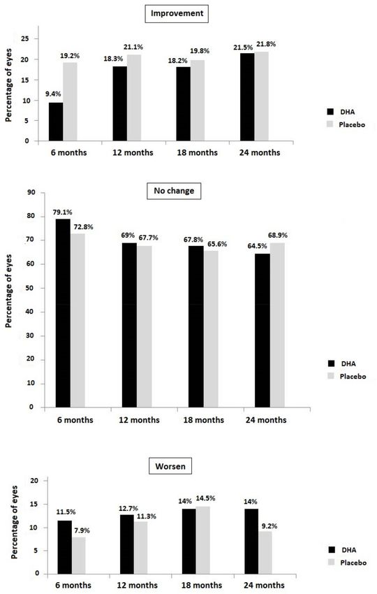
Antioxidants 2022, 11, 116                                                                                          2 of 15

                             prevalence of NPDR of 15.1% in patients with diabetes and 27% in patients with type 2
                             diabetes [7,8]. In a recent comprehensive review based on 90 studies with 204,189 patients
                             with diabetes, a prevalence of NPDR of 24.8% was reported [9]. Approximately half of the
                             patients with severe NPDR will progress to PDR within a year [4], so the prevention of
                             disease progression to more advanced stages with potential visual loss and the identification
                             of people at high risk of progression and greatest potential to benefit from treatment is of
                             the utmost importance.
                                  The treatment of mild NPDR may not be necessary, but understanding the multifac-
                             torial pathogenic mechanisms of DR could optimize the treatment of moderate/severe
                             NPDR and slow the progression to PDR or DME [10]. Several mechanisms, by which hyper-
                             glycemia causes retinal capillary damage, include increased polyol and hexosamine path-
                             ways, increased non-enzymatic glycation with advanced glycation end-products (AGE),
                             abnormal activation of signaling cascades such as protein kinase C pathway (PKC), in-
                             creased oxidative stress, increased expression of adhesion molecules, local inflammatory
                             activity with upregulation of proinflammatory mediators, interleukins, and critically im-
                             portant growth factors promoting angiogenesis, breakdown of the blood–retinal barrier,
                             and retinal neurodegeneration [11–16]. Omega-3 long-chain polyunsaturated fatty acids
                             (n-3 PUFAs), particularly docosahexaenoic (DHA) acid, have demonstrated consistent anti-
                             inflammatory, antiproliferative, antiangiogenic, and antioxidant properties on pathways
                             leading to DR [17]. These include promoting vascular integrity [18], reduction of oxidative
                             stress-induced apoptosis of photoreceptors [19], neuroprotection [20], inhibition of the
                             nuclear factor-kappa B (NF-κB) signaling pathway [21], reduction of proinflammatory
                             cytokine production (IL-6, IL-10, IL-1β, FNTα) and intercellular and vascular adhesion
                             molecules [22], reduction of pathological retinal angiogenesis [23], and increase of E3-
                             derived anti-inflammatory mediators (E3 eicosanoids, B5 leukotrienes) and pro-resolving
                             mediators (protectin D1, resolvins E1, D1) [24].
                                  These pleomorphic effects of DHA support the rationale of dietary supplementation
                             with DHA in DR. However, there is little experience in the use of dietary supplemen-
                             tation with DHA in early stages of DR. In a prospective controlled study of 12 asymp-
                             tomatic patients with NPDR and 12 healthy controls, high rich DHA triglyceride (DHA-TG)
                             (1050 mg/day) supplementation for 90 days was associated with the progressive and
                             significant improvement of macular function measured by microperimetry in eyes from
                             DHA-treated subjects compared with controls [25].
                                  To our knowledge, clinical studies on the effect of DHA dietary supplementation with
                             antioxidant activity in diabetic patients with NPDR have not been previously reported.
                             Therefore, a randomized controlled study was conducted to determine whether dietary
                             supplementation with high dose DHA for 2 years could slow the progression of any
                             pre-proliferative stage of NPDR in patients with diabetes.

                             2. Materials and Methods
                             2.1. Study Design and Participants
                                  A prospective randomized double-blind placebo-controlled study (PAOXRED study,
                             “Protección AntiOXidante en la REtinopatía Diabética”, Antioxidant Protection in Diabetic
                             Retinopathy) was conducted at the Service of Ophthalmology of a regional hospital in
                             Sevilla, Spain. Type 1 and type 2 diabetic patients of both sexes aged >18 years, diagnosed
                             with NPDR (any stage) by four specialized ophthalmologists (P.P.G., F.J.H.M., N.A.L.,
                             E.T.S.), were invited to participate in the study during a routine ophthalmological appoint-
                             ment at the study center. NPDR was diagnosed using non-contact slit lamp biomicroscopy
                             examination, and classified into mild, moderate, and severe stages, in the absence of neo-
                             vascularization [26]. Mild NPDR was characterized by microaneurysm(s) only; moderate
                             NPDR by at least one hemorrhage or microaneurysm and/or at least one of the following:
                             retinal hemorrhages, hard exudates, cotton-wood spots, venous beading; and severe NPDR
                             by any of the following but no signs of PDR (4-2-1 rule): >20 intraretinal hemorrhages
                             in each of the four quadrants, definite venous beading in two or more quadrants, and
Antioxidants 2022, 11, 116 3 of 15
prominent intraretinal microvascular abnormality (IRMA) in one or more quadrants. The
exclusion criteria were as follows: the presence of PDR and/or DME documented on optical
coherence tomography (OCT), previous surgery for morbid obesity, chronic diarrhea of any
cause, anticoagulation, known allergy to fish proteins, use of dietary supplementation with
vitamin/minerals or fatty acids, pregnant women, cognitive impairment, patients unable
to participate according to the criteria of the ophthalmologist, and those who refused to
give written consent.
This study protocol was approved by the Clinical Research Ethics Committee of
Hospital San Juan de Dios del Aljarafe (Sevilla, Spain) (study code PAOXRED Vo2, approval
date 20 January 2017). All participants provided written informed consent.
2.2. Study Intervention
Eligible patients were assigned to the DHA supplementation group (experimental) or
the control group using pseudorandom numbers generated by the data collection computer
server at the time of entering the first datum of a patient, with p = 0.5 so that each patient
had a 50% probability of being randomized to one of the two study groups. Randomization
was implemented without restriction or additional procedures to balance the sample size
in each study group.
The patients in the DHA group received a high rich DHA triglyceride (1050 mg/day)
nutraceutical formulation (Brudyretina 1.5 g, Brudy Lab, S.L., Barcelona, Spain). This is
a concentrated DHA triglyceride having a high antioxidant activity patented to prevent
cellular oxidative damage [27]. Table 1 shows the composition of the nutraceutical formula-
tion, which includes DHA, eicosapentaenoic acid (EPA), vitamins (B-complex, C, E), lutein,
zeaxanthin, glutathione, and minerals.
Table 1. Composition of Brudyretina 1.5 g per capsule.
Per % Recommended Daily Per Three % Recommended Daily
Composition
Capsule Amount Capsules Amount
Concentrated oil in ω-3 fatty acids 500 mg 1500 mg
TG-DHA 70% 350 mg - 1050 mg -
EPA 8.5% 42.5 mg - 127 mg -
DPA 6% 30 mg - 90 mg -
Vitamins
Vitamin B1 (thiamine) 0.37 mg 33 1.1 mg 100
Vitamin B2 (riboflavin) 0.47 mg 33 1.4 mg 100
Vitamin B3 (niacin/niacinamide) 5.3 mg NE 33 16 mg NE 100
Vitamin B6 (pyridoxine) 0.47 mg 33 1.4 mg 100
Vitamin B9 (folic acid) 66.7 µg 33 200 µg 100
Vitamin B12 (cobalamin) 0.83 µg 33 2.5 µg 100
Vitamin C (ascorbic acid) 26.7 mg 33 80 mg 100
Vitamin E (d-α-TE) 4 mg α-TE 33 12 mg α-TE 100
Essential trace elements
Zinc 1.66 mg 16.66 5 mg 50
Cooper 0.16 mg 16.66 0.5 mg 50
Selenium 9.16 µg 16.66 27.5 µg 50
Magnesium 0.33 mg 16.66 1 mg 50
Other components
Lutein 3 mg - 9 mg -
Zeaxanthin 0.3 mg - 0.9 mg -
Glutathione 2 mg - 6 mg -
Energetic value (Kcal) 5.7 17.1
TG-DHA: triglyceride-bound DHA, DHA: docosahexaenoic acid, EPA: eicosapentaenoic acid, DPA: docosapen-
taenoic acid, NE: niacin equivalent, TE: tocopherol equivalent.Antioxidants 2022, 11, 116 4 of 15
Patients in the placebo group were treated with identically appearing olive oil capsules
(also labeled Brudyretina 1.5 g). All patients were instructed to take three capsules of
Brudyretina 1.5 g once daily, preferably in the morning with food and a glass of water.
2.3. Study Procedures
Patients were recruited between March 2017 and December 2020, and were followed
for 24 months, with control visits every 6 months. At the baseline visit, the inclusion criteria
were checked, the written informed consent was signed, and the nutraceutical formulation
(Brudyretina 1.5 g) was delivered for the initial 6-month treatment period.
The ophthalmological studies included non-contact (Volk SuperField NC® ) slit lamp
biomicroscopy examination of the optic fundus with mydriasis, measurement of best-
corrected visual acuity (BCVA) using an ETDRS optotype at 2 m distance from the observer,
and assessment of the macular condition by optical coherence tomography (OCT) (Stratus
OCT, Carl Zeiss Meditec, Dublin, CA, USA). Retinography was also performed, as well as
OCT to exclude DME. Visual acuity (VA) was expressed in a decimal scale and in logarithm
of the minimum angle of resolution (logMAR). A peripheral venous blood sample was
drawn after at least 8 h fasting to measure the serum levels of glycosylated hemoglobin
(HbA1c) as an indicator of metabolic control, which was defined as an HbA1c value of
7–8% following the clinical practice guidelines of redGDPS (Network of Diabetes Study
Groups in Primary Care) for patients with long-standing diabetes or comorbidity [28]. The
serum levels of HbA1c were measured using a high-performance liquid chromatography
(HPLQ) analyzer (Menarini Diagnostics, Badalona, Barcelona, Spain).
The same examinations were performed at each study visit, at 6, 12, 18, and 24 months
after enrolment. At the 6-, 12-, and 18-month visits, the nutraceutical formulation was de-
livered for the next 6-month treatment period. The ophthalmologists paid special attention
to insist on the importance of compliance with the dietary supplement and the benefit that
the patient may receive from the supplement. At each visit, the patients were interviewed
about gastrointestinal tolerability to the nutraceutical formulation and other side effects.
Compliance with the nutraceutical supplementation was assessed at the study visits by
return of supplementation tablet counts. The ophthalmologists who evaluated the results
of the treatment (P.P.G., F.J.H.M., N.A.L., and E.T.S.) were blinded to which subjects were
assigned to the experimental or the placebo group. All data were anonymized and recorded
by the researchers on a specific website with access through a personal password.
2.4. Outcomes
The primary outcome was the change of the stages of NPDR during the study in the
DHA and the placebo groups, in particular the number of eyes in each NPDR stage at
24 months compared with baseline. Changes of the stages of NPDR were also assessed in
terms of improvement (decrease in any NPDR stage), unchanged (remaining in the same
NPDR stage), and worsening (increase in any NPDR stage). Changes of VA and the serum
levels of HbA1c were the secondary outcome variables.
2.5. Statistical Analysis
The per-protocol (PP) data set was analyzed—that is, all randomized patients who
attended the follow-up visits and complete the 2-year study period. Categorical variables
are expressed as frequencies and percentages, and continuous variables as mean and
standard deviation (SD). Categorical variables were compared with the chi-square (χ2 ) test
or the Fisher’s exact test according to the conditions of application. Quantitative variables
were compared with the non-parametric Mann-Whitney U test. Within-group differences
of VA and the serum levels of HbA1c during the study period were analyzed with the
Wilcoxon signed-rank test, and changes of NPDR stages using the McNemar–Browker
test. Statistical significance was set at p < 0.05. Statistical analyses were performed with
the Statistical Package for the Social Sciences, version 26.0 software (IBM Corp., Armonk,
NY, USA).Antioxidants 2022, 11, 116 5 of 15
3. Results
3.1. Characteristics of the Study Patients
A total of 170 patients with diabetes (type 1, 15 patients; type 2, 155 patients) were
included in the study and were assigned to the DHA group (n = 83) or the placebo group
(n = 87). There were 130 men and 40 women, with a mean age (SD) of 61.7 (11.3) years. As
shown in Table 2, there were no significant differences in the baseline characteristics of the
patients assigned to the DHA or the placebo groups. More than 60% of the patients from
each group had mild NPRD, and only 3.2% and 1.3% of patients in the DHA and placebo
groups, respectively, presented with a severe stage. Visual acuity and the serum levels of
HbA1c were similar in both study groups. Most patients received antidiabetic treatment,
with oral antidiabetic drugs administered in half of the patients and combined with insulin
in 30% of cases.
Table 2. Demographic and clinical characteristics of the study population.
Study Group
Variables p Value
DHA (n = 83) Placebo (n = 87)
Gender, males 67 (80.7) 63 (72.4) 0.212
Age, years, mean (SD) 61.7 (10.8) 61.8 (11.8) 0.889
Height, cm, mean (SD) 167.1 (9.0) 168.1 (9.1) 0.393
Weight, kg, mean (SD) 81.9 (12.7) 85.4 (14.8) 0.141
Diabetes
Type 1 6 (7.2) 9 (10.3)
0.474
Type 2 77 (92.8) 78 (89.7)
Duration of diabetes, years, mean
14.9 (9.9) 14.1 (8.4) 0.869
(SD)
Duration of NPDR, years, mean
1.0 (1.7) 1.6 (3.2) 0.817
(SD)
Smoking history
Current smoker 20 (24.1) 14 (16.1)
Ex-smoker 34 (41.0) 37 (42.5) 0.397
Never smoker 29 (34.9) 36 (41.4)
Physical exercise
Sedentary (none) 22 (26.8) 30 (34.5)
Moderate (1 h/day) 36 (43.9) 32 (36.8)
Comorbidities
Hypertension 57 (68.7) 61 (70.1) 0.839
Dyslipidemia 56 (67.5) 49 (56.3) 0.135
Heart disease 8 (9.6) 7 (8.0) 0.714
Nephropathy 1 (1.2) 1 (1.1) 1
Peripheral vascular disease 5 (6.0) 2 (2.3) 0.269
Patients with eyes affected
Both eyes 72 77
Left/right 5/6 6/4
NPDR stage, total eyes 154 163
Mild 95 (61.7) 101 (61.9) 0.806
Moderate 54 (35.1) 60 (36.8) 0.668
Severe 5 (3.2) 2 (1.3) 0.678Antioxidants 2022, 11, 116 6 of 15
Table 2. Cont.
Study Group
Variables p Value
DHA (n = 83) Placebo (n = 87)
BCVA, mean (SD)
Left eye
Decimal 0.748 (0.222) 0.723 (0.261) 0.563
LogMAR 0.160 (0.171) 0.182 (0.172) 0.393
Right eye
Decimal 0.716 (0.208) 0.752 (0.237) 0.245
LogMAR 0.167 (0.142) 0.161 (0.165) 0.359
HbA1c level, %, mean (SD) 8.38 (1.80) 7.95 (1.68) 0.173
Antidiabetic medication
Oral antidiabetic agents 43 (51.8) 44 (50.6)
Insulin 15 (18.1) 9 (10.3)
0.293
Both 25 (30.1) 33 (37.9)
No medication 0 1 (1.1)
DHA: docosahexaenoic acid; SD: standard deviation; NPDR: non-proliferative diabetic retinopathy. LogMAR:
logarithm of the minimum angle of resolution; BCVA: best-corrected visual acuity.
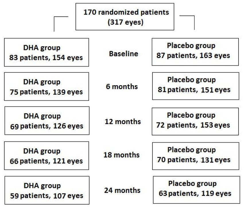
A total of 83 patients (154 eyes) were assigned to the DHA group and 59 (71.1%)
patients (107 eyes, 69.5%) completed the study, whereas of the 87 patients (163 eyes)
assigned to the placebo group, 63 (72.4%) (119 eyes, 73.0%) completed the study (Figure 1).
However, statistically significant differences in the baseline characteristics between the
patients who were lost to follow-up and those who completed the study were not found
(Table 3).
Figure 1. Distribution of patients thought the 2-year study period. Number of patients attending
each follow-up visit and number of eyes analyzed.Antioxidants 2022, 11, 116 7 of 15
Table 3. Demographic and clinical characteristics of patients who completed the study and patients
who were lost to follow-up.
Completed the Study Lost to Follow-Up
Variables p Value
(n = 122) (n = 48)
Gender, males 91 (74.6) 38 (79.2) 0.489
Age, years, mean (SD) 62.4 (11.1) 60.2 (11.8) 0.402
Height, cm, mean (SD) 167.9 (8.7) 166.5 (9.9) 0.339
Weight, kg, mean (SD) 83.5 (14.0) 84.2 (13.7) 0.597
Smoking history
Current smoker 19 (15.6) 15 (31.2)
Ex-smoker 54 (44.3) 17 (35.4) 0.071
Never smoker 49 (40.2) 16 (33.3)
Physical exercise
Sedentary (none) 39 (32.2) 13 (27.1)
Moderate (1 h/day) 45 (37.2) 23 (47.9)
Missing 1
Comorbidities
Hypertension 80 (65.6) 38 (79.2) 0.083
Dyslipidemia 79 (64.8) 26 (54.2) 0.201
Heart disease 12 (9.8) 3 (6.2) 0.558
Nephropathy 2 (1.6) 0 1
Peripheral vascular disease 6 (4.9) 1 (2.0) 0.675
Patients with eyes affected by
NPDR
Both eyes 104 45
Left/right 9/9 2/1
NPDR stage, total eyes 225 92
Mild 142 (63.1) 54 (58.7) 0.576
Moderate 76 (53.2) 38 (41.3) 0.331
Severe 7 (4.9) 0
BCVA, mean (SD)
Left eye
Decimal 0.756 (0.242) 0.688 (0.238) 0.081
LogMAR 0.163 (0.174) 0.192 (0.165) 0.142
Right eye
Decimal 0.740 (0.226) 0.718 (0.219) 0.344
LogMAR 0.167 (0.142) 0.170 (0.137) 0.319
HbA1c level, %, mean (SD) 8.08 (1.55) 8.35 (2.18) 0.854
Antidiabetic medication
Oral antidiabetic agents 57 (46.7) 30 (62.5)
Insulin 17 (13.9) 7 (14.6)
0.293
Both 47 (38.5) 11 (22.9)
No medication 1 0
DHA: docosahexaenoic acid; SD: standard deviation; NPDR: non-proliferative diabetic retinopathy. LogMAR:
logarithm of the minimum angle of resolution; BCVA: best-corrected visual acuity.Antioxidants 2022, 11, 116 8 of 15
3.2. Changes of NPDR Stage
In relation to the primary outcome of the study, the percentages of mild NPDR in-
creased from 61.7% at baseline to 75.7% at the end of the study in the DHA group, and
from 61.9% to 73.1% in the placebo group. Moderate NPDR stages decreased from 35.1% at
baseline to 18.7% at the end of the study in the DHA group, and from 36.8% to 26.0% in the
placebo group. In the DHA group, there were 5 eyes with severe NPDR at baseline, which
increased to 1 more at the end of the study. In the placebo group, of 2 eyes with severe
NPDR at baseline, 1 eye remained at the end of the study. As shown in Table 4, differences
of changes in NPDR stages at each study visit as compared with baseline were not statis-
tically significant in any of the study groups, except for the within-group comparison of
visits at 6 and 12 months vs. baseline in the placebo group.
Table 4. Changes of NPDR stage in the two study groups.
Study Group Total NPDR Stage, Number of Eyes (%) Within-Group
and Visits Patients/Eyes Mild Moderate Severe p Value *
DHA group
Baseline 83/154 95 (61.7) 54 (35.1) 5 (3.2)
6 months 75/139 87 (62.6) 46 (33.1) 6 (4.3) 0.902
12 months 69/126 89 (70.6) 30 (23.8) 7 (5.5) 0.171
18 months 66/126 84 (69.4) 29 (24.0) 8 (6.6) 0.189
24 months 59/121 81 (75.7) 20 (18.7) 6 (5.6) 0.120
Placebo group
Baseline 87/163 101 (61.9) 60 (36.8) 2 (1.3)
6 months 81/151 111 (73.5) 40 (26.5) 0 0.025
12 months 72/133 98 (73.7) 29 (21.8) 6 (4.5) 0.004
18 months 70/131 88 (67.2) 43 (32.8) 0 0.275
24 months 63/119 87 (73.1) 31 (26.0) 1 (0.8) 0.084
* McNemar–Bowker test.
Figure 2 shows the percentages of eyes in which the stages of NPDR improved,
remained unchanged, or worsened at each study visit compared with baseline. Overall
changes in NPDR stages (improvement, no change, worsening) were statistically significant
in the placebo group at 6 months (p = 0.045) compared with the DHA group, but between-
group differences at 12 months (p = 0.825), 18 months (p = 0.931), and 24 months (p = 0.526)
did not reach statistical significance.
3.3. Secondary Outcome Variables
The results of secondary outcome variables are shown in Table 5. In both study groups,
VA almost remained unchanged and significant differences between the DHA and placebo
groups along the study were not found. Changes of the serum levels of HbA1c showed
significant decreases at the 12- and 18-month visits vs. baseline in patients treated with
DHA, and at the 12- and 24-month visits vs. baseline in patients treated with the placebo,
although the between-group differences were not statistically significant (Table 5).
In relation to the tolerability of the study supplements, gastrointestinal discomfort
was recorded in 13 patients (7.6%), regurgitation in 11 (6.5%), nausea in 5 (2.9%), diarrhea
in 2 (1.2%), and vomiting in 1 (0.6%), and other complaints in 13 (7.6%). There were no
significant differences in the occurrence of adverse events between the study groups. Based
upon the pill count, all participants had taken at least 80% of their capsules.Antioxidants 2022, 11, 116 9 of 15
Figure 2. NPDR stage at each study visit in the DHA and placebo groups regarding the percentage of
eyes with improvement, no change, or worsening of NPDR stage.Antioxidants 2022, 11, 116 10 of 15
Table 5. Results of secondary outcome variables: visual acuity (VA) and serum levels of HbA1c in
the two study groups.
Outcomes Baseline 6 Months 12 Months 18 Months 24 Months
VA in decimal system
DHA group, no. eyes 152 139 127 121 107
Mean (SD) 0.73 (0.22) 0.71 (0.24) 0.68 (0.21) 0.64 (0.19) 0.65 (0.22)
Within-group p value * 0.147 0.003Antioxidants 2022, 11, 116 11 of 15
visits. The mean value of HbA1c of 7–8% indicated an adequate metabolic control in our
patients, who had a mean duration of diabetes of 14 years and a high percentage of them
presented with associated hypertension and dyslipidemia. We also found that HbA1c
values slightly decreased over the study period in both study groups, but differences
between the DHA and placebo groups were not observed.
Although it has been shown that increased dietary intake or active supplementation
with antioxidants, including n-3 PUFAs, has a protective effect on diabetes complications
including retinopathy [29–33], there is little information on the potential benefits of antioxi-
dant supplementation, particularly n-3 PUFAs in the early stages of DR. The retina is highly
susceptive to oxidative stress, due principally to the high content of PUFAs, high oxygen
uptake, and glucose oxidation. The set of processes triggered by hyperglycemia, such as a
formation of AEG, activation of PKC, and the polyol and hexosamine pathways, provoke
oxidative stress, which are also reinforced by oxidative stress in a vicious circle, causing
a continuous increase in reactive oxygen species (ROS) and the consequent activation of
pathophysiological mechanisms underlying the progression of DR [34–36]. However, a few
studies have evaluated the effect of supplementation with antioxidants in NPDR, and the
results obtained have been inconsistent due to differences in the design and study variables,
the characteristics of nutraceutical products, or the duration of supplementation.
The nutraceutical product used in the present study includes a high dose of DHA (1 g),
EPA, a mixture of B vitamins, vitamins C and E, lutein, zeaxanthin, and minerals, but none
of the previous studies reported in the literature used a nutraceutical supplement of similar
composition. In relation to lutein, one of the dietary xanthophyll carotenoids with antioxi-
dant properties, in a randomized double-blind, placebo-controlled trial of 31 patients with
NPDR assigned to 10 mg/day of lutein or identical placebo for 36 weeks, a slight improve-
ment in VA was observed in the lutein group [37]. Interestingly, lutein supplementation
was shown to improve macular pigment optical density in 100 healthy subjects (200 eyes)
from a Mediterranean population, being significantly increased in the presence of DHA,
which supports the adjunctive role of DHA for a better lutein availability [38]. In our study,
lutein was a component of the nutraceutical compound and was administered at a dose of
9 mg/day, but no apparent effects on visual performance were observed. In a retrospective
study of 72 patients with NPDR treated with zeaxanthin for 4 months, the addition of lutein
in 36 patients compared to 36 patients who did not receive lutein did not show significant
differences in VA, contrast sensitivity, or glare sensitivity [39]. In a study of 97 patients with
NPRD followed for 5 years, 56 of whom received an antioxidant supplementation and 41
were included in the placebo group, no changes in BCVA were found [40]. To assess the
progression of DR, the authors scored NPDR from 1 (mild), 2 (moderate) to 3 (severe), but
the mean differences in the final score compared with baseline were similar in both groups,
although in the placebo group statistical significance was reached (supplementation group
2.29 (0.66) vs. 2.53 (0.73), p = 0.08; placebo group 2.26 (0.76) vs. 2.65 (0.76), p < 0.01) [40].
In the study, the supplementation product did not include n-3 PUFAs, and the authors
did not evaluate progression according to the characteristic retinography features of the
three stages of NPDR, as was done in our study. In another study of 62 patients with
mild to moderate NPDR assigned to two matched-age groups, a 6-month treatment with a
combination of vitamin E, pycnogenol, and coenzyme Q10 was associated with a reduction
of ROS levels [41], but the clinical translation into the reduced progression of NPDR was
not evaluated. Finally, in a study of 67 patients randomized to an active multi-component
formula containing xanthophyll pigments, antioxidants, and selected botanical extracts
(n = 39) or placebo (n = 28) for 6 months, better visual function (contrast sensitivity, macular
pigment optical density, color discrimination, macular threshold perimetry) was reported
in the supplemented group [42]. However, the number of patients with mild or moderate
NPDR was small, since DR was absent in 37 (55.2%) of the diabetic subjects.
Supplementation with high dose DHA triglyceride was used in none of the afore-
mentioned studies. In a previous experience with the same DHA-based nutraceutical
compound given for 90 days to patients with NPDR, improved macular function assessedAntioxidants 2022, 11, 116 12 of 15
by microperimetry was found [25]. In the present study, however, it may be argued that
although DHA supplementation did not appear to significantly affect the progression of
the NPDR stages, improvements in macular function could have been observed since the
supplementation product was administered for a prolonged period of time, but measure-
ment of macular function-related variables was not included in the study protocol. On
the other hand, total plasma antioxidant capacity (TAC) as an indicator of the antioxidant
effect of DHA supplementation was not measured, either. In patients with advanced
diabetic retinal disease as DME, intravitreal ranibizumab treatment combined with dietary
supplementation with the same high rich DHA triglyceride or placebo was associated with
the anatomical improvement of DME (decrease in central subfield macular thickness on
OCT) in the DHA group after 3 years of treatment [43,44]. Moreover, differences in plasma
TAC and erythrocyte membrane DHA content were statistically significant in favor of the
DHA supplementation group.
Compliance with the nutraceutical supplementation was adequate, and none of the
patients discontinued the study because of adverse effects, which occurred in a small
percentage of patients. Forty-eight patients (24.7%) discontinued the study, especially
between 12 and 24 months after enrolment. The reasons for non-attendance to the study
visits were unknown, but significant differences in the baseline data compared to patients
who completed the study were not observed.
The present findings should be interpreted considering the limitations of the study,
including the single-center design, which may account for the slow rate of recruitment.
Moreover, other variables that could reflect the effect of DHA supplementation, particularly
improvements in macular function, TAC, or IL-6 levels, were not measured, as the present
study was focused on the assessment of progression of any pre-proliferative stage of NPDR
in patients with diabetes. At baseline, between-group differences in antidiabetic medication
were not found, and although it seems unlikely, a potential influence of diabetes treatment
masking the DHA effect cannot be discarded. On the other hand, the higher frequency
of type 2 diabetes is explained by the mean age of the population (around 60 years), but
baseline differences between the study groups in the percentages of patients with type 1 and
type 2 diabetes were not found. It seems unlikely that the type of diabetes may introduce a
bias into the study findings.
The strengths of the study are the study design (randomized controlled trial), and
the assessment of the long-term effects of a highly concentrated DHA plus xanthophyll
carotenoid multivitamin product exclusively in diabetic patients with NPDR, especially
given the paucity of studies on antioxidant supplementation in NDPR. The increase in the
percentages of patients with mild NPDR at the end of the study was 14% in the DHA group
vs. 11.2% in the placebo group, whereas moderate NPDR decreased by 16.4% in the DHA
group vs. 10.8% in the placebo group. These differences may indicate a trend towards a
greater effect of DHA in slowing the progression of the early stages of NPDR. It may be
suggested that the antioxidant and other effects of DHA and other compounds may not be
sufficiently selective for targeting the specific underlying pathophysiological mechanisms
involved in the incipient stages of NPDR.
5. Conclusions
In the present randomized double-blind and placebo-controlled clinical study, the
use of a nutraceutical supplement of 1050 g/day of DHA triglyceride, EPA, vitamins,
minerals, zeaxanthin, and lutein for 2 years did not appear to influence the slowing of the
progression of NPDR. At the end of the study, the increase in eyes with mild NPDR stage
and the decrease in eyes with the moderate stage compared with baseline were higher
in the DHA group, but differences with the placebo did not reach statistical significance.
Among the eyes with a severe NPDR stage, there was an increase of one eye in the DHA
group and a decrease of one eye in the placebo group, which is difficult to interpret given
the between-group disbalance of severe NPDR stage at baseline. Further studies in patientsAntioxidants 2022, 11, 116 13 of 15
with NPDR are necessary to clarify the role of antioxidant supplementation in the early
stages of DR.
Author Contributions: Conceptualization and methodology, P.P.G.; collection of data, P.P.G., F.J.H.M.,
N.A.L., L.C.T., and M.E.T.S.; supervision and original draft preparation, P.P.G. All authors have read
and agreed to the published version of the manuscript.
Funding: This research received no external funding. The APC was founded by Brudy Lab, S.L.,
Barcelona, Spain.
Institutional Review Board Statement: The study was conducted according to the guidelines of the
Declaration of Helsinki, and approved by the Clinical Research Ethics Committee of Hospital San
Juan de Dios del Aljarafe (Sevilla, Spain) (study code PAOXRED Vo2, approval date 20 January 2017).
Informed Consent Statement: Informed consent was obtained from all subjects involved in the study.
Data Availability Statement: The data presented in this study are available in article.
Acknowledgments: The authors thank Jaume Borrás and Paloma Morata for their coordination and
monitoring of the study; Sergi Mojal, biostatistician, for statistical analysis; and Marta Pulido for
editing the manuscript and for her editorial assistance.
Conflicts of Interest: The authors declare no conflict of interest.
References
1. De Boer, I.H.; Bangalore, S.; Benetos, A.; Davis, A.M.; Michos, E.D.; Muntner, P.; Rossing, P.; Zoungas, S.; Bakris, G. Diabetes and
hypertension: A position statement by the American Diabetes Association. Diabetes Care 2017, 40, 1273–1284. [CrossRef]
2. Diabetes Control and Complications Trial Research Group. The effect of intensive diabetes treatment on the progression of
diabetic retinopathy in insulin-dependent diabetes mellitus. The Diabetes Control and Complications Trial. Arch. Ophthalmol.
1995, 113, 36–51. [CrossRef] [PubMed]
3. Fullerton, B.; Jeitler, K.; Seitz, M.; Horvath, K.; Berghold, A.; Siebenhofer, A. Intensive glucose control versus conventional glucose
control for type 1 diabetes mellitus. Cochrane Database Syst. Rev. 2014, 2014, CD009122. [CrossRef]
4. Sivaprasad, S.; Pearce, E. The unmet need for better risk stratification of non-proliferative diabetic retinopathy. Diabet. Med. 2019,
36, 424–433. [CrossRef] [PubMed]
5. Thomas, R.L.; Halim, S.; Gurudas, S.; Sivaprasad, S.; Owens, D.R. IDF Diabetes Atlas: A review of studies utilising retinal
photography on the global prevalence of diabetes related retinopathy between 2015 and 2018. Diabetes Res. Clin. Pract. 2019, 157,
107840. [CrossRef]
6. Sato, Y.; Lee, Z.; Hayashi, Y. Subclassification of preproliferative diabetic retinopathy and glycemic control: Relationship between
mean hemoglobin A1C value and development of proliferative diabetic retinopathy. Jpn J. Ophthalmol. 2001, 45, 523–527.
[CrossRef]
7. Song, P.; Yu, J.; Chan, K.Y.; Theodoratou, E.; Rudan, I. Prevalence, risk factors and burden of diabetic retinopathy in China: A
systematic review and meta-analysis. J. Glob. Health 2018, 8, 010803. [CrossRef]
8. Yang, Q.H.; Zhang, Y.; Zhang, X.M.; Li, X.R. Prevalence of diabetic retinopathy, proliferative diabetic retinopathy and non-
proliferative diabetic retinopathy in Asian T2DM patients: A systematic review and meta-analysis. Int. J. Ophthalmol. 2019, 12,
302–311. [PubMed]
9. Hashemi, H.; Rezvan, F.; Pakzad, R.; Ansaripour, A.; Heydarian, S.; Yekta, A.; Ostadimoghaddam, H.; Pakbin, M.;
Khabazkhoob, M. Global and regional prevalence of diabetic retinopathy; A comprehensive systematic review and meta-analysis.
In Seminars in Ophthalmology; Taylor & Francis: Oxfordshire, UK, 2021; pp. 1–16. [CrossRef]
10. Wykoff, C.C. Management of Diabetes-Related Retinopathy. In Prevention and Management of Diabetes-Related Eye Disease; American
Diabetes Association: Arlington VA, USA, 2019. Available online: https://www.ncbi.nlm.nih.gov/books/NBK544520/ (accessed
on 27 August 2021).
11. Cai, J.; Boulton, M. The pathogenesis of diabetic retinopathy: Old concepts and new questions. Eye 2002, 16, 242–260. [CrossRef]
12. Joy, S.S.; Siddiqui, K. Molecular and pathophysiological mechanisms of diabetic retinopathy in relation to adhesion molecules.
Curr. Diabetes Rev. 2019, 15, 363–371. [CrossRef] [PubMed]
13. Stem, M.S.; Gardner, T.W. Neurodegeneration in the pathogenesis of diabetic retinopathy: Molecular mechanisms and therapeutic
implications. Curr. Med. Chem. 2013, 20, 3241–3250. [CrossRef]
14. Safi, S.Z.; Qvist, R.; Kumar, S.; Batumalaie, K.; Ismail, I.S. Molecular mechanisms of diabetic retinopathy, general preventive
strategies, and novel therapeutic targets. BioMed Res. Int. 2014, 2014, 801269. [CrossRef]
15. Chang, Y.C.; Chuang, L.M. The role of oxidative stress in the pathogenesis of type 2 diabetes: From molecular mechanism to
clinical implication. Am. J. Transl. Res. 2010, 2, 316–331. [PubMed]
16. Rochette, L.; Zeller, M.; Cottin, Y.; Vergely, C. Diabetes, oxidative stress and therapeutic strategies. Biochim. Biophys. Acta 2014,
1840, 2709–2729. [CrossRef]Antioxidants 2022, 11, 116 14 of 15
17. Lafuente, M.; González-Herrero, M.R.; Villadóniga, S.R.; Domingo, J.C. Antioxidant activity and neuroprotective role of
docosahexaenoic acid (DHA) supplementation in eye diseases that can lead to blindness: A narrative review. Antioxidants
2021, 10, 386. [CrossRef]
18. Connor, K.M.; SanGiovanni, J.P.; Lofqvist, C.; Aderman, C.M.; Chen, J.; Higuchi, A.; Hong, S.; Pravda, E.A.; Majchrzak, S.;
Carper, D.; et al. Increased dietary intake of omega-3-polyunsaturated fatty acids reduces pathological retinal angiogenesis. Nat.
Med. 2007, 13, 868–873. [CrossRef]
19. Rotstein, N.P.; Politi, L.E.; German, O.L.; Girotti, R. Protective effect of docosahexaenoic acid on oxidative stress-induced apoptosis
of retina photoreceptors. Investig. Opthalmol. Vis. Sci. 2003, 44, 2252–2259. [CrossRef] [PubMed]
20. Mukherjee, P.K.; Marcheselli, V.L.; Serhan, C.N.; Bazan, N.G. Neuroprotectin D1: A docosahexaenoic acid-derived docosatriene
protects human retinal pigment epithelial cells from oxidative stress. Proc. Natl. Acad. Sci. USA 2004, 101, 8491–8496. [CrossRef]
[PubMed]
21. Yang, Y.C.; Lii, C.K.; Wei, Y.L.; Li, C.C.; Lu, C.Y.; Liu, K.L.; Chen, H.W. Docosahexaenoic acid inhibition of inflammation is partially
via cross-talk between Nrf2/heme oxygenase 1 and IKK/NF-κB pathways. J. Nutr. Biochem. 2013, 24, 204–212. [CrossRef]
22. Solanki, P.; Aminoshariae, A.; Jin, G.; Montagnese, T.A.; Mickel, A. The effect of docosahexaenoic acid (DHA) on expression of
IL-1ß, IL-6, IL-8, and TNF-α in normal and lipopolysaccharide (LPS)-stimulated macrophages. Quintessence Int. 2013, 44, 393.
[CrossRef] [PubMed]
23. Fu, Z.; Löfqvist, C.; Shao, Z.; Sun, Y.; Joyal, J.-S.; Hurst, C.G.; Cui, R.Z.; Evans, L.P.; Tian, K.; SanGiovanni, J.P.; et al. Dietary ω-3
polyunsaturated fatty acids decrease retinal neovascularization by adipose-endoplasmic reticulum stress reduction to increase
adiponectin. Am. J. Clin. Nutr. 2015, 101, 879–888. [CrossRef]
24. Calder, P.C. Omega-3 fatty acids and inflammatory processes. Nutrients 2010, 2, 355–374. [CrossRef]
25. González-Herrero, M.E.R.; Ruiz, M.; Román, F.J.L.; Sánchez, J.M.M.; Domingo, J.C. Supplementation with a highly concentrated
docosahexaenoic acid plus xanthophyll carotenoid multivitamin in nonproliferative diabetic retinopathy: Prospective controlled
study of macular function by fundus microperimetry. Clin. Ophthalmol. 2018, 12, 1011–1020. [CrossRef] [PubMed]
26. Early Treatment Diabetic Retinopathy Study Research Group. Grading diabetic retinopathy from stereoscopic color fundus
photographs—An extension of the modified Airlie House classification: ETDRS report number 10. Ophthalmology 1991, 98
(Suppl. 5), 786–806. [CrossRef]
27. Brudy Technology, S.L. Use of DHA for Treating a Pathology Associated with Cellular Oxidative Damage. European Patent EP 1
962 825 B1, 2 April 2008. Available online: https://patentimages.storage.googleapis.com/55/e3/28/c2f8a893be6c93/EP1962825
B1.pdf (accessed on 10 November 2021).
28. Alemán Sánchez, J.J.; Artola Menéndez, S.; Ávila Lachica, L.; Barrot de la Puente, J.; Barutell Rubio, L.; Benito Badorrey, B.;
Buil Cosiales, P.; Carramiñana Barrera, F.; Carrillo Fernández, L.; Cebrián Cuenca, A. Guía de la Diabetes Tipo 2 Para Clínicos. Re-
comendaciones de redGDPS; Fundación redGDPS: Madrid, Spain, 2018. Available online: https://www.redgdps.org/publicaciones-
redgdps/ (accessed on 26 August 2021).
29. Sasaki, M.; Kawasaki, R.; Rogers, S.; Man, R.E.K.; Itakura, K.; Xie, J.; Flood, V.M.; Tsubota, K.; Lamoureux, E.L.; Wang, J.J. The
associations of dietary intake of polyunsaturated fatty acids with diabetic retinopathy in well-controlled diabetes. Investig.
Ophthalmol. Vis. Sci. 2015, 56, 7473–7479. [CrossRef]
30. Hu, F.B.; Cho, E.; Rexrode, K.M.; Albert, C.M.; Manson, J.E. Fish and long-chain omega-3 fatty acid intake and risk of coronary
heart disease and total mortality in diabetic women. Circulation 2003, 107, 1852–1857. [CrossRef] [PubMed]
31. Shimizu, H.; Ohtani, K.-I.; Tanaka, Y.; Sato, N.; Mori, M.; Shimomura, Y. Long-term effect of eicosapentaenoic acid ethyl (EPA-E)
on albuminuria of non-insulin dependent diabetic patients. Diabetes Res. Clin. Pract. 1995, 28, 35–40. [CrossRef]
32. Chewcharat, A.; Chewcharat, P.; Rutirapong, A.; Papatheodorou, S. The effects of omega-3 fatty acids on diabetic nephropathy: A
meta-analysis of randomized controlled trials. PLoS ONE 2020, 15, e0228315. [CrossRef]
33. Tao, M.; McDowell, M.A.; Saydah, S.H.; Eberhardt, M.S. Relationship of polyunsaturated fatty acid intake to peripheral neuropathy
among adults with diabetes in the National Health and Nutrition Examination Survey (NHANES) 1999 2004. Diabetes Care 2008,
31, 93–95. [CrossRef] [PubMed]
34. Kowluru, R.A.; Chan, P.S. Oxidative stress and diabetic retinopathy. Exp. Diabetes Res. 2007, 2007, 43603. [CrossRef] [PubMed]
35. Kang, Q.; Yang, C. Oxidative stress and diabetic retinopathy: Molecular mechanisms, pathogenetic role and therapeutic
implications. Redox Biol. 2020, 37, 101799. [CrossRef]
36. Li, C.; Miao, X.; Li, F.; Wang, S.; Liu, Q.; Wang, Y.; Sun, J. Oxidative stress-related mechanisms and antioxidant therapy in diabetic
retinopathy. Oxid. Med. Cell. Longev. 2017, 2017, 9702820. [CrossRef]
37. Zhang, P.C.; Wu, C.R.; Wang, Z.L.; Wang, L.Y.; Han, Y.; Sun, S.L.; Li, Q.S.; Ma, L. Effect of lutein supplementation on visual
function in nonproliferative diabetic retinopathy. Asia Pac. J. Clin. Nutr. 2017, 26, 406–411. [PubMed]
38. Zanón-Moreno, V.; Pedrol, J.C.D.; Sanz-González, S.M.; Raga-Cervera, J.; Salazar-Corral, J.; Pinazo-Durán, M.D. Feasibility
study of a docosahexaenoic acid optimized nutraceutical formulation on the macular levels of lutein in a healthy Mediterranean
population. Ophthalmic Res. 2020, 64, 1068–1076. [CrossRef]
39. Ren, Y.B.; Qi, Y.X.; Su, X.J.; Luan, H.Q.; Sun, Q. Therapeutic effect of lutein supplement on non-proliferative diabetic retinopathy:
A retrospective study. Medicine 2019, 98, e15404. [CrossRef]
40. Garcia-Medina, J.J.; Pinazo-Duran, M.D.; Garcia-Medina, M.; Zanon-Moreno, V.; Pons-Vazquez, S. A 5-year follow-up of
antioxidant supplementation in type 2 diabetic retinopathy. Eur. J. Ophthalmol. 2011, 21, 637–643. [CrossRef]Antioxidants 2022, 11, 116 15 of 15
41. Domanico, D.; Fragiotta, S.; Cutini, A.; Carnevale, C.; Zompatori, L.; Vingolo, E.M. Circulating levels of reactive oxygen species in
patients with nonproliferative diabetic retinopathy and the influence of antioxidant supplementation: 6-month follow-up. Indian
J. Ophthalmol. 2015, 63, 9–14. [PubMed]
42. Chous, A.P.; Richer, S.P.; Gerson, J.D.; Kowluru, R.A. The Diabetes Visual Function Supplement Study (DiVFuSS). Br. J. Ophthalmol.
2016, 100, 227–234. [CrossRef]
43. Lafuente, M.; Ortín, L.; Argente, M.; Guindo, J.L.; López-Bernal, M.D.; López-Román, F.J.; García, M.J.; Domingo, J.C.; Lajara, J.
Combined intravitreal ranibizumab and oral supplementation with docosahexaenoic acid and antioxidants for diabetic macular
edema: Two-year randomized single-blind controlled trial results. Retina 2017, 37, 1277–1286. [CrossRef] [PubMed]
44. Lafuente, M.; Ortín, L.; Argente, M.; Guindo, J.L.; López-Bernal, M.D.; López-Román, F.J.; Domingo, J.C.; Lajara, J. Three-year out-
comes in a randomized single-blind controlled trial of intravitreal ranibizumab and oral supplementation with docosahexaenoic
acid and antioxidants for diabetic macular edema. Retina 2019, 39, 1083–1090. [CrossRef]You can also read