SUBMIT ABSTRACTS BY 18 AUGUST 2021 - 20-24 February 2022 Town and Country Resort & Convention Center San Diego, CA, USA - spie.org/mi22call ...
←
→
Page content transcription
If your browser does not render page correctly, please read the page content below

20–24 February 2022
Town and Country Resort & Convention Center
                                                          022 ·
San Diego, CA, USA                                    72–2
                                                  · 19

SUBMIT ABSTRACTS
BY 18 AUGUST 2021

                                           spie.org/mi22call
                                        #SPIEMedicalImaging
Conferences and Courses
                                              20–24 February 2022
                                              Town and Country Resort & Convention Center
                                              San Diego, California, USA

                                                                                                                                                                                     022 ·
                                                                                                                                                                                 72–2
                                                                                                                                                                             · 19

                                                                                               An invitation to participate
                                                                                               Present and discuss the latest research at SPIE Medical Imaging
                                                                                               where you’ll experience five days of thought-provoking sessions,
                                                                                               plenary talks, and networking events that will bring the imaging
                                                                                               community together again.
      Present your work in San Diego                                                           This meeting is the internationally recognized forum for reporting

      at Medical Imaging 2022                                                                  state-ofthe-art research and development in medical imaging.
                                                                                               The event focuses on the latest innovations found in underlying
      The leading conference that explores the science                                         fundamental scientific principles, technology developments,
      of medical imaging                                                                       scientific evaluation, and clinical application. The symposium
                                                                                               covers the full range of medical imaging modalities focusing
      SPIE Medical Imaging is the conference where the latest                                  on image acquisition, display, processing, analysis, perception,
      information is presented by leading researchers in image                                 decision support, and informatics.
       processing, physics, computer-aided diagnosis, perception,                              We hope you will consider submitting an abstract. We welcome
      image-guided procedures, biomedical applications, ultrasound,                            your participation as we look to build another outstanding
      informatics, radiology, and digital and computational                                    program. SPIE Medical Imaging 2022 marks our 50th
      pathology. This also includes an increased focus on areas like                           Anniversary—please join us as we celebrate this significant
      deep learning, artificial intelligence (AI), machine learning,                           milestone.
      information fusion, augmented/ virtual reality, and global health
                                                                                               Until then,
      initiatives.
                                                                                               Your 2022 Medical Imaging Chairs
      Review the nine topic areas and see where your research fits best.

      ABSTRACT SUBMISSIONS ARE DUE 18 AUGUST.

                                                                                             SYMPOSIUM CHAIRS:

                                                                                                             Metin N. Gurcan                                     Robert M. Nishikawa
                                                                                                             Wake Forest Baptist                                 Univ. of Pittsburgh (USA)
                                                                                                             Medical Ctr. (USA)

 ii                  SPIE MEDICAL IMAGING 2022 • www.spie.org/mi22call                      Tel: +1 360 676 3290   •   help@spie.org   •   #SPIEMedicalImaging                               1
www.spie.org/mi22call
THE MULTIDISCIPLINARY EVENT                                                                                                                                                 CALL FOR PAPERS
                                                 FOR THE ADVANCEMENT OF
                                                 IMAGING TECHNOLOGIES                         Contents
                                                                                              CONFERENCE MI101.  .  .  .  .  .  .  .  .  .  .  .  .  .  .  .  .  .  .  .  . PAGE 5   CONFERENCE MI106 .  .  .  .  .  .  .  .  .  .  .  .  .  .  .  .  .  .  . PAGE 13
Executive Organizing Committee                                                                Physics of Medical Imaging                                                             Biomedical Applications in Molecular,
Nick Bottenus, Univ. of Colorado Boulder         Claudia Mello-Thoms, Univ. Iowa Carver       Chair(s): Lifeng Yu; Wei Zhao; Rebecca Fahrig                                          Structural, and Functional Imaging
Oliver Colliot, Ctr. National de la Recherche      College of Medicine                                                                                                               Chair(s): Barjor S. Gimi; Andrzej Krol
                                                                                              CONFERENCE MI102. .  .  .  .  .  .  .  .  .  .  .  .  .  .  .  .  .  .  .  . PAGE 6
 Scientifique                                    Brian Park, Oregon Health & Science Univ.                                                                                           CONFERENCE MI107. .  .  .  .  .  .  .  .  .  .  .  .  .  .  .  .  .  .  . PAGE 14
                                                                                              Image Processing
Thomas Doserno, Technische Univ.                 Nicole Ruiter, Karlsruher Institut für                                                                                              Imaging Informatics for Healthcare
                                                                                              Chair(s): Olivier Colliot; Ivana Išgum
  Braunschweig                                     Technologie
                                                                                                                                                                                     Chair(s): Thomas M. Deserno; Brian J. Park
Karen Drukker, The Univ. of Chicago              Jeffrey Siewerdsen, Johns Hopkins Univ.      CONFERENCE MI103. .  .  .  .  .  .  .  .  .  .  .  .  .  .  .  .  .  .  .  . PAGE 8
Barjor Gimi, Cooper Medical School, Rowan        Sian Taylor-Phillips, The Univ. of Warwick   Computer-Aided Diagnosis                                                               CONFERENCE MI108. . . . . . . . . . . . . . . . . . . . PAGE 16
 Univ.                                           John Tomaszewski, Univ. at Buffalo           Chair(s): Karen Drukker; Khan M. Iftekharuddin                                         Ultrasonic Imaging and Tomography
Khan Iftekharuddin, Old Dominion Univ.           Aaron Ward, The Univ. of Western Ontario                                                                                            Chair(s): Nick Bottenus; Nicole V. Ruiter
                                                                                              CONFERENCE MI104 .  .  .  .  .  .  .  .  .  .  .  .  .  .  .  .  .  .  . PAGE 10
Ivana Išgum, Amsterdam UMC                       Lifeng Yu, Mayo Clinic
                                                                                              Image-Guided Procedures, Robotic                                                       CONFERENCE MI109 .  .  .  .  .  .  .  .  .  .  .  .  .  .  .  .  .  .  . PAGE 17
Andrzej Krol, SUNY Upstate Medical Univ.         Wei Zhao, Stony Brook Univ.                  Interventions, and Modeling                                                            Digital and Computational Pathology
Cristian Linte, Rochester Institute of
                                                                                              Chair(s): Cristian A. Linte; Jeffrey H. Siewerdsen                                     Chair(s): John E. Tomaszewski; Aaron D. Ward
  Technology
                                                                                              CONFERENCE MI105. .  .  .  .  .  .  .  .  .  .  .  .  .  .  .  .  .  .  . PAGE 12
                                                                                              Image Perception, Observer                                                             General Information.  .  .  .  .  .  .  .  .  .  .  .  .  .  .  .  .  .  .  .  .  . 19
Cooperating Organizations                                                                     Performance, and Technology                                                            Submission of Abstracts.  .  .  .  .  .  .  .  .  .  .  .  .  .  .  .  .  . 21
                                                                                              Assessment
                                                                                              Chair(s): Claudia R. Mello-Thoms;
                                                                                              Sian Taylor-Phillips

2022 Plenary Presentation

                                                                                                      2022 STUDENT PAPER AWARDS INFORMATION
                                                                                                      ATTENTION STUDENTS
Emerging Clinical Applications of Medical Extended Reality (MXR)                                      Submission instructions and eligibility requirements for the 2022 All Conference Best
                                                                                                      Student Paper Awards will be available in July 2021.
                             JENNIFER N. AVARI SILVA
                          MD, Director, Pediatric Electrophysiology,                                  See 2021 Award Winners online: www.spie.org/awards2021
                     Washington University School of Medicine in St. Louis

                                                                                                                                      Nominations now being accepted for the
           SPIE remains committed to advancing light-based research and meeting                                                       SPIE HARRISON H. BARRETT AWARD
             the needs of our constituents by providing you with an opportunity
            for sharing your work and connecting you with the global science and                                                      Nominate a colleague in recognition of outstanding accomplishments
           engineering community. SPIE Medical Imaging 2022 is scheduled to take                                                      in medical imaging. Deadline for 2022 nominations is 1 July 2021.
                 place as planned, and we look forward to your participation.                                                         View award details and past winners online.

                                                                                                                                                                  2021 AWARD RECIPIENT
                                    Stay Up to Date via Email                                                                                                          Kevin Berbaum
                                                                                                                                               University of Iowa, Iowa City, Iowa, USA
                    Sign up to receive emails about SPIE Medical Imaging.
                                    www.spie.org/signup

2                     SPIE MEDICAL IMAGING 2022 • www.spie.org/mi22call                        Tel: +1 360 676 3290                     •    help@spie.org                 •    #SPIEMedicalImaging                                                                      3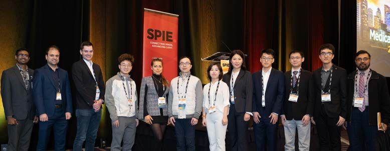
MEDICAL IMAGING 2022                                                                                                                                                                      CALL FOR PAPERS

Physics of Medical Imaging (MI101)
Conference Chairs: Lifeng Yu, Mayo Clinic (United States); Wei Zhao, Stony Brook Univ. (United States)         TOPIC AREAS (FOR THIS CONFERENCE
                                                                                                               ONLY):
Conference Co-Chair: Rebecca Fahrig, Siemens Healthcare GmbH (Germany)
                                                                                                               During the submission process, you will be asked to
Program Committee: Shiva Abbaszadeh, Univ. of Illinois at Urbana-Champaign (United States);
                                                                                                               choose three different topics to assist in the review
Adam M. Alessio, Michigan State Univ. (United States); Hilde Bosmans, UZ Leuven (Belgium);
                                                                                                               process.
Seungryong Cho, KAIST (Korea, Republic of); Mini Das, Univ. of Houston (United States);
Mats E. Danielsson, KTH Royal Institute of Technology (Sweden); Maria Drangova, Robarts                        • ALG - Algorithmic developments, simulations,
Research Institute (Canada); Thomas G. Flohr, Siemens Healthcare GmbH (Germany);                                 calibration, classification, etc. (for reconstruction
Arundhuti Ganguly, Varex Imaging Corp. (United States); Yongshuai Ge, Shenzhen Institutes                        and machine learning use dedicated categories)
of Advanced Technology, Chinese Academy of Sciences (China); Taly Gilat Schmidt, Marquette                     • CARD - Cardiovascular imaging
Univ. (United States); Stephen J. Glick, U.S. Food and Drug Administration (United States), Univ. of           • CLIM - Clinical evaluation
Massachusetts Medical School (United States); Marc Kachelriess, Deutsches Krebsforschungszentrum               • CON - Physics of contrast enhancement using
(Germany); Karim S. Karim, Univ. of Waterloo (Canada); Patrick J. La Riviere, The Univ. of                       contrast media / nanoparticles
Chicago (United States); Ke Li, Univ. of Wisconsin School of Medicine and Public Health (United States);       • CT - All conventional and multi-energy CT topics
Quanzheng Li, Massachusetts General Hospital (United States); Joseph Y. Lo, Duke Univ. (United                   (for cone beam use dedicated category)
States); Peter B. Noël, Univ. of Pennsylvania (United States); Frédéric Noo, The Univ. of Utah (United         • CTCB - Cone beam CT
States); Jinyi Qi, Univ. of California, Davis (United States); John M. Sabol, GE Healthcare (United            • DET - Detector technology; scintillators,
States); Ioannis Sechopoulos, Radboud Univ. Medical Ctr. (Netherlands); Behrouz Shabestari,                      photoconductors, diodes, TFT
National Institute of Biomedical Imaging and Bioengineering (United States); Joseph W. Stayman,                • DIAG - Diagnostic imaging
Johns Hopkins Univ. (United States); Anders Tingberg, Lund Univ. (Sweden); Adam S. Wang,
                                                                                                               • DOSE - Radiation dose, dosimetry, and dose
Stanford Univ. School of Medicine (United States); Yuxiang Xing, Tsinghua Univ. (China);
                                                                                                                 effects
John Yorkston, Carestream Health, Inc. (United States)
                                                                                                               • IGI - Image guided interventions
                                                                                                               • IMG - Imaging methods including optical, MR,
This conference will cover all aspects of image forma-    • Multi-energy (spectral) x-ray and CT imaging         ultrasound, etc. (for x-ray, CT, or nuclear based
tion in medical imaging, including systems using ion-     • Computer simulation of imaging systems               methods use dedicated categories)
izing radiation (x-rays, gamma rays) or non-ionizing        including models for radiation sources, imaged     • MAM - Imaging of the breast (any device)
techniques (ultrasound, optical, thermal, magnetic          objects, physical interactions, and detectors      • METR - Measurement methods (MTF, NPS, DQE,
resonance, or magnetic particle imaging). Papers of       • Phantoms (physical and numerical)                    eDQE, gDQE,
a theoretical nature or papers reporting new experi-      • Photon counting                                      Spectra, ...)
mental results and applications using Artificial Intel-                                                        • ML – Machine Learning applied to imaging
                                                          • Proton based imaging
ligence techniques are invited. Topics of particular                                                             physics (reconstruction, corrections, evaluations,
interest include novel methods for image formation,       • Imaging method for radiation therapy
                                                                                                                 etc…)
experimental methods and results regarding image          • Radiation (e.g., optical) and signal transport
                                                                                                               • MULTI – Multi modality imaging
performance, algorithms for image reconstruction          • Radiation dose, dosimetry, and dose effects
                                                            (risk), as well as possible stratification         • NEURO - Neuroimaging
and correction, detector materials and electronic de-
                                                                                                               • NUC - Nuclear medical imaging innovations
sign, analytical and computer modeling of imaging         DEVICES
systems, and physics of contrast media. Work di-                                                               • ONC - Oncology
                                                          • Advanced multi-slice or cone beam CT systems       • OTHER - Other methodology, systems or
rected toward the imaging of human subjects, small
                                                          • Advanced radiographic, fluoroscopic, or              applications
animals, or tissue specimens are welcome. The con-
                                                            angiographic systems (including phase contrast     • PCI - Photon counting imaging
ference will also cover dedicated approaches for var-
                                                            and diffraction)
ious imaging applications resulting from the above                                                             • PER - Observer or perception-based
mentioned general imaging framework, for example          • Ultrasound, MRI, optical, thermal, magnetic          performance evaluations of systems
cardiovascular or neuroimaging applications.                particle imaging (and other non-ionizing
                                                                                                               • PHS - Phase contrast imaging
                                                            radiation systems)
Original papers are especially requested in the fol-                                                           • PHT - Work involving development of phantoms
                                                          • Small animal imaging systems
lowing areas:                                                                                                    or anatomical simulation models
                                                          • Nuclear medicine imaging methods (SPECT and
                                                                                                               • PRI - Proton based imaging
IMAGING SCIENCE                                             PET)
                                                                                                               • RECON - Image reconstruction including CT,
• Physics of signal detection, image formation and        • Multi-modality imaging devices
                                                                                                                 SPECT, PET, OCT and tomosynthesis
  signal degradation                                      • Low-cost imaging devices with global health
                                                                                                               • SMAX - Small animal or microscopic imaging
• Object characterization and contrast                      applications
  mechanisms                                                                                                   • TSY - Tomosynthesis
                                                          APPLICATIONS                                         • VCT - Virtual clinical trials
• Characterization of detector and system
                                                          • Cardiovascular imaging                             • XIM - X-ray imaging, x-ray sources, scatter
  performance (MTF, NPS, DQE, task- and
  observer-based)                                         • Neuroimaging                                         reduction techniques
• Virtual imaging and virtual clinical trials             • Mammographic imaging                               • XME - Multi-energy radiography or
                                                          • Interventional imaging                               mammography
TECHNOLOGY                                                • Imaging applications in therapy (e.g., radiation
• Novel medical imaging systems and methods                 therapy, surgery, in-vivo verification)
  including contrast media / nanoparticles                • Advanced applications (clinical, translational,
• Properties of scintillating, photoconductive, or          preclinical, basic science, biomarkers)
  other sensor materials                                  • Novel medical imaging for precision medicine
• Novel sources of radiation                                applications
• Image reconstruction methods (e.g., for CT,
  tomosynthesis, SPECT and PET, optical imaging,
  MRI, etc.)
• Machine learning approaches to image
  formation                                                                                                                                   ABSTRACTS DUE: 18 AUGUST 2021

                                                                                                                        Submit your abstract today: www.spie.org/mi22call

4                         SPIE MEDICAL IMAGING 2022 • www.spie.org/mi22call                                    Tel: +1 360 676 3290       •   help@spie.org     •   #SPIEMedicalImaging                 5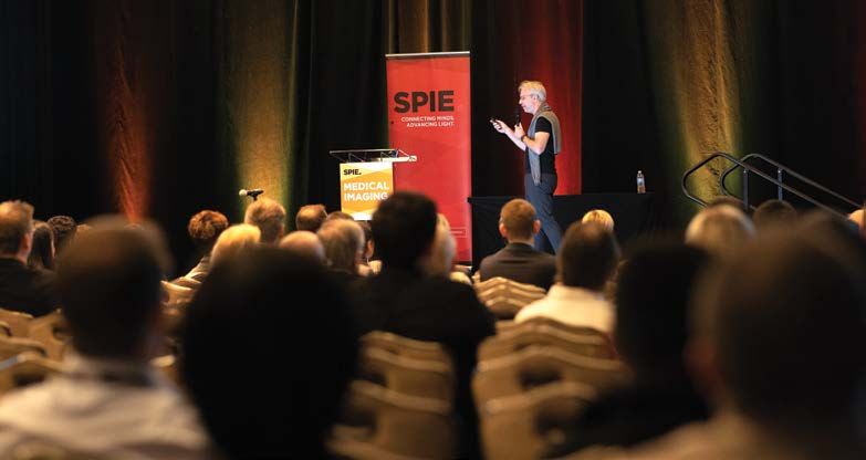
MEDICAL IMAGING 2022                                                                                                                                                                  CALL FOR PAPERS

Image Processing (MI102)
Conference Chairs: Olivier Colliot, Ctr. National de la Recherche Scientifique (France);
Ivana Išgum, Amsterdam UMC (Netherlands)                                                                     TOPIC AREAS (FOR THIS CONFERENCE
Program Committee: Elsa D. Angelini, Imperial College London (United Kingdom), Columbia Univ.                ONLY):
(United States), Télécom ParisTech (France); Meritxell Bach-Cuadra, Univ. de Lausanne (Switzerland);         To assist the reviewers, choose up to three keywords
Ulas Bagci, Univ. of Central Florida (United States); Antong Chen, Merck & Co., Inc. (United States);        in order of relevance from the following list.
Tolga Çukur, Bilkent Univ. (Turkey); Benoit M. Dawant, Vanderbilt Univ. (United States);                     • Augmented/virtual reality
Marleen de Bruijne, Erasmus MC (Netherlands); Lotta Maria Ellingsen, Univ. of Iceland (Iceland);             • Classification
Alexandre X. Falcão, Univ. Estadual de Campinas (Brazil); Aaron Fenster, Robarts Research
                                                                                                             • Compressed sensing, sparse reconstruction
Institute (Canada); James Fishbaugh, NYU Tandon School of Engineering (United States);
                                                                                                               methods
Alejandro F. Frangi, Univ. of Leeds (United Kingdom); Yu Gan, The Univ. of Alabama (United States);
Mona K. Garvin, The Univ. of Iowa (United States); James C. Gee, Univ. of Pennsylvania (United               • Computational anatomy and atlases
States); Miguel Angel González Ballester, Univ. Pompeu Fabra (Spain); Hayit Greenspan, Tel                   • Computer vision
Aviv Univ. (Israel); David R. Haynor, Univ. of Washington (United States); Tobias Heimann, Siemens           • Connectome analysis
Healthineers (Germany); Bulat Ibragimov, Univ. of Copenhagen (Denmark); Leigh Johnston, The                  • Deep learning
Univ. of Melbourne (Australia); Stefan Klein, Erasmus MC (Netherlands); Bennett A. Landman,                  • Deformable geometry
Vanderbilt Univ. (United States); Tianhu Lei, MD Imaging Research (United States); Tim Leiner, Univ.         • Diffusion MRI analysis
Medical Ctr. Utrecht (Netherlands); Karim Lekadir, Univ. de Barcelona (Spain); Boudewijn P. F.               • Functional imaging analysis
Lelieveldt, Leiden Univ. Medical Ctr. (Netherlands); Natasha Lepore, The Univ. of Southern California        • Generating virtual populations
(United States); Marius George Linguraru, Children’s National Medical Ctr. (United States);
                                                                                                             • Generative/adversarial learning
Murray H. Loew, The George Washington Univ. (United States); Cristian Lorenz, Philips Research
(Germany); Frederik Maes, Katholieke Univ. Leuven (Belgium); Vincent A. Magnotta, The Univ.                  • Image representation and compression
of Iowa Hospitals and Clinics (United States); Rashindra Manniesing, Radboud Univ. Medical Ctr.              • Image restoration and enhancement
(Netherlands); Diana Mateus, Ecole Centrale de Nantes (France); Jhimli Mitra, GE Global Research             • Image synthesis
(United States); Sunanda D. Mitra, Texas Tech Univ. (United States); Marc Modat, King’s College              • Imaging genetics
London (United Kingdom); Albert Montillo, Univ. of Texas Southwestern Medical Ctr. (United States);          • Machine learning and pattern recognition
Kensaku Mori, Nagoya Univ. (Japan); Mads Nielsen, Niels Bohr Institute (Denmark); Ipek Oguz,                 • Methods for training and validation, including
Vanderbilt Univ. (United States); Dzung L. Pham, Henry Jackson Foundation/USU (United States),                 ground truth generation
National Institutes of Health (United States), Johns Hopkins Univ. (United States); Jerry L. Prince, Johns   • Model-based image analysis
Hopkins Univ. (United States); Xin Qi, Rutgers, The State Univ. of New Jersey (United States);               • Motion/time series analysis
Maryam E. Rettmann, Mayo Clinic (United States); Letícia Rittner, Univ. Estadual de Campinas
                                                                                                             • Multi-scale imaging (from single cell to whole
(Brazil); Mirabela Rusu, Stanford Univ. School of Medicine (United States); Nishant Ravikumar, Univ.
                                                                                                               body)
of Leeds (United Kingdom); Punam K. Saha, The Univ. of Iowa (United States); Lin Shi, The Chinese
Univ. of Hong Kong (China); Rachel E. Sparks, King’s College London (United Kingdom);                        • Open software for medical image processing
Marius Staring, Leiden Univ. Medical Ctr. (Netherlands); Martin A. Styner, The Univ. of North Carolina       • Population/clinical studies
at Chapel Hill (United States); Kenji Suzuki, Tokyo Institute of Technology (Japan); Tanveer F. Syeda-       • Quantitative image analysis/quantitative
Mahmood, IBM Research - Almaden (United States); Raphael Sznitman, Univ. Bern (Switzerland);                   imaging biomarkers
Zeike A. Taylor, Univ. of Leeds (United Kingdom); Yubing Tong, Univ. of Pennsylvania (United                 • Registration methodologies
States); Jayaram K. Udupa, Univ. of Pennsylvania (United States); Koen Van Leemput, Harvard                  • Radiomics and texture representation/analysis
Medical School (United States), Massachusetts General Hospital {United States); Tomaž Vrtovec, Univ.         • Segmentation methodologies
of Ljubljana (Slovenia); Wolfgang Wein, ImFusion GmbH (Germany)                                              • Shape representation and analysis
                                                                                                             • Statistical methodology
Original papers are invited on all aspects of the       •   Diffusion MRI analysis                           • Visualization methods
processing and analysis of medical, small animal,       •   Functional imaging analysis                      • Voxel/deformation/tensor-based morphometry
or cellular images, with applications in medicine,      •   Generative/adversarial learning                  DEEP-DIVE SESSION
biological, and pharmaceutical research. Of interest    •   Image representation and compression
are algorithms applied to all imaging modalities, in-                                                        A limited number of papers selected for oral presen-
                                                        •   Image restoration and enhancement                tation will be chosen for a novel deep-dive session.
cluding x-ray, DSA, CT, MRI, neuroimaging, nuclear
medicine, optical, ultrasound, macroscopic, and mi-     •   Image synthesis                                  This session will comprise of very few oral presenta-
croscopic imaging. Papers dealing with the challeng-    •   Imaging genetics                                 tions followed by a longer more in-depth discussion
es of bringing advances in research laboratories into   •   Machine learning and pattern recognition         that will be led by experienced researchers. In order
clinical application are particularly welcomed.         •   Methods for training and validation, including   to be considered for this Deep-dive session, please
                                                            ground truth generation                          enter the following text in the custom tracking title
Papers typically involve research that includes one                                                          when prompted, “Deep-dive”.
or more of the following categories (in alphabetical    •   Model-based image analysis
order):                                                 •   Motion/time series analysis
                                                                                                             OPEN-SOURCE SOFTWARE
• Augmented/virtual reality                             •   Open software for medical image processing
                                                        •   Population/clinical studies                      We are supporting development of open source soft-
• Classification
                                                        •   Quantitative image analysis/quantitative         ware. Accepted manuscripts describing open source
• Compressed sensing, sparse reconstruction                                                                  code via public repositories will be indicated in the
  methods                                                   imaging biomarkers
                                                                                                             program.
• Computational anatomy and atlases                     •   Registration methodologies
• Computer vision                                       •   Radiomics and texture representation/analysis
• Connectome analysis                                   •   Segmentation methodologies
• Deep learning                                         •   Shape representation and analysis
• Deformable geometry                                   •   Statistical methodology
                                                        •   Visualization methods
                                                        •   Voxel/deformation/tensor-based morphometry
                                                                                                                                           ABSTRACTS DUE: 18 AUGUST 2021

                                                                                                                      Submit your abstract today: www.spie.org/mi22call

6                        SPIE MEDICAL IMAGING 2022 • www.spie.org/mi22call                                   Tel: +1 360 676 3290      •   help@spie.org    •   #SPIEMedicalImaging                 7
MEDICAL IMAGING 2022                                                                                                                                                                               CALL FOR PAPERS

Computer-Aided Diagnosis (MI103)
Conference Chairs: Karen Drukker, The Univ. of Chicago (United States); Khan M. Iftekharuddin,                            TOPIC AREAS: FOR THIS CONFERENCE
Old Dominion Univ. (United States)                                                                                        ONLY
Program Committee: Sameer K. Antani, U.S. National Library of Medicine (United States);                                   During the submission process, you will be asked to
Samuel G. Armato III, The Univ. of Chicago (United States); Susan M. Astley, The Univ. of                                 choose the 2-3 most appropriate keywords (one ‘ap-
Manchester (United Kingdom); Ulas Bagci, Univ. of Central Florida (United States); Esther E. Bron,                        plication’ and up to two ‘topics’) from the following
Erasmus MC (Netherlands); Matthew S. Brown, Univ. of California, Los Angeles (United States);                             lists to assist in the review process.
Kenny H. Cha, U.S. Food and Drug Administration (United States); Heang-Ping Chan, Univ. of                                Choose one keyword from the following applications
Michigan (United States); Weijie Chen, U.S. Food and Drug Administration (United States);                                 list:
Thomas M. Deserno, Technische Univ. Braunschweig (Germany); Jan Ehrhardt, Univ. zu Lübeck                                 • Applications: Abdomen
(Germany); Catalin Fetita, Télécom SudParis (France); Maryellen L. Giger, The Univ. of Chicago
                                                                                                                          • Applications: Breast
(United States); Hayit Greenspan, Tel Aviv Univ. (Israel); Lubomir M. Hadjiiski, Univ. of Michigan
(United States); Horst K. Hahn, Fraunhofer MEVIS (Germany), Jacobs Univ. Bremen (Germany);                                • Applications: Cardiovascular
Takeshi Hara, Gifu Univ. School of Medicine (Japan); Helen Hong, Seoul Women’s Univ. (Korea,                              • Applications: COVID-19 (involving, e.g., lung,
Republic of); Nico Karssemeijer, Radboud Univ. Nijmegen Medical Ctr. (Netherlands);                                          heart, brain)
JongHyo Kim, Seoul National Univ. Hospital (Korea, Republic of); Despina Kontos, Penn Medicine                            • Applications: Eye, retina
(United States); Zhengrong Jerome Liang, Stony Brook Univ. (United States); Marius George                                 • Applications: Head & neck
Linguraru, Children’s National Medical Ctr. (United States); Hongbing Lu, Fourth Military Medical                         • Applications: Lung
Univ. (China); Maciej A. Mazurowski, Duke Univ. (United States); Fabrice Meriaudeau, Univ. de                             • Applications: Musculoskeletal
Bourgogne (France); Kensaku Mori M.D., Nagoya Univ. (Japan); Chisako Muramatsu, Shiga Univ.                               • Applications: Multiple organ systems
(Japan); Janne J. Näppi, Massachusetts General Hospital (United States), Harvard Medical School                           • Applications: Neurology
(United States); Noboru Niki, Univ. of Tokushima (Japan); Carol L. Novak, Siemens Healthineers
                                                                                                                          • Applications: Novel applications
(United States); Nicholas A. Petrick, U.S. Food and Drug Administration (United States);
Prateek Prasanna, Stony Brook Univ. (United States); Letícia Rittner, Univ. Estadual de Campinas                          • Applications: Pediatrics, fetal
(Brazil); Ravi K. Samala, U.S. Food and Drug Administration (United States); Clarisa I. Sánchez,                          • Applications: Skin
Radboud Univ. Nijmegen Medical Ctr. (Netherlands); Ronald M. Summers, National Institutes of                              • Applications: Other (please specify)
Health (United States); Kenji Suzuki, Illinois Institute of Technology (United States); Jonas Teuwen,                     Choose up to two keywords from the following topics
Netherlands Cancer Institute (Netherlands), Radboud Univ. Medical Ctr. (Netherlands); Pallavi Tiwari,                     list:
Case Western Reserve Univ. (United States); Rafael Wiemker, Philips Research (Germany);                                   • Classification
Axel Wismüller, Univ. of Rochester Medical Ctr. (United States); Shandong Wu, Univ. of Pittsburgh                         • Comparison and/or fusion of CAD systems
(United States); Xiaofeng Yang, Emory Univ. (United States); Hiroyuki Yoshida, Massachusetts
                                                                                                                          • Content-based image retrieval, reference
General Hospital (United States), Harvard Medical School (United States); Chuan Zhou, Univ. of Michigan
                                                                                                                             libraries
Health System (United States)
                                                                                                                          • Data harmonization
                                                                                                                          • Deep learning, novel learning methods
This conference will provide a forum for researchers      NEW FOR 2022:JOINT SESSION WITH THE                             • Detection
involved in development and application of comput-        IMAGE PERCEPTION CONFERENCE                                     • Imaging biomarkers
er-aided detection and diagnosis (CAD) systems in         “Translation of CAD-AI methods to clinical practice;            • Performance evaluation
medical imaging. Original papers are requested on all     are we there yet?” We invite papers on comparisons              • Precision medicine
novel CAD methods and applications, including both        in performance between CAD-AI and humans, retro-
‘conventional’ and deep learning approaches. CAD                                                                          • Prognosis, outcome prediction
                                                          spective studies comparing CAD-AI output to origi-              • Quantitative imaging
has found increasing medical applications since its       nal clinical decisions, reader studies, and studies of
inception a few decades ago and it continues to be                                                                        • Radiomics
                                                          CAD-AI in clinical practice.
a hot topic, especially with the proliferation of arti-                                                                   • Radiogenomics, multi-omics
ficial intelligence (AI) in many aspects of daily life.                                                                   • Risk assessment
Thus, the CAD conference is soliciting papers in the      LIVE DEMONSTRATIONS WORKSHOP                                    • Segmentation
broad sense of CAD-AI, including topics also beyond       A workshop featuring real-time demonstrations of algo-          • Staging, treatment response monitoring
detection and diagnosis with an emphasis on novel         rithms and systems will be held during the conference. This     • System quality, validation
methods, applications, learning paradigms, -omics         workshop is intended to be a forum for developers to ex-        • Topics: Other (please specify)
integration, and performance evaluation. A detailed       hibit their software, find new collaborators, and inspire the
list of topics can be found below. Applications in all    attendees. All participants of the SPIE Medical Imag-
medical imaging modalities are encouraged, includ-        ing Symposium are invited to submit a proposal for a
ing but not limited to X-ray, computed tomography,        demonstration. More information will be provided at a
magnetic resonance imaging, nuclear medicine,             later date.
molecular imaging, optical imaging, ultrasound, en-
doscopy, macroscopic and microscopic imaging, and
multi-modality technologies.

                                                                                                                                                        ABSTRACTS DUE: 18 AUGUST 2021

                                                                                                                                   Submit your abstract today: www.spie.org/mi22call
                                                                                                                                                                            CONTINUED NEXT PAGEÆ

8                        SPIE MEDICAL IMAGING 2022 • www.spie.org/mi22call                                                Tel: +1 360 676 3290      •   help@spie.org    •   #SPIEMedicalImaging                 9
MEDICAL IMAGING 2022                                                                                                                                                                     CALL FOR PAPERS

Image-Guided Procedures, Robotic
Interventions, and Modeling (MI104)                                                                             TOPIC AREAS: FOR THIS CONFERENCE
Conference Chairs:Cristian A. Linte, Rochester Institute of Technology (United States);                         ONLY
Jeffrey H. Siewerdsen, Johns Hopkins Univ. (United States)                                                      During the submission process, you must choose no
Program Committee: Purang Abolmaesumi, The Univ. of British Columbia (Canada);                                  more than three topics from the following list to as-
Kristy K. Brock, The Univ. of Texas M.D. Anderson Cancer Ctr. (United States); Matthieu Chabanas,               sist in the review process.
Univ. Grenoble Alpes (France); Elvis C. S. Chen, Robarts Research Institute (Canada);                           • Abdominal procedures
Sandy Engelhardt, Ruprecht-Karls-Univ. Heidelberg (Germany); Rebecca Fahrig, Siemens                            • Calibration
Healthineers (Germany); Baowei Fei, The Univ. of Texas at Dallas (United States), The Univ. of Texas            • Cardiac procedures
Southwestern Medical Ctr. (United States); Gabor Fichtinger, Queen’s Univ. (Canada); Ryan J. Halter,            • Pelvic procedures
Thayer School of Engineering at Dartmouth (United States); David R. Haynor, Univ. of Washington                 • AI and Deep Learning applications for therapy
(United States); William E. Higgins, The Pennsylvania State Univ. (United States); David R. Holmes                planning/ modeling/ monitoring
III, Mayo Clinic (United States); Pierre Jannin, Univ. de Rennes 1 (France); David M. Kwartowitz,
                                                                                                                • Diagnosis and disease characterization
Grand Canyon Univ. (United States); Shuo Li, Western Univ. (Canada); Michael I. Miga, Vanderbilt
Univ. (United States); Kensaku Mori, Nagoya Univ. (Japan); Parvin Mousavi, Queen’s Univ. (Canada);              • Image-based characterization and differentiation
Jack H. Noble, Vanderbilt Univ. (United States); Maryam E. Rettmann, Mayo Clinic (United States);                 of COVID vs. non-COVID-induced lung disease
Eric J. Seibel, Univ. of Washington (United States); Amber L. Simpson, Queen’s Univ. (Canada);                  • Endoscopic procedures
Stefanie Speidel, National Ctr. for Tumor Diseases Dresden (Germany); Tamas Ungi,                               • Human factors
Queen’s Univ. (Canada); Satish E. Viswanath, Case Western Reserve Univ. (United States);                        • Image-guided therapy
Robert J. Webster III, Vanderbilt Univ. (United States); Andrew D. Wiles, Northern Digital Inc.                 • Data integration for the clinic/OR
(Canada); Ivo Wolf, Hochschule Mannheim (Germany); Ziv R. Yaniv, National Institute of Allergy and              • Intra-operative imaging
Infectious Diseases (United States); Terry S. Yoo, The Univ. of Maine (United States)                           • Localization and tracking technologies
                                                                                                                • Machine Learning and AI for surgical/
This conference is primarily concerned with applica-    • Machine Learning & Artificial Intelligence              interventional applications
tions of medical imaging data in the engineering of       for Surgical / Interventional / Therapeutic           • Medical robotics
therapeutic systems. Original papers are requested in     Applications                                          • COVID-related robot-assisted tools for patient
the following topic areas:                              • Interventional / Therapeutic Assessment and             management in hazardous environments
• Image-guided procedures                                 Prediction                                            • Geometric / Physiological / Therapeutic
• Minimally invasive surgery                            • Surgical / Interventional / Therapeutic Data            Modeling
• Computer-assisted therapy and therapy                   Science                                               • COVID-related modeling to better understand
  planning                                              • Safety and standards for image-guided and               disease and guide/predict therapy
• Robotic interventions and surgical tools                robotic procedures                                    • Monitoring and feedback
• COVID-related robot-assisted tools for patient        • Other related areas.                                  • Multimodality display
  management in hazardous environments                  Submissions that cross over between this conference     • Neurosurgical procedures
• Localization technologies and navigation              and others at SPIE Medical Imaging, and which would     • Registration
  systems                                               be appropriate for combined sessions, are also wel-     • Segmentation
• Tracking and calibration                              comed.                                                  • Stereoscopic display
• Intra-operative imaging                                                                                       • Surgical / Interventional / Therapeutic data
• Novel image-to-patient registration for surgery       JOINT SESSIONS:                                           science
  and intervention                                                                                              • Treatment / therapy modeling and (surgical)
                                                        We plan to continue co-hosting joint sessions with
• Mathematical modeling to guide and understand                                                                   simulation
                                                        US Imaging and Tomography (MI108) conference
  therapy                                                                                                       • Treatment and therapy planning
                                                        and Imaging Informatics for Healthcare, (MI107)
• COVID-related modeling to better understand           conference.                                             • Ultrasound image guidance
  disease and guide/predict therapy                                                                             • Validation/evaluation
• Modeling of intra-procedural changes                  AWARDS:                                                 • Virtual, augmented, and mixed reality
• Modeling and analysis of procedures and                                                                         visualization and interaction.
  procedure workflows                                   The Image-guided procedures, Robotic Interventions
• Techniques in population-specific and patient-        and Modeling conference features three awards:
  specific model generation                             • the Intuitive Best Student Paper Award, for
• Image-based models for characterization of              which all first author students are eligible to
  tissue and disease properties                           apply;
• Image-based characterization and differentiation      • the Siemens Young Scientist Award, for which all
  of COVID vs. non-COVID-induced lung disease             first author early career scientists (students and
• Medical image-based simulation and training             postdoctoral fellows) are eligible to apply, and;
• Augmented / Virtual / Mixed Reality                   • the NDI Poster award, for which all posters on
  Visualization                                           display are automatically eligible.
• 3D / stereoscopic visualization                       • Starting with SPIE Medical Imaging 2021, we
                                                          also intend to give special recognition to authors
• Novel interfaces for therapy and visualization of
                                                          whose conference proceedings are accompanied
  data
                                                          by open source access to datasets and software.
• Clinical applications and technology integration
• High performance computing for real-time              Limited student travel awards are also available, for
  modeling and/or large dataset visualization           which all student authors of SPIE Medical Imaging
                                                        papers are eligible to apply.

                                                                                                                                              ABSTRACTS DUE: 18 AUGUST 2021

                                                                                                                        Submit your abstract today: www.spie.org/mi22call

10                       SPIE MEDICAL IMAGING 2022 • www.spie.org/mi22call                                      Tel: +1 360 676 3290      •   help@spie.org    •   #SPIEMedicalImaging                11
MEDICAL IMAGING 2022 CALL FOR PAPERS
Image Perception, Observer Performance, and Biomedical Applications in Molecular, Structural,
Technology Assessment (MI105) and Functional Imaging (MI106)
Conference Chairs: Claudia R. Mello-Thoms, Univ. Iowa Carver College of Medicine (United States), Conference Chairs: Barjor S. Gimi, Cooper Medical School, Rowan Univ. (United States);
Univ. of Pittsburgh (United States); Andrzej Krol, SUNY Upstate Medical Univ. (United States)
Sian Taylor-Phillips, The Univ. of Warwick (United Kingdom)
Program Committee: Amir A. Amini, Univ. of Louisville (United States); Juan R. Cebral,
Program Committee: Craig K. Abbey, Univ. of California, Santa Barbara (United States); Mark A. George Mason Univ. (United States); Nancy L. Ford, The Univ. of British Columbia (Canada);
Anastasio, Washington Univ. in St. Louis (United States); Susan M. Astley, The Univ. of Manchester Alejandro F. Frangi, Univ. of Leeds (United Kingdom); Xavier Intes, Rensselaer Polytechnic Institute
(United Kingdom); Jongduk Baek, Yonsei Univ. (Korea, Republic of); François O. Bochud, Ctr. (United States); Ciprian N. Ionita, Univ. at Buffalo (United States); Vikram Kodibagkar, Arizona
Hospitalier Univ. Vaudois (Switzerland); Jovan G. Brankov, Illinois Institute of Technology (United State Univ. (United States); Changqing Li, Univ. of California, Merced (United States);
States); Yan Chen, Loughborough Univ. (United Kingdom); Brandon D. Gallas, U.S. Food and Drug Armando Manduca, Mayo Clinic College of Medicine (United States); Nicholas J. Tustison, Univ. of
Administration (United States); Howard C. Gifford, Univ. of Houston (United States); Virginia (United States); John B. Weaver, Dartmouth Hitchcock Medical Ctr. (United States);
Stephen L. Hillis, The Univ. of Iowa (United States); Elizabeth A. Krupinski, Emory Univ. School David L. Wilson, Case Western Reserve Univ. (United States); Axel Wismüller, Univ. of Rochester
of Medicine (United States); Matthew A. Kupinski, College of Optical Sciences, The Univ. of Arizona Medical Ctr. (United States); Baohong Yuan, The Univ. of Texas at Arlington (United States)
(United States); Mark F. McEntee, Univ. College Cork (Ireland); Robert M. Nishikawa, Univ. of
Pittsburgh (United States); Ljiljana Platiša, Univ. Gent (Belgium); Ingrid S. Reiser, The Univ. of
Chicago (United States); Frank W. Samuelson, U.S. Food and Drug Administration (United States); This conference will cover all aspects of observing, • Soft tissue imaging: deformation, quantification,
Pontus A. Timberg, Scanias Univ. Hospital (Sweden) measuring, quantifying and modeling molecular, segmentation, detection, analysis
structural and functional parameters from biomedi- • Preclinical and clinical imaging, small animal
This conference focuses on a broad understanding of medical image perception, observer-performance as-
cal images. imaging, molecular imaging, fluorescence
sessment, and the application of these methods to the evaluation of medical technology. Areas of traditional
interest include, but are not limited to, optimizing image acquisition, display and workstations, psychophysical Descriptions of work based on any imaging technol- tomography, bioluminescence tomography,
and vision-science based models of human observer performance including observer variability, perceptual ogy, including multidimensional and multimodality, x-ray phase contrast tomography, photoacoustic
are invited. Techniques, methods, and systems for tomography, Cerenkov luminescence imaging,
evaluation and interpretation of structure-function X-ray fluorescence computed tomography
factors that affect the diagnostic process, eye-move- TOPIC AREAS: FOR THIS CONFERENCE relationships and interrelationships from images of (XFCT), X-ray luminescence computed
ment studies, observer performance methods, hu- ONLY intact, living tissues, are of particular interest. Work tomography (XLCT)
man-computer interaction, medical decision-making in emerging areas such as novel imaging probes, • Physiologic modeling applied to imaging:
strategies, statistical models for evaluation of observ- To assist the reviewers, choose up to three keywords small animal imaging, optical or electrical impedance metabolism, receptor-ligand binding
er performance, and the development of task-based in order of relevance from the following list. tomography, and multi-modality imaging is also of • Pharmacokinetic models applied to imaging
performance assessment methods for technology • Image Display special interest. • Vessel and airway imaging: detection,
evaluation. The conference welcomes new areas of • Image Perception Original papers are requested in, but not limited to, segmentation, modeling, trees, reactivity, blood
research related to medical image perception and • Observer Performance Evaluation the following areas: flow, perfusion
observer performance assessment. Standardized, • ROC & Other Assessment Methodologies • Breast imaging
stand-alone performance measurements for the pur-
• Model Observers • Bone and skeletal imaging: micro-structure, TOPIC AREAS: FOR THIS CONFERENCE
poses of developing imaging technologies may be
more relevant to other device-specific conferences. • Technology Assessment orthopedic, finite-element models, and ONLY
• Technology Impact segmentation
Original papers and posters are requested in the fol- During the submission process, you will be asked to
• Human Factors • Biomechanical imaging and modeling choose no more than three topics from the following
lowing areas:
• Cardiac structure and function: perfusion, list to assist in the review process.
• Impact of AI technologies on provider clinical
NEW FOR 2022: JOINT SESSION WITH modeling, electrophysiology • Breast imaging
decisions & workflow
THE COMPUTER-AIDED DETECTION • Electrical impedance, electrical impedance • Bone and skeletal imaging
• Evaluation of AI technologies in head-to-head spectroscopy (EIS), terahertz or microwave
comparison to human observers examining the CONFERENCE • Cardiac imaging
imaging
same images/in the same study; “Translation of CAD-AI methods to clinical practice; • Imaging agents/molecular probes
• Functional neuroimaging and brain mapping,
• Observer performance evaluation of new are we there yet?” We invite papers on comparisons fMRI, rsfMRI, fcMRI, PET, SPECT, tractography, • Image processing, detection, segmentation,
technologies (acquisition devices, CAD/AI/ML, in performance between CAD-AI and humans, retro- connectome registration, and analysis for quantifying and
display devices etc.) spective studies comparing CAD-AI output to orig- modeling molecular, structural and functional
• Image processing, detection, segmentation,
• Technology assessment using medical image inal clinical decision, reader studies, and studies of parameters.
registration, and analysis for quantifying and
perception & observer performance techniques CAD-AI in clinical practice. • Magnetic particle imaging (MPI)
modeling molecular, structural and functional
• Diagnostic-performance evaluation parameters • Nanoparticle imaging
methodologies (ROC, FROC and alternatives) • Machine learning, deep learning, deep • Neuroimaging, neurochemistry, brain mapping,
• Cognitive aspects of image interpretation convolutional neural networks in molecular, fMRI, brain PET, brain SPECT
• Visual search of medical images structural, and functional imaging • Novel imaging methods
• Perceptual and performance factors in • Magnetic resonance imaging (MRI) • Machine learning, deep learning, deep
diagnostic workstation and environmental • MRI quantitation of fat, diffusion and CEST, MRI convolutional neural networks in molecular,
design spectroscopy structural, and functional imaging
• Perceptual and performance factors in • Multimodality imaging, hybrid imaging • Ocular imaging
new modalities (e.g., digital pathology and • Nanoparticle, biosensors and magnetic particle • Optical imaging
telemedicine) imaging (MPI) • Pulmonary structure and function imaging:
• Human observer models for various diagnostic • Ocular imaging, segmentation perfusion, ventilation, mechanics, segmentation
tasks including detection, discrimination, and • Vascular imaging
• Novel physiological imaging agents/
localization
probes: quantum dots, nanoparticles,
• The nature of reader expertise radiopharmaceuticals
• Sources of observer variability • Novel molecular and functional imaging
• Human Factors technologies
• Nuclear medicine: PET, SPECT, molecular
breast imaging (MBI), molecular brain imaging,
ABSTRACTS DUE: 18 AUGUST 2021
scintigraphy, Cerenkov luminescence imaging
• Optical imaging, optical coherence tomography
Submit your abstract today: www.spie.org/mi22call (OCT), diffuse optical tomography, NIRS
12 SPIE MEDICAL IMAGING 2022 • www.spie.org/mi22call Tel: +1 360 676 3290 • help@spie.org • #SPIEMedicalImaging 13MEDICAL IMAGING 2022 CALL FOR PAPERS
Imaging Informatics for Healthcare (MI107)
Conference Chairs: Thomas M. Deserno, Technische Univ. Braunschweig (Germany), Hannover
Medical School (Germany); Brian J. Park, Oregon Health & Science Univ. (United States) THEME 4: ADVANCED VISUALIZATION AND 3D THEME 7: MOBILE IMAGING & IMAGE-BASED
Program Committee: Peter R. Bak, McMaster Univ. (Canada); Po-Hao Chen, Cleveland Clinic PRINTING VITAL DATA
(United States); Tessa S. Cook, The Univ. of Pennsylvania Health System (United States); Three-dimensional (3D) image data can be visual- Medical imaging becomes mobile. Small devices
Steven C. Horii, The Univ. of Pennsylvania Health System (United States); Maria Y. Law, Hong Kong ized and handled in actual 3D space. Technology in integrate ultrasound, endoscopy, fundoscopy and
Sanatorium and Hospital (Hong Kong, China); Heinz U. Lemke, Computer Assisted Radiology and augmented reality (AR) juxtaposes medical imaging other imaging modalities. The use of smart phones
Surgery (Germany); Brent J. Liu, The Univ. of Southern California (United States); Eliot L. Siegel, data with the real world, while virtual reality (VR) can for medical imaging is rapidly increasing. However,
Univ. of Maryland Medical Ctr. (United States); Wyatt M. Tellis, Univ. of California, San Francisco create entirely immersive environments. 3D printing mobile data is different from clinic-based diagnostics
(United States); Shandong Wu, Univ. of Pittsburgh (United States); Hiroyuki Yoshida, Massachusetts provides new ways to simulate procedures or pro- with respect to type, data quality and management.
General Hospital (United States), Harvard Medical School (United States) totype personalized medical devices. New technical Furthermore, vital signs are computed from mobile
milestones or clinical applications involving the use acquired photographs and videos. This data needs
of 3D objects, either physical or virtual, are welcome. management, integration, and evaluation.
Born as “Digital Radiology” Conference, we proudly THEME 1: PACS-INTEGRATION OF MULTIMEDIA • 3D model generation and printing • Mobile and handheld imaging
celebrate the 40th anniversary of picture archiving DATA • Virtual reality for simulation and training • Image-based vital sign monitoring
and communication (PACS) Conference, as part of Data generated in cardiology, pathology, ophthal- • Augmented reality visualization • Management of mobile data
SPIE Medical Imaging Symposium, which 50 years mology, dermatology, and surgery has been widely • References and ground truth for mobile data
• Device assessment
ago was called “Application of Optical Instrumen- used in screening, diagnosis, treatment, and rehabil- • Integration of advanced visualization • Integrating mobile and stationary imaging
tation in Medicine”. Today, “Imaging Informatics” itation, and it often becomes part of the electronic
has turned into a multidisciplinary field targeting technologies
medical record. Compared to radiology-centric im-
not only radiologists, but also patients, healthy sub- aging practices, the data acquisition methods, work- THEME 5: DIGITAL OPERATING THEATRES
jects, caregivers, and other health professionals. To flow operations and management of these non-ra- The DICOM standard has broadened its scope of in-
improve healthcare outcomes, current research and diological images are quite different. teroperability to include use cases within radiation
applications emphasize the development and evalu-
• PACS integration and standardization oncology, optical imaging, and digital pathology.
ation of new and efficient means of managing the
• Migration of imaging databases and big data Furthermore, imaging has made the digital operating
ever-increasing volumes of imaging data. In the era
• Data security and block-chains room possible via surgical PACS. Research aims at
of advanced modalities, there is a need for interoper-
• Multimedia data in clinical practices bridging the gaps between diagnostic and interven-
ability of data workflow, sophisticated visualizations,
• Social media for medical imaging tional imaging.
and accurate as well as reliable analytics. Also, the
growing demand for personalized, precision medi- • Intelligent surgical instruments and robotics
THEME 2: DATA MANAGEMENT FOR PRECISION • Situation-aware robotic devices for therapeutics
cine requires the integration of clinical information,
MEDICINE • Surgical cockpit systems
molecular and genomic data, imaging results, and
pathology. Imaging informatics supports new tech- Precision medicine involves using detailed, pa- • Therapeutic navigated control
nical solutions that can accommodate the needs of tient-specific molecular, genetic and imaging infor- • Intelligent infrastructure and processes
all imaging-rich clinical specialties, not just radiology, mation to diagnose and categorize disease, then
guide treatment to improve clinical outcome. The THEME 6: IMAGES FOR EDUCATION
while keeping patient data both accessible to health
professionals and safe from malicious agents. This combination of medical imaging, genomics, and mo- The new generation of learning professional work
track focuses on new methods for obtaining, trans- lecular markers presents a new opportunity to link through interconnected, immersive, and self-direct-
ferring, managing, analyzing, and visualizing data for observations made at the cellular or molecular levels ed environments, have been made possible through
healthcare, biomedical, and educational applications. to macroscopic phenotypes but also requires novel technology. Additionally, modern patients reviewing
The conference will include but is not limited to the strategies for data management. their own medical imaging and diagnostic reports
following themes. • Imaging informatics for translational research can take a more active role in their medical decisions
• Correlative analytics of genomics, imaging, and with proper technology providing timely and clear
TOPIC AREAS (FOR THIS CONFERENCE clinical phenotypes explanations. This theme welcomes research and
ONLY): • Molecular diagnostic and biomarkers technical breakthroughs about the education of stu-
• Combined quantitative and functional imaging dents, patients, and other healthcare professionals.
During the submission process, you will be asked to • Context-sensitive reference tool
choose no more than three topics from the following • Application of translational research
• Massive-online classroom
list to assist in the review process: THEME 3: BIG DATA & ANALYTICS • Simulations and immersive learning environment
• PACS-Integration of Multimedia Data The cloud and “big data” technologies have made • Educational multimedia database and repository
• Data Management for Precision Medicine image data management, modeling, sharing, and • Reference tools
• Big Data & Analytics collaboration possible at scale. Advances in artificial
• Advanced Visualization and 3D Printing intelligence (AI) are poised to change health care
• Digital Operating Theatres profoundly. However, integrating AI into the clinical
• Images for Education environment requires active communications with
• Mobile Imaging & Image-based Vital Data? traditional PACS and the wider electronic health re-
cords (EHR) of the health system.
• FAIR data management
• Crowd-sourced, cloud-based, or collaborative
image use
• Content-based image retrieval and indexing
• Non-imaging applications of AI in healthcare
informatics
• Integration of AI with PACS and EHR
ABSTRACTS DUE: 18 AUGUST 2021
Submit your abstract today: www.spie.org/mi22call
14 SPIE MEDICAL IMAGING 2022 • www.spie.org/mi22call Tel: +1 360 676 3290 • help@spie.org • #SPIEMedicalImaging 15MEDICAL IMAGING 2022 CALL FOR PAPERS
Ultrasonic Imaging and Tomography (MI108) Digital and Computational Pathology (MI109)
Conference Chairs: Nick Bottenus, Univ. of Colorado Boulder (United States); Nicole V. Ruiter, Conference Chairs: John E. Tomaszewski, Univ. at Buffalo (United States); Aaron D. Ward, The
Karlsruher Institut für Technologie (Germany) Univ. of Western Ontario (Canada)
Program Committee: Mark A. Anastasio, Washington Univ. in St. Louis (United States); Program Committee: Selim Aksoy, Bilkent Univ. (Turkey); Ulysses J. Balis, Univ. of Michigan
Jeffrey C. Bamber, The Royal Marsden NHS Foundation Trust (United Kingdom); Johan G. Bosch, Health System (United States); Rohit Bhargava, Univ. of Illinois at Urbana-Champaign (United States);
Erasmus Univ. Rotterdam (Netherlands); Brett C. Byram, Vanderbilt Univ. (United States); Ulf-Dietrich Braumann, Hochschule für Technik, Wirtschaft und Kultur Leipzig (Germany);
Marvin M. Doyley, Univ. of Rochester (United States); Aaron Fenster, Robarts Research Institute Bradley Brimhall, The Univ. of Texas Health Science Ctr. at San Antonio (United States);
(Canada); James F. Greenleaf, Mayo Clinic (United States); Peter E. Huthwaite, Imperial College Weijie Chen, U.S. Food and Drug Administration (United States); Wei-Chung Cheng, U.S. Food and
London (United Kingdom); Michael Jaeger, Univ. Bern (Switzerland); Jørgen Arendt Jensen, Drug Administration (United States); Eric Cosatto, NEC Labs. America, Inc. (United States);
Technical Univ. of Denmark (Denmark); David H. Kim, Pohang Univ. of Science and Technology Scott Doyle, Rutgers, The State Univ. of New Jersey (United States); Michael D. Feldman, The Univ.
(Korea, Republic of); Cuiping Li, Delphinus Medical Technologies, Inc. (United States); Bilal H. Malik, of Pennsylvania Health System (United States); Marios A. Gavrielides, AstraZeneca Pharmaceuticals
QT Ultrasound LLC (United States); Stephen A. McAleavey, Univ. of Rochester (United States); LP (United States); April Khademi, Ryerson Univ. (Canada); Tom R. L. Kimpe, Barco N.V. (Belgium);
Mohammad Mehrmohammadi, Wayne State Univ. (United States); Svetoslav I. Nikolov, BK Elizabeth A. Krupinski, Emory Univ. School of Medicine (United States); Richard M. Levenson,
Medical (Denmark); Olivier Roy, Barbara Ann Karmanos Cancer Institute (United States); Univ. of California, Davis (United States); Olivier Lezoray, Univ. de Caen Basse-Normandie (France);
François Varray, CREATIS (France); James W. Wiskin, QT Ultrasound LLC (United States) Geert Litjens, Radboud Univ. Medical Ctr. (Netherlands); Anant Madabhushi, Case Western
Reserve Univ. (United States); Derek R. Magee, Univ. of Leeds (United Kingdom); Anne L. Martel,
Sunnybrook Research Institute (Canada); Erik Meijering, The Univ. of New South Wales (Australia);
This conference provides a forum for in-depth dis- TOPIC AREAS: FOR THIS CONFERENCE James P. Monaco, Inspirata, Inc. (United States); Mehdi Moradi, IBM Research (United States);
cussions related to medical ultrasound engineering, ONLY Bahram Parvin, Lawrence Berkeley National Lab. (United States); Nasir M. Rajpoot, The Univ. of
imaging and clinical applications. We are soliciting Warwick (United Kingdom); Berkman Sahiner, U.S. Food and Drug Administration (United States);
original contributions related to the following top- During the submission process, you will be asked to Pinaki Sarder, Univ. at Buffalo (United States); Chukka Srinivas, Amazon Lab126 (United States);
ics: physics of ultrasound wave propagation, image choose no more than three topics from the following Darren Treanor, Univ. of Leeds (United Kingdom); Jeroen van der Laak, Radboud Univ. Nijmegen
reconstruction techniques, hardware and system list to assist in the review process. Medical Ctr. (Netherlands); Mitko Veta, Technische Univ. Eindhoven (Netherlands); Martin J. Yaffe,
design, novel transducer technologies, ultrasound • Physics and computer simulations Sunnybrook Research Institute (Canada); Bülent Yener, Rensselaer Polytechnic Institute (United States)
image analysis strategies, ultrasound functional im- • Transducer technologies
aging, contrast agents and biological and biomedical • Ultrasound hardware development and
applications of new ultrasound imaging modalities. evaluation This conference will address digital and computa- TOPIC AREAS: FOR THIS CONFERENCE
tional pathology, from acquisition of pathology data ONLY
• Beamforming techniques
to its management, analysis, and interpretation by
• Ultrasound tomography and reconstruction observers. With the recent advances in whole slide During the submission process, you will be asked to
• Tissue characterization scanners and novel instrumentation for multispec- choose no more than three topics from the following
• Elastography tral, multiparametric tissue imaging the use of digital list to assist in the review process.
• Motion and deformation imaging pathology data is growing in importance. Both the IMAGE ACQUISITION, STORAGE AND DISPLAY
• Blood flow imaging pre-clinical and clinical modeling of disease states • Acquisition, storage, display and processing of
• Contrast imaging are addressed by the developing field of computa- digital microscopy images
• US assisted drug delivery tional pathology. The evolving concepts of human • Image mosaicking of nontraditional near-real-
• Ultrafast imaging intelligence-artificial intelligence interactions in our time microscopy (OCT, confocal)
understanding of image data are foundational in
• Shear-wave imaging • Multispectral imaging
computational pathology. There is evidence that
• High frequency imaging digital and computational pathology can improve di- • High-dimensional multiplexed staining and
• Ultrasound image processing and analysis agnosis and grading of cancer and other pathology imaging of tissues
• Photoacoustic imaging tasks, but there are still limitations and challenges • Multi-focus volume imaging
• Acoustic microscopy that must be addressed before it can be fully incor- • Compression
• Ultrasound therapeutics porated into the clinical workflow. • Methodologies for the objective technical
• High intensity focused ultrasound methods and Although there has been great progress in the devel- assessment of digital pathology systems
applications opment and application of computational pathology including color calibration
• Ultrasound procedure guidance methods over recent years, there are several signifi- • Whole slide imaging
• Clinical applications of ultrasound and US cant computational challenges specific to pathology • Strategies for data storage and remote
tomography imaging that distinguish it from its radiological coun- processing
• New applications of ultrasound in medicine and terpart. There are also unique challenges in terms of QUANTITATIVE IMAGE ANALYSIS
biology how digitized pathology specimens and correlated
• Computer-aided diagnosis, prognosis and
data are presented to, modified and interpreted by
predictive analysis
clinicians and computers.
• Automated quantification of tissue biomarkers
We invite submissions that address specific problems • Grading and classification of pathology images
related to image acquisition, display, interpretation,
• Segmentation of cellular and tissue structures
computer-aided diagnosis, and quantitative image
analysis of pathology specimens. We particularly • Shape analysis and morphology in pathology
welcome contributions that identify and address imaging
challenges encountered in digital pathology imaging • Architectural feature extraction and
as well as in new approaches for image capture and quantification
analysis. • Multispectral- and volume-based segmentation
• Content-based image retrieval
• High-performance computing for whole-slide
tissue image analysis
• Multi-stain and multiplexed image analysis
• Correlative microscopy
• Understanding of image data across scale.
ABSTRACTS DUE: 18 AUGUST 2021
• Machine learning trends in digital pathology:
handcrafted features versus deep learning
Submit your abstract today: www.spie.org/mi22call continued
16 SPIE MEDICAL IMAGING 2022 • www.spie.org/mi22call Tel: +1 360 676 3290 • help@spie.org • #SPIEMedicalImaging 17You can also read