Splice-specific lyn knockout mice reveal a dominant function of LynB in preventing autoimmunity
←
→
Page content transcription
If your browser does not render page correctly, please read the page content below
Splice-specific lyn knockout mice reveal a dominant function of LynB in preventing autoimmunity

Ben F. Brian IV1, Monica L. Sauer2, Brian Ruis3, Jennifer L. Auger4, S. Erandika Senevirathne1, Whitney L.
Swanson1, Myra G. Nunez1, Branden S. Moriarity 3, Clifford A. Lowell5, Bryce A. Binstadt4,6, and Tanya S.
Freedman1,6,7,*

Department of Pharmacology, University of Minnesota; Minneapolis, MN 55455, United States.
1

Graduate Program in Biochemistry, Molecular Biology, and Biophysics; University of Minnesota; Minneapolis,
2

MN, 55455, United States.
Center for Genome Engineering, University of Minnesota; Minneapolis, MN, 55455, United States.
3

4
 Department of Pediatrics, Division of Rheumatology, Allergy & Immunology, University of Minnesota; Minneap-
olis, MN, 55455, United States.
Department of Laboratory Medicine, University of California, San Francisco; San Francisco, CA, 94143, United
5

States.
Center for Immunology, University of Minnesota; Minneapolis, MN, 55455, United States.
6

Masonic Cancer Center and Center for Autoimmune Diseases Research, University of Minnesota; Minneapolis,
7

MN 55455, United States.
*To whom correspondence should be addressed: tfreedma@umn.edu

Abstract: The unique roles of the Src-family kinases LynA and LynB in immune activating and inhibitory sig-
naling have eluded definition. Here we report that LynB, the shorter splice product of lyn, carries the dominant
immunosuppressive function. We used CRISPR/Cas9 gene editing to constrain lyn splicing and expression to
a single product: LynAKO or LynBKO mice. While activities of both isoforms regulate homeostatic Lyn expres-
sion, only LynB protects against autoimmune disease. LynBKO monocytes and dendritic cells are TLR4-hyper-
responsive, and TLR4 expression increases with age in LynBKO myeloid and B cells. These changes are ac-
companied by the development of an inflammatory disease that resembles human systemic lupus erythemato-
sus (SLE). The interplay between LynB and TLR4 likely underlies the autoimmunity risk associated with LYN
hypomorph and TLR4 hypermorph alleles.

Main Text
In myeloid and B cells the Src-family kinase (SFK) Lyn phosphorylates immunoreceptor tyrosine-based activa-
tion motifs (ITAMs) and Syk kinase to activate antimicrobial signaling (1, 2). Lyn is also inhibitory, dampening
ITAM (2, 3) and Toll-like receptor (TLR) (4) signaling. Hypomorphic polymorphisms of LYN are risk alleles for
human systemic lupus erythematosus (SLE) (5, 6), and patients can have functional deficiencies in Lyn ex-
pression, signaling, or trafficking (7, 8). Lyn knockout (KO) mice develop lupus, with production of anti-nuclear
antibodies (ANA), glomerulonephritis, myeloproliferation, and splenomegaly (9, 10). Factors controlling
whether Lyn initiates pro-inflammatory vs. immunosuppressive signaling have remained a mystery and a bar-
rier to therapeutic development.
Lyn is expressed as the alternatively-spliced isoforms LynA (56 kDa) and LynB (53 kDa) (11), which differ only
in a 21-residue insert in LynA, encoded by the 3’ region of exon 2 (Fig. 1A, Fig. S1A) (11). Using overexpres-
sion/reconstitution in Lyn-deficient cells, previous studies have suggested that the roles of LynA and LynB may
be non-overlapping (12, 13). We have observed that selective degradation of LynA results in signaling loss,
and thus also have inferred non-overlapping functions for the two isoforms of Lyn (14, 15). To unmask any iso-
form-specific functions of Lyn, we used CRISPR/Cas9 to generate splice-fixed LynAKO and LynBKO mice.
For LynAKO, we injected embryos with Cas9 and two guide (g)RNAs, excising part of lyn exon 2 and triggering
repair by non-homologous end joining (NHEJ) (Fig. 1B); shortened PCR-screening candidates were se-
quenced and bred to homozygosity. LynAKO(CRISPR) resulted in a frameshift and a premature stop codon in
LynA (Fig. S1B), with no effect on LynB (Fig. S1C).

                                                        1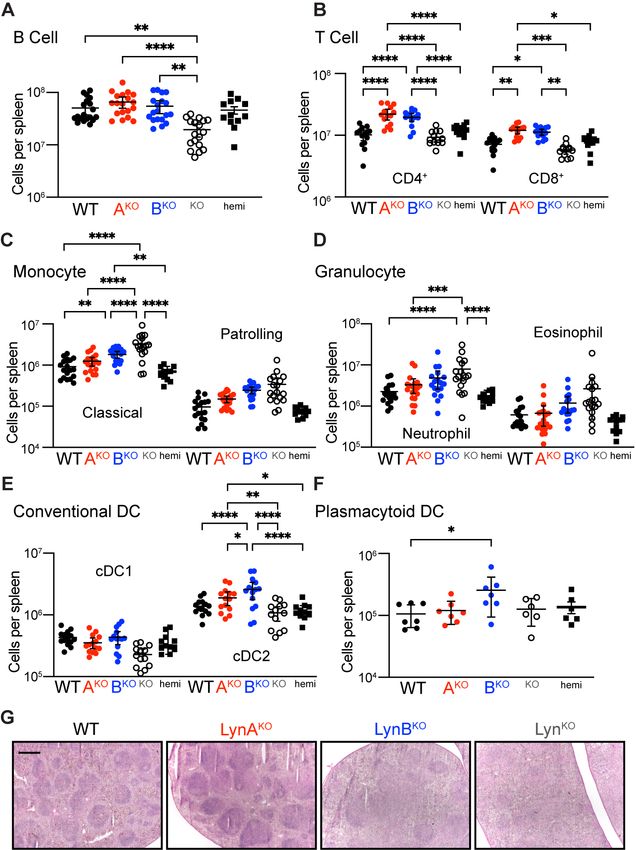
Fig. 1. LynAKO and LynBKO. (A) LynA and LynB splice junctions in WT lyn, highlighting a SnaBI site near the
cryptic LynB splice donor in exon 2. (B) Strategy for LynAKO via double cutting and NHEJ within the LynA in-
sert. (C) Strategy for LynBKO via HDR, ablating the LynB splice donor. (D) Breeding scheme for LynAKO (AKO),
LynBKO (BKO), LynABhemi (hemi), and total LynKO (KO) mice, showing parental CRISPR and Neomycin (Neo) (16)
knockouts. For progeny LynAKOLynB+/+=biallelic LynB expression, LynAKOLynB+/-=monoallelic LynB expression
(isoforms reversed in LynBKO). (E) Immunoblot showing SFK expression in WT/knockout BMDMs; Erk1/2
shows loading. (F) Quantification (17) of LynA and LynB protein; in subsequent figures LynAKOLynB+/- and Lyn-
BKOLynA+/- are referred to as LynAKO and LynBKO, respectively.

For LynBKO, we used a single gRNA and donor oligonucleotide to template homology-directed repair (HDR),
inducing one mutation ablating the LynB splice donor and a second (silent) mutation ablating a SnaBI re-
striction site (Fig. 1C). In LynBKO(CRISPR), ablation of the LynB splice donor caused a V24L substitution in
LynA (Fig. S2). We performed control experiments to confirm that activation and regulation of LynAV24L was
                                                      2
comparable to wild type (WT) LynA (Supplementary Text, Fig. S3A to D). We also generated LynAKOLynB+/+ x
LynBKO LynA+/+ F1 (LynABhemi) mice (Fig. 1D, Fig. S3E), which have WT-like expression levels of LynAV24L and
LynB (Fig. 1E) and are phenotypically indistinguishable from WT (e.g. Fig. S3F). We therefore consider
LynAV24L an acceptable substitute for WT LynA.
LynA and LynB protein were absent as expected in LynAKOLynB+/+ and LynBKOLynA+/+ bone-marrow-derived
macrophages (BMDMs) (Fig. 1D and E), with preserved interferon (IFN)-γ-dependent upregulation (Fig. S4A
and B) (15). Expression of the remaining Lyn isoform, however, was elevated 2.5x; other SFKs were unaf-
fected (Fig. 1E and F). This suggests a feedback mechanism that senses and independently tunes basal LynA
and LynB expression. To achieve WT-like expression of the remaining isoform, we generated LynKO x homozy-
gous LynAKO(CRISPR) or LynBKO(CRISPR) F1 mice (Fig. 1D), which expressed the remaining Lyn isoform
from only one allele, with levels comparable to WT (Fig. 1E and F). These LynAKOLynB+/- and LynBKOLynA+/-
mice are hereafter referred to as LynAKO and LynBKO.
Expression of LynA or LynB alone reversed the B-cell deficiency found in total LynKO mice (Fig. 2A). Surpris-
ingly, expression of either Lyn isoform also increased CD4+ and CD8+ T-cell numbers over WT (Fig. 2B);
LynABhemi was indistinguishable from WT. LynBKO spleens had elevated numbers of classical monocytes and
trended toward increased patrolling monocytes and granulocytes (Fig. 2C and D). Activation of dendritic cells
(DC) drives autoimmune disease in LynKO mice (4, 18-20), but in contrast to monocytes and granulocytes, DC
populations are similar in LynKO and WT mice (19, 21). LynBKO mice, however, had increased conventional-
type-2-DC (cDC2) and plasmacytoid-DC (pDC) numbers (Fig. 2E and F). cDC2 regulate CD4+ T-cell activation
and promote germinal center formation, while pDC secrete cytokines and drive autoimmune disease (22). We
assessed spleen organization and found that WT and LynAKO spleens had well-formed lymphoid follicles,
whereas LynBKO and LynKO had follicular effacement (23) (Fig. 2G). Together, these data suggest that LynB
has a unique negative-regulatory role, restraining DC and monocyte proliferation and maintaining spleen or-
ganization; LynA may therefore have a counterbalancing effect.

                                                      3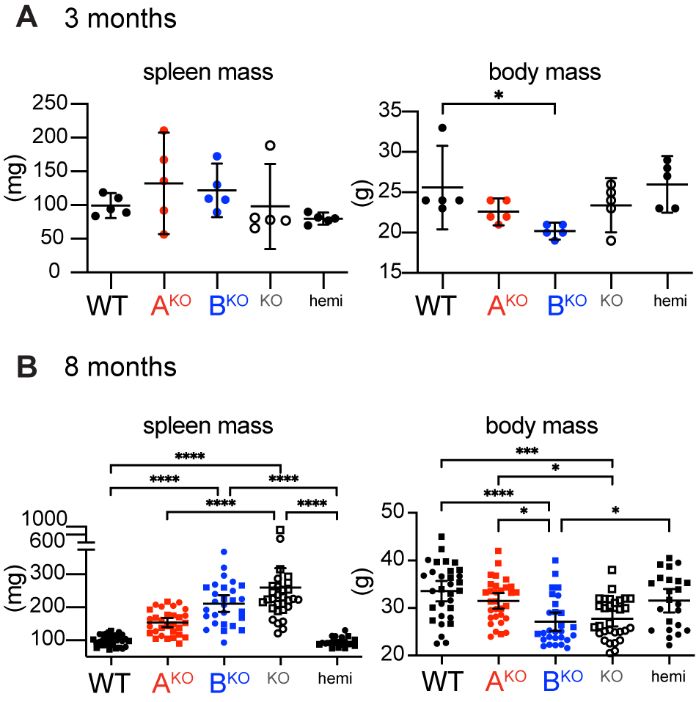
Fig. 2. Spleen cellularity and architecture in aged LynBKO mice. Spleens harvested from 8-month-old WT
and Lyn knockout mice analyzed via flow cytometry or histology. (A to F) Cellularity of recovered splenocytes,
reported as total number per spleen; points describe data from different individuals compiled from 3 independ-
ent cohort analyses. Cell populations include (A) B cell, (B) T cell, (C) monocyte, (D) granulocyte, (E) cDC1,
cDC2, and (F) pDC. Statistical tests and cell markers described in the supplement/methods. (G) Spleen sec-
tions visualized by H&E staining; bar=500 µm.

TLR4 initiates pro-inflammatory signaling in response to lipopolysaccharides (LPS) (24). Functional increases
in TLR4 signaling, due to increased activity (25) or receptor overexpression (26), are risk factors for human
SLE and drivers of lupus in mice (25-27). Lyn can positively or negatively regulate TLR4 signaling in myeloid
cells; in DCs it has a primarily negative-regulatory role (4, 19, 20, 28). Because DCs are key regulators of the
adaptive immune response and drivers of lupus in LynKO mice, we investigated TLR4 signaling in bone-mar-
row-derived dendritic cells (BMDCs) treated with LPS. Like LynKO (4), LynBKO BMDCs were hyperresponsive to
LPS, with increased phosphorylation of IκB kinase (IKK) and Erk1/2 (Fig. 3A); like WT, LynAKO WT BMDCs
were less responsive to LPS. LynB, therefore, carries the TLR4-suppressive function historically attributed to
Lyn. LynBKO similarly increased LPS-induced production of tumor necrosis factor (TNF) in splenocytes (Fig.
3B). Furthermore, TLR4 expression on circulating B and myeloid cells increased significantly in older LynKO
and LynBKO mice (Fig. 3C), consistent with observations in human autoimmune diseases (29).

                                                       4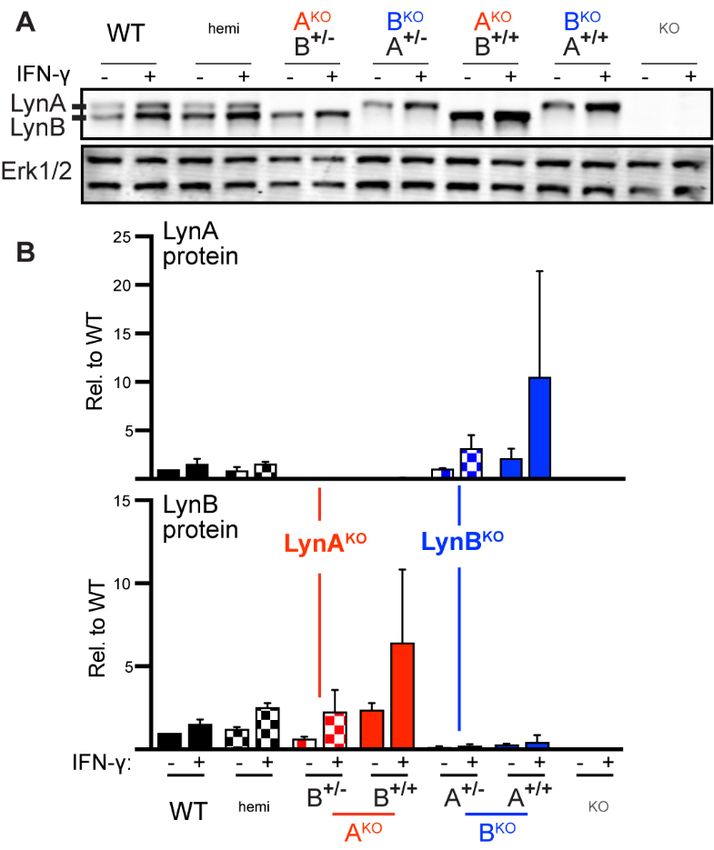
Fig. 3. Loss of LynB promotes TLR4 signaling. (A) Immunoblots showing phosphorylation of IKK and Erk in
response to LPS; β-Actin reflects loading, representative of 3 independent experiments. (B) Geometric Mean
Fluorescence Intensity (gMFI) showing surface expression of TLR4 in circulating classical and patrolling mono-
cytes in 3-month-old mice. (C) Splenocytes from 3-month-old mice treated 4 h with LPS, stained for cell mark-
ers/TNF and analyzed via flow cytometry. Points indicate intracellular-TNFhi monocyte populations in prepara-
tions from different spleens. (C) Blood leukocytes from 8-month-old mice with flow-cytometric quantification of
relative TLR4 surface expression.
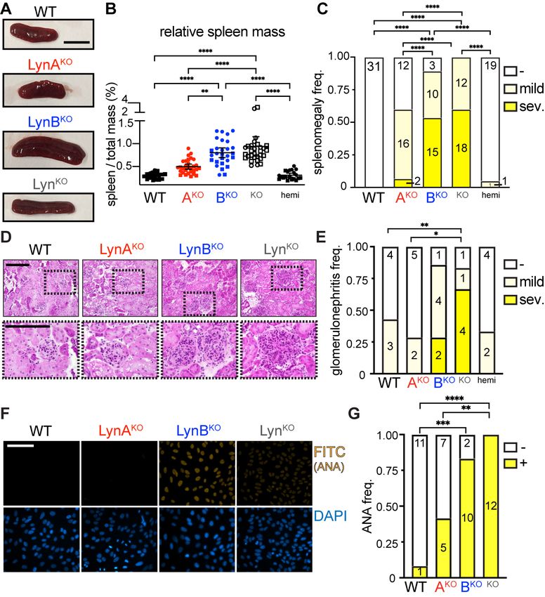
Like LynKO mice (9, 10, 30), LynBKO mice lost body mass and developed splenomegaly (Fig. 4A, Fig. S5A and
B). LynAKO mice trended toward mild splenomegaly, whereas LynABhemi mice were indistinguishable from WT
(Fig. 4B). Most LynKO and LynBKO mice develop severe splenomegaly, in contrast to only 7% of LynAKO mice
(Fig. 4C).
LynKO and LynBKO kidney sections showed severe immune infiltration, expanded glomeruli, and tubular inflam-
mation (Fig.4D and E), in contrast to no/mild inflammation in WT, LynAKO and LynABhemi mice, and most Lyn-
BKO and LynKO mice produced ANA, unlike LynAKO and WT (Fig. 4F and G). No sex-specific differences were
observed (Fig. S6). Together, these data indicate that the lupus-like disease in aging LynKO mice is caused by
the loss of LynB function, suggesting that LynB is the dominant immunosuppressive/protective isoform.
Although it has long been appreciated that Lyn is a critical regulator of immune signaling, dissecting the contri-
butions of the two isoforms, LynA and LynB, has been stymied by the lack of experimental tools. LynB has no
unique sequence, making development of LynB-specific antibodies and RNA silencing reagents infeasible;
LynB production from a cryptic, intra-exon splice site rendered traditional knockout and recombination/excision
approaches intractable due to residual effects on LynA.
5Fig. 4. LynBKO is the dominant driver of autoimmune disease. WT and Lyn knockout mice aged 8 months
and tested for indicators of autoimmune disease. (A) Representative spleens; bar=1 cm. (B) Relative spleen
mass (spleen mass / total mass) of male (square) and female (circle) mice. (C) Frequency of mice with no (-,
spleen mass 0.75%). (D) H&E-stained kid-
ney sections scored for glomerulonephritis by a blinded pathologist; bars=200 µm, bottom enlarged from
boxes. (E) Frequency of glomerulonephritis; numbers are scores from individual mice. (F) Serum probed for
IgG-ANA; bar=100 µm, representative of 3 experiments. (G) Frequency of ANA negativity (-) and positivity (+);
numbers are sera from individual mice.
We pioneered two CRISPR/Cas9 strategies to create splice-fixed LynA- or LynB-only mouse strains: (i) delet-
ing/frameshifting the unique LynA insert (LynAKO) or (ii) ablating the LynB splice donor (LynBKO), both of which
can express the remaining isoform from one allele or two, for physiological or supraphysiological expression.
We found that protein levels of LynA and LynB are coregulated by a feedback mechanism that senses the ba-
sal activity of either isoform. This Lyn-specific feedback, independent of other SFKs, highlights the unique im-
portance of balancing the activating and inhibitory functions of Lyn for proper control of cellular responses and
maintenance of immune homeostasis. While expression of LynA or LynB alone is sufficient to prevent the B-
cell lymphopenia observed in LynKO mice, LynB supplies the primary immunoregulatory activity that protects
against autoimmune disease, with its loss leading to increased TLR4 signaling and both innate- and adaptive-
immune dysregulation. Disease indicators and hyperresponsive signaling in LynBKO mice and cells are compa-
rable to total LynKO, suggesting that LynA and LynB functions are distinct, not additive. This supports a model
in which LynA tunes the sensitivity of cells to pro-inflammatory activation (14, 15) and LynB directs suppressive
signaling. Definition of LynA-specific and LynB-specific signaling pathways and their respective contributions to
immune regulation will inspire new ways of understanding and manipulating these signaling pathways to re-
store immune balance in SLE and other autoimmune patients.
6Materials and Methods
Citation Practices
Please refer to our previous publications (30, 14, 15) for comprehensive referencing of materials, methodolo-
gies, and primary literature.
Generation of LynAKO and LynBKO mice
LynAKO was induced using two gRNAs to delete 77 bp encompassing portions of the unique LynA insert in
mouse lyn exon 2 (5'-GAUCUCUCACAUAAAUAGUU-3') and the following intron 2 (5'-
CCAUGCUCCGAUCCUACUGU-3'). NHEJ then induced a frame shift that resulted in a premature stop codon
after amino-acid residue 77.
LynBKO was induced using one gRNA (5'-GUUCGGUCAGUAUUACGUAC-3') to make a cut near the LynB
splice site in exon 2. A donor oligonucleotide (5'-AAAAGGAAAGACAATCTCAATGACGATGAAGTAGAT-
TCGAAGACTCAACCAcTgCGTAA-
TACTGACCCAACTATTTATGTGAGAGATCCAACGTCCAATAAACAGCAAAGGCCAGTAAG-3') was supplied
to induce two single-nucleotide substitutions via HDR (lower-case letters in the sequence) that ablated the
LynB splice site and a SnaBI restriction site. gRNA’s were designed using CRISPOR.org.
3-week-old C57BL/6J female mice were purchased from The Jackson Laboratory and adapted to University of
Minnesota animal facilities 3-4 days before the first hormone injection. The stud C57BL/6J male mice were ob-
tained from the Jackson Laboratory directly. The recipient females CD-1 mice at 38-49 days old was pur-
chased from the Charles River Laboratory. The animal protocols used for all mutant mice generation were ap-
proved by the Institutional Animal Care and Use Committee (IACUC).
Microinjection of embryos: The C57BL/6J females at 3-4 weeks old were superovulated by intraperitoneal in-
jection of 5 IU pregnant mare serum gonadotropin, followed by injection of 5 IU human chorionic gonadotropin
48 hrs. later (both hormones from National Hormone & Peptide Program, Torrance, CA, USA), and they were
immediately crossed to C57BL/6J stud males. The next day, mouse embryos were obtained from superovu-
lated C57BL/6J females. For LynAKO, the embryos were injected with mixture of 30 ng/ul Cas9 protein, 3.5
ng/ul sgRNA each of 2 gRNAs. For LynBKO, the embryos were injected with mixture of 30 ng/ul Cas9 protein,
3.5 ng/ul of gRNA, and 7 ng/ul of single-stranded oligonucleotide (120bp) donor as the point mutation template.
CRISPR reagents were designed by University of Minnesota Genome Engineering Shared Resources and pur-
chased from IDT. All CRISPR reagents were tested and validated on NIH 3T3 cells before the embryo injec-
tion. And all the CRISPR reagents were resuspended in the injection buffer (10 mM Tris-HCl, 0.25 mM EDTA,
pH 7.4) for the embryo injection
Toe DNA from the resulting pups was isolated using DNeasy Blood & Tissue Kit (Qiagen, Hilden, Germany)
and subjected to and intermediate topo cloning step (Invitrogen, Carlsbad, CA) to insert PCR products into a
plasmid for PCR sequencing (GeneWiz, South Plainfield, NJ) and digestion. A 500 bp segment in wild-type lyn
encompassing the LynB splice site and the LynA unique insert was amplified using the following PCR primers:
Forward 5’-acaaccgagatgtcctgct-3’ Reverse 5’-agccagattatccctaaaatctctaca-3’. SnaBI (New England Biolabs,
Ipswich, MA) cleavage of this product (recognition site TAC/GTA) generated two 250 bp fragments. In LynAKO
a Cas9 double-cut deletion yields a shorter (~423 bp) PCR product that retains sensitivity to SnaBI cleavage.
In LynBKO an HDR-induced mutation in the SnaBI site yields a 500 bp PCR product insensitive to SnaBI.
Other mouse strains and housing
C57BL/6-derived Csk AS mice are hemizygous for the Csk AS BAC transgene on a Csk -/- background, as de-
scribed previously (15). LynKO mice (29) were maintained on a C57BL/6 background. All mice were housed in
specific pathogen-free conditions and genotyped using real-time PCR (Transnetyx, Inc, Memphis, TN). All ani-
mal use complies with University of Minnesota (UMN) and National Institutes of Health (NIH) policy (Animal
Welfare Assurance Number A3456-01). UMN is accredited by AAALAC, experiments involving mice were ap-
proved by the UMN Institutional Animal Care and Use Committee (IACUC, protocol # 1603-33559A). Animals
are kept under supervision of a licensed Doctor of Veterinary Medicine and supporting veterinary staff under
strict NIH guidelines.
7Jurkat cell lines and transfection The Jurkat T-cell strain JCaM1.6 (Lck-deficient) was a gift from the laboratory of Y. Shimizu (University of Min- nesota). Cell lines were authenticated by STR profiling and tested negative for mycoplasma (ATCC, Manas- sas, Virginia). JCaM1.6 was cultured in RPMI-1640 medium supplemented with 5-10% fetal bovine serum (FBS) (Omega Scientific, Inc, Tarzana, CA) and 2 mM glutamine, penicillin and streptomycin (Sigma-Aldrich, St. Louis, MO). JCaM1.6 cells were transiently transfected via electroporation. Briefly, cells were grown over- night in antibiotic-free RPMI-1640 medium supplemented with 10% FBS (Omega Scientific) and 2 mM gluta- mine (RPMI10). Batches of 15 M cells were resuspended in RPMI10 with 10–15 µg plasmid DNA per con- struct. Cells were rested, electroporated at 285 V for 10 ms in a BTX square-wave electroporator (Harvard Ap- paratus, Holliston, MA), resuspended in RPMI10, and allowed to recover overnight. One million live cells were then resuspended in phosphate-buffered saline (PBS), rested for 30 min at 37°C, and stimulated. Preparation of BMDMs Bone marrow was extracted from femura/tibiae of mice. After hypotonic lysis of erythrocytes, BMDMs were de- rived on untreated plastic plates (BD Falcon, Bedford, MA) by culturing in Dulbecco's Modified Eagle Medium (Corning Cellgro, Manassas, VA) containing approximately 10% heat-inactivated FBS (Omega Scientific, Tar- zana, CA), 0.11 mg/ml sodium pyruvate (UCSF Cell Culture Facility), 2 mM penicillin/streptomycin/L-glutamine (Sigma-Aldrich, St. Louis, MO), and 10% CMG-12-14-cell-conditioned medium as a source of M-CSF. After 6 or 7 days, cells were resuspended in enzyme-free ethylenediaminetetraacetic acid (EDTA) buffer and replated in untreated 6-well plates (BD Falcon) at 1 M cells per well in unconditioned medium ±25 U/ml IFN-γ (Pepro- tech, Rocky Hill, NJ) (14, 30). Flow cytometry Spleens were excised from mice and cut into
CD4                        GK1.5              BD Biosciences            BUV395
 CD8a                       53-6.7             BD Biosciences            BUV737

Histology
Kidneys were snap-frozen in optimal cutting temperature compound and 5-μm sections were H&E stained. The
presence and severity of nephritis was evaluated in a blinded fashion (as previously described by one of the
authors. Scoring of glomerulonephritis and interstitial nephritis on a 0-3 scale (0=absent, 1=mild, 2=moderate,
3=severe) for glomeruli was based on glomerular size, glomerular hypercellularity, and presence of glomerular
sclerosis, and for interstitial disease was based on the degree of inflammatory infiltrate and alteration in tissue
architecture.

Anti-nuclear antibody staining
Kallestad HEp-2 (Bio-Rad, Cat. No. 30472) was used to detect Anti-Nuclear antibody staining according to the
manufacturer’s instruction, modified to detect mouse IgG. Briefly, wells were incubated with 40 μL mouse se-
rum, diluted 40x in phosphate-buffered saline + 1% Bovine-serum albumin, at room temperature for 20 min.
After incubation, slides were washed in PBS for 10 minutes and incubated with 30 μL FITC-labeled goat-anti-
mouse IgG (Jackson ImmunoResearch, Cat. No. 115-095-164) diluted 200x and DAPI for 20 min. at room tem-
perature, followed by a 10 min. PBS wash. Slides were then mounted and coverslipped and viewed on an
Olympus BX51 fluorescent microscope equipped with a digital camera and DP-BSW software (Olympus). Im-
ages were quantified, pseudocolored, and merged using ImageJ software (NIH) with the Fiji plugin.

Splenocyte stimulation ex vivo
Spleens were excised from mice and placed in ice-cold RPMI, 2% FBS, 2 mM penicillin and streptomycin.
Spleen tissue was mechanically disrupted using a 100-µm cell strainer (Corning, Corning, NY) on a 50 ml tube,
using a plunger of a 1 ml syringe. During and following processing of the spleen, strainers were flushed with
ice-cold RPMI to acquire all cells. After processing, single-cell suspensions were counted and kept at 4˚C be-
fore proceeding with the in vitro stimulation. Stimulation and staining of cells were performed in 96-wells V-bot-
tom plates (Nunc, Roskilde Denmark), using 2x10⁶ cells per well in 50 μL RPMI. Cells were placed in a 5%
CO2 37 °C incubator for 1 hr prior to treatment. LPS (Invivogen, San Diego, CA) was diluted in RPMI 2-5%
FBS and added by pipetting 50 µl (at 2x concentration) to the wells containing 50 µl of sample. After 30 min,
Brefeldin A and monensin (eBioscience, San Diego, CA) were added. For the last 30 min of stimulation,
Live/Dead stain (Tonbo Biosciences, San Diego, CA) was added. Signaling was quenched at 4 h by adding
paraformaldehyde (PFA) to a final concentration of 1.6% and fixing at room temperature for 15 min in the dark.
Following fixation, cells were washed twice with BD Biosciences Fix/Perm diluent and by centrifuging at 0.4 x g
for 5 min. and dumping supernatant. Cells were then mixed with staining master mix diluted in BD Biosciences
Fix/Perm diluent overnight at 4 °C. Following intracellular staining, cells were washed twice with permeabiliza-
tion buffer and resuspended in PBS, 0.5% PFA. Data was acquired using a BD Fortessa X-30 and analyzed
using Flowjo.

Preparation of BMDCs
Bone marrow was extracted from femura/tibiae of mice. After hypotonic lysis of erythrocytes, BMDCs were de-
rived on 10 cm tissue-culture-treated, plastic plates (Celltreat, Pepperell, MA) in Dulbecco's Modified Eagle
Medium (DMEM) (Corning Cellgro, Manassas, VA) with ~10% heat-inactivated FBS (Omega), 0.11 mg/ml so-
dium pyruvate (UCSF Cell Culture Facility), 5 mM penicillin/streptomycin/L-glutamine (Sigma-Aldrich, St. Louis,
MO), 10 ng/mL murine GM-CSF (PeproTech, Cranbury, NJ), and 10 ng/mL murine IL-4 (PeproTech, Cranbury,
NJ). Media was replaced on day 3 or 4. After 6 or 7 days, cells were resuspended in enzyme-free ethylenedia-
minetetraacetic acid (EDTA) buffer and replated in TC-treated 12-well plates (Celltreat, Pepperell, MA) at 1 M
cells per well in 10%FBS-DMEM.

BMDC stimulation
BMDCs were replated in 12-well tissue-culture-treated plates and rested overnight in a 37°C incubator. LPS
(Invivogen) was added to BMDCs at a final concentration of 100 ng/ml and incubated at 37°C for 30, 60, or 90
min. Samples were collected and prepared for immunoblotting using a standard protocol (30).

Immunblotting
                                                         9
0.025 M cell equivalents were run in each lane of a 7% Nupage Tris-Acetate gel (Invitrogen, Carlsbad, CA) and then transferred to an Immobilon-FL PVDF membrane (EMD Millipore, Burlington, MA). REVERT Total Protein Stain (TPS, LI-COR Biosciences, Lincoln, NE) was used according to the standard protocol to quantify lane loading. After destaining, membranes were treated with Odyssey Blocking Buffer (TBS) for 1 hr. Blotting was performed using standard procedures, and blots were imaged on an Odyssey CLx near-infrared imager (LI- COR). Primary Antibodies: Mouse anti-β-Actin (8H10D10) (Cell Signaling, Cat. #3700, 1:5000), Rabbit anti- pErk1/2 (phosphoThr202/phosphoTyr204, D13.14.4E) (Cell Signaling, Cat. #4370, 1:5000), Rabbit anti-p- IKKα/β (Ser176/180, 16A6) (Cell Signaling, Cat. #2697, 1:1000), Mouse anti-LynA + LynB (Lyn-01) (Abcam, Cat. #ab1890, 1:1000). Secondary Antibodies were either IRDye (680/800) Donkey anti-Mouse IgG or Donkey anti-Rabbit IgG (Li-COR, Biosciences, 1:10000). Cell Markers in Fig. 2 (A) B cell (B220+), (B) CD4+ and CD8+ T cell (B220-NK1.1-TCRβ+), (C) classical monocyte (Ly6G-CD64+MerTK- Ly6C+), patrolling monocyte (Ly6G-CD64+MerTK-Ly6C-), (D) neutrophil (Ly6Ghigh), eosinophil (CD64-CD11c - SSChi), (E) cDC1 (Ly6G-CD64-CD11c +MHCIIhiXCR1+CD11blo), cDC2 (Ly6G-CD64- CD11c +MHCIIhiXCR1loCD11bhi), and (F) pDC (Ly6G-CD64-CD11c +PDCA-1+Ly6C+). Statistical Analyses Fig. 2B- Significance (Sig.) assessed using 1-way ANOVA with Tukey’s multiple comparison test (ANOVA1w - Tukey); error bars reflect 95% confidence intervals (95%CI), ****P
8.     F. Flores-Borja, P. S. Kabouridis, E. C. Jury, D. A. Isenberg, R. A. Mageed, Decreased Lyn expression
       and translocation to lipid raft signaling domains in B lymphocytes from patients with systemic lupus
       erythematosus. Arthritis Rheum 52, 3955-3965 (2005).
9.     M. L. Hibbs et al., Multiple defects in the immune system of Lyn-deficient mice, culminating in
       autoimmune disease. Cell 83, 301-311 (1995).
10.    K. W. Harder et al., Gain- and loss-of-function Lyn mutant mice define a critical inhibitory role for Lyn in
       the myeloid lineage. Immunity 15, 603-615 (2001).
11.    T. L. Yi, J. B. Bolen, J. N. Ihle, Hematopoietic cells express two forms of lyn kinase differing by 21
       amino acids in the amino terminus. Mol Cell Biol 11, 2391-2398 (1991).
12.    D. Alvarez-Errico et al., Functional analysis of Lyn kinase A and B isoforms reveals redundant and
       distinct roles in Fc epsilon RI-dependent mast cell activation. J Immunol 184, 5000-5008 (2010).
13.    G. Tornillo et al., Dual Mechanisms of LYN Kinase Dysregulation Drive Aggressive Behavior in Breast
       Cancer Cells. Cell Rep 25, 3674-3692 e3610 (2018).
14.    B. F. Brian et al., Unique-region phosphorylation targets LynA for rapid degradation, tuning its
       expression and signaling in myeloid cells. eLife 8, e46043 (2019).
15.    T. S. Freedman et al., LynA regulates an inflammation-sensitive signaling checkpoint in macrophages.
       eLife 4, e09183 (2015).
16.    V. W. Chan, F. Meng, P. Soriano, A. L. DeFranco, C. A. Lowell, Characterization of the B lymphocyte
       populations in Lyn-deficient mice and the role of Lyn in signal initiation and down-regulation. Immunity
       7, 69-81 (1997).
17.    B. F. Brian, C. R. Guerrero, T. S. Freedman, Immunopharmacology and Quantitative Analysis of
       Tyrosine Kinase Signaling. Current protocols in immunology / edited by John E. Coligan ... [et al.] 130,
       e104 (2020).
18.    C. L. Chu, C. A. Lowell, The Lyn tyrosine kinase differentially regulates dendritic cell generation and
       maturation. J Immunol 175, 2880-2889 (2005).
19.    C. Lamagna, P. Scapini, J. A. van Ziffle, A. L. DeFranco, C. A. Lowell, Hyperactivated MyD88 signaling
       in dendritic cells, through specific deletion of Lyn kinase, causes severe autoimmunity and
       inflammation. Proc Natl Acad Sci U S A 110, E3311-3320 (2013).
20.    T. Ban et al., Lyn Kinase Suppresses the Transcriptional Activity of IRF5 in the TLR-MyD88 Pathway to
       Restrain the Development of Autoimmunity. Immunity 45, 319-332 (2016).
21.    C. Lamagna, Y. Hu, A. L. DeFranco, C. A. Lowell, B cell-specific loss of Lyn kinase leads to
       autoimmunity. J Immunol 192, 919-928 (2014).
22.    M. Swiecki, M. Colonna, The multifaceted biology of plasmacytoid dendritic cells. Nat Rev Immunol 15,
       471-485 (2015).
23.    S. A. Elmore, Histopathology of the lymph nodes. Toxicol Pathol 34, 425-454 (2006).
24.    K. Takeda, S. Akira, TLR signaling pathways. Semin Immunol 16, 3-9 (2004).
25.    S. A. Summers et al., TLR9 and TLR4 are required for the development of autoimmunity and lupus
       nephritis in pristane nephropathy. J Autoimmun 35, 291-298 (2010).
26.    B. Liu et al., TLR4 up-regulation at protein or gene level is pathogenic for lupus-like autoimmune
       disease. J Immunol 177, 6880-6888 (2006).
27.    C. Richez, P. Blanco, I. Rifkin, J. F. Moreau, T. Schaeverbeke, Role for toll-like receptors in
       autoimmune disease: the example of systemic lupus erythematosus. Joint Bone Spine 78, 124-130
       (2011).
28.    M. Avila, A. Martinez-Juarez, A. Ibarra-Sanchez, C. Gonzalez-Espinosa, Lyn kinase controls TLR4-
       dependent IKK and MAPK activation modulating the activity of TRAF-6/TAK-1 protein complex in mast
       cells. Innate Immun 18, 648-660 (2012).
29.    L. M. Ganley-Leal, Y. Liang, M. Jagannathan-Bogdan, F. A. Farraye, B. S. Nikolajczyk, Differential
       regulation of TLR4 expression in human B cells and monocytes. Mol Immunol 48, 82-88 (2010).
30.    P. Scapini, S. Pereira, H. Zhang, C. A. Lowell, Multiple roles of Lyn kinase in myeloid cell signaling and
       function. Immunol Rev 228, 23-40 (2009).
Acknowledgments: We thank Anindya Bagchi, Yun You, University of Minnesota Mouse Genetics Laboratory
and University of Minnesota Genome Engineering Shared Resource for technical assistance. Many thanks
also to Kaylee Schwertfeger, Michael Farrar, Carol Lange, John Connett and Marc Jenkins for valuable feed-
back and discussion of the project or manuscript.

                                                        11
Funding:
National Institutes of Health grant R01AR073966 (TSF)
National Institutes of Health grant R03AI130978 (TSF)
National Institutes of Health grant T32DA007097 (BFB)
University of Minnesota Center for Immunology New Mouse Award (TSF)
University of Minnesota Foundation Equipment Award E-0918-01 (TSF)
University of Minnesota Center for Autoimmune Diseases Research Pilot Grant (TSF)
Author contributions:
Conceptualization: BFB, TSF
Methodology: BFB, JLA, BLR, BSM, CAL, BAB, TSF
Investigation: BFB, MLS. JLA, MGN, SES, WLS, TSF
Visualization: BFB, JLA, TSF
Funding acquisition: BFB, TSF
Project administration: TSF
Supervision: TSF
Writing – original draft: BFB, TSF
Writing – review & editing: BFB, MLS, JLA, MGN, SES, WLS, BLR, BSM, CAL, BAB, TSF
Competing interests: Authors declare that they have no competing interests.
Data and materials availability: All data are available in the main text or the supplementary materials.
Supplementary Text
To confirm that V24L substitution does not impair LynA, wild-type (WT) LynA- and LynAV24L- initiated signaling
was tested in JCaM1.6 (Jurkat T) cells, which depend on introduced LynA for signal initiation (14). Specific in-
hibition of cotransfected memCsk AS with the small molecule 3-IB-PP1 induces spontaneous SFK/LynA activa-
tion and downstream signaling (14). LynAV24L activation led to Erk phosphorylation (pErk) similarly to WT (Fig.
S3A and B). To confirm that V24L substitution does not impair activation-induced LynA degradation, we in-
duced SFK/LynA activation by treating bone-marrow-derived macrophages (BMDMs) from transgenic Csk AS
(14) Csk ASLynBKO mice with 3-IB-PP1. Rates of degradation of WT Lyn and LynAV24L were indistinguishable,
demonstrating that the V24L substitution does not impair Y32 phosphorylation, interaction with c-Cbl, or other
signal-initiation and regulatory machinery (Fig. S3C and D).
12A. WT mouse lyn (genomic DNA sequence)
5’ region of exon 2 (shared by LynA & LynB)
exon 2 LynB splice donor site (GT) & SnaBI restriction site (TACGTA)2
3’ region of exon 2 (LynA only)
intron
exon 3 (shared)
exon 4
ATGGGATGTATTAAATCAAAAAGGAAAGACAATCTCAATGACGATGAAGTAGATTCGAA-
GACTCAACCA GTACGTA ATACTGACCGAACTATTTATGTGAGAGATCCAACGTCCAATAAACAGCAAAGGCCAgtaagtagactagtctcaggggagaa
atcccacagtaggatcggagcatggctgtgcagcctaaaccccaatcttt-
gcatgccatatatatatacacatatataaattga…ttgctgctgttaggaaaatgatatgcatgcgagaagaaaactatgatcatgacttttgttcctc
taacaaatactcttccatagGTTCCTGAATTTCATCTTTTACCAGGACAGAGAT-
TTCAAACAAAAGgtatgtttcctaaccaacataattcatgttttgtttgtcattgcggaaaaatacacacaacacaagtttatcacagtttcagtcact
gttttcaatgtcaagttctgaacaggaagtaca…ATCCAGAGGAACAAGGTGACATTGTGGTGGCCTTATACCCTTATGATGG-
CATCCACCCAGATGACTTGTCCTTCAAGAAAGGAGAAAAGATGAAAGTTCTAGAAGA…
B. LynAKO(CRISPR) – LynA sequence
DNA notation (connecting mRNA splice junctions)
WT ATGGGATGTATTAAATCAAAAAGGAAAGACAATCTCAATGACGATGAAGTAGATTCGAAGACTCAACCA GTACGTA ATACT
AKO ATGGGATGTATTAAATCAAAAAGGAAAGACAATCTCAATGACGATGAAGTAGATTCGAAGACTCAACCA GTACGTA ATACT
*********************************************************************************
WT GACCGAACTATTTATGTGAGAGATCCAACGTCCAATAAACAGCAAAGGCCAGTTCCTGAATTTCATCTTTTACCAGGACAG…
AKO GACCGAACAGAGAC-------------------------------------GTTCCTGAATTTCATCTTTTACCAGGACAG…
******** ******************************
Translation
WT MGCIKSKRKDNLNDDEVDSKTQP VR NTDRTIYVRDPTSNKQQRPVPEFHLLPGQRFQTKDPEEQGDIVVALYPYDGIHPDD…
AKO MGCIKSKRKDNLNDDEVDSKTQP VR NTDRTETFLNFIFYQDRDFKQKIQRNKVTLWWPYTLMMASTQMTCPSRKEKR-
******************************* 77
1 30
C. LynAKO(CRISPR) – LynB sequence
DNA notation (connecting mRNA splice junctions)
WT ATGGGATGTATTAAATCAAAAAGGAAAGACAATCTCAATGACGATGAAGTAGATTCGAAGACTCAACCAGTTCCTGAATTT…
AKO ATGGGATGTATTAAATCAAAAAGGAAAGACAATCTCAATGACGATGAAGTAGATTCGAAGACTCAACCAGTTCCTGAATTT…
*********************************************************************************
Translation
WT MGCIKSKRKDNLNDDEVDSKTQPVPEFHLLPGQRFQTKDPEEQGDIVVALYPYDGIHPDDLSFKKGEKMKVLE…
AKO MGCIKSKRKDNLNDDEVDSKTQPVPEFHLLPGQRFQTKDPEEQGDIVVALYPYDGIHPDDLSFKKGEKMKVLE…
*************************************************************************
1 73
Fig. S1. Nucleotide and protein sequence generated by LynAKO(CRISPR). (A) Genomic sequence of exon
2 (yellow) and exon 3 (green) of wild-type (WT) mouse lyn, illustrating the cryptic LynB splice site (boxed) and
LynA unique region (cyan). (B) LynAKO (AKO) sequences following CRISPR/Cas9 gene editing, illustrating the
frameshift and premature stop codon. Nucleotide sequences are shown in DNA notation for ease of compari-
son to genomic sequence above. Translated sequences were generated using Expasy Translate Tool
(https://web.expasy.org/translate/). (C) The sequence of LynB is unaffected by these mutations.
13LynAKO(CRISPR) – LynB sequence
(n/a, mRNA splicing cannot occur)
LynAKO(CRISPR) – LynA sequence
DNA notation (connecting mRNA splice junctions)
WT ATGGGATGTATTAAATCAAAAAGGAAAGACAATCTCAATGACGATGAAGTAGATTCGAAGACTCAACCA GTACGTA ATACTG
BKO ATGGGATGTATTAAATCAAAAAGGAAAGACAATCTCAATGACGATGAAGTAGATTCGAAGACTCAACCA CTGCGTA ATACTG
********************************************************************* * **********
WT ACCGAACTATTTATGTGAGAGATCCAACGTCCAATAAACAGCAAAGGCCAGTTCCTGAATTTCATCTTTTACCAGGACAGA…
BKO ACCGAACTATTTATGTGAGAGATCCAACGTCCAATAAACAGCAAAGGCCAGTTCCTGAATTTCATCTTTTACCAGGACAGA…
*********************************************************************************
Translation
WT MGCIKSKRKDNLNDDEVDSKTQP VR NTDRTIYVRDPTSNKQQRPVPEFHLLPGQRFQTKDPEEQGDIVVALYPYDGIHPDD…
BKO MGCIKSKRKDNLNDDEVDSKTQP LR NTDRTIYVRDPTSNKQQRPVPEFHLLPGQRFQTKDPEEQGDIVVALYPYDGIHPDD…
***********************:**********************************************************
1 81
Fig. S2. Nucleotide and protein sequence generated by LynBKO(CRISPR). LynB is not expressed in the
LynBKO due to a splice-site mutation. LynA has a single amino-acid substitution (V24L).
14Fig. S3. Function is preserved in LynAV24L . (A) Representative immunoblot of lysates from JCaM1.6 cells
transiently transfected with cDNAs encoding LynA and memCsk AS and treated with 3-IB-PP1. Phosphorylation
of Erk (pT202/pY204) occurs downstream of LynA (WT and V24L)-initiated signaling, in contrast to kinase-
dead LynAT410K+Y397F ; total Erk reflects protein loading. (B) Quantification of Lyn-dependent Erk phosphorylation
during 3-IB-PP1 treatment, corrected for the initial amount of transfected LynA protein and shown relative to
WT basal. (C) Representative immunoblot of lysates from LynA-expressing Csk AS and LynAV24L-expressing
Csk ASLynBKO BMDMs treated with 3-IB-PP1. Syk Y352 is an SFK substrate, and β-Actin reflects protein load-
ing. (D) Quantification of LynA degradation in Csk AS and Csk ASLynBKO BMDMs. (E) Lyn protein expression in
parental and LynABhemi mice. (F) Protection of LynAV24L-expressing LynABhemi from the mild splenomegaly oc-
curring in LynAKO mice at 8 months.
15Figure S4. Lyn upregulation in response to IFN-γ is preserved in LynAKO and LynBKO BMDMs. (A) Rep-
resentative immunoblot of WT and Lyn knockout BMDMs rested (-) or incubated overnight with low-dose IFN-γ
(+). (B) Quantification of LynA and LynB protein in BMDMs before and after IFN-γ treatment; n=2.
16Figure S5. Splenomegaly and weight loss develop over time in LynBKO mice. Spleen and body mass in mice aged (A) 3 months or (B) 8 months. Sig. ANOVA1-Tukey: ****P
Figure S6. LynBKO increases the incidence of autoimmune disease in male and female mice. Frequency
of autoimmune indicators in male and female mice aged 8 months. Numbers represent individual tests, which
may result in multiple counts from an individual. There were no significant differences between males and fe-
males within any genotype, as assessed via two-sided Fisher's exact test.

                                                     18
You can also read