Significance of Angiotensin II Receptor Blocker Lipophilicities and Their Protective Effect against Vascular Remodeling
←
→
Page content transcription
If your browser does not render page correctly, please read the page content below
593

                                                                                                                   Hypertens Res
                                                                                                                   Vol.28 (2005) No.7
                                                                                                                   p.593-600

Original Article

            Significance of Angiotensin II Receptor Blocker
               Lipophilicities and Their Protective Effect
                     against Vascular Remodeling
         Shinji TAKAI*1, Kazuyoshi KIRIMURA*1,2, Denan JIN*1, Michiko MURAMATSU*1,
                Katsuhiro YOSHIKAWA*1,3, Yoshiki MINO*3, and Mizuo MIYAZAKI*1

          Although the lipophilicities of the various angiotensin II receptor blockers (ARBs) are very different, the rela-
          tionship between lipophilicity and the protective effect against vascular remodeling is unclear. In this study,
          we compared the protective effects of a highly lipophilic ARB, telmisartan, and an ARB with low lipophilicity,
          losartan, on vascular function and oxidative stress in stroke-prone spontaneously hypertensive rats (SHR-
          SP). SHR-SP received oral placebo, 1 mg/kg telmisartan, or 10 mg/kg losartan for 2 weeks. The blood pres-
          sure (BP) in SHR-SP was significantly higher than that in Wistar-Kyoto (WKY) rats before treatment, and the
          BP was reduced equally in telmisartan- and losartan-treated SHR-SP compared to placebo-treated SHR-SP.
          Acetylcholine-induced vasorelaxation in isolated carotid arteries was significantly weaker in SHR-SP than
          in WKY rats, but in both telmisartan- and losartan-treated SHR-SP, acetylcholine-induced vasorelaxation
          was significantly higher than in placebo-treated SHR-SP. Moreover, acetylcholine-induced vasorelaxation in
          telmisartan-treated rats was significantly stronger than in losartan-treated SHR-SP. The expression of the
          endothelial nitric oxide synthase gene was significantly higher in telmisartan- and losartan-treated rats than
          in placebo-treated SHR-SP, and was significantly higher in telmisartan-treated rats than in losartan-treated
          rats. In contrast, the expression of the NAD(P)H oxidase subunit p22phox gene in telmisartan-treated SHR-SP
          was significantly lower than that in losartan-treated SHR-SP. Immunohistochemistry showed that angio-
          tensin II expression in the aorta was significantly lower in telmisartan-treated SHR-SP than in losartan-
          treated SHR-SP. In conclusion, a highly lipophilic ARB, telmisartan, may be useful for preventing NAD(P)H
          oxidase activity, and thereby for conferring vascular protection. (Hypertens Res 2005; 28: 593–600)

          Key Words: angiotensin II receptor blocker, lipophilicity, nitric oxide, NAD(P)H oxidase

                                                                           from acting, two types of agents have been developed: one
                                                                           type prevents angiotensin II formation and includes the ACE
                        Introduction
                                                                           inhibitors and the renin inhibitors; the other type blocks the
Angiotensin II is formed from angiotensin I by angiotensin-                binding of angiotensin II to AT1 receptors, and includes the
converting enzyme (ACE) and non-ACE angiotensin II-form-                   angiotensin II receptor blockers (ARBs). ACE inhibitors were
ing enzymes, such as chymase. Angiotensin I is formed from                 the first among these two types of agents to be developed and
angiotensinogen by renin. Angiotensin II plays an important                widely used in clinical practice. However, ACE inhibitors
role in increasing blood pressure (BP) by stimulating angio-               contribute not only to the inhibition of angiotensin II forma-
tensin II type 1 (AT1) receptors. To prevent angiotensin II                tion but also to bradykinin degradation, and thus are associ-

From the *1Department of Pharmacology, Osaka Medical College, Takatsuki, Japan; *2Environmental Biological Life Science Research Center (BILIS),
Kota, Japan; and *3Department of Environmental Analysis, Osaka University of Pharmaceutical Sciences, Takatsuki, Japan.
Address for Reprints: Shinji Takai, Ph.D., Department of Pharmacology, Osaka Medical College, 2−7 Daigaku-machi, Takatsuki 569−8686, Japan. E-
mail: pha010@art.osaka-med.ac.jp
Received April 6, 2005; Accepted in revised form May 26, 2005.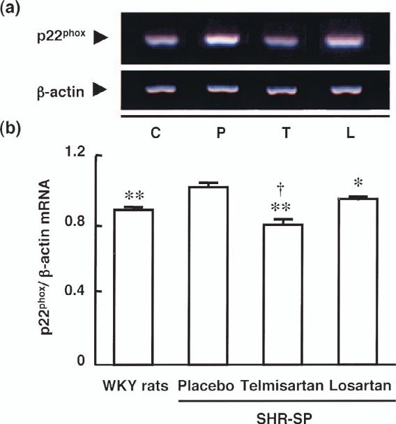
594       Hypertens Res Vol. 28, No. 7 (2005)

ated with side effects, such as cough. On the other hand,           tissue samples were obtained.
ARBs do not affect bradykinin degradation, and therefore
have fewer side effects than ACE inhibitors. Moreover, since
                                                                    Plasma Renin Activity (PRA) and Angiotensin II
ARBs block the effects of angiotensin II, one would also
                                                                    Concentration
expect that they could decrease the risk of coronary artery dis-
ease, cardiac failure, renal dysfunction, and cerebral artery       PRA was determined using an SRL renin kit (TFB Co.,
diseases (1). In fact, studies have shown that ARBs, like ACE       Tokyo, Japan). Angiotensin II concentration was measured
inhibitors, do significantly reduce these risks, and that their     using an enzyme immunoassay kit (Peninsula Laboratories
mechanisms of action may involve the blocking of angio-             Inc., Belmont, USA).
tensin II-related functions, such as by inducing growth factors
and cytokines, in addition to their hypotensive effect.
                                                                    Acetylcholine-Induced Vasorelaxation in Isolated
   Various ARBs have been widely used to treat hypertensive
                                                                    Rat Arteries
patients. Though the ARBs vary in their lipophilicities (2), the
significance of the different lipophilicities of the various        Isolated rat carotid arteries were cut into helical strips, 10 mm
ARBs has hardly been discussed. In contrast, the significance       in length and 1.0 mm in width. The artery strip was placed on
of the different lipophilicities of the various ACE inhibitors      a myograph under a resting tension of 1.0 g. The bathing
has been discussed extensively (3−6). For example, it has           medium was Tyrode’s solution consisting of 137 mmol/l
been clearly shown that ACE inhibitors with high lipophilic-        NaCl, 2.7 mmol/l KCl, 1.8 mmol/l CaCl2, 1.1 mmol/l MgCl2,
ity are more useful for inhibiting tissue ACE activity than         0.42 mmol/l NaH2PO4, 12 mmol/l NaHCO3 and 5.7 mmol/l
ACE inhibitors with low lipophilicity, which resulted in dif-       glucose, pH 7.4. The medium was maintained at 37°C and
ferent antihypertensive and tissue-protective effects (3−6).        was bubbled continuously with 5% CO2 in oxygen. Vasocon-
These reports suggest that different lipophilicities might          striction to 50 mmol/l KCl was first obtained, and then the
result in different protective effects, even when the hypoten-      bathing medium was washed out. Relaxation with acetylcho-
sive effects are the same.                                          line was assessed after contraction to a steady-state tension
   In general, the lipophilicity index is defined as the ratio of   using 1 μmol/l norepinephrine.
octanol to buffer, and the values of log P are: - 2.45 with EXP
3174 (an active metabolite of losartan); - 0.96 with cande-
                                                                    Reverse Transcription (RT)–Polymerase Chain
sartan; - 0.95 with valsartan; +1.48 with irbesartan; and +3.20
                                                                    Reaction (PCR)
with telmisartan (2). Thus, the ARB with the lowest lipophi-
licity is EXP 3174, and the ARB with the highest lipophilicity      The total RNA of the aorta was extracted using Trizol reagent
is telmisartan. In the present study, we compared the effects       (Life Technologies, Rockville, USA) and dissolved in 0.1%
on vascular remodeling in stroke-prone spontaneously hyper-         diethyl pyrocarbonate-treated water. RT to cDNA was
tensive rats (SHR-SP) of an ARB with high lipophilicity             accomplished by analyzing 5 μg of the total RNA sample
(telmisartan, 1 mg/kg per day) and an ARB with low lipophi-         with SuperScript II reverse transcriptase and oligo(dT)12−18
licity (losartan, 10 mg/kg per day) at doses which showed           primer (Invitrogen, Carlsbad, USA). The reaction was carried
equal hypotensive effects based on our preliminary study            out in the presence of first-strand buffer, 1 mmol/l dNTPs and
(data not shown).                                                   20 mol/l dithiothreitol, at 42°C for 50 min. The PCR mixture
                                                                    contained 1 μl of the cDNA reaction mixture, 20 pmol/l prim-
                                                                    ers, PCR buffer, 0.4 mmol/l dNTPs, and 2.5 U Taq poly-
                          Methods
                                                                    merase. The reaction was carried out with a RoboCycler
                                                                    (Stratagene, La Jolla, USA). The sequences of the oligonucle-
Animals
                                                                    otide primers for PCR were as follows: an endothelial nitric
Twelve-week-old male Wistar-Kyoto (WKY) rats (n= 8) and             oxide synthase (eNOS) sense primer, 5′-GGCATCACCAG
SHR-SP (n= 24) were obtained from Japan SLC Inc. (Shi-              GAAGAAGAC-3′, and antisense primer, 5′-CGAACACA
zuoka, Japan). The experimental procedures were in accor-           CAGAACCTGACC-3′, were used for the amplification of
dance with the Guide for the Care and Use of Laboratory             eNOS; a p22phox sense primer, 5′-GCTCATCTGTCTGCTG
Animals (Animal Research Laboratory, Osaka Medical Col-             GAGTA-3′, and antisense primer, 5′-ACGACCTCATCTGT
lege).                                                              CACTGGA-3′, were used for the amplification of p22phox;
  SHR-SP were given placebo (n= 8), 1 mg/kg telmisartan             and a β-actin sense primer, 5′-CCAAGGCCAACCGC
(n= 8), or 10 mg/kg losartan (n= 8) daily for 2 weeks. Systolic     GAGAAGATGAC-3′, and antisense primer, 5′-AGGG
BP (SBP) was monitored by tail-cuff plethysmography (BP-            TACATGGTGGTGCCGCCAGAC-3′, were used for the
98; Softron Co., Tokyo, Japan). After placebo or ARBs were          amplification of β-actin for the calibration of sample loading.
administered for 2 weeks, the body weights of the rats were         The PCR products were separated by electrophoresis on 2%
measured. The animals were anesthetized with 35 mg/kg of            agarose gel stained with ethidium bromide, and the samples
sodium pentobarbital intraperitoneally, and then blood and          were then visualized by ultraviolet transillumination.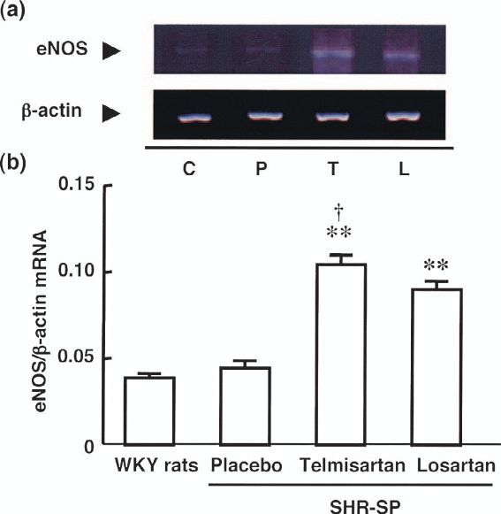
Takai et al: Significance of ARB Lipophilicities    595

Table 1. Effects of Telmisartan and Losartan on Body Weight, Heart Weight and Ratio of Heart Weight to Body Weight
                                                                                                             24 h after last p.o.
             Parameters                            Groups                     Pretreatment
                                                                                                              administration
  Body weight (g)                               WKY rats                        312±2.8**                         348±6.1**
                                                Placebo                         250±3.2                           281±11
                                                Telmisartan                     250±3.1                           285±8.7
                                                Losartan                        247±3.1                           283±10
  Heart weight (g)                              WKY rats                           ⎯                             0.91±0.01**
                                                Placebo                            ⎯                             1.20±0.03
                                                Telmisartan                        ⎯                             1.06±0.08**,#
                                                Losartan                           ⎯                             1.12±0.01**
  Heart weight/body weight (mg/g)               WKY rats                           ⎯                             2.62±0.04**
                                                Placebo                            ⎯                             4.29±0.11
                                                Telmisartan                        ⎯                             3.70±0.03**,#
                                                Losartan                           ⎯                             3.94±0.07**
WKY rats, Wistar-Kyoto rats. **p
596 Hypertens Res Vol. 28, No. 7 (2005)
Fig. 2. PRA and angiotensin II concentration in WKY rats (C), and placebo (P)-, telmisartan (T)-, and losartan (L)-treated
SHR-SP. **p< 0.01 vs. placebo-treated SHR-SP. ††p< 0.01 vs. losartan-treated SHR-SP.
Expressions of eNOS and p22phox
The expression of eNOS in the aortas of SHR-SP is shown in
Fig. 4. The expression of eNOS in the aortas of placebo-
treated SHR-SP and WKY rats was very weak, though the
eNOS expression in WKY rats tended to be lower than that in
SHR-SP. On the other hand, the eNOS expressions in both the
telmisartan- and the losartan-treated SHR-SP were signifi-
cantly higher than those in the placebo-treated SHR-SP and
the WKY rats. However, eNOS expression in the telmisartan-
treated SHR-SP resulted in a significantly greater induction
than that seen in the losartan-treated SHR-SP (Fig. 4).
Fig. 3. Acetylcholine-induced vasorelaxation in noradrena- In contrast, a significant induction of p22phox expression
line-precontracted carotid arteries in WKY rats, and pla- was observed in the placebo-treated SHR-SP rather than in
cebo-, telmisartan-, and losartan-treated SHR-SP. The the WKY rats. However, p22phox expression in both the telm-
results are given as the percentages of the maximal relax- isartan- and the losartan-treated SHR-SP was significantly
ation for papaverine. *p< 0.05 and **p< 0.01 vs. the pla- lower than that in the placebo-treated SHR-SP (Fig. 5). Nev-
cebo-treated SHR-SP. †p< 0.05 vs. losartan-treated SHR-SP. ertheless, p22phox expression in the telmisartan-treated SHR-
SP was significantly lower than that in the losartan-treated
SHR-SP (Fig. 5).
tan- and losartan-treated groups than in the placebo-treated
SHR-SP (Fig. 2). However, at 3 h after the last dose, these
Immunohistochemistry
levels in the telmisartan-treated SHR-SP were significantly
lower than those in the losartan-treated SHR-SP, whereas at Anti-angiotensin II antibody-positive cells in the aortas of
24 h the levels in the telmisartan-treated SHR-SP were higher WKY rats were observed only on the intimal side of the
than those in the losartan-treated SHR-SP (Fig. 2). medial lesions, but these cells in the aortas of placebo-treated
SHR-SP were observed not only on the intimal side but also
over the whole medial lesion, including the adventitial side
Vascular Responses
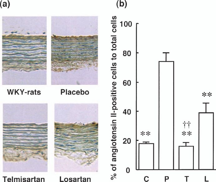
(Fig. 6). The anti-angiotensin II antibody-positive cells in
In all rats, acetylcholine-induced vasorelaxation was both the telmisartan- and the losartan-treated SHR-SP were
observed (Fig. 3). The vasorelaxation in the placebo-treated fewer in number than in the placebo-treated SHR-SP (Fig. 6).
SHR-SP was significantly lower than that in the WKY rats Positive cells in the aortas of the telmisartan-treated SHR-SP,
(Fig. 3). Vasorelaxation in the telmisartan- and the losartan- as in the WKY rats, were observed only on the intimal side,
treated SHR-SP was significantly greater than that in the pla- but those in the losartan-treated SHR-SP, as in the placebo-
cebo-treated SHR-SP. Of note, vasorelaxation in the telmisar- treated SHR-SP, were observed over the whole medial lesion
tan-treated SHR-SP was significantly greater than that in the (Fig. 6). The ratios of anti-angiotensin II antibody-positive
losartan-treated SHR-SP (Fig. 3). cells to total cells in both the telmisartan- and the losartan-
treated SHR-SP were significantly lower than in the placebo-
treated SHR-SP (Fig. 6). This ratio in the telmisartan-treatedTakai et al: Significance of ARB Lipophilicities 597
Fig. 4. (a) Typical photographs of eNOS and β-actin
expressions in aortas obtained from WKY rats (C), and pla- Fig. 6. (a) Typical photographs of anti-angiotensin II anti-
cebo (P)-, telmisartan (T)-, and losartan (L)-treated SHR-SP. body-stained aortas in WKY rats, and placebo-, telmisartan-,
(b) Ratios of eNOS expression to β-actin in aortas obtained and losartan-treated SHR-SP. (b) Ratio of anti-angiotensin II
from WKY rats, and placebo-, telmisartan-, and losartan- antibody-stained cells to total cells in WKY rats (C), and pla-
treated SHR-SP. **p< 0.01 vs. placebo-treated SHR-SP. cebo (P)-, telmisartan (T)-, and losartan (L)-treated SHR-SP.
†
p< 0.05 vs. losartan-treated SHR-SP. **p< 0.01 vs. the placebo-treated SHR-SP. ††p< 0.01 vs. the
losartan-treated SHR-SP.
although the ratio in the losartan-treated SHR-SP was signifi-
cantly lower than that in the placebo-treated SHR-SP, the
ratio in the losartan-treated SHR-SP was significantly higher
than that in the telmisartan-treated SHR or the WKY rats (Fig.
6).
Discussion
In the present study, we evaluated the protective effects
against vascular remodeling of telmisartan, an ARB with high
lipophilicity, and losartan, an ARB with low lipophilicity.
Oxidative stress caused by angiotensin II has been thought to
play an important role in the development and progression of
vascular remodeling, and the expression of the NAD(P)H oxi-
dase subunit p22phox is regarded as a typical marker of oxida-
tive stress (8−11). For example, an increase of p22phox
Fig. 5. (a) Typical photographs of p22phox and β-actin expression reflects an increase in NAD(P)H oxidase activity,
expressions in aortas obtained from WKY rats (C), and pla- which is well known to be closely related to oxidative stress
cebo (P)-, telmisartan (T)-, and losartan (L)-treated SHR-SP. caused by angiotensin II in the vascular tissue of hypertensive
(b) Ratios of p22phox expression to β-actin in aortas obtained rats (8, 9). In the aortas of SHR-SP, p22phox expression was
from WKY rats, and placebo-, telmisartan-, and losartan- significantly higher than in the aortas of WKY rats in our
treated SHR-SP. *p< 0.05 and **p< 0.01 vs. the placebo- study, as has also been previously reported (10). Brosnan et
treated SHR-SP. †p< 0.05 vs. losartan-treated SHR-SP. al. (11) demonstrated that the expression of p22phox in the aor-
tas of irbesartan-treated SHR-SP was significantly lower than
in vehicle SHR-SP. We also observed a significant reduction
SHR-SP was the same as that in the WKY rats, and the ratio of p22phox expression in the aortas of telmisartan- and losar-
in the telmisartan-treated SHR-SP was significantly lower tan-treated SHR-SP compared with placebo-treated SHR-SP.
than that in the placebo-treated SHR-SP. On the other hand, However, a highly lipophilic ARB, telmisartan, significantly598 Hypertens Res Vol. 28, No. 7 (2005) suppressed the expression of p22phox compared to an ARB In the present study, acetylcholine-induced vasorelaxation, with low lipophilicity, losartan, despite the two ARBs exhib- which is used as an index of endothelial function, was signif- iting the same degree of BP lowering. One possible explana- icantly lower in SHR-SP than in WKY rats, as has also been tion for the present finding that p22phox expression was lower previously reported (22). Endothelial dysfunction is directly in the telmisartan-treated SHR-SP may be the different lipo- affected by nitric oxide (NO) bioavailability, and its dysfunc- philicities of the two ARBs. NAD(P)H oxidase activity, eval- tion is associated not only with hypertension but also with uated using isolated mononuclear leukocytes in vitro, was vascular remodeling. It is thought that endothelial dysfunc- found to be significantly reduced by treatment with a more tion is induced in hypertension by the reduction of eNOS highly lipophilic ACE inhibitor, such as quinapril, but not expression and an increase of oxidative stress in the vascula- with ACE inhiitors having lower lipophilicity, such as enala- ture. However, the reduction of NO bioavailability has been pril and lisinopril (12). Similarly, more highly lipophilic related to increased oxidative stress rather than to decreased angiotensin II-blocking agents may be able to more easily eNOS expression. In the present study, we did not observe a penetrate into the tissues or the cells. This would result in a significant difference in eNOS expression between SHR-SP greater reduction of p22phox in the vascular tissues by telmi- and WKY rats, although eNOS expression in SHR-SP tended sartan treatment than by losartan treatment. to be higher than that in WKY rats. Although a relative defi- In clinical studies, ARBs were expected to prevent cardio- ciency in NO has been associated with hypertension, data as vascular events such as myocardial infarction in addition to to the regulation of eNOS expression in SHR-SP are not con- suppressing BP in hypertensive patients. In fact, in the Evalu- sistent, in that decreased, increased, and unchanged eNOS ation of Losartan in the Elderly (ELITE) II trial, the Valsartan expression have been reported (23−26). Although the under- Heart Failure Trial (Val-HeFT), and the VALsartan In Acute lying causes of these discrepancies are unknown, it is possible myocardial iNfarcTion (VALIANT) trial, ARBs were shown that they may be related to the use of different age groups of to confer a level of protection against cardiovascular events SHR-SP in these experiments. For example, in SHR-SP, almost equal to that of ACE inhibitors, which have shown baseline eNOS expression in 8- to 12-week-old rats was more cardiovascular protection than any other antihyperten- increased, that in 12- to 16-week-old rats was unchanged, and sive agents in clinical studies (13−15). On the other hand, in that in 31-week-old rats was decreased (23−25). In spontane- the Valsartan Antihypertensive Long-term Use Evaluation ously hypertensive rats (SHR), it has been reported that eNOS (VALUE) trial, the BP-lowering effect of amlodipine was expression in younger age groups was increased, while eNOS greater than that of valsartan, and amlodipine was also found expression was decreased in older age groups (27, 28). to be superior to valsartan in preventing cardiovascular events Together with the present results, these findings indicate that (16). This would suggest that a strong hypotensive effect may endothelial dysfunction may not be directly affected by eNOS also be an important factor for preventing cardiovascular expression. We also observed that eNOS expression in SHR- events. However, not only the degree of the hypotensive SP was significantly increased with telmisartan and losartan effect, but also its duration may be involved in preventing car- treatment compared to placebo treatment, but that the degree diovascular events, and the different half-lives of valsartan (9 of increase was significantly greater with telmisartan treat- h) and amlodipine (over 35 h) may play a role (17, 18). Simi- ment than with losartan treatment. Although there was a larly, in the present study, the two ARBs not only had differ- greater increase in eNOS expression with telmisartan treat- ent lipophilicities (i.e., telmisartan has high lipophilicity and ment than with losartan treatment, this may have been partly losartan has low lipophilicity) but they also had very different due to a greater improvement of endothelial function with durations of action. The half-life of telmisartan is approxi- telmisartan, which would have limited the contribution of mately 24 h, while that of losartan is 2.1 h and that of eNOS expression. EXP3147 (an active metabolite of losartan) is approximately On the other hand, in the present study, the expression of 6.3 h (19, 20). However, in the present study, we used losar- p22phox was significantly higher with telmisartan treatment tan at 10 times the dose of telmisartan, which resulted in both than with losartan treatment. Hamilton et al. (10) have drugs having equal hypotensive effects throughout the exper- reported that treatment with inhibitors of NAD(P)H oxidase iment. Therefore, the difference noted between telmisartan improved endothelial function of isolated blood vessels not and losartan with respect to the preventive effects of vascular only in SHR-SP but also in humans. NAD(P)H oxidase is damage in this study was not due to any hypotensive effects. known to be activated by mechanical forces and angiotensin Jinno et al. (21) reported that treatment with 0.5 mg/kg or 3 II (29, 30). In human vascular smooth muscle cells (VSMCs), mg/kg of olmesartan did not affect the BP in normotensive the binding of angiotensin II to AT1 receptors leads to a phos- mice, but that treatment with 3 mg/kg olmesartan, but not 0.3 phorylation of p47phox, initiating the translocation of this sub- mg/kg olmesartan, significantly reduced p22phox expression. unit to the cell membrane and assembly of the enzyme Therefore, this would suggest that the different degree of complex (31). Angiotensin II also increases the expression of p22phox expression inhibition between telmisartan and losartan all NAD(P)H oxidase subunits, including p22phox, which was found in the present study was likely independent of BP evaluated in the present study (32). Previously, we reported reduction. that ACE activity in the aorta was significantly higher in
Takai et al: Significance of ARB Lipophilicities   599

SHR-SP than in WKY rats, although plasma ACE activity                differences in lipophilicity among ARBs may also be
was lower in SHR-SP (33). In genetically hypertensive mod-           involved in the different levels of protection against cardiac
els, such as SHR and SHR-SP, expressions of ACE and                  hypertrophy conferred by these agents.
angiotensin II concentration in the vasculature are upregu-            In conclusion, ARBs with different lipophilicities show dif-
lated and angiotensin II action against blood vessels is             ferent protective effects against vascular remodeling in SHR-
strengthened (34, 35). In the present study, we observed a sig-      SP. Although it is accepted that all ARBs show antihyperten-
nificant increase of anti-angiotensin II antibody-positive cells     sive effects and vascular protective effects, different ARBs
in placebo-treated SHR-SP compared with WKY rats, sug-               may exhibit different vascular protective effects in various
gesting an increase of angiotensin II formation in SHR-SP.           experimental models and clinical studies. Therefore, further
On the other hand, both telmisartan and losartan treatments          studies are needed to assess the significance of lipophilicity
resulted in a reduction of anti-angiotensin II antibody-posi-        among ARBs.
tive cells. This finding suggests that both telmisartan and
losartan block angiotensin II binding to AT1 receptors, and                                    References
that this results in a decrease of angiotensin II effects, such as
NAD(P)H oxidase expression in the vasculature. Moreover,             1.    Unger T, Culman J, Gohlke P: Angiotensin II receptor
                                                                           blockade and end-organ protection: pharmacological ratio-
the fact that telmisartan blocks the binding of angiotensin II to
                                                                           nale and evidence. J Hypertens Suppl 1998; 16: S3−S9.
vascular AT1 receptors more strongly than does losartan              2.    Wienen W, Entzeroth M, van Meel JC, et al: A review on
could result in the lower p22phox expression by telmisartan,               telmisartan: a novel, long-acting angiotensin II-receptor
and this difference between the two ARBs may have been                     antagonist. Cardiovasc Drug Rev 2000; 18: 127−156.
involved in the finding that improvement of endothelial func-        3.    Unger T, Ganten D, Lang RE, Scholkens BA: Persistent
tion by telmisartan treatment was significantly greater than               tissue converting enzymeinhibition following chronic treat-
that by losartan treatment.                                                ment with Hoe498 and MK421 in spontaneously hyperten-
   Cardiac hypertrophy was observed in all SHR-SP, but the                 sive rats. J Cardiovasc Pharmacol 1985; 7: 36−41.
hypertrophy in the telmisartan-treated SHR-SP, but not in the        4.    Miyazaki M, Kawamoto T, Okunishi H: Vascular affinity of
losartan-treated SHR-SP, was significantly lower than in the               trandolapril. Am J Hypertens 1995; 8: 63S−67S.
placebo-treated SHR-SP. Our findings agree with Wagner et            5.    Pilote L, Abrahamowicz M, Rodrigues E, Eisenberg MJ,
                                                                           Rahme E: Mortality rates inelderly patients who take dif-
al. (36), who reported that telmisartan, but not losartan,
                                                                           ferent angiotensin-converting enzyme inhibitors after acute
reduced cardiac hypertrophy, despite the similar BP reduction              myocardial infarction: a class effect? Ann Intern Med 2004;
between the two drugs. Therefore, since the hypotensive                    141: 102−112.
effects seen with telmisartan and losartan are similar, they         6.    Takai S, Jin D, Sakaguchi M, Miyazaki M: Significant tar-
cannot account for the differences seen in the reduction of                get organs for hypertension and cardiac hypertrophy by
cardiac hypertrophy. Although we have not studied the mech-                angiotensin-converting enzyme inhibitors. Hypertens Res
anism of cardiac hypertrophy reduction, it is possible that the            2004; 27: 213−219.
higher lipophilicity of telmisartan may be a contributing fac-       7.    Jin D, Ueda H, Takai S, et al: Effect of chymase inhibition
tor for reducing cardiac hypertrophy. In the present study,                on the arteriovenous fistula stenosis in dog. J Am Soc Neph-
both the PRA and angiotensin II concentration were signifi-                rol 2005; 16: 1024−1034.
cantly higher in the losartan-treated group than in the telmi-       8.    Fukui T, Ishizaka N, Rajagopalan S, et al: p22phox mRNA
                                                                           expression and NADPH oxidase activity are increased in
sartan-treated group at 3 h after the last doses, but they were
                                                                           aortas from hypertensive rats. Circ Res 1997; 80: 45−51.
significantly higher in the telmisartan-treated group than in        9.    Ulker S, McMaster D, McKeown PP, Bayraktutan U:
the losartan-treated group at 24 h after the last doses. In gen-           Impaired activities of antioxidant enzymes elicit endothelial
eral, increases of PRA and angiotensin II concentration after              dysfunction in spontaneous hypertensive rats despite
ARB treatment were observed, and were reflected in the tis-                enhanced vascular nitric oxide generation. Cardiovasc Res
sue angiotensin II blockade. Therefore, the findings of the                2003; 59: 488−500.
present study may be dependent on the longer duration of tis-        10.   Hamilton CA, Brosnan MJ, Al-Benna S, Berg G, Dominic-
sue angiotensin II blockade caused by telmisartan than by                  zak AF: NAD(P)H oxidase inhibition improves endothelial
losartan. In SHR, there was a significant positive correlation             function in rat and human blood vessels. Hypertension
between left ventricular weight and tissue angiotensin II con-             2002; 40: 755−762.
centration, which is related to lipophilicity (37). We recently      11.   Brosnan MJ, Hamilton CA, Graham D, Lygate CA, Jardine
                                                                           E, Dominiczak AF: Irbesartan lowers superoxide levels and
reported that an ACE inhibitor with high lipophilicity, tran-
                                                                           increases nitric oxide bioavailability in blood vessels from
dolapril, but not an ACE inhibitor with low lipophilicity,                 spontaneously hypertensive stroke-prone rats. J Hypertens
enalapril (both of which produced the same hypotensive                     2002; 20: 281−286.
effect), reduced cardiac hypertrophy (6). Izumi et al. (38)          12.   van der Giet M, Erinola M, Zidek W, Tepel M: Captopril
reported that an increase of mitogen-activated protein                     and quinapril reduce reactive oxygen species. Eur J Clin
kinases, which are activated by AT1 receptor stimulation,                  Invest 2002; 32: 732−737.
contributed to cardiac hypertrophy in SHR-SP. Therefore, the         13.   Pitt B, Poole-Wilson PA, Segal R, et al: Effect of losartan
600 Hypertens Res Vol. 28, No. 7 (2005)
compared with captopril on mortality in patients with symp- nist reduces oxidative stress by upregulating Cu/Zn super-
tomatic heart failure: randomised trial⎯the Losartan Heart oxide dismutase in stroke-prone spontaneously
Failure Survival Study ELITE II. Lancet 2000; 355: 1582− hypertensive rats. Hypertens Res 2004; 27: 877−885.
1587. 27. Vaziri ND, Ni Z, Oveisi F: Upregulation of renal and vascu-
14. Cohn JN, Tognoni G: A randomized trial of the angio- lar nitric oxide synthase in young spontaneously hyperten-
tensin-receptor blocker valsartan in chronic heart failure. N sive rats. Hypertension 1998; 31: 1248−1254.
Engl J Med 2001; 345: 1667−1675. 28. Crabos M, Coste P, Paccalin M, et al: Reduced basal NO-
15. Pfeffer MA, McMurray JJ, Velazquez EJ, et al: Valsartan, mediated dilation and decreased endothelial NO-synthase
captopril, or both in myocardial infarction complicated by expression in coronary vessels of spontaneously hyperten-
heart failure, left ventricular dysfunction, or both. N Engl J sive rats. J Mol Cell Cardiol 1997; 29: 55−65.
Med 2003; 349: 1893−1906. 29. Seshiah PN, Weber DS, Rocic P, Valppu L, Taniyama Y,
16. Julius S, Kjeldsen SE, Weber M, et al: Outcomes in hyper- Griendling KK: Angiotensin II stimulation of NAD(P)H
tensive patients at high cardiovascular risk treated with reg- oxidase activity: upstream mediators. Circ Res 2002; 91:
imens based on valsartan or amlodipine: the VALUE 406−413.
randomised trial. Lancet 2004; 363: 2022−2031. 30. Ying CJ, Xu JW, Ikeda K, Takahashi K, Nara Y, Yamori Y:
17. McInnes GT: Angiotensin II antagonism in clinical prac- Tea polyphenols regulate nicotinamide adenine dinucle-
tice: experience with valsartan. J Cardiovasc Pharmacol otide phosphate oxidase subunit expression and ameliorate
1999; 33 (Suppl 1): S29−S32. angiotensin II-induced hyperpermeability in endothelial
18. Stopher DA, Beresford AP, Macrae PV, Humphrey MJ: cells. Hypertens Res 2003; 26: 823−828.
The metabolism and pharmacokinetics of amlodipine in 31. Touyz RM, Yao G, Schiffrin EL: c-Src induces phosphory-
humans and animals. J Cardiovasc Pharmacol 1988; 12 lation and translocation of p47phox: role in superoxide gener-
(Suppl 7): S55−S59. ation by angiotensin II in human vascular smooth muscle
19. Burnier M, Maillard M: The comparative pharmacology of cells. Arterioscler Thromb Vasc Biol 2003; 23: 981−987.
angiotensin II receptor antagonists. Blood Press 2001; 1 32. Touyz RM, Chen X, Tabet F, et al: Expression of a
(Suppl): 6−11. functionally active gp91phox-containing neutrophil-type
20. Lo MW, Goldberg MR, McCrea JB, Lu H, Furtek CI, NAD(P)H oxidase in smooth muscle cells from human
Bjornsson TD: Pharmacokinetics of losartan, an angiotensin resistance arteries: regulation by angiotensin II. Circ Res
II receptor antagonist, and its active metabolite EXP3174 in 2002; 90: 1205−1213.
humans. Clin Pharmacol Ther 1995; 58: 641−649. 33. Takai S, Sakonjo H, Miyazaki M: Beneficial effect of tran-
21. Jinno T, Iwai M, Li Z, et al: Calcium channel blocker dolapril on the lifespan of a severe hypertensive model.
azelnidipine enhances vascular protective effects of AT1 Hypertens Res 2001; 24: 559−564.
receptor blocker olmesartan. Hypertension 2004; 43: 263− 34. Morishita R, Higaki J, Nakamura F, et al: Regression of
269. hypertension-induced vascular hypertrophy by an ACE
22. Hashimoto Y, Kurosawa Y, Minami K, Fushimi K, Narita inhibitor and calcium antagonist in the spontaneously
H: A novel angiotensin II-receptor antagonist, 606A, hypertensive rat. Blood Press Suppl 1992; 3: 41−47.
induces regression of cardiac hypertrophy, augments endo- 35. Fernandez-Alfonso MS, Kreutz R, Zeh K, Liu Y, Ganten D,
thelium-dependent relaxation and improves renal function Paul M: Differential regulation of vascular angiotensin I-
in stroke-prone spontaneously hypertensive rats. Jpn J converting enzyme in hypertension. Hypertension 1994; 24:
Pharmacol 1998; 76: 185−192. 280−286.
23. Kerr S, Brosnan MJ, McIntyre M, Reid JL, Dominiczak AF, 36. Wagner J, Drab M, Bohlender J, Amann K, Wienen W,
Hamilton CA: Superoxide anion production is increased in Ganten D: Effects of AT1 receptor blockade on blood pres-
a model of genetic hypertension: role of the endothelium. sure and the renin-angiotensin system in spontaneously
Hypertension 1999; 33: 1353−1358. hypertensive rats of the stroke prone strain. Clin Exp Hyper-
24. Kimoto-Kinoshita S, Nishida S, Tomura TT: Decrease of tens 1998; 20: 205−221.
endothelial nitric oxide synthase in stroke-prone spontane- 37. Mizuno K, Niimura S, Katoh K, Fukuchi S: TCV-116, a
ously hypertensive rat cerebral cortex. Neurosci Lett 2000; newly developed angiotensin II receptor antagonist, induces
288: 103−106. regression of cardiac hypertrophy through suppression of
25. Tanabe A, Naruse M, Seki T, et al: Gene expression of the tissue renin-angiotensin system in spontaneously hyper-
endothelin-1 and endothelial-type nitric oxide synthase in tensive rats. Life Sci 1994; 54: 1987−1994.
cardiovascular tissues of stroke-prone spontaneously hyper- 38. Izumi Y, Kim S, Murakami T, Yamanaka S, Iwao H: Car-
tensive rats/Izm: effects of the angiotensin-converting diac mitogen-activated protein kinase activities are chroni-
enzyme inhibitor aracepril. J Cardiovasc Pharmacol 1998; cally increased in stroke-prone hypertensive rats.
31 (Suppl 1): S395−S398. Hypertension 1998; 31: 50−56.
26. Umemoto S, Tanaka M, Kawahara S, et al: Calcium antago-You can also read


