SHARE @ Children's Mercy - Children's Mercy Kansas City
←
→
Page content transcription
If your browser does not render page correctly, please read the page content below
Children's Mercy Kansas City
SHARE @ Children's Mercy

Manuscripts, Articles, Book Chapters and Other Papers

8-2021

Re-tubularization of highly-ischemic anti-mesenteric border
(ReHAB)
Joseph Lopez
Children's Mercy Hospital

Richard J. Hendrickson
Children's Mercy Hospital

Follow this and additional works at: https://scholarlyexchange.childrensmercy.org/papers

Recommended Citation
Lopez, JJ and Hendrickson, RJ. Re-tubularization of highly-ischemic anti-mesenteric border (ReHAB).
Journal of Pediatric Surgery Case Reports. 2021; 71. doi:10.1016/j.epsc.2021.101882.

This Article is brought to you for free and open access by SHARE @ Children's Mercy. It has been accepted for
inclusion in Manuscripts, Articles, Book Chapters and Other Papers by an authorized administrator of SHARE @
Children's Mercy. For more information, please contact library@cmh.edu.
Journal of Pediatric Surgery Case Reports 71 (2021) 101882

                                                                      Contents lists available at ScienceDirect

                                                       Journal of Pediatric Surgery Case Reports

Re-tubularization of highly-ischemic anti-mesenteric border (ReHAB)
Joseph J. Lopez, Richard J. Hendrickson *
Children’s Mercy Hospital, Department of Pediatric Surgery, Kansas City, Missouri, USA

ARTICLE INFO                                                ABSTRACT

Keywords:                                                   Complex gastroschisis and other etiologies of intestinal compromise present a difficult medical and surgical chal-
Ischemic bowel                                              lenge in the neonatal and pediatric population. Aggressive resection of intestinal length in these cases can lead to
Short bowel syndrome                                        significant short bowel syndrome and prolonged or lifelong dependence on parenteral nutrition. This method of
Complex gastroschisis
                                                            bowel preservation previously described has led to additional patients achieving enteral autonomy and we pre-
Bowel preservation
                                                            sent their cases and most recent outcomes.

1. Introduction                                                                              suscitation, he developed worsening respiratory status and abdominal
                                                                                             distention. A CT scan of his abdomen/pelvis demonstrated diffuse
    In neonatal and pediatric patients, enteral nutrition is vital to the                    pneumatosis and portal venous gas. As such, operative intervention was
growth and development. This population of patients is vulnerable to                         indicated. A midline laparotomy incision was made and there was an
congenital anomalies and physiologic insults which can compromise in-                        immediate return of dark serous fluid that was foul smelling yet no suc-
testinal function and integrity. One such anomaly, gastroschisis, is the                     cus encountered. From the jejunum to the ascending colon, there were
most common abdominal wall defect seen across the world [1]. These                           signs of an inflammatory process as the bowel appeared purple with no
cases are often managed by placement of extruded abdominal viscera in                        signs of necrosis. There was no perforation. Due to the unstable nature
silos and slowly reducing until abdominal domain is achieved. Complex                        of the patient, a silo (Specialty Surgical Products, Inc, Montana, US)
cases involving long segments of compromised bowel warrant salvage                           was placed to keep the abdomen open as well as to visualize the bowel.
techniques to prevent the development of short bowel syndrome and in-                        In 48 hours, a second-look operation was performed. There were many
testinal failure. Many complications related to long-term parenteral nu-                     areas of poor perfusion and some patchy areas of frank necrosis, al-
trition are described in the literature. In addition, the financial cost of                  though no perforation was seen. The stomach, duodenum, and proxi-
home parenteral nutrition has been described to be very steep for fami-                      mal jejunum appeared healthy. The sigmoid and rectum was identified
lies [2,3].                                                                                  and appeared healthy as well. The left colon did not show any areas of
    A previously described technique involving resection of signifi-                         ischemia, but the mid-transverse and right colon appeared frankly
cantly ischemic anti-mesenteric border of intestine followed by re-                          necrotic. Patchy anti-mesenteric necrosis of jejunum was resected and
tubularization, also known as ReHAB, has provided some benefit of                            reapproximated. Frank necrosis of 90 cm of jejunum was resected.
these patients that would otherwise develop intestinal failure and short                     Patchy ischemia of the ileum was left in situ. The resultant anatomy
bowel syndrome [4]. This method is based on the previously described                         was: from the ligament of Treitz, there was 40 cm of jejunum that was
techniques of intestinal lengthening to improve intestinal function in                       stapled off distally, an island of 5 cm of jejunum stapled off on both
these patients. In these patients, salvage of native bowel provides pa-                      ends, a 15 cm segment of ileum that was stapled off on both ends, and
tients with more physiologically active length as well as improved like-                     finally transverse colon present down to the rectum. A silo was then
lihood of enteral autonomy [5–7].                                                            placed. Upon operative exploration the next day, proximal jejunum ap-
                                                                                             peared viable but had patchy necrosis of the anti-mesenteric surface.
1.1. Case one                                                                                The distal jejunum appeared frankly necrotic. A resection of 9 cm of je-
                                                                                             junum was performed using the GIA stapler (Covidien Surgical, Mans-
   An 11-month-old male presented with 32% TBSA severe submersion                            field, MA, US). The areas of anti-mesenteric necrosis were excised and
burns to his extremities. Within the first 48 hours of admission and re-                     the bowel was reapproximated with running 5–0 PDS suture. This was

 * Corresponding author. Pediatric Transplantation Department of Pediatric Surgery Children's Mercy Hospital - Kansas City 2401 Gillham Road, Kansas City, MO

64108, USA.
   E-mail address: rjhendrickson@cmh.edu (R.J. Hendrickson).

https://doi.org/10.1016/j.epsc.2021.101882
Received 22 March 2021; Received in revised form 21 April 2021; Accepted 21 April 2021
Available online 8 May 2021
2213-5766/© 2021 The Authors.             Published by Elsevier Inc.          This is                  an   open    access   article   under   the   CC   BY-NC-ND     license
(http://creativecommons.org/licenses/by-nc-nd/4.0/).
J.J. Lopez and R.J. Hendrickson Journal of Pediatric Surgery Case Reports 71 (2021) 101882
performed for a 20 cm segment (Fig. 1). A jejunostomy and mucous fis-
tula were created, a gastrostomy tube was placed for enteral access, and
the abdomen was closed. Over the subsequent weeks, parenteral nutri-
tion was required, and trophic tube feeds were tolerated. The decision
was made to take the patient back to the operating room for stoma re-
versal. A 3 cm segment of bowel that had undergone the previous Re-
HAB procedure was noted to be severely stenosed. This segment was re-
sected, and stoma takedown was performed as planned. At the conclu-
sion, 25 cm of small bowel and the transverse colon remained. In the
subsequent 5 months, transition to partial parenteral nutrition as well
as enteral feeds was made, and the patient was discharged home. He
did return later in the month with an intravenous line malfunction and
infection, which was treated successfully with antibiotics, and was dis-
charged home with parenteral nutrition as well as enteral tube feeds via
the gastrostomy tube. He is being followed by the gastroenterology and
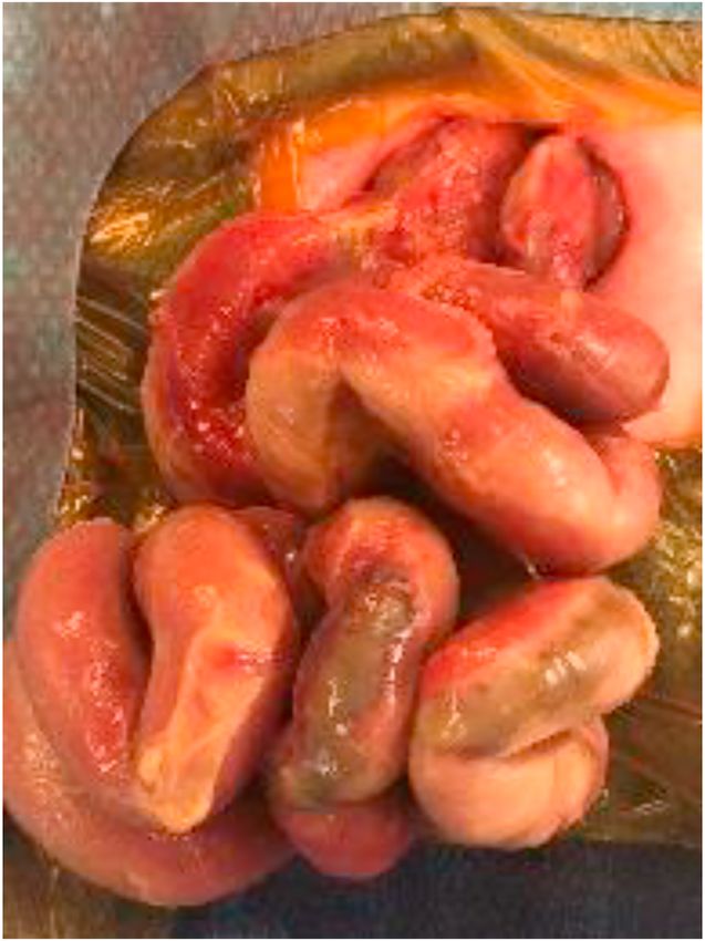
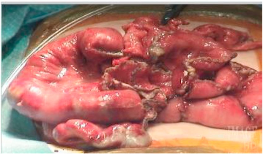
Fig. 2. Small bowel after resection of highly-ischemic mesenteric border with
nutrition teams as an outpatient. At the most recent follow-up appoint-
re-tubularization in a patient that presented with severe extremity burns and
ment, weight gain is at the 50th percentile.
subsequent hypotension.
1.2. Case two
other silo was placed. In 48 hours, another operative exploration was
performed. Ischemia was again noted but no clear demarcations for re-
A full-term female with prenatally detected gastroschisis was born
section were identified and a silo was placed. On day of life 12, opera-
to a G3P1 mother via spontaneous vaginal delivery. At birth, the infant
tive exploration was repeated. Areas of ischemia on the anti-mesenteric
cried spontaneously (Apgar score at 1 minute: 8/10). Gastric decom-
border were identified. A wedge resection of the ischemic portion of the
pression, fluid resuscitation, and spring-loaded silo placement were the
proximal jejunum and ileum (ReHAB) was performed. The bowel was
next steps in initial management. The anus was patent. On examination
then closed transversely with running 6–0 Prolene sutures. Bowel was
of the gastroschisis, small intestine, large intestine, and stomach were
flushed with saline to ensure no leak and a silo was then replaced. In 48
outside abdominal wall, and noted to be pink and perfused. In the sub-
hours, operative exploration was again performed. One area in the
sequent days, attempted reductions were difficult. An increase of ab-
proximal colon appeared necrotic, which had been noted previously.
dominal pressure, need for increased respiratory support, and findings
The anti-mesenteric necrotic area was excised. Due to the extension of
suspicious for ischemic intestine necessitated operative exploration on
this to the mesentery, the bowel was then brought together with multi-
day of life 4. Once eviscerated the small bowel was examined and the
ple silk interrupted sutures and the silo replaced. In the subsequent
anti-mesenteric portion of a significant portion of the small bowel ap-
weeks, attempts to control entero-atmospheric fistulas were made uti-
peared partially ischemic (Fig. 2). There was a frankly necrotic area in
lizing a wound vac. With abdominal domain becoming a challenge, the
the very proximal small bowel at the jejunum. This was resected with a
decision was made to return to the operating room to perform a cre-
GIA stapler. The remainder of the bowel was examined and there was
ation of fascial and skin flaps. Additional wound vac therapy was at-
no perforation but rather 6 skip lesions of anti-mesenteric ischemia. An-
tempted. At 3 months of life, she was noted to have developed multiple
other silo was placed. In 48 hours, a second-look laparotomy was per-
areas of intussusception. She returned to the operating room for ex-
formed and the anti-mesenteric ischemia was again noted in addition to
ploratory laparotomy, extensive lysis of adhesions, reduction of 16 in-
some frank necrosis at the mid-ileum. This was resected after decom-
tussuscepted and fistulized bowel segments with resection and primary
pression of thick meconium within the small bowel. After irrigation, an-
anastomosis, creation of jejunostomy and mucus fistula with Ladd's pro-
cedure, feeding gastrostomy, and appendectomy. She healed in the sub-
sequent months and progressed to discharge home. Through multi-
disciplinary care and intestinal rehabilitation, she was weaned from
parenteral nutrition, tolerating enteric tube feeds as well as oral solid
foods, and had gained approximately 33 g per day in the month since
her last follow up visit.
1.3. Previous case follow-up
In a previous series, we presented two patients with complex gas-
troschisis that underwent ReHAB procedures as a means of intestinal
salvage [4]. In summary, the first patient was a formerly 34-week gesta-
tional age male who developed short bowel syndrome secondary to
complex gastroschisis which was complicated by intestinal perforation
and subsequent small bowel resections. He had undergone ReHAB pro-
cedure to attempt bowel salvage. This was complicated by small bowel
obstruction due to adhesive disease which required an operative lysis of
adhesions. Subsequently, the patient had progressed to discharge home.
Early follow up this year was notable for the patient tolerating regular
diet, passage of flatus and normal bowel function, and gaining weight
appropriately.
The second patient reported was a 37-week gestational age male
born with complex gastroschisis who underwent intestinal salvage with
a ReHAB procedure. This was initially managed with silo but compli-
Fig. 1. Complex gastroschisis with patchy “skip” areas of ischemia and necrosis
cated by intestinal sepsis and intestinal ischemia. The patient's post-
after attempted spring-loaded silo reduction.
2J.J. Lopez and R.J. Hendrickson Journal of Pediatric Surgery Case Reports 71 (2021) 101882
operative course was complicated by a failure to advance on oral feeds. Authorship
A subsequent exploration and extensive lysis of adhesions as well as a
Ladd's procedure with gastrostomy tube placement for feeding was per- All authors attest that they meet the current ICMJE criteria for Au-
formed. The remainder of the post-operative course was unremarkable, thorship.
and he progressed to discharge. The most recent follow-up appointment
was notable for the patient having continued enteral autonomy; how- Consent
ever, there are some behavioral challenges including distractions dur-
ing meals, which are being addressed. Normal bowel function and Patient consents were obtained prior to submission of this work.
weight gain are being documented. This report does not contain any personal information that could lead to
the identification of the patients.
2. Conclusion
Declaration of competing interest
Herein, we describe additional cases and updated outcomes of pa-
tients who underwent intestinal salvage via re-tubularization of highly- The authors declare that they have no known competing financial
ischemic anti-mesenteric border (ReHAB) [4]. Although these patients interests or personal relationships that could have appeared to influ-
have had differing post-operative and clinical courses, each had an in- ence the work reported in this paper.
creased potential for enteral autonomy, nutritional stability, and
growth. Counseling these patients and parents about potential short References
and long-term complications is important moving forward. Follow-up
[1] Centers for Disease Control and Prevention (CDC). Hospital stays, hospital charges,
with intestinal rehabilitation clinics and programs can also optimize
and in-hospital deaths among infants with selected birth defects--United States, 2003.
nutritional status and weight gain in this population. Our technique has MMWR Morb Mortal Wkly Rep 2007;56:25–9.
shown promise for not only gastroschisis patients, but also any patient [2] D’Antonio F, Virgone C, Rizzo G, Khalil A, Baud D, Cohen-Overbeek TE, et al.
who sustains an ischemic intestinal insult which would otherwise por- Prenatal risk factors and outcomes in gastroschisis: a meta-analysis. Pediatrics 2015;
136:e159–69.
tend extensive resection and short bowel syndrome. [3] Kosar C, Steinberg K, de Silva N, Avitzur Y, Wales PW Cost of ambulatory care for the
Our experience with this technique will involve continuing to follow pediatric intestinal failure patient: one-year follow-up after primary discharge. J
these patients in the long-term to determine outcomes. With the multi- Pediatr Surg 2016;51:798–803.
[4] Hendrickson RJ, Poola AS, Gonzalez KW, Lim J, Oyetunji TA Re-tubularization of
disciplinary approach to treatment and nutritional optimization, it is
highly-ischemic anti-mesenteric border (ReHAB): a novel bowel preservation
our hope that we can provide a durable surgical option in selected pa- technique in complex gastroschisis. J Neonatal Surg 2017;6(3):63.
tients at risk for intestinal failure and short bowel syndrome. [5] Bianchi A Intestinal loop lengthening—a technique for increasing small intestinal
length. J Pediatr Surg 1980;15:145–51.
[6] Kim HB, Fauza D, Garza J, Oh J-T, Nurko S, Jaksic T Serial transverse enteroplasty
Funding (STEP): a novel bowel lengthening procedure. J Pediatr Surg 2003;38:425–9.
[7] McCullagh M, Garvie DC, Dykes EH Papers Presented at the 25th Annual Meeting of
No funding or grant support was obtained for this work. the Canadian Association of Paediatric Surgeons. A new method of intestinal salvage
for severe small bowel ischemia. J Pediatr Surg 1994;29:1231–3.
3You can also read