Safety assessment of Bauhinia cheilantha Bong. Steud leaves extract: acute, sub-acute toxicity, antioxidant, and antihemolytic evaluations
←
→
Page content transcription
If your browser does not render page correctly, please read the page content below
Toxicology Research, 2021, 10, 613–626

                                                                              doi: 10.1093/toxres/tfab044
                                                                              Advance Access Publication Date: 29 May 2021
                                                                              Paper

PA P E R

Safety assessment of Bauhinia cheilantha Bong. Steud

                                                                                                                                                         Downloaded from https://academic.oup.com/toxres/article/10/3/613/6288551 by guest on 23 November 2021
leaves extract: acute, sub-acute toxicity, antioxidant,
and antihemolytic evaluations
Alanne Lucena de Brito,1 Carla Mirele Tabósa Quixabeira,1
Lidiane Mâcedo Alves de Lima,3 Silvana Tavares Paz,2
Ayala Nara Pereira Gomes,3 Thiago Antônio de Souza Araújo,5
Ulysses Paulino de Albuquerque,4 Dayane Aparecida Gomes,1
Tania Maria Sarmento Silva3 and Eduardo Carvalho Lira1,∗
1 Laboratório  de Neuroendocrinologia e Metabolismo, Departamento de Fisiologia e Farmacologia, Centro de
Biociências, Universidade Federal de Pernambuco, Avenida Prof. Moraes Rego, 1235, 50670-901, Recife,
Pernambuco, Brasil, 2 Departamento de Patologia, Centro de Biociências, Universidade Federal de Pernambuco,
Avenida Prof. Moraes Rego, 1235, 50670-901, Recife, Pernambuco, Brasil, 3 Departamento de Química,
Universidade Federal Rural de Pernambuco, Rua Dom Manoel de Medeiros, S/N, 52171-900, Recife, Pernambuco,
Brasil, 4 Laboratório de Ecologia e Evolução de Sistemas Socioecológicos, Departamento de Botênica, Centro de
Biociências, Universidade Federal de Pernambuco, Avenida Prof. Moraes Rego, 1235, 50670-901, Recife,
Pernambuco, Brazil and 5 Departamento de saúde, Centro Universitário Maurício de Nassau, Rua Jonathas de
Vasconcelos, 92, 51021-140, Recife, Pernambuco, Brazil
∗ Correspondence address. Departamento de Fisiologia e Farmacologia, Centro de Biociências, Universidade Federal de Pernambuco, Av. Prof. Moraes Rego,
1235, Cidade Universitária, Recife, PE, 50670-901, Brasil. Tel: +55-81-2126-8530; Fax: 55-81-2126-8976; E-mail: eduardo.clira2@ufpe.br

Abstract
Bauhinia cheilantha (Fabaceae), known popularly as pata-de-vaca and mororó has been largely recommended treating several
diseases in folk medicine. However, information on safe doses and use is still scarce. The goal was to evaluate in-vitro
antioxidant and antihemolytic and also acute and sub-acute toxicity effects of hydroalcoholic extract from B. cheilantha
leaves (HaEBcl). The identification of the compounds in the HaEBcl was performed by ultra-performance liquid
chromatography coupled with a diode array detector and quadrupole time-of-f light mass spectrometry. Antioxidant and
hemolytic activity of HaEBcl was evaluated in vitro. To study acute toxicity, female mice received HaEBcl in a single dose of
300 and 2.000 mg/kg. Later, sub-acute toxicity was introduced in both female and male mice by oral gavage at 300, 1000, or
2000 mg/kg for 28 consecutive days. Hematological and biochemical profiles were created from the blood as well as from
histological analysis of the liver. HaEBcl is rich in f lavonoids (quercitrin and afzelin), has no hemolytic effects and moderate
antioxidant effects in vitro. Acute toxicity evaluation showed that lethal dose (LD50 ) of HaEBcl was over 2000 mg/kg.
Sub-acute toxicity testing elicited no clinical signs of toxicity, morbidity, or mortality. The hematological and biochemical
parameters discounted any chance of hepatic or kidney toxicity. Furthermore, histopathological data did not reveal any
disturbance in liver morphology in treated mice. Results indicate that HaEBcl has no hemolytic and moderate antioxidant
effects in vitro. In addition, HaEBcl dosage levels up to 2000 mg/kg are nontoxic and can be considered safe for mammals.

Received: 9 November 2020; Revised: 8 April 2021; Accepted: 27 April 2021

© The Author(s) 2021. Published by Oxford University Press. All rights reserved. For Permissions, please email: journals.permissions@oup.com

                                                                                                                                                 613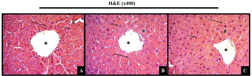
614     Toxicology Research, 2021, Vol. 10, No. 3

Graphical Abstract

                                                                                                                                              Downloaded from https://academic.oup.com/toxres/article/10/3/613/6288551 by guest on 23 November 2021
Key words: quercitin, afzelin, acute toxicity, sub-acute toxicity, Bauhinia cheilantha

Introduction                                                           lead to toxicity, or even death, in both humans and animals [5,
                                                                       6]. For example, pyrrolizidine alkaloids are naturally occurring
The genus Bauhinia (Fabaceae, Leguminoseae) is composed of
                                                                       toxins produced by over 6000 plant species whose toxicity is
large and diversified pantropical plants with ∼300–350 species,
                                                                       largely documented [7]. Other alkaloids include tropane alkaloids
which are known as cow’s paw or cow’s hoof, because of the
                                                                       such as atropine and scopolamine, and hyoscyamine, which are
shape of their leaves [1, 2]. Traditionally, this genus has been
                                                                       used as anticholinergics in Food and Drug Administration (FDA)
used in folk medicine as a remedy for different kinds of diseases,
                                                                       approved drugs and homeopathic remedies [8]. This molecule
mainly diabetes, pain in general, inflammation, and infections
                                                                       may be very dangerous at high doses. Various plants can cause
[2, 3].
                                                                       toxic effects when ingested. For example, progressive interstitial
    Bauhinia cheilantha (Bong.) Steud. is a shrub or small tree with
                                                                       nephritis leading to end stage of renal disease and urothelial
simple and alternate, leaves split at the apex up to the middle
                                                                       malignancy in users of Aristolochia fangchi [9] has been docu-
(similar to cow’s foot). It is native to the Caatinga (seasonal dry
                                                                       mented. Atropa belladonna, popularly known as belladonna or
forest), a specific Brazilian biome, where it grows mainly on stony
                                                                       deadly nightshade, ranks among one of the most poisonous
soils, poor in nutrients, in open formations, and at high altitudes
                                                                       plants in Europe and other parts of the world. Thus, it is manda-
[1]. The areal parts and stem-bark are used in folk medicine in
                                                                       tory to trace the toxicological profile of popularly-used herbal
many communities living in the semiarid region of Northeastern
                                                                       medicines.
Brazil [2]. Some are used to treat diabetes, influenza, pain in
                                                                           The aim of this study was to provide scientific data on the
general, diabetes, intestinal, stomach, and renal problems, as
                                                                       safe use of B. cheilantha, focusing on its in-vitro antihemolytic
well as a in decoction, alcoholic infusion, aqueous infusion,
                                                                       and antioxidant activity and also acute and sub-acute toxicities
poultice, or juice [2, 4]. In addition, B. cheilantha has economic
                                                                       through the evaluation of behavioral, biochemical, hematolog-
and nutritional importance [1]. Bauhinia cheilantha seeds have a
                                                                       ical, and histological parameters of the hydroalcoholic extract
high protein content (∼36%), reasonable essential amino acids
                                                                       from B. cheilantha leaves (HaEBcl).
profile, and low levels of antinutritional compounds [4]. For is
reason, this species is widely used for different proposes in local
communities in the semiarid region.
    However, the general perception that plants, because of their      Materials and Methods
nutritional, medicinal, and economic importance, are absent
from adverse effects is not only false, but also dangerous. Plants
                                                                       Plant material and preparation of extract
produce bioactive compounds known as secondary metabolites,            Bauhinia cheilantha leaves were collected in March 2019 in the
which play an essential role in defending the plants from biotic       town of Altinho, (8◦ 35 13,5 S; 36◦ 5 34,6 W), in the state of
and abiotic stress conditions [5]. Even though these molecules         Pernambuco, Brazil, and identified by Dr. Ulysses Paulino de
can be used for biopharmaceutical purposes, some of them may           Albuquerque (UFPE). A voucher specimen (PEUFR 48 653, 46 183)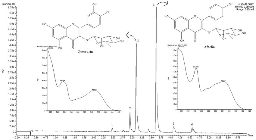
Alanne Lucena de Brito et al.        615

has been deposited at the Herbarium of the Universidade Federal       450 μl of the solution of DPPH˙ (23.6 mg/ml in ethanol, EtOH)
Rural de Pernambuco (UFRPE).                                          were transferred to 0.5-ml Eppendorf tubes and the volume was
    The hydroalcoholic extract of B. cheilantha leaves (HaEBcl)       completed with EtOH, following homogenization. Samples were
was prepared by maceration from dried and powdered leaves             sonicated for 30 min and placed in a 96-well plate to record
(100 g) in 100 ml of ethanol 80% at room temperature (28◦ C)          the amount of DPPH˙ on a UV–Vis (Biochrom EZ Read 2000® )
overnight. The solvent from HaEBcl was filtered and concen-           device at a wavelength of 517 nm. Ascorbic acid was used as a
trated using a rotary evaporator to provide an aqueous ethanolic      positive control and all concentrations were tested in triplicate.
extract After preparation, the dried powder extract was stored at     The percentage scavenging activity (% SA) was calculated from
room temperature. The HaEBcl extract was used for preliminary         the equation:
phytochemical analysis and toxicological assays.

                                                                                %SA = 100 × Abscontrol − Abssample /Abscontrol

Phytochemical analysis
                                                                      where Abscontrol is the absorbance of the control containing only

                                                                                                                                             Downloaded from https://academic.oup.com/toxres/article/10/3/613/6288551 by guest on 23 November 2021
Determination of total phenolic content. The total phenolic con-
                                                                      the ethanol solution of DPPH, and Abssample is the absorbance
tent of the samples was determined using the Folin–Ciocalteau
                                                                      of the radical in the presence of the sample or standard ascor-
reagent according to the method of Slinkard and Singleton [10]
                                                                      bic acid.
modified using gallic acid as a standard phenolic compound. Ini-
tially, the samples were solubilized in ethanol at concentrations
                                                                      2,2 -azino-bis (3-ethylbenzothiazoline-6-sulfonic acid) (ABTS˙+ )
of 1.0 mg/ml. A 50-μl aliquot of each solution was transferred to
                                                                      scavenging activity assay. The 2,2 -azino-bis (3-ethylbenz-
an Eppendorf tube with 20 μl of the Folin–Ciocalteau reagent and
                                                                      othiazoline-6-sulfonic acid) (ABTS˙+ ) radical scavenging of
870 μl of distilled water was added and stirred for 1 min. Then,
                                                                      HaEBcl was carried out following the methodology described by
60 μl of the Na2 CO3 solution (15%) was added to the mixture and
                                                                      Re et al. [14], in a UV–Vis (Biochrom EZ Read 2000® ) device, using
stirred for 30 s, resulting in a final concentration of 100 μg/ml
                                                                      Trolox as the standard compound. The starting concentrations
for the samples. Absorbance was measured at 760 nm on a
                                                                      of the solutions of the samples were 0.1–1.0 mg/ml, with
spectrophotometer. The total amount of phenolic compounds
                                                                      the addition of 450 μl of the radical ABTS˙+ solution to give
was determined in equivalent micrograms of gallic acid, using
                                                                      final concentrations of 2.5–100.0 μg/ml samples. Samples were
the equation obtained in the standard gallic acid graph.
                                                                      protected from light and sonicated for 6 min. Absorbance of the
                                                                      samples and the positive control were measured at a wavelength
                                                                      of 734 nm using a microplate of 96 wells. Each concentration was
Total flavonoid content                                               tested in triplicate. The percentage of free radical scavenging
The determination of flavonoids followed the methodology pro-         activity of ABTS˙+ was calculated by the equation:
posed by Woisky and Salatino [11] with modifications, using
quercetin as the standard. Initially, ethanol solutions (1.0 mg/ml)
                                                                                %SA = 100 × Abscontrol − Abssampl /Abscontrol
of the Bauhinia extract were prepared. For the test, 50 μl of
sample (final concentration 50 μg/ml) and 500 μl of 5% alu-
minum chloride solution (AlCl3 ) were placed in an Eppendorf          where Abscontrol is the absorbance of the control containing only
tube and the volume was made up to 1000 μl with methanol.             the ethanol solution of ABTS˙+ and Abssample is the absorbance
After 30 min, the absorbance was measured using an Elisa UV–Vis       of the radical in the presence of the sample or standard ascor-
spectrophotometer at 425 nm, using 96-well plates. The analyses       bic acid.
were performed in triplicate. The content of total flavonoids was         The antiradical efficiency was established using linear regres-
determined by interpolating the sample absorbance against a           sion analysis and the 95% confidence interval (CI; P < 0.05)
calibration curve constructed with quercetin standard solutions       obtained using the statistical program GraphPad Prism 5.0® . The
in various concentrations (2.5–20 μg/ml) and expressed as an          results were expressed through the value of the half maximal
equivalent milligram. Quercetin per gram of extract (mg EQ/g),        effective concentration (EC50 ), which represents the concentra-
considering the standard deviation (SD).                              tion of the sample necessary to sequester 50% of the radicals.
    The phytochemical analysis was obtained in negative elec-
trospray mode using an XEVO-G2XSQTOF mass spectrometer
(Waters, Manchester, UK) connected to an ACQUITY UPLC system          In vitro hemolytic assay
(Waters, Milford® , MA, USA). The analytical detector was a Waters
Acquity PDA detector, which was set to a wavelength range of          The evaluation of the osmotic fragility of red blood cells (RBC) was
200–400 nm. The conditions for obtaining the data by UPLC-ESI-        performed on a fresh blood sample collected from a healthy rat,
qTOF-MS/MS were set according to Cabrera et al. [12].                 using dethylenediaminetetraacetic acid (EDTA) as anticoagulant
                                                                      [15]. The blood sample was carefully mixed with 0.90% NaCl
                                                                      solution and the mixture was centrifuged (1500 rpm, 15 min). The
                                                                      supernatant was discarded and this procedure was repeated. The
The antioxidant activity of HaEBcl.                                   concentrate of RBC (5 ml) was added to 1.0 ml of plant extract
2,2-diphenyl-1-picrylhydrazyl (DPPH) radical scavenging activity      in different concentrations (corresponding to 0, 25, 50, 100, and
assay. The radical scavenging activity of HaEBcl was performed        200 mg of plant/ml in 0.9% NaCl solution) and incubated for
against the free radical 2,2-diphenyl-1-picrylhydrazyl (DPPH)         60 min at room temperature, with two repetitions for each con-
following the methodology of Silva et al. [13]. Stock solution        centration. Extracts were also prepared of 200 mg/ml containing
was prepared from the extracts and ethanol fraction in different      different concentrations of NaCl (0 up to 0.9%) and incubated
concentrations (0.10–5.0 mg/ml). Through preliminary analysis,        with blood samples. The hemolytic percentage was determined
appropriate quantities of stock solutions of the samples and          by measurement of hemoglobin in the supernatants using a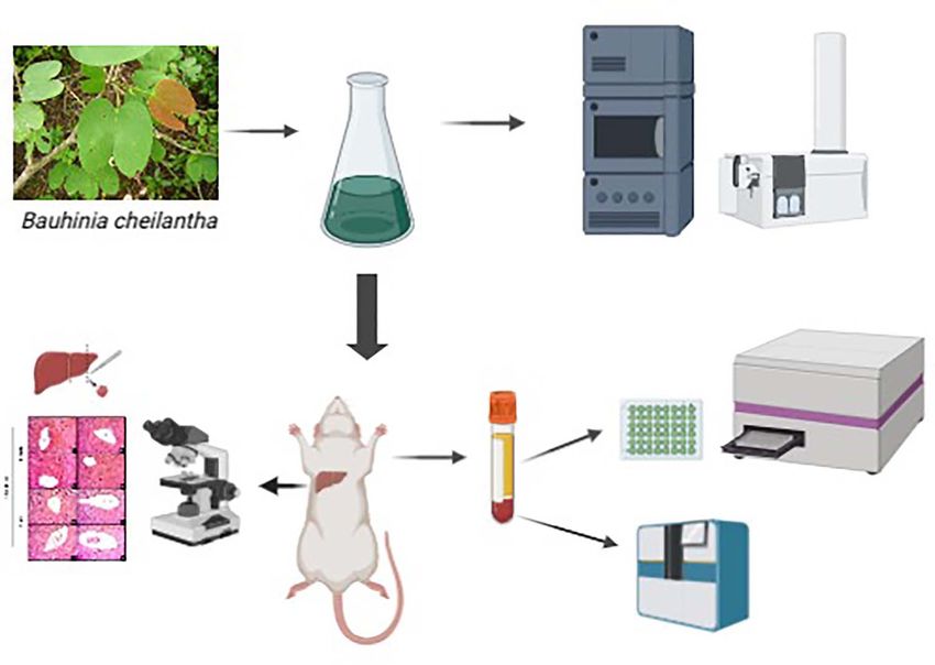
616     Toxicology Research, 2021, Vol. 10, No. 3

commercial kit (BioClin® , Belo Horizonte, MG, Brazil) and a spec-     with saline 0.9% (Control); (ii) treated with 300 mg/kg of HaEBcl
trophotometer at 540 nm. The experiments were analyzed with            (HaEBcl300); (iii) treated with 1000 mg/kg of HaEBcl (HaEBcl1000);
paired t-test to verify potential differences between hypotonic        and (iv) treated with 2000 mg/kg of HaEBcl (HaEBcl2000).
and isotonic phases (% concentrations of NaCl) versus relative             Animals were weighed weekly and observed for behavioral
hemolysis (% hemolysis). The hemolysis rate was calculated as          changes, food and water intake, and general morphological
follows:                                                               aspect. At the end of the treatment, all animals were anes-
                                                                       thetized by intraperitoneal administration of ketamine and
                                                               xylazine mixture (80 mg and 10 mg/kg, respectively), and
Hemolysis % = Abssample − Absbasal / AbsTotal − AbsBasal ×100
                                                                       euthanized by cervical dislocation. Blood samples were collected
                                                                       for biochemical and hematological analysis and the organs were
                                                                       weighed and stored for histopathology (liver).
Experimental animals and ethical statement.
Adult female and male Swiss albino mice (35 ± 3 g, aged 8–
10 weeks) were obtained from Animal House of the Physiology

                                                                                                                                                Downloaded from https://academic.oup.com/toxres/article/10/3/613/6288551 by guest on 23 November 2021
                                                                       Biochemical and hematological analysis
and Pharmacology Department at Federal University of Pernam-
buco. Mice were housed with a 12/12 light–dark cycle, 22 ± 3◦ C        Blood collecting. On the 14th and 28th days after treatment, fasted
with free access to water and conventional lab chow diet (Purina,      mice rats were anesthetized as described previously; blood was
Labina® , Brazil). All experiments were performed between 8:00         drawn by the retro-orbital technique with or without heparin for
and 10:00 am. The protocols were approved by the Animal Ethics         both hematological and serum biochemical analysis.
Committee of the Federal University of Pernambuco (CEUA-UFPE,
process #: 23076.004793/2015-42) and conducted in accordance           Hematological analysis. On the 14th and 28th days, hematologi-
with Guide for the Care and Use Laboratory Animal published by         cal parameters included RBC, hemoglobin concentration (HBG),
the US National Institutes of Health (NIH Publications No. 8023,       hematocrit (HCT), red cell volume distribution (RDW), platelet
revised 1978).                                                         count (PLT), mean platelet volume (MPV), mean corpuscular vol-
                                                                       ume (MCV), mean corpuscular hemoglobin (MCH), mean corpus-
                                                                       cular hemoglobin concentration (MCHC), lymphocytes (LYM), and
Acute oral toxicity                                                    white blood cells (WBC). These were determined by an automatic
                                                                       hematology analyzer (SDH-20, Labtest® , Brazil).
The acute toxicity test was performed according to Guideline
423 (acute toxic class method) of the Organization of Economic
Co-operation and Development (OECD) for testing chemicals for          Serum biochemistry. The serum levels of alanine (ALT) and aspar-
acute oral toxicity [16]. After 5 days of acclimation in a propylene   tate aminotransferase (AST), albumin, total protein (TP), blood
cage, nulliparous and non-pregnant female mice (three for each         urea nitrogen (BUN), and creatinine (CRE) were measured with
group) were fasted for 3 h and weighed before extract administra-      kits from Labtest® in accordance with manufacture’s protocol.
tion. The HaEBcl was dissolved in saline 0.9% and administered
by gavage in mice in single dose using animal feeding needles          Organ mass and histopathological analysis. On the 14th and 28th
(100 μl/100 g b.w.). Animals were randomly divided into three          days, all mice were necropsied after blood collection for anatom-
groups with three animals each: (i) treated with saline 0.9%           ical localization and gross examination of visible changes in the
(Control); (ii) treated with 300 mg/kg of HaEBcl (HaEBcl300); and      organs (aspect, color, and size). Selected organs including lungs,
(iii) treated with 2000 mg/kg of HaEBcl (HaEBcl2000).                  heart, liver, kidney, testicles, uterus, ovaries, spleen, adipose tis-
     In the first 4 h, all animals were closely observed for pilo-     sue, stomach, soleus, and extensor digitorum longus (EDL) mus-
erection, changes in skin, fur and eyes, toxic effects on the          cle. Were carefully excised and trimmed of fat and connective
mucous membrane, behavior pattern disorientation, hypoactiv-           tissue before being weighed. The organ weight values were nor-
ity, hyperventilation, asthenia, lethargy, sleep, diarrhea, tremors,   malized by tibia length. On the 14th and 28th day of treatment,
salivation, convulsion, coma, motor activity, or death. After this,    the specimens of liver were fixed in 10% formalin buffer solution
the animals were continuously observed every 24-h daily during         for 24 h at room temperature and washed for 4 h with tap
14 days. Parameters such as body weight, food and water intake         water. They were then dehydrated step-wise using 70–100% EtOH.
were monitored daily. On the 14th day, female mice were euth-          Dehydrated tissues were made transparent through addition of
anized by injection of a xylazine (80 mg/kg, i.p.) and ketamine        xylene. Tissues were added to pre-heated paraffin for sufficient
(10 mg/kg, i.p.) solution, and the blood and organs were drawn         infiltration, then cooled, and formed into paraffin blocks before
for biochemical and macroscopic analysis. Based on mortality in        being cut into 5-μm sections using a microtome (Leica® RM 2025,
each group, the LD50 was estimated [17].                               Heidelberger, Germany). The paraffin was removed and speci-
                                                                       mens were dehydrated and stained with hematoxylin–eosin for
                                                                       observation with an inverted optical microscope (Leica® , Heidel-
Sub-acute oral toxicity test                                           berger, Germany) coupled to a video camera (Leica® DFC 280, Wet-
The sub-acute toxicity test was conducted according to the             zlar, Germany) and a computer monitor. The specimens of liver
Guideline 407 (repeated dose 28-day oral toxicity study in             were photographed and analyzed by an experienced pathologist
rodent) of the OECD to test chemicals for acute oral toxicity          for any cellular damage or change in morphology.
[18]. After 5 days of acclimation in propylene cages, male and
nulliparous and non-pregnant female mice were fasted for               Statistical analysis. The data were expressed as mean ± stan-
3 h and weighed before extract administration. The mice were           dard error of the mean (SEM). The one-way analysis of variance
divided into groups of 10, 5 male and 5 female each, to be treated     (ANOVA) followed by the Bonferroni test was employed to ana-
daily over 28 consecutive days by gavage using animal feeding          lyze the data between treated groups and their respective control
needles (100 μl/100 g b.w.) The groups were as follows: (i) treated    groups. P value < 0.05 was considered statistically significant.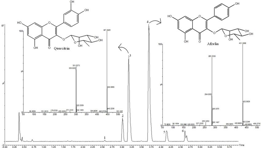
Alanne Lucena de Brito et al.   617

                                                                                                                                                           Downloaded from https://academic.oup.com/toxres/article/10/3/613/6288551 by guest on 23 November 2021
Figure 1: Chromatogram of hydroalcoholic extract of Bauhinia cheilantha (Bong.) Steud. leaves analyzed by UPLC-DAD.

 Table 1: Total content of phenols and flavonoids in the hydroalco-                 compared with the available literature. Peaks 3 and 4 were
 holic extract of Bauhinia cheilantha (Bong.) Steud. leaves                         compared with the standard and identified as quecitrin and
 Phenolic content                                    Flavonoid content              afzelin, respectively. The mass spectra are shown in Fig. 2.
 (mg EGA/g sample)                                   (mg EQ/g sample)               Compounds 1 and 2 were identified as quercetin hexoside, m/z
                                                                                    463.0818 and quercetin–pentoside m/z 433.0710 [M−H]− with
 184.10 ± 7.54                                       202.39 ± 4.43                  specific fragmentation at m/z 301.0218 [M−H−hexose]− and m/z
                                                                                    301.0281 [M−H−pentose]− , corresponding to radical loss from
                                                                                    deprotonated molecules, respectively. Compounds 5 and 6 at
                                                                                    m/z 599.1011 [M−H]− and 583.1001 [M−H]− were identified as
Results                                                                             quercetin and kaempferol galloyl rhamnosides, respectively. The
Phytochemical analysis                                                              main fragmentation at m/z 301.0291 and m/z 285.0348 resulted
                                                                                    from the loss of a galloyl-rhamnoside moiety.
Total phenols and flavonoids content. The results showed no sta-
tistically significant difference (Table 1) between total phenols
and flavonoids content, which may suggest that the phenolic                         Antioxidant activity of HaEBcl. As shown in Table 3, the radical
compounds found in the B. cheilantha extract are of the flavonoid                   scavenging activity of the HaEBcl was performed using the radi-
type, similar to quercetin.                                                         cal scavenging abilities of DPPH and ABTS. The HaEBcl was able
                                                                                    to inhibit the activity of DPPH radicals (EC50 = 409.5 μg/ml) and
Ultra-performance liquid chromatography. The hydroalcoholic                         also perform an antioxidant activity for ABTS (EC50 = 104 μg/ml).
extract from B. cheilantha was analyzed by ultra-performance
liquid chromatography coupled with diode array detection and                        Hemolytic activity. The result presented in Fig. 3 show that HaEBcl
quadrupole time-of-flight mass spectrometry for the profiling                       did not disrupt the red cell membrane but preserved the integrity
and structural characterization of the compounds. The UPLC-                         of the erythrocyte membrane in various NaCl concentrations.
diode array detector (DAD) chromatograms at 340 nm and base
peak ion (BPI) chromatograms of extract are presented in Figs 1                     Acute oral toxicity (single dose evaluation of HaEBcl). In acute
and 2, respectively. The compounds were identified based on                         toxicity testing, administration of a single oral dose of HaEBcl
their characteristic UV–Vis spectra peaks and mass detection                        did not produce mortality or any toxic signs or symptoms at the
as well as the accurate mass measurement of the precursor                           different doses under study. No skin irritation, fur, eyes, mucous
and product ions. All the compounds detected are listed on                          membrane toxic effects, piloerection, disorientation, hypoactiv-
Table 2. The principal compounds 3 (quercitrin) and 4 (afzelin)                     ity, hyperventilation, asthenia, lethargy, sleep, diarrhea, tremors,
were compared with standard samples (Figs 1 and 2).                                 salivation, convulsion, coma, motor activity, or death were regis-
    Four compounds were identified as quercetin glycosides (1, 2,                   tered. In addition, body weight gain and food and fluid consump-
3, and 5) and two were identified as glycosides of kaempferol (4                    tion differences among the groups treated with HaEBcl were
and 6). Observation of glycosidic residues pentoside (136 Da),                      not observed (Fig. 4). Likewise, the macroscopic analysis revealed
rhamnosyl (146 Da), and glucosyl (162 Da) were cleaved                              that HaEBcl did not produce any alteration in organ color, size,
sequentially and generated characteristic aglycone fragments                        shape, or texture, compared with the control.
618 Toxicology Research, 2021, Vol. 10, No. 3
Downloaded from https://academic.oup.com/toxres/article/10/3/613/6288551 by guest on 23 November 2021
Figure 2: ESI BPI chromatogram of hydroalcoholic extract of Bauhinia cheilantha (Bong.) Steud. leaves analyzed by UPLC-qTOF-MS.
Table 2: Characterization of compounds from hydroalcoholic extract of Bauhinia cheilantha (Bong.) Steud. leaves by UPLC/qTOF-MSE
Compound RT (min) UV λmax [M−H]− (m/z) [M−H]− (m/z) MS/MS Identification
(nm) experimental calculated
1 2.55 349 463.0818 463.0882 301.0218 [M−H−hexose]− Quercetin hexoside
2 3.01 352 433.0710 433.0776 301.0281 [M−H−pentose]− , 300.0197 Quercetin pentoside
[M−2H−pentose]− , 271.0391
[M−2H–CO–pentose]−
3 3.17 255, 349 447.0983 447.0932 301.0277 [M−H−rhamnose]− , 300.0207 Quercitrin∗
[M−2H–rhamnose]− , 271.0187
[M−2H–CO–rhamnose]−
4 3.68 263, 344 431.0914 431.0983 285.0342 [M−H–rhamnose]− , 284.0269 Afzelin∗
[M−2H–rhamnose]− , 255.0257
[M−2H–CO–rhamnose]−
5 4.14 348 599.1011 599.1042 445.1307 [M−2H–galloyl]− , 301.0291 Quercetin-galloyl-
[M−2H–galloyl–rhamnose]− rhamnoside
6 4.61 265, 347 583.1001 583.1093 285.0348 [M−2H–galloyl–rhamnose]− Kaempferol–galloyl–
rhamoside
Table 3: Determination of antioxidant activity according to EC50 As shown in Table 4, the oral acute toxicity of single dose of
(μg/ml) values of hydroalcoholic extract of Bauhinia cheilantha (Bong.) the HaEBcl did not produce relative weight organ alteration when
Steud. leaves, ascorbic acid and TROLOX samples by DPPH and ABST
compared with control group. In addition, neither any biochem-
methods
ical nor hematological parameters evaluated were statistically
Sample DPPH ABTS different in oral acute and a single dose treated mice when
EC50 μg/ml (CI) EC50 μg/ml (CI) compared with control group at 14th day of experiment (Tables 5
and 6).
HaEBcl 409.5 (401.4–420.6) 104.0 (93.0–114.1)
Histopathological examination of organs from animals
Ascorbic Acid 1.6 (1.4–1.8) –
TROLOX – 4.1 (3.7–5.8) treated with animals with different doses showed normal archi-
tecture, suggesting non-harmful changes, and morphological
∗ Ascorbic acid and TROLOX (6-hydroxy-2,5,7,8-tetramethylchroman-2- disorders induced by single oral dose of HaEBcl at 14th day of
carboxylic acid) were used as reference antioxidants for DPPH and ABTS treatment (Fig. 5).
analysis, respectively.Alanne Lucena de Brito et al. 619
Table 4: Effects of hydroalcoholic extract of Bauhinia cheilanthaπ (Bong.) Steud. leaves by an oral acute toxicity test on absolute total weight gain
(g) and relative organ weight in female mice treated at 14th day
Parameters Groups
Control 300 mg/kg HaEBcl 2.000 mg/kg HaEBcl
Total weight gain (g) 28.31 ± 0.58 28.90 ± 0.36 30.24 ± 0.39
Tibia (mm) 18.67 ± 0.7 18.25 ± 0.25 18.25 ± 0.48
Liver (mg/mm) 10.0 ± 0.67 10.0 ± 0.5 102.2 ± 0.5
Kidneys (mg/mm) 2.12 ± 0.17 2.51 ± 0.19 2.26 ± 0.11
Heart (mg/mm) 0.73 ± 0.03 0.80 ± 0.02 0.80 ± 0.06
Lung (mg/mm) 1.04 ± 0.07 1.06 ± 0.10 1.05 ± 0.11
Stomach (mg/mm) 1.42 ± 0.07 1.87 ± 0.13 1.90 ± 0.11
Spleen (mg/mm) 0.73 ± 0.03 0.82 ± 0.6 0.86 ± 0.6
Uterus (mg/mm) 0.49 ± 0.07 0.44 ± 0.04 0.38 ± 0.05
Downloaded from https://academic.oup.com/toxres/article/10/3/613/6288551 by guest on 23 November 2021
Ovaries (mg/mm) 0.15 ± 0.01 0.15 ± 0.02 0.10 ± 0.01
Adipose tissue (mg/mm) 2.68 ± 0.28 3.53 ± 0.36 3.40 ± 0.15
EDL (mg/mm) 0.04 ± 0.01 0.05 ± 0.01 0.05 ± 0.01
Soleus (mg/mm) 0.03 ± 0.01 0.04 ± 0.01 0.04 ± 0.01
Results are expressed as mean ± SEM. One way ANOVA variance test was performed followed by Bonferroni test. The difference among groups were considered
statically when P < 0.05.
Table 5: Effects of hydroalcoholic extract of Bauhinia cheilantha (Bong.) Steud. leaves on oral acute toxicity test in biochemical parameters in
female mice treated at 14th day
Parameters Groups
Control HaEBcl300 HaEBcl2.000
ALT (U/L) 69.10 ± 10.24 61.30 ± 1.85 61.47.50 ± 2.92
AST(U/L) 109.40 ± 11.41 93.52 ± 12.08 100.40.71 ± 4.44
AST: ALT ratio 1.40 ± 0.50 1.53 ± 0.20 1.53 ± 0.04
TP (g/dl) 5.76 ± 0.23 5.45 ± 0.05 5.65 ± 0.06
Albumin (g/dl) 2.16 ± 0.17 1.90 ± 0.04 2.02 ± 0.23
BUN (mg/dl) 73.13 ± 3.18 70.80 ± 6.664 70.50 ± 4.76
CRE (mg/dl) 0.50 ± 0.005 0.55 ± 0.03 0.45 ± 0.05
Results are expressed as Mean ± SEM. One way ANOVA variance test was performed followed by Bonferroni test. The difference among groups were considered statically
when P < 0.05.
Table 6: Effects of hydroalcoholic extract of Bauhinia cheilantha (Bong.) Steud. leaves on oral acute toxicity test in hematological parameters in
female mice treated at 14th day
Parameters Groups
Control HaEBcl300 HaEBcl2.000
RBC (106 /mm3 ) 7.15 ± 0.23 5.33 ± 0.36 6.71 ± 0.26
HGB (g/dl) 9.73 ± 0.48 7.68 ± 0.57 8.65 ± 0.30
MCH (%) 13.60 ± 0.31 13.05 ± 0.13 12.90 ± 0.20
MCHC (g/dl) 25.27 ± 0.52 12.90 ± 0.20 24.90 ± 0.21
MCV (fL) 54.00 ± 0.00 53.00 ± 0.71 51.75 ± 0.48
HCT (%) 38.53 ± 1.41 31.18 ± 2.17 34.65 ± 1.33
RDW% (%) 15.23 ± 0.07 14.85 ± 0.21 12.75 ± 1.16
PLT (103 /mm3 ) 619.67 ± 72.65 582.25 ± 18.89 716.75 ± 49.30
MPV (fL) 6.90 ± 0.55 6.80 ± 0.85 6.15 ± 0.61
WBC (109 /l) 3.70 ± 0.70 2.93 ± 0.91 3.50 ± 1.14
LYM (mm3 ) 8.83 ± 0.15 8.65 ± 1.47 8.39 ± 0.50
Results are expressed as Mean ± SEM. One way ANOVA variance test was performed followed by Bonferroni test. The difference among groups were considered statically
when P < 0.05.
Sub-acute oral test (repeated dose 28-day oral toxicity) Body weight gain, food and fluid intake, and relative organ weight.
Body weight gain, food, and fluid intake were compatible with
General signs and mortality. All animals survived until scheduled
physiological development for females and males during 28 days
necropsy. The sub-acute oral test was conducted over 4 weeks
of treatment (Fig. 6). The relative tissue weights were not altered
(28 days) with three different doses: 300, 1000, and 2000 mg/kg/-
by hydroalcoholic extract of B. cheilantha leaves (Table 7). The
day and control group. The daily observation did not reveal any
organ mass analysis of the target tissues of the treated animals
sign of toxicity, altered behavior or mortality.620 Toxicology Research, 2021, Vol. 10, No. 3
Table 7: Effect of sub-acute oral toxicity evaluation of hydroalcoholic extract of Bauhinia cheilantha (Bong.) Steud. leaves on absolute total body
weight gain and relative organ weight in female and male Swiss mice treated for 28 consecutive days
Sex Parameters Groups
Control HaEBcl300 HaEBcl1.000 HaEBcl2.000
Female Total weight gain (g) 36.66 ± 0.49 33.15 ± 0.52 29.59 ± 0.48 35.58 ± 0.88
Tibia (mm) 19.3 ± 0.33 16.25 ± 0.48 19.25 ± 0.48 19.20 ± 0.37
Liver (mg/mm) 8.36 ± 0.60 9.51 ± 0.38 6.71 ± 0.23 7.38 ± 0.67
Kidneys (mg/mm) 2.09 ± 0.09 1.84 ± 0.04 1.90 ± 0.03 1.99 ± 0.14
Heart (mg/mm) 0.82 ± 0.03 0.73 ± 0.05 0.73 ± 0.02 0.89 ± 0.08
Lung (mg/mm) 1.48 ± 0.13 0.96 ± 0.06 1.04 ± 0.17 1.49 ± 0.25
Stomach (mg/mm) 1.99 ± 0.09 1.80 ± 0.09 0.98 ± 0.03 1.57 ± 0.31
Spleen (mg/mm) 0.52 ± 0.09 0.69 ± 0.05 0.42 ± 0.08 0.54 ± 0.08
Uterus (mg/mm) 0.62 ± 0.05 0.56 ± 0.04 0.67 ± 0.13 0.72 ± 0.34
Downloaded from https://academic.oup.com/toxres/article/10/3/613/6288551 by guest on 23 November 2021
Ovaries (mg/mm) 0.11 ± 0.02 0.23 ± 0.04 0.16 ± 0.02 0.34 ± 0.18
Adipose tissue 8.91 ± 1.32 7.93 ± 0.95 12.70 ± 1.46 9.29 ± 2.17
(mg/mm)
EDL (mg/mm) 0.89 ± 0.17 0.69 ± 0.18 0.75 ± 0.23 0.98 ± 0.16
Soleus (mg/mm) 0.47 ± 0.06 0.35 ± 0.03 0.27 ± 0.06 0.41 ± 0.06
Male Total weight gain (g) 37.99 ± 0.95 38.00 ± 0.79 36.97 ± 0.23 36.68 ± 0.87
Tibia (mm) 17.20 ± 0.80 20.20 ± 0.86 18.60 ± 0.93 18.50 ± 0.87
Liver (g/100 g) 13.04 ± 2.86 13.89 ± 0.83 13.44 ± 0.63 13.79 ± 0.72
Kidneys (mg/mm) 5.22 ± 0.23 4.78 ± 0.34 4.45 ± 0.34 4.06 ± 0.49
Heart (mg/mm) 1.64 ± 0.08 1.37 ± 0.04 1.17 ± 0.07 1.44 ± 0.03
Lung (mg/mm) 3.31 ± 0.33 2.98 ± 0.11 2.29 ± 0.10 2.59 ± 0.25
Stomach (mg/mm) 3.57 ± 0.49 2.49 ± 0.27 2.56 ± 0.25 3.30 ± 0.72
Spleen (mg/mm) 1.15 ± 0.15 0.96 ± 0.08 0.76 ± 0.09 1.03 ± 0.13
Testicles (mg/mm) 1.35 ± 0.07 1.29 ± 0.09 1.37 ± 0.12 1.08 ± 0.13
Adipose tissue 7.83 ± 0.43 8.59 ± 0.82 5.71 ± 0.60 6.36 ± 0.36
(mg/mm)
EDL (mg/mm) 0.11 ± 0.07 0.37 ± 0.050 0.31 ± 0.07 0.26 ± 0.13
Results are expressed as Mean ± SEM. One way ANOVA variance test was performed followed by Bonferroni test. The difference among groups were considered statically
when P < 0.05. Weight gain, weight of heart, liver, and kidneys as normalized by tibial length, adipose tissue, soleus muscles and EDL.
Hematological parameters. The oral sub-acute toxicity study did
not produce any significant effect on the hematological parame-
ters, after 28 days daily administration (Table 9).
Histopathological parameters. Bauhinia cheilantha leaves from the
sub-acute oral toxicity treatment did not produce any significant
(P < 0.05) effect on the histopathological examinations of tissues
on any of the harvested organs, indicating no treatment-related
changes in the treated and control groups (Fig. 7).
Discussion
Species belonging to the Bauhinia genus are used around the
world in traditional medicine for treating dysentery, diarrhea,
colds, diabetes, and inflammation [2, 19–21]. Despite their posi-
tive biological effects, the absence of toxicological activity of this
Figure 3: In-vitro hemolytic effect of hydroalcoholic extract of Bauhinia cheilantha and other vegetal species should always be confirmed. Extract
(Bong.) Steud. leaves (0–200 mg/ml) on female mice erythrocyte. of B. cheilantha has been used in traditional medicine as a hypo-
glycemic agent [22]. Despite the popular use and the confirmed
biological effects [4, 22], there are no studies about its toxicolog-
ical safety. This study is the first report on the safe use of the
did not show significant (P < 0.05) changes when compared with
hydroalcoholic extract of B. cheilantha leaves in mice. We provided
the control group for either sex (Table 7).
clear evidence that HaEBcl did not show any sign of toxicity,
with neither acute nor sub-acute symptoms in vivo. There was
Biochemical parameters. The treatment with HaEBcl during 28 moderate antioxidant without hemolytic activity in vitro.
consecutive days in doses of 300, 1000, and 2000 mg/kg did not Assessment of toxic effect of natural products plays a crucial
change in any biochemical profile in mice of both sexes, as shown role in the guarantee for traditional use, in which hemolytic
in Table 8. activity is first step towards providing information about theAlanne Lucena de Brito et al. 621
Downloaded from https://academic.oup.com/toxres/article/10/3/613/6288551 by guest on 23 November 2021
Figure 4: Effect of hydroalcoholic extract of Bauhinia cheilantha (Bong.) Steud. leaves through an oral acute toxicity test (single dose) on body weight gain (A), food intake
(B) and fluid intake (C) in female Swiss mice.
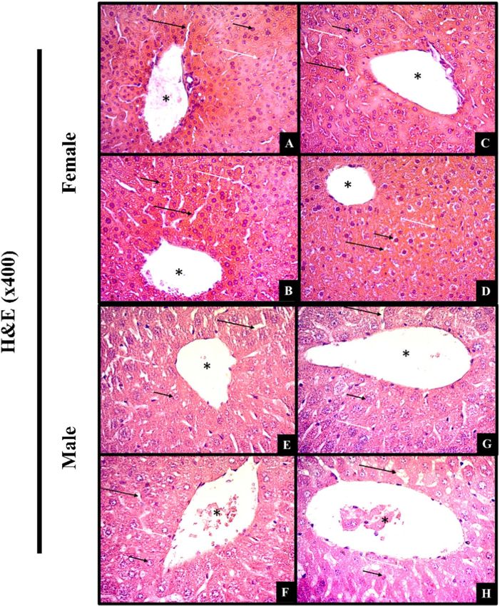
Figure 5: Histological examination of liver sections from mice treated orally with hydroalcoholic extract of Bauhinia cheilantha (Bong.) Steud. leaves (300 or 2000 mg/kg)
at 14 day in acute single dose toxicity study.
interaction between the active compounds and biological enti- not the aim of this study, it should be noted that the antihe-
ties at cellular levels [23]. HaEBcl did not display hemolytic activ- molytic activity can be associated with an anti-inflammatory
ity, probably because it stabilized the RBC membrane, reducing property [26]. This suggests that HaEBcl, then, may be used in
hypotonic solution-induced hemolysis. Mammalian cytotoxicity the future to combat inflammation with no toxic effects.
assays were also performed by hemolysis test [24]. Non-toxic Considering the complexity of the phytochemical composi-
effects of the plant extract, shown by absence of hemolytic tion of plant extract, total antioxidant properties need to be eval-
activity, have also been reported by other researchers [24, 25]. It uated, using at least two methods [28]. Here, we performed DPPH
is not clear about what the precise mechanism of plant extract is radical and ABTS scavenging antioxidant tests, which showed a
that stabilizes the erythrocyte membrane. The alteration of the moderate antioxidant activity which may be explained, at least in
erythrocyte membrane permeability and increase in movement part, by the presence of flavonoids (quercitrin and afzelin), well-
of water and ions cause excessive accumulation of fluid in the known natural antioxidant molecules [19, 29, 30]. Both DPPH and
cell, allowing the release of hemoglobin [26]. It is possible that ABTS scavenging activities reflect a hydrogen-donating ability to
HaEBcl and its constituents may interreact with phospholipid form non radical species which in turn presents the lipid peroxi-
and protein cellular membrane of the RBC. It is well-known that dation of cellular components [19]. The antioxidant property has
flavonoids protect erythrocyte membrane stability against hypo- been strongly demonstrated in Bauhinia genus, such methanolic
tonic lysis [27]. In addition, stabilization of the erythrocyte mem- extract of B. variegate bark [20] and B. vahalii leaves [19]. There
brane, by reducing the release of lytic enzymes, may contribute are also studies of the hydroalcoholic extract of B. forficata and
towards its anti-inflammatory property. Although his aspect was B. microstachya leaves [31, 32]. Here, we show that HAEBcl has the622 Toxicology Research, 2021, Vol. 10, No. 3
Downloaded from https://academic.oup.com/toxres/article/10/3/613/6288551 by guest on 23 November 2021
Figure 6: Effect of sub-acute oral toxicity evaluation of (28 days) of hydroalcoholic extract of Bauhinia cheilantha (Bong.) Steud. leaves on body weight gain (A and B), food
intake (C and D) and fluid intake (E and F) in female and male Swiss mice.
ability to scavenge free radicals, and may be used as potential toxicological properties of any substance intended for prolonged
natural antioxidant to prevented biological damage caused by use [36]. Thus, repeated-dose oral toxicity (sub-acute) may be
oxidative stress. This reinforces the potential use of the HaEBcl evaluated after initial information on toxicity has been obtained
in biomedical applications. by acute testing [37]. This strategy may determine the absolute
Rodent models have been instrumental in helping to answer toxic dose and target organ toxicity, and highlight biochemical,
questions related to traditional use of the plants: in this case, hematological and histopathological repercussions in mice. Sub-
toxicity [33]. Here, HaEBcl had no acute toxic effect in the eval- acute toxicity testing, in our study elicited no clinical signs of
uated doses for Swiss mice with respect to behavior, biochemi- toxicity, morbidity, or mortality in any of the evaluated doses
cal, histological, hemolysis activity, and body weight gain, food (300, 1000, and 2000 mg/kg), which may lead to the inference
intake or sudden death. In addition, the non-acute toxicity effect that these doses could be safely employed in disease treatment.
could also suggest that the LD50 of this extract is greater than Our data corroborates other studies by showing no signs of sub-
2.000 mg/kg administered by oral route in mice. In accordance acute toxicity by hydroalcoholic extracts Piper cernuum [15], B.
with OECD guidelines, HaEBcl is classified in category 5, as non- purpurea [21, 38] Verbena litoralis up to 1000 mg/kg and 2000 mg/kg,
toxic [16]. To the best our knowledge, there is no scientific report respectively, during at least 28 days.
about toxicity of B. cheilantha, however B holophylla [34], B. acumi- The 28-day oral sub-acute test revealed that HaEBcl provoked
nata [35] and B. purpurea [21] have no acute toxic effect either. no deaths or clinical signs of toxicity in any dose evaluated. Body
Regulatory authorities require precise biochemical, hema- weight and food intake were similar for all the doses evaluated.
tological, and histopathological analyses to characterize the HaEBcl compromised neither the metabolism of macronutrientsAlanne Lucena de Brito et al. 623
Table 8: Effect of sub-acute oral toxicity evaluation of hydroalcoholic extract of Bauhinia cheilantha (Bong.) Steud. leaves on biochemical
parameters in female and male Swiss mice treated for 28 consecutive days
Sex Parameters Groups
Control HaEBcl300 HaEBcl1.000 HaEBcl2.000
Female ALT (U/L) 103.52 ± 6.91 88.5 ± 2.57 100.60 ± 5.73 83.50 ± 5.22
AST (U/L) 118.70 ± 4.33 100.10 ± 8.67 110.23 ± 9.65 90.71 ± 8.97
AST: ALT Ratio 1.16 ± 0.06 1.82 ± 0.03 1.14 ± 0.03 1.25 ± 0.07
TP (g/dl) 7.08 ± 0.52 6.01 ± 0.4 6.57 ± 0.17 6.92 ± 0.29
Albumin (g/dl) 1.95 ± 0.09 1.82 ± 0.10 2.62 ± 0.09 2.70 ± 0.07
BUN (mg/dl) 65.92 ± 1.05 54.37 ± 3.43 46.13 ± 3.05 54.8 ± 2.52
CRE (mg/dl) 0.35 ± 0.04 0.26 ± 0.01 0.25 ± 0.02 0.38 ± 0.01
Male ALT (U/L) 105.58 ± 6.88 98.88 ± 6.04 101.21 ± 1.04 99.39 ± 3.16
AST(U/L) 145.50 ± 8.94 144.77 ± 14.38 135.15 ± 12.75 147.25 ± 13.00
Downloaded from https://academic.oup.com/toxres/article/10/3/613/6288551 by guest on 23 November 2021
AST: ALT Ratio 1.39 ± 0.10 1.55 ± 0.16 1.39 ± 0.11 1.47 ± 0.16
TP (g/dl) 3.56 ± 0.04 3.57 ± 0.03 3.53 ± 0.04 3.48 ± 0.04
Albumin (g/dl) 2.11 ± 0.13 1.99 ± 0.04 2.08 ± 0.15 1.84 ± 0.04
BUN (mg/dl) 78.76 ± 3.47 73.03 ± 4.11 65.21 ± 2.29 81.38 ± 3.21
CRE (mg/dl) 0.85 ± 0.01 0.69 ± 0.01 0.81 ± 0.05 0.58 ± 0.05
Results are expressed as Mean ± SEM. One way ANOVA variance test was performed followed by Bonferroni test. The difference among groups were considered statically
when P < 0.05.
Table 9: Effect of sub-acute oral toxicity evaluation of hydroalcoholic extract of Bauhinia cheilantha (Bong.) Steud. leaves on hematological
parameters in female and male Swiss mice treated for 28 consecutive days
Sex Parameters Groups
Control HaEBcl300 HaEBcl1.000 HaEBcl2.000
Female RBC (106 /mm3 ) 8.01 ± 0.10 5.68 ± 0.13 7.61 ± 0.11 7.70 ± 0.22
HGB (g/dl) 15.21 ± 0.20 6.28 ± 0.21 13.40 ± 0.10 13.71 ± 0.34
MCH (%) 18.90 ± 0.22 11.13 ± 0.50 32.91 ± 0.60 30.45 ± 0.60
MCHC (g/dl) 31.41 ± 0.70 20.88 ± 1.16 34.06 ± 0.90 30.31 ± 0.92
MCV (fL) 60.01 ± 1.72 53.50 ± 0.50 53.61 ± 1.63 59.10 ± 1.75
HCT (%) 48.30 ± 1.61 30.30 ± 0.89 40.80 ± 0.91 45.24 ± 1.00
RDW% (%) 15.00 ± 0.30 13.03 ± 0.59 13.15 ± 0.43 16.94 ± 0.15
PLT (103 /mm3 ) 474.00 ± 69.41 933.00 ± 135.25 532.25 ± 134.66 522.20 ± 199.95
MPV (fL) 4.90 ± 0.05 6.37 ± 2.26 4.95 ± 0.15 4.87 ± 0.30
WBC (109 /l) 5.50 ± 0.70 3.35 ± 1.25 2.50 ± 0.10 3.71 ± 0.40
LYM (mm3 ) 5.40 ± 0.70 4.70 ± 0.52 4.31 ± 0.3 4.60 ± 0.6
Male RBC (106 /mm3 ) 7.86 ± 0.12 8.29 ± 0.17 7.81 ± 0.17 8.42 ± 0.14
HGB (g/dl) 14.2 ± 0.27 14.4 ± 0.13 13.76 ± 0.42 14.7 ± 0.26
MCH (%) 18.4 ± 0.40 17.38 ± 0.32 17.86 ± 0.79 17.45 ± 0.26
MCHC (g/dl) 35.42 ± 0.27 34.54 ± 0.39 36.02 ± 0.85 35.00 ± 0.44
MCV (fL) 51.02 ± 0.90 50.34 ± 0.56 50.02 ± 1.28 49.92 ± 0.69
HCT (%) 40.08 ± 0.74 41.74 ± 0.44 39.1 ± 0.69 42.05 ± 0.45
RDW (%) 17.00 ± 0.16 17.18 ± 0.21 16.6 ± 0.23 17.68 ± 0.33
PLT (mm3 ) 673.8 ± 22.89 766.8 ± 33.44 709.0 ± 13.59 719.5 ± 51.33
MPV (fL) 4.84 ± 0.06 4.84 ± 0.04 4.66 ± 0.15 4.875 ± 0.06
WBC (109 /l) 1.38 ± 0.28 1.82 ± 0.11 1.96 ± 0.27 2.02 ± 0.30
LYM (mm3 ) 1.32 ± 0.25 1.78 ± 0.11 1.98 ± 0.34 1.85 ± 0.25
Results are expressed as Mean ± SEM. One way ANOVA variance test was performed followed by Bonferroni test. The difference among groups were considered statically
when P < 0.05.
and animal growth nor affected intra and extracellular dehydra- of the hematological parameters evaluated, illustrating that this
tion in mice [39]. In addition, the absence of organ weight mod- extract did not adversely affect animal health.
ifications, which are a simple and sensitive means for detecting The liver plays a pivotal role in drug metabolism and bio-
harmful effects of xenobiotics [15] reaffirmed the non-toxicity transformation, and its function and structure may be evaluated
effect of HaEBcl in subacute oral tested mice. for toxicity. Serum biomarkers are used to assess liver function
The analysis of blood parameters is relevant for risk evalua- and damage [17]. An increase in serum content of alanine and
tion, since changes in the hematological system have a higher aspartate transaminases, as well as reduction in albumin and
predictive value for human toxicity, when data are translated TP, are strong indicators of hepatic injury [40]. HaEBcl did not
from animal studies [17, 38]. In this sub-chronic study, HaEBcl produce any alteration in any serum biomarker of the liver func-
did not elicit any changes in either white or RBC, nor in any tion, which was confirmed by the preservation of the normal liver624 Toxicology Research, 2021, Vol. 10, No. 3
Downloaded from https://academic.oup.com/toxres/article/10/3/613/6288551 by guest on 23 November 2021
Figure 7: Histological examination of liver sections from mice treated orally with hydroalcoholic extract of Bauhinia cheilantha (Bong.) Steud. leaves (300 or 2000 mg/kg)
after 28-day sub-acute toxicity study.
architecture. An increase in serum CRE and urea levels indicates is up to 2000 mg/kg in oral acute and up to 1000 mg/kg in oral
impaired kidney functions [40]. Similarly, HaEBcl did not alter sub chronic doses for both female and male mice. In general,
the kidney functions in any dose evaluated in the oral acute HaEBcl may be classified to be safe, with a broad safety margin for
and sub-chronic experiments with mice. The protective effect therapeutic use. In addition, HaEBcl has a potential as a radical
of the B. variegate and B. hookeri in acute liver and kidney injury scavenger and to detect the presence of quercitrin and afzelin.
induced by thioacetamide [41] and carbon tetrachloride [42] has This work provides valuable data for the safe use of HaEBcl which
been demonstrated in rats. Taken together, our results suggest should be essential for future pharmacological studies. HaEBcl
that daily administration of HaEBcl in the tested doses does not has a high potential for use in food and drug products, with
produce any significant toxicity in female and male mice. remarkable benefits for human health.
Conclusion Acknowledgments
Based on oral acute and sub chronic administration in mice, it The authors are grateful for the financial support of
may be concluded that hydroalcoholic extract from B. cheilanta Coordenação de Aperfeiçoamento de Pessoa de Nível
leaves has no clinical signs of toxicity effect or mortality in Superior (CAPES), Conselho Nacional de Desenvolvimento
evaluated doses administered to mice. The DL50 value of HaEBcl Científico e Tecnológico (CNPq, grant no. 424800/2018-7). WeAlanne Lucena de Brito et al. 625
acknowledge the National Institute of Science and Technol- 9. Vanherweghem J-L, Depierreux M, Tielemans C et al. Rapidly
ogy—Ethnobiology, Bioprospecting and Nature Conservation progressive interstitial renal fibrosis in young women:
(INCT), which is certified by CNPq and financially supported association with slimming regimen including Chinese herbs.
by the Science and Technology Support Foundation of the Lancet, 1993;341:387–91.
State of Pernambuco (FACEPE, grant no. APQ-0562-2.01/17) for 10. Slinkard K, Singleton VL. Total phenol analyses: automation
the partial funding of the study. The English text of this paper and comparison with manual methods. Am J Enol Viticult
has been revised by Sidney Pratt, Canadian, MAT (The Johns 1977;28:49–55.
Hopkins University), RSAdip—TESL (Cambridge University). 11. Woiky RG, Salatino A. Analysis of propolis: some parameters
and procedures for chemical quality control. J Apicult Res
1998;37:99–105.
12. Cabrera SP, Camara CA, Silva TMS. Chemical constituents of
Glossary flowers from Geoffroea spinosa Jacq. (Leguminosae), a plant
Alanine aminotransferase (ALT), aspartate aminotransferase species visited by bees. Biochem Syst Ecol 2020;88:1–3.
(AST), 2,2’-azino-bis-3-ethylbenzothiazoline-6-sulfonic acid 13. Silva JP, Areias FM, Proença FM, et al. Oxidative stress protec-
Downloaded from https://academic.oup.com/toxres/article/10/3/613/6288551 by guest on 23 November 2021
(ABTS), blood urea nitrogen (BUN), creatinine (CRE), 2,2- tion by newly synthesized nitrogen compounds with phar-
diphenyl-1-picrylhydrazyl (DPPH), diode array detector macological potential. Life Sci 2006;78:1256–67.
14. Re R, Pellegrini N, Proteggente A et al. Antioxidant activity
(DAD), dimethyl sulfoxide (DMSO), extensor digitorum
applying in improved ABTS radical cation. Decolorization
longus (EDL), dethylenediaminetetraacetic acid (EDTA),
assay. Free Radic Biol Med 1999;26:1231–7.
half maximal effective concentration (EC50), hematocrit
15. Wolff FR, Broering MF, Jurcevic JD et al. Safety assessment
(HCT), hemoglobin concentration (HBG), high performance
of Piper cernuum Vell. (Piperaceae) leaves extract: acute, sub-
liquid chromatography (HPCL), hydroalcoholic extract of
acute toxicity and genotoxicity studies. J Ethnopharmacol
B. cheilantha (HaEBcl), lethal dose (LD50), lymphocytes 2018;10:109–16.
(LYM), mean corpuscular hemoglobin concentration (MCHC), 16. Organization for economic cooperation and development
mean corpuscular hemoglobin (MCH), mean corpuscular (OECD) Guidelines for the Testing of Chemicals, OECD 423.
volume (MCV), mean platelet volume (MPV), organization Acute Oral Toxicity-Acute Toxic Class Method. Paris: Organiza-
for economic cooperation and development (OECD), platelet tion for Economic Cooperation and Development, 2001.
count (PLT), red blood cells (RBC), red cell volume distribution 17. Barbosa HM, Nascimento JN, Araújo TA et al. Acute
(RDW), total protein (TP), and white blood cells (WBC). toxicity and cytotoxicity effect of ethanolic extract of
Spondias tuberosa Arruda bark: hematological, biochemi-
cal and histopathological evaluation. An Acad Bras Cienc
Conf licts of Interest 2016;88:1993–2004.
18. Organization for economic cooperation and development
The authors declare there are no conflicts of interest.
(OECD) Guidelines for the Testing of Chemicals, OECD
407. Acute Oral Toxicity-Acute Toxic Class Method. Paris:
Organization for Economic Cooperation and Development,
References 2001.
1. Silva ACC, Oliveira DG. Population structure and spatial dis- 19. Sowndhararajan K, Kang SC. Free radical scavenging activity
tribution of Bauhinia cheilantha (Bong.) Steud. in two frag- from different extracts of leaves of Bauhinia vahlii Wight &
ments at different regeneration stages in the caatinga, in Arn. Saudi J Biol Sci 2013;20:319–25.
Sergipe, Brazil. Rev Arv Viçosa 2015;39:431–7. 20. Sharma N, Sharma A, Bhatia G et al. Isolation of phytochemi-
2. Filho VC. Chemical composition and biological poten- cals from Bauhinia variegata bark and their in vitro antioxidant
tial of plants from the genus Bauhinia. Phytother Res and cytotoxic potential. Antioxidants 2019;8:492.
2009;23:1347–54. 21. Kumar S, Kumar R, Gupta YK, et al. In vivo anti-arthritic activ-
3. Cartaxo SL, Souza MMA, Albuquerque UP. Medicinal plants ity of Bauhinia purpurea Linn bark extract, Indian. J Pharmacol
with bioprospecting potential used in semi-arid northeast- 2019;51:25–30.
ern Brazil. J Ethnopharmacol 2010;131:326–62. 22. Almeida ER, Guedes MC, Albuquerque JFC, et al. Xavier HH.
4. Teixeira DC, Farias DF, Carvalho AFU et al. Chemical com- Hypoglycemic effect of Bauhinia cheilandra in rats. Fitoterapia
position, nutritive value, and toxicological evaluation of 2006;77:276–8.
Bauhinia cheilantha seeds: a legume from semiarid regions 23. Suganthy N, Muniasamy S, Archunan G. Safety assessment
widely used in folk medicine. Biomed Res Int 2013;67881:1–7. of methanolic extract of Terminalia chebula fruit, Termina-
5. Isah T. Stress and defense responses in plant secondary lia arjuna bark and its bioactive constituent 7-methyl gal-
metabolites production. Biol Res 2019;52:39. lic acid: in vitro and in vivo studies. Regul Toxicol Pharmacol
6. Dicson SM, Samuthirapandi M, Govindaraju A, et al. Eval- 2018;92:347–57.
uation of in vivo and in vitro safety profile of the Indian 24. Silva SCC, Braz EMA, Carvalho FAA et al. Antibacterial and
traditional medicinal plant Grewia tiliaefolia. Regul Toxicol cytotoxic properties from esterified Sterculia gum. J Biol Macro-
Pharmacol 2015;73:241–7. mol 2020;164:606–15.
7. Jiang Ma, Qingsu Xia, Peter P. Fu, et al., Pyrrole-protein 25. Aquino DFS, Monteiro TA, Claudia ALC et al. Investiga-
adducts – A biomarker of pyrralizidine alkaloid-induced hep- tion of the antioxidant and hypoglycemiant properties of
atotoxicity. J Food Drug Anal, 2018, 26(3), 965–972. Alibertia edulis (L.C. Rich) A.C. Rich. leaves. J Ethnopharmocol
8. Kohnen-Johannsen KL, Kayser O. Tropane alkaloids: chem- 2020;253:1–9.
istry, pharmacology, biosynthesis and production. Molecules 26. Javeda F, Jabeena Q, Aslamb N, et al. Pharmacological
2019;24:1–23. evaluation of analgesic, anti-inflammatory and antipyretic626 Toxicology Research, 2021, Vol. 10, No. 3
activities of ethanolic extract of Indigofera argentea Burm. F. J 35. Padgaonkar AV, Suryavanshi SV, Londhe VY, et al. Acute toxi-
Ethnopharmocology 2020;15:256. city study and anti-nociceptive activity of Bauhinia acuminata
27. Chaudhuri S, Banerjee A, Basu K et al. Interaction of Linn. Leaf extracts in experimental animal models. Biomed
flavonoids with red blood cell membrane lipids and Pharmacother 2018;97:60–6.
proteins: antioxidant and antihemolytic effects. Int J Biol 36. Branquinho LS, Santos JA, Cardoso CAL et al. Anti-
Macromol 2007;41:42–8. inflamatory and toxicological evaluation of essential oil
28. Rejeb IB, Dhen N, Gargouri M, et al. Chemical composition, from Piper glabratum leaves. J Ethnopharmacol 2017;198:
antioxidant potential and enzymes inhibitory properties of 372–8.
Globe artichoke by-products. Chem Biodivers 2020;6. 37. Nazari S, Rameshrad M, Hosseinzadeh H. Toxicological
29. Silva KL, Filho VC. Plantas do gênero Bauhinia: com- effects of Glycyrrhiza glabra (Licorice): a review. Phytother Res
posição química e potencial farmacológico. Quim Nova 2017;31:1635–50.
2002;25:449–54. 38. Lima R d, Guex CG, Silva ARH et al. Acute and subacute toxi-
30. Sezer ED, Oktay LM, Karadadas E et al. Assessing Anticancer city and chemical constituents of the hydroethanolic extract
Potential of Blueberry Flavonoids, Quercetin, Kaempferol, of Verbena litoralis Kunth. J Ethnopharmacol 2018;224:76–84.
Downloaded from https://academic.oup.com/toxres/article/10/3/613/6288551 by guest on 23 November 2021
and Gentisic Acid, Through Oxidative Stress and Apoptosis 39. Aouachria S, Boumerfeg S, Benslama A et al. Acute, sub-
Parameters on HCT-116 Cells. J Med Food 2019;22:1118–26. acute toxicity and antioxidant activities (in vitro and in
31. Menezes PR, Schwarz EA, Santos C.A.M. In vitro antiox- vivo) of Reichardia picroide crude extract. J Ethnopharmacol
idant activity of species collected in Paraná. Fitoterapia 2017;208:105–16.
2004;75:398–400. 40. Senior JR. Alanine aminotransferase: a clinical and regula-
32. Miceli N, Buongiorno LP, Celi MG et al. Role of the flavonoid- tory tool for detecting liver injury—past, present, and future.
rich fraction in the antioxidant and cytotoxic activities of Clin Pharmacol Ther 2012;92:332–9.
Bauhinia forficata Link. (Fabaceae) leaves extract. Nat Prod Res 41. Bashandy SAE, El Awdan SA, Mohamed SM, et al. Allium
2016;30:1229–39. porrum and Bauhinia variegata mitigate acute liver failure and
33. Atsafack SS, Kuiate J-R, Mouokeu RS et al. Toxicological nephrotoxicity induced by thioacetamide in male rats. Indian
studies of stem bark extract from Schefflera barteri Harms J Clin Biochem 2020;35:147–57.
(Araliaceae). BMC Complement Altern Med 2015;15:44. 42. Al-Sayed E, Abdel-Daim MM, Kilany OE et al. Protective
34. Rozza AL, Cesar DA, Pieroni LG et al. Antiulcerogenic activity role of polyphenols from Bauhinia kookeri against carbon
and toxicity of Bauhinia holophylla hydroalcoholic extract. Evid tetrachloride-induced hepato- and nephrotoxicity in mice.
Based Complement Alternat Med 2015;2015:1–9. Ren Fail 2015;37:1198–207.You can also read