Role of Stress-Related Dopamine Transmission in Building and Maintaining a Protective Cognitive Reserve - MDPI
←
→
Page content transcription
If your browser does not render page correctly, please read the page content below
brain
              sciences
Review
Role of Stress-Related Dopamine Transmission in Building and
Maintaining a Protective Cognitive Reserve
Simona Cabib 1,2, * , Claudio Latagliata 1 and Cristina Orsini 2

                                          1   Department of Experimental Neurosciences, IRCCS Fondazione Santa Lucia, 00179 Roma, Italy;
                                              claudio.latagliata@gmail.com
                                          2   Department of Psychology, Sapienza University of Rome, 00185 Roma, Italy; cristina.orsini@uniroma1.it
                                          *   Correspondence: simona.cabib@uniroma1.it

                                          Abstract: This short review presents the hypothesis that stress-dependent dopamine (DA) transmis-
                                          sion contributes to developing and maintaining the brain network supporting a cognitive reserve.
                                          Research has shown that people with a greater cognitive reserve are better able to avoid symptoms of
                                          degenerative brain changes. The paper will review evidence that: (1) successful adaptation to stres-
                                          sors involves development and stabilization of effective but flexible coping strategies; (2) this process
                                          requires dynamic reorganization of functional networks in the adult brain; (3) DA transmission is
                                          amongst the principal mediators of this process; (4) age- and disease-dependent cognitive impairment
                                          is associated with dysfunctional connectivity both between and within these same networks as well
                                          as with reduced DA transmission.

                                          Keywords: avoidance; cognitive reserve; connectivity; controllability; coping; dopamine; large-scale
                                          brain networks


Citation: Cabib, S.; Latagliata, C.;
Orsini, C. Role of Stress-Related         1. Introduction
Dopamine Transmission in Building
                                                The concept of cognitive reserve (CR) was developed to explain why brain damage
and Maintaining a Protective
                                          does not always predict the severity of cognitive dysfunctions. It was associated with
Cognitive Reserve. Brain Sci. 2022, 12,
                                          a history of positive life experiences (higher education and work milestones, active and
246. https://doi.org/10.3390/
brainsci12020246
                                          rewarding lifestyles) and IQ. Hence, these factors are considered proxies and used as a mea-
                                          sure of CR in the absence of a validated marker. CR proxies are associated with a reduced
Academic Editors: Suelen Boschen,         risk of cognitive decline in normal aging and dementia [1–3]. Moreover, Alzheimer’s dis-
Paolo D’Aquila and Deranda Lester         ease (AD) patients with CR proxies can show more advanced AD-type neuropathology but
Received: 25 January 2022                 comparable clinical severity of cognitive impairment than those without CR proxies [2,4].
Accepted: 8 February 2022                 Studies have reported similar findings in different neurodegenerative diseases such as
Published: 11 February 2022               multiple sclerosis [5], Parkinson’s disease [6], and schizophrenia [7]. Therefore, there is an
                                          increasing interest in developing interventions capable of fostering CR in the treatment of
Publisher’s Note: MDPI stays neutral
                                          cognitive decline. This goal requires identifying neural processes and mechanisms involved
with regard to jurisdictional claims in
                                          in developing and maintaining this phenotype [8].
published maps and institutional affil-
                                                It has been proposed that CR involves efficient and flexible utilization of different
iations.
                                          brain networks. An authoritative CR definition posits that proxies “ . . . result in individual
                                          differences in the flexibility and adaptability of brain networks that may allow some
                                          people to cope better than others with age- or dementia-related brain changes” [1]. This
Copyright: © 2022 by the authors.         definition could well refer to the outcome of a history of successful coping with acute
Licensee MDPI, Basel, Switzerland.        stressful experiences. Indeed, stressors are rarely traumatic; they are primarily changes in
This article is an open access article    lifestyles that require the adaptation of behavioral, affective, and cognitive strategies [9].
distributed under the terms and           Thus, marriage and personal achievement score 50 and 25/100, respectively, on a standard
conditions of the Creative Commons        stress severity scale [10,11]. Moreover, human and animal studies have demonstrated that
Attribution (CC BY) license (https://     whereas traumatic, repeated, or chronic uncontrollable/unavoidable stress experiences
creativecommons.org/licenses/by/          foster dysfunctional adaptation, successful coping experiences make adults resilient to
4.0/).

Brain Sci. 2022, 12, 246. https://doi.org/10.3390/brainsci12020246                                           https://www.mdpi.com/journal/brainsci
Brain Sci. 2022, 12, 246 2 of 11
further stress. Being stress-resilient does not mean being insensitive to stressful events but
capable of successfully adapting to the specific challenge encountered [12–17].
Results from animal or human studies also support the prominent role of dopamine
(DA) transmission in developing and stabilizing successful coping strategies [12,18–20].
Conversely, reduced DA transmission has been associated with depressive symptoms,
including anhedonia and apathy, which characterize both AD and frontotemporal demen-
tia [21–25]. Moreover, data collected in AD patients and animal models of AD support
impaired DA transmission in cognitive impairment, and age-related decline in striatal
DA has been associated with the cognitive decline seen in normal aging [26–29]. Finally,
evidence from genetic AD models indicates reduced DA availability in the limbic systems
that largely precedes the development of the classical AD profile [30].
This brief review proposes a role of DA transmission associated with successful stress
coping in fostering the brain network needed to support CR. Therefore, it will be focused
on the dynamic of coping with novel stress, the responses and plasticity of brain circuits
engaged by successful coping with stressful experiences, and the ability of DA to drive,
guide, and control the reorganization of these circuits. Moreover, the evidence discussed
will mostly come from research in animal models because of the difficulties of measuring
changes in DA neurotransmission in human subjects and the advantage of testing causal
influences of brain DA transmission on behavioral phenotypes offered by animal models.
2. Learning to Cope with Stress
Organisms behaviorally deal with stressful events through coping strategies that
terminate the physiological defensive responses needed but are expensive and dangerous
in the long run (allostatic overload) [31]. Thus, most coping strategies aim to remove or
avoid the source of stress, and they are successful when stressors are susceptible to the
organisms’ actions (removable, escapable/avoidable, controllable). Nonetheless, some
stressors cannot be removed or escaped, such as losing a loved one. In these cases, coping
strategies aim to control the emotional arousal responsible for maintaining physiological
stress responses [9,32].
Studies in animal models investigate controlling stress severity and duration and the
interaction between stressors and individuals. Results obtained by these studies indicate
that coping strategies are developed through trial and error and stabilized through learning-
related processes. Virtually all the aversively motivated learning tasks used in animal
research are stressful situations that can be escaped, avoided, or controlled by specific
coping strategies. Rodents learn to escape from a water maze by swimming toward a
hidden platform that can be found in specific locations and learn to avoid a shock by
performing specific actions when a conditioned stimulus (CS) predicts its arrival. These
are strategies rather than reactions to aversive stimuli. Indeed, finding a hidden platform
may require the use of complex cognitive abilities [33], while learning to avoid a shock
predicted by a CS requires inhibition of the freezing response, an evolutionary preserved
defensive reaction toward potential threats, as well as control over the impulse to escape
before CS presentation [34–36].
Coping strategies must be flexible to adapt to new experiences. Thus, freezing is
usually the immediate response to an unpredicted change in the environment and allows
the organism to evaluate impending threats while reducing the risks of being detected [37].
Escape is the subsequent reaction because it requires the appraisal of actual risk and
inhibition of the freezing response [17]. However, organisms need to inhibit both of these
reactive coping responses to develop proactive strategies that are adaptive in the specific
context [38]. Finally, findings from animal studies indicate that adapting to a novel stressor
requires extinction processes that involve learning to inhibit the expression of previously
acquired successful strategies appraised as ineffective in the new context [39].
The experience of successful coping fosters the learning of a specific response and pro-
tects the organism from dysfunctional outcomes of subsequent encounters with pathogenic
stressors. Rats that learn to temporarily control a shock experience by an instrumentalBrain Sci. 2022, 12, 246 3 of 11
escape response (wheel turning) do not show the behavioral and neurochemical effects
of a social defeat experience occurring seven days later in a different context [16]. These
findings were obtained with the so-called ‘triadic’ protocol developed by S.F. Maier [16,40].
In this protocol, pairs of rats are exposed to tail-shocks delivered at random intervals,
while control rats are exposed to the apparatus without receiving shocks. Only one rat can
temporarily block the shock delivery (exposed to escapable stress: ES) for both members
of a shocked couple. Thus, the yoked rats (exposed to inescapable stress: IS) share with
the ES rats the experience of the physical aspects of the stressor (intensity, duration, tem-
poral distributions) but not the experience of successfully controlling it [16,40]. Results
obtained with this protocol demonstrated the expression of the behavioral indices of a
severe anxiety-depression syndrome by IS but not by ES rats. Moreover, whereas ES rats
readily and persistently extinguished a newly acquired conditioned freezing when the CS
was disassociated from the aversive experience, the IS rats acquired conditioned freezing
as an inflexible and relapsing response. Finally, implementing a triadic-like protocol in
human experiments (using a loud noise as a stressor) yielded similar findings [40].
The reviewed evidence supports the conclusion that the experience of successful
coping with stressful events fosters the ability to develop flexible strategies and protect or-
ganisms against the risk of developing phenotypes associated with anxiety and depression
through subsequent interactions with uncontrollable/unavoidable stressors.
3. The Neurocircuitry of Stress Coping
Findings from studies in animal models indicate that brain responses to a novel
stressor allow the organism to stabilize the coping strategy that is most effective for that
situation. Hormonal and neurochemical stress responses engage different brain networks
by sequentially activating and inhibiting the different loops connecting prefrontal cortices
with limbic and striatal targets. The outcomes of the attempts to cope with the stressful
experience determine the dynamic of this process [15,41].
The amygdala orchestrates long-term learning stabilization (memory consolidation)
under emotional arousal, by modulating the mnemonic activity and synaptic plasticity in
several brain regions [42]. Noradrenergic stimulation of the amygdala, typically fostered
by arousing experiences, enhances the consolidation of both striatum-dependent and
hippocampus-dependent memory. Moreover, noradrenergic activation in the basolateral
amygdala (BLA) is required for stress hormones (glucocorticoids) to influence memory
processing dependent on prelimbic cortex (PL) interactions with the anterior insular cortex
(aIC) and dorsal hippocampus [43]. On the other hand, partially overlapping competitive
circuits allow for flexible adaptation of acquired responses to the ongoing experience. A
circuit connecting the infralimbic cortex (IL) with a GABAergic nucleus of the amygdala
supports the extinction of a consolidated freezing response when the CS ceases to predict the
aversive experience, a process moderated by the IC [44,45]. Moreover, a circuit connecting
the PL with the ventral striatum through the BLA allows for escape/avoidance responses
to overcome freezing when needed [36,38].
As discussed, learning to successfully cope with adverse experiences fosters resilience
to subsequent stress experiences. Thus, rats who have learned to stop shock delivery
temporarily (ES rats) are protected against the dysfunctional sequalae fostered by the
stressor and by subsequent experiences of uncontrollable stress. A PL-centered circuit
supports these protective effects [16,46]. Indeed, a PL connection with the dorsal raphe
nucleus (DRN) was shown to inhibit serotonin (5-HT) release from local neurons and
prevent the dysfunctional outcomes of the stressful experience in ES. Moreover, a PL
connection with the dorsomedial striatum (DMS) is responsible for the appraisal of the
situation as ‘controllable’, which fosters structural plasticity (increase in dendritic spines)
in the PL. This neuroplastic event, in turn, is required for the PL-DMS circuit to be activated
in response to subsequent uncontrollable stressors [47]. Finally, stressors used to model
psychiatric disturbances in experimental animals foster a decrease rather than an increase
in structural connectivity within the PL [48–50].Brain Sci. 2022, 12, 246 4 of 11
Human neuroimaging data collected in recent years indicate that exposure to an
experimental stressor fosters a reallocation of resources to a large-scale neurocognitive net-
work known as the salience network (SN). Large-scale networks consist of self-organized
co-activation of brain areas. They are identified through resting-state functional magnetic
resonance imaging by correlating fluctuations in the blood oxygen signal in different brain
areas. According to the results of the meta-analyses of functional neuroimaging data, SN
nodes respond consistently to different salient stimuli, including aversive material, condi-
tioned stimuli, and pain. Thus, the network is proposed to play a crucial role in identifying
the most relevant internal and external stimuli to guide behavior appropriately [51–53].
Although the brain areas included in a specific network can differ slightly, due to the
rapid increase in data that characterizes this area of research, major nodes of the SN net-
work are the dorsal anterior Cingulate Cortex (daCC), the aIC, the striatum, the amygdala,
and the DAergic brainstem nuclei [53–55]. Together, these structures complete a discrete
cortico-striatal-thalamic-cortical loop evident at functional and structural levels [55,56].
Finally, substantial evidence indicates that human aCC and rodents PL play the same role in
supporting threat responses and connect with the same limbic/striatal areas [57]. Therefore,
the SN encompasses the circuits that mediate the development and stabilization of coping
strategies in animal models.
SN is only one of the identified large-scale networks. Still, it appears to dynamically
control the communication between the default-mode (DMN) and the central-executive
(CEN) networks in both young and elderly healthy subjects [58]. DMN network is more
activated during internally directed cognitive activities, such as self-monitoring and social
functions. In contrast, CEN seems to be more activated by externally controlled higher-order
cognitive functions, such as attention, working memory, and decision-making. Evidence
supports the hypothesis that functional coupling between the large-scale brain networks is
essential for successful cognition and coping across health and disease [54,58–61]. More-
over, a pivotal role for the activity of DMN and SN in individual coping styles has been
reported [62]. Most importantly, for the topic of the present review, higher connectivity
between major networks in the brain is associated with an effective CR in Alzheimer’s dis-
ease and very high cognitive performance in healthy aging [63–66]. Finally, although most
of these reports are based on functional connectivity data, there is evidence of structural
effects of CR proxies [3,65,67].
Together, these considerations support the conclusion that coping with acute stress
is mediated by a brain network centered around PL and IC that engages the amygdala,
the striatum, and DA transmission. Moreover, stabilization of flexible adaptive coping
strategies requires neuroplasticity within specific nodes of these circuits. On the other hand,
data from human studies indicate that SN, a large-scale network including daCC, aIC, the
amygdala, the striatum, and the brain DA systems, is engaged by stress experiences and
coping. Finally, functional and structural plasticity within and between the SN and other
primary brain networks exert protection against cognitive deterioration fostered by aging
ad disease.
4. Dopamine
Neural responses to environmental changes enable organisms to rapidly detect threats,
respond adequately, restore homeostasis through successful coping, and better prepare
for future challenges. Stress-sensitive hormones and neurotransmitters are dynamically
activated and inhibited to modulate the neural excitability of different brain circuits. This
complex response develops according to a highly preserved pattern across species and is
sensitive to the outcomes of the ongoing interaction between the organism and the stressor.
There are several informative reviews on stress hormones, norepinephrine (NE), 5-HT, and
their effects on brain functioning and plasticity [20,68–73]. Therefore, the present review
focuses on DA transmission.
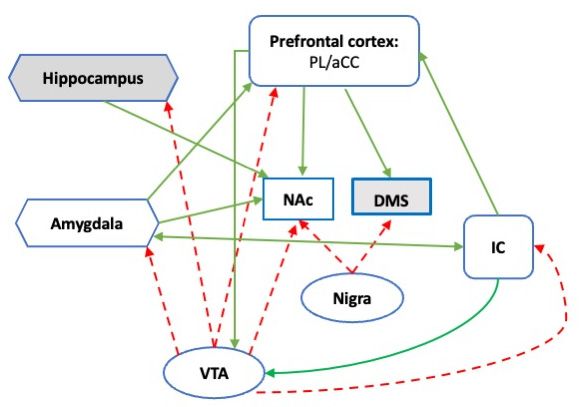
The DA circuit involved in successful coping with acute stress is the mesocorticolimbic
system made up by ventral tegmental area (VTA) DA neurons projecting to the PL, theBrain Sci. 2022, 12, 246 5 of 11
amygdala, the ventral striatum (NAc), and the hippocampus. A recent review defined
the role of the mesocorticolimbic circuitry: “ . . . as a hub linking circuits involved in
the emotional-motivational appraisal of salient information with networks underlying
executive functioning” [20]. Indeed, the mesocorticolimbic circuit includes core regions of
the SN, and it is in the position to engage the dorsal striatum through a dynamic ‘spiraling’
connection with the substantia nigra [74], the major source of striatal DA (Figure 1). Data
collected in animal models point to a rapid but temporary increase in extracellular DA in
PL, amygdala, and NAc in response to novel stress [19,75–77]. The NAc DA response to
novel stressors is dependent on increased NE transmission in the PL and is constrained by
DA transmission in the same brain area [19,78–81]. DA transmission in the NAc supports
active coping by stimulating DA receptors of the D2 type [9,19,82] and is dependent on
Brain Sci. 2022, 12, x FOR PEER REVIEW 6 of 1
the expectancy of successful active coping. Indeed, during the experience of a novel
stressful situation as inescapable and uncontrollable, DA release in the NAc is progressively
inhibited by a sharp reduction in NE and a parallel increase in DA in the PL [19].
Figure1.1.Schematic
Figure Schematic representation
representation of theofmesocorticolimbic
the mesocorticolimbic
DA systemDAassociated
system associated with stress cop
with stress coping.
ing. Dotted
Dotted lines indicate
lines indicate DA afferences;
DA afferences; solid linessolid lines
indicate indicateconnectivity;
functional functional white-filled
connectivity; white-filled
areas
areas
are partare partSNofinthe
of the SN inand
humans humans
rodentsand rodents
[53]. [53]. PL
PL prelimbic prelimbic
cortex; cortex; cingulate
aCC: Anterior aCC: Anterior
cortex; cingulat
cortex:
IC: InsulaIC: Insula
cortex; NAc: cortex; NAc:
Nucleus Nucleus accumbens/ventral
accumbens/ventral striatum; DMS: striatum; DMS:striatum;
Dorsomedial Dorsomedial
Nigra: striatum
nigra: Substantia
Substantia nigra parsnigra pars compacta;
compacta; VTA: Ventral VTA: Ventral
tegmental tegmental area.
area.
One
An of the stress
acute hypothesized mechanisms
experience by which brainofDA
or the administration acts is in mediating
anxiogenic compounds theincrease
dynamic balance between processes of “flexible updating and cognitive stabilization” [83].
DA output in the PL/IL area of Roman high-avoidance (RHA/Verh) but not of Roman low
As discussed, a PL-DMS circuit is engaged by the appraisal of stress controllability. Inac-
avoidance
tivation ratsbrain
of each (RLA/Verh).
area doesRHA/Verh display
not influence eitherrobust and long-lasting
the expression active coping
or the acquisition of with
novel
the stressors,
specific copingwhereas
responseRLA/Verh rats the
used to control show frequent
stressor. freezing
Instead, that interferes
connectivity between with ac
quiring the active avoidance response [86]. DA transmission in the
PL and DMS is required for the protective effects of the previous experience of controlPL does not mediat
the acquisition
against of aoutcomes
dysfunctional positively reinforcedinescapable-uncontrollable
of subsequent instrumental response, stressors
but it is[16].
necessary
A fo
PL-DMS
learningconnection is also involvedassociations
about action–outcome in the acquisition
[50].ofThus,
positively valued goal-directed
DA transmission within the PL
instrumental
could guideactions, which
the choice flexibly
of the mostadapt to changes
effective copinginstrategies
the valuein ofnovel
the goal. Instead,
stressful situations
connectivity between motor cortices and the dorsolateral striatum (DLS) is
In line with this hypothesis, a specific PL-DMS pathway is involved in decision-making required to
acquire a habitual instrumental response elicited by conditioned stimuli regardless of the
about motivationally conflicting options [87].
goal value [84]. All instrumental responses are acquired in parallel by the two circuits,
Rats with experience of control over a stressor show increased dendritic spine
which then compete to control the expression of instrumental responses. DA transmission
within
in DMS orPLDLS
[47]. Asdetermine
will discussed, experimental
whether the organismstressful experiences
expresses used
the acquired to reproduce dys
goal-directed
or habitual response [84]. It is thus possible that rats exposed to controllable stress engage in stri
functional phenotypes in animal models foster increased structural neuroplasticity
atal/limbic targets but reduced plasticity in PL [48–50,88] and the PL-DMS circuit is dis
rupted by the experience of these stressors [89]. Structural plasticity in the adult brain
relies on the experience-dependent proliferation and pruning of dendritic spines within
specific networks, leading to reorganization of connectivity between neuronal populaBrain Sci. 2022, 12, 246 6 of 11
the circuit responsible for habitual behavior to express a specific escape response but engage
the circuit responsible for goal-directed behavior to adapt to novel stressful conditions. In
line with this hypothesis, six weeks of voluntary wheel running, which protects rats against
the adverse effects of uncontrollable stress as the acute experience of control does, were
shown to significantly enhance DA release induced by an uncontrollable shock in the DMS
but not in DLS [85].
An acute stress experience or the administration of anxiogenic compounds increases
DA output in the PL/IL area of Roman high-avoidance (RHA/Verh) but not of Roman
low-avoidance rats (RLA/Verh). RHA/Verh display robust and long-lasting active coping
with novel stressors, whereas RLA/Verh rats show frequent freezing that interferes with
acquiring the active avoidance response [86]. DA transmission in the PL does not mediate
the acquisition of a positively reinforced instrumental response, but it is necessary for
learning about action–outcome associations [50]. Thus, DA transmission within the PL
could guide the choice of the most effective coping strategies in novel stressful situations.
In line with this hypothesis, a specific PL-DMS pathway is involved in decision-making
about motivationally conflicting options [87].
Rats with experience of control over a stressor show increased dendritic spines within
PL [47]. As discussed, experimental stressful experiences used to reproduce dysfunctional
phenotypes in animal models foster increased structural neuroplasticity in striatal/limbic
targets but reduced plasticity in PL [48–50,88] and the PL-DMS circuit is disrupted by
the experience of these stressors [89]. Structural plasticity in the adult brain relies on
the experience-dependent proliferation and pruning of dendritic spines within specific
networks, leading to reorganization of connectivity between neuronal populations. DA is
required for glutamate-induced spinogenesis and exerts dichotomous effects on neurons
expressing the D1 or D2 receptor subtypes so that they store memories of reward and
reward omission, respectively, through cell-type-specific spine enlargement [90,91]. Finally,
reduced DA availability due to degeneration of DA cells interferes with spinogenesis while
increasing pruning [92].
Moreover, there is strong evidence that DA transmission modulates SN connectiv-
ity [93–95]. DA modulates aCC activity during executive tasks and the synaptic availability
of DA may be directly related to efficient IC function. On the other hand, a high correla-
tion between the binding of the D2/D3 ligand [18F]-fallypride and grey matter density
has been observed in the aCC, IC, and midbrain, suggesting that reduced grey matter
observed in the SN of schizophrenics is directly associated with DA deficits [95]. Moreover,
DA synthesis capacity has been recently associated with greater SN connectivity, partic-
ularly in brain regions that act as information-processing hubs [94] and there is strong
evidence for VTA functional connectivity with the posterior cingulate/precuneus, a central
hub node of the DMN [96]. On the other hand, progressive depletion of striatal DA is
the hallmark of Parkinson’s disease (PD). Around 30% of PD patients present cognitive
deficits long before the appearance of classic motor symptoms and evidence from clinical
and preclinical studies on cognitive deficits in PD points to a role of reduced striatal DA
availability [6,97]. There is strong evidence that reduced DA availability in the ventral
striatum (NAc) precedes typical motor deficits in PD and several studies found a reduced
connectivity between the aCC and the ventral striatum [98]. Finally, PD patients with mild
cognitive impairment, who are susceptible to developing dementia, are characterized by
severe DA depletion in the associative striatum and reduced D2 receptor availability in the
IC [99]. These findings seem coherent with the dynamic relationship between PL and NAc
DA observed in stressed animals and bridge dysfunctional connectivity within SN and
between large brain networks with cognitive impairment and reduced DA transmission.
Together, the discussed evidence points to a significant role of DA transmission in
cortical, limbic, and striatal nodes in the immediate and long-term adaptive effects of
successful coping with stress challenges. Moreover, the reviewed evidence suggests that
DA exerts this role by modulating both functional and structural brain connectivity within
the SN and supports a strong influence of this modulation on cognitive capacity.Brain Sci. 2022, 12, 246 7 of 11
5. Conclusions
This brief review discussed evidence of an overlap between brain circuitry support-
ing the development and stabilization of adaptive stress coping strategies and cognitive
functioning in aging and disease. Moreover, we pointed to a significant moderating role of
DA transmission in specific nodes of the SN in circuital functioning and plasticity in the
adult brain. Based on these considerations, it is reasonable to hypothesize that experiences
of successful coping with life events, through DA transmission-induced modulation of
brain functional and structural connectivity, contribute to establishing a rich CR. Instead,
reduced brain DA availability, due to genetic predisposition or to a history of uncontrol-
lable/unavoidable experiences, can undermine the development and maintenance of CR,
leading to more severe and rapid cognitive deterioration in response to neurodegeneration.
Finally, the proposed hypothesis opens new opportunities to translational research by
overcoming the problems of reproducing classic CR proxies in animal models.
Author Contributions: S.C. proposed the topic and prepared a first sketch of the manuscript; S.C.,
C.L. and C.O. discussed and equally contributed to the submitted versions of the paper. All authors
have read and agreed to the published version of the manuscript.
Funding: This work was founded by Italian Ministry of Health: NoGR 2018 1236 5991.
Data Availability Statement: Not applicable.
Conflicts of Interest: The authors declare no conflict of interest.
References
1. Stern, Y. What is cognitive reserve? Theory and research application of the reserve concept. J. Int. Neuropsychol. Soc. 2002, 8,
448–460. [CrossRef] [PubMed]
2. Stern, Y. How Can Cognitive Reserve Promote Cognitive and Neurobehavioral Health? Arch. Clin. Neuropsychol. 2021, 36,
1291–1295. [CrossRef] [PubMed]
3. Pettigrew, C.; Soldan, A. Defining Cognitive Reserve and Implications for Cognitive Aging. Curr. Neurol. Neurosci. Rep. 2019, 19,
1–12. [CrossRef] [PubMed]
4. Cabeza, R.; Albert, M.; Belleville, S.; Craik, F.I.M.; Duarte, A.; Grady, C.L.; Lindenberger, U.; Nyberg, L.; Park, D.C.; Reuter-Lorenz,
P.A.; et al. Maintenance, reserve and compensation: The cognitive neuroscience of healthy ageing. Nat. Rev. Neurosci. 2018, 19,
701–710. [CrossRef]
5. Estrada-Lopez, M.; Garcia-Martin, S.; Canton-Mayo, I. Cognitive Dysfunction in Multiple Sclerosis: Educational Level as a
Protective Factor. Neurol. Int. 2021, 13, 335–342. [CrossRef]
6. Loftus, A.M.; Gasson, N.; Lopez, N.; Sellner, M.; Reid, C.; Cocks, N.; Lawrence, B.J. Cognitive Reserve, Executive Function, and
Memory in Parkinson’s Disease. Brain. Sci. 2021, 11, 992. [CrossRef]
7. Herrero, P.; Contador, I.; Stern, Y.; Fernandez-Calvo, B.; Sanchez, A.; Ramos, F. Influence of cognitive reserve in schizophrenia: A
systematic review. Neurosci. Biobehav. Rev. 2020, 108, 149–159. [CrossRef]
8. Perneczky, R.; Kempermann, G.; Korczyn, A.D.; Matthews, F.E.; Ikram, M.A.; Scarmeas, N.; Chetelat, G.; Stern, Y.; Ewers,
M. Translational research on reserve against neurodegenerative disease: Consensus report of the International Conference on
Cognitive Reserve in the Dementias and the Alzheimer’s Association Reserve, Resilience and Protective Factors Professional
Interest Area working groups. BMC Med. 2019, 17, 47.
9. Ventura, R.; Cabib, S.; Babicola, L.; Andolina, D.; Di Segni, M.; Orsini, C. Interactions Between Experience, Genotype and Sex in
the Development of Individual Coping Strategies. Front. Behav. Neurosci. 2021, 15, 785739. [CrossRef]
10. Holmes, T.H.; Rahe, R.H. The Social Readjustment Rating Scale. J. Psychosom. Res. 1967, 11, 213–218. [CrossRef]
11. Scully, J.; Tosi, H.; Banning, K. Life Event Checklists: Revisiting the Social Readjustment Rating Scale after 30 Years. Educ. Psychol.
Meas. 2000, 60, 864–876. [CrossRef]
12. Charney, D.S. Psychobiological mechanisms of resilience and vulnerability: Implications for successful adaptation to extreme
stress. Am. J. Psychiatry 2004, 161, 195–216. [CrossRef] [PubMed]
13. Daskalakis, N.P.; Bagot, R.C.; Parker, K.J.; Vinkers, C.H.; de Kloet, E.R. The three-hit concept of vulnerability and resilience: Toward
understanding adaptation to early-life adversity outcome. Psychoneuroendocrinology 2013, 38, 1858–1873. [CrossRef] [PubMed]
14. Taylor, A.E.; Stanton, A.L. Coping resources, coping processes, and mental health. Annu. Rev. Clin. Psychol. 2007, 3,
377–401. [CrossRef]
15. Cabib, S.; Campus, P.; Conversi, D.; Orsini, C.; Puglisi-Allegra, S. Functional and Dysfunctional Neuroplasticity in Learning to
Cope with Stress. Brain. Sci. 2020, 10, 127. [CrossRef]
16. Maier, S.F. Behavioral control blunts reactions to contemporaneous and future adverse events: Medial prefrontal cortex plasticity
and a corticostriatal network. Neurobiol. Stress 2015, 1, 12–22. [CrossRef]Brain Sci. 2022, 12, 246 8 of 11
17. Cabib, S.; Campus, P.; Latagliata, E.C.; Orsini, C.; Tarmati, V. Repetitive and Inflexible Active Coping and Addiction-like
Neuroplasticity in Stressed Mice of a Helplessness-Resistant Inbred Strain. Behav. Sci. 2021, 11, 174. [CrossRef]
18. Southwick, S.M.; Vythilingam, M.; Charney, D.S. The psychobiology of depressione and resilience to stress: Implications for
Prevention and Treatment£™. Annu. Rev. Clin. Psychol. 2005, 1, 255–291. [CrossRef]
19. Cabib, S.; Puglisi-Allegra, S. The mesoaccumbens dopamine in coping with stress. Neurosci. Biobehav. Rev. 2012, 36,
79–89. [CrossRef]
20. Douma, E.H.; de Kloet, E.R. Stress-induced plasticity and functioning of ventral tegmental dopamine neurons. Neurosci. Biobehav.
Rev. 2020, 108, 48–77. [CrossRef]
21. Delva, N.C.; Stanwood, G.D. Dysregulation of brain dopamine systems in major depressive disorder. Exp. Biol. Med. 2021, 246,
1084–1093. [CrossRef] [PubMed]
22. Goldstein Ferber, S.; Weller, A.; Yadid, G.; Friedman, A. Discovering the Lost Reward: Critical Locations for Endocannabi-
noid Modulation of the Cortico-Striatal Loop That Are Implicated in Major Depression. Int. J. Mol. Sci. 2021, 22, 1867.
[CrossRef] [PubMed]
23. Wang, S.; Leri, F.; Rizvi, S.J. Anhedonia as a central factor in depression: Neural mechanisms revealed from preclinical to clinical
evidence. Prog. Neuropsychopharmacol. Biol. Psychiatry 2021, 110, 110289. [CrossRef] [PubMed]
24. McLaurin, K.A.; Harris, M.; Madormo, V.; Harrod, S.B.; Mactutus, C.F.; Booze, R.M. HIV-Associated Apathy/Depression and
Neurocognitive Impairments Reflect Persistent Dopamine Deficits. Cells 2021, 10, 2158. [CrossRef] [PubMed]
25. Noworyta, K.; Cieslik, A.; Rygula, R. Neuromolecular Underpinnings of Negative Cognitive Bias in Depression. Cells 2021,
10, 3157. [CrossRef]
26. Backman, L.; Lindenberger, U.; Li, S.C.; Nyberg, L. Linking cognitive aging to alterations in dopamine neurotransmitter
functioning: Recent data and future avenues. Neurosci. Biobehav. Rev. 2010, 34, 670–677. [CrossRef]
27. Pan, X.; Kaminga, A.C.; Wen, S.W.; Wu, X.; Acheampong, K.; Liu, A. Dopamine and Dopamine Receptors in Alzheimer’s Disease:
A Systematic Review and Network Meta-Analysis. Front. Aging Neurosci. 2019, 11, 175. [CrossRef]
28. D’Amelio, M.; Puglisi-Allegra, S.; Mercuri, N. The role of dopaminergic midbrain in Alzheimer’s disease: Translating basic
science into clinical practice. Pharmacol. Res. 2018, 130, 414–419. [CrossRef]
29. Koch, G.; Di Lorenzo, F.; Bonni, S.; Giacobbe, V.; Bozzali, M.; Caltagirone, C.; Martorana, A. Dopaminergic modulation of cortical
plasticity in Alzheimer’s disease patients. Neuropsychopharmacology 2014, 39, 2654–2661. [CrossRef]
30. Nobili, A.; Latagliata, E.C.; Viscomi, M.T.; Cavallucci, V.; Cutuli, D.; Giacovazzo, G.; Krashia, P.; Rizzo, F.R.; Marino, R.; Federici,
M.; et al. Dopamine neuronal loss contributes to memory and reward dysfunction in a model of Alzheimer’s disease. Nat.
Commun. 2017, 8, 14727. [CrossRef]
31. McEwen, B.S.; Gianaros, P.J. Stress- and allostasis-induced brain plasticity. Annu. Rev. Med. 2011, 62, 431–445.
[CrossRef] [PubMed]
32. de Kloet, E.R.; Molendijk, M.L. Coping with the Forced Swim Stressor: Towards Understanding an Adaptive Mechanism. Neural.
Plast. 2016, 2016, 6503162. [CrossRef] [PubMed]
33. Vorhees, C.V.; Williams, M.T. Assessing spatial learning and memory in rodents. ILAR J. 2014, 55, 310–332. [CrossRef] [PubMed]
34. Piantadosi, P.T.; Yeates, D.C.M.; Floresco, S.B. Cooperative and dissociable involvement of the nucleus accumbens core and
shell in the promotion and inhibition of actions during active and inhibitory avoidance. Neuropharmacology 2018, 138, 57–71.
[CrossRef] [PubMed]
35. Wenzel, J.M.; Oleson, E.B.; Gove, W.N.; Cole, A.B.; Gyawali, U.; Dantrassy, H.M.; Bluett, R.J.; Dryanovski, D.I.; Stuber, G.D.;
Deisseroth, K.; et al. Phasic Dopamine Signals in the Nucleus Accumbens that Cause Active Avoidance Require Endocannabinoid
Mobilization in the Midbrain. Curr. Biol. 2018, 28, 1392–1404.e1395. [CrossRef]
36. Diehl, M.M.; Bravo-Rivera, C.; Quirk, G.J. The study of active avoidance: A platform for discussion. Neurosci. Biobehav. Rev. 2019,
107, 229–237. [CrossRef]
37. Roelofs, K. Freeze for action: Neurobiological mechanisms in animal and human freezing. Philos. Trans. R. Soc. Lond. B Biol. Sci.
2017, 372, 20160206. [CrossRef]
38. Boeke, E.A.; Moscarello, J.M.; LeDoux, J.E.; Phelps, E.A.; Hartley, C.A. Active Avoidance: Neural Mechanisms and Attenuation of
Pavlovian Conditioned Responding. J. Neurosci. 2017, 37, 4808–4818. [CrossRef]
39. Moscarello, J.M.; Maren, S. Flexibility in the face of fear: Hippocampal-prefrontal regulation of fear and avoidance. Curr. Opin.
Behav. Sci. 2018, 19, 44–49. [CrossRef]
40. Maier, S.F.; Seligman, M.E. Learned helplessness at fifty: Insights from neuroscience. Psychol. Rev. 2016, 123, 349–367. [CrossRef]
41. Cabib, S.; Campus, P.; Colelli, V. Learning to cope with stress: Psychobiological mechanisms of stress resilience. Rev. Neurosci.
2012, 23, 659–672. [CrossRef] [PubMed]
42. Roozendaal, B.; McEwen, B.S.; Chattarji, S. Stress, memory and the amygdala. Nat. Rev. Neurosci. 2009, 10, 423–433.
[CrossRef] [PubMed]
43. Barsegyan, A.; Mirone, G.; Ronzoni, G.; Guo, C.; Song, Q.; van Kuppeveld, D.; Schut, E.H.S.; Atsak, P.; Teurlings, S.; McGaugh,
J.L.; et al. Glucocorticoid enhancement of recognition memory via basolateral amygdala-driven facilitation of prelimbic cortex
interactions. Proc. Natl. Acad. Sci. USA 2019, 116, 7077–7082. [CrossRef] [PubMed]
44. Namkung, H.; Thomas, K.L.; Hall, J.; Sawa, A. Parsing neural circuits of fear learning and extinction across basic and clinical
neuroscience: Towards better translation. Neurosci. Biobehav. Rev. 2021, 134, 104502. [CrossRef] [PubMed]Brain Sci. 2022, 12, 246 9 of 11
45. Klein, A.S.; Dolensek, N.; Weiand, C.; Gogolla, N. Fear balance is maintained by bodily feedback to the insular cortex in mice.
Science 2021, 374, 1010–1015. [CrossRef]
46. Maier, S.F.; Watkins, L.R. Role of the medial prefrontal cortex in coping and resilience. Brain. Res. 2010, 1355, 52–60. [CrossRef]
47. Baratta, M.V.; Gruene, T.M.; Dolzani, S.D.; Chun, L.E.; Maier, S.F.; Shansky, R.M. Controllable stress elicits circuit-specific patterns
of prefrontal plasticity in males, but not females. Brain. Struct. Funct. 2019, 224, 1831–1843. [CrossRef]
48. Willner, P. The chronic mild stress (CMS) model of depression: History, evaluation and usage. Neurobiol. Stress. 2017, 6,
78–93. [CrossRef]
49. Dias-Ferreira, E.; Sousa, J.C.; Melo, I.; Morgado, P.; Mesquita, A.R.; Cerqueira, J.J.; Costa, R.M.; Sousa, N. Chronic stress causes
frontostriatal reorganization and affects decision-making. Science 2009, 325, 621–625. [CrossRef]
50. Woon, E.P.; Sequeira, M.K.; Barbee, B.R.; Gourley, S.L. Involvement of the rodent prelimbic and medial orbitofrontal cortices in
goal-directed action: A brief review. J. Neurosci. Res. 2020, 98, 1020–1030. [CrossRef]
51. Li, W.; Wang, Y.; Ward, B.D.; Antuono, P.G.; Li, S.J.; Goveas, J.S. Intrinsic inter-network brain dysfunction correlates with symptom
dimensions in late-life depression. J. Psychiatr. Res. 2017, 87, 71–80. [CrossRef] [PubMed]
52. Hermans, E.J.; Henckens, M.J.; Joels, M.; Fernandez, G. Dynamic adaptation of large-scale brain networks in response to acute
stressors. Trends. Neurosci. 2014, 37, 304–314. [CrossRef] [PubMed]
53. Seeley, W.W. The Salience Network: A Neural System for Perceiving and Responding to Homeostatic Demands. J. Neurosci. 2019,
39, 9878–9882. [CrossRef] [PubMed]
54. Kim, J.; Kim, Y.K. Crosstalk between Depression and Dementia with Resting-State fMRI Studies and Its Relationship with
Cognitive Functioning. Biomedicines 2021, 9, 82. [CrossRef]
55. Menon, V. Large-scale brain networks and psychopathology: A unifying triple network model. Trends. Cogn. Sci. 2011, 15,
483–506. [CrossRef]
56. Peters, S.K.; Dunlop, K.; Downar, J. Cortico-Striatal-Thalamic Loop Circuits of the Salience Network: A Central Pathway in
Psychiatric Disease and Treatment. Front. Syst. Neurosci. 2016, 10, 104. [CrossRef]
57. Laubach, M.; Amarante, L.M.; Swanson, K.; White, S.R. What, If Anything, Is Rodent Prefrontal Cortex? eNeuro 2018, 5. [CrossRef]
58. Chand, G.B.; Wu, J.; Hajjar, I.; Qiu, D. Interactions of the Salience Network and Its Subsystems with the Default-Mode and the
Central-Executive Networks in Normal Aging and Mild Cognitive Impairment. Brain. Connect. 2017, 7, 401–412. [CrossRef]
59. Putcha, D.; Ross, R.S.; Cronin-Golomb, A.; Janes, A.C.; Stern, C.E. Salience and Default Mode Network Coupling Predicts
Cognition in Aging and Parkinson’s Disease. J. Int. Neuropsychol. Soc. 2016, 22, 205–215. [CrossRef]
60. van Leeuwen, J.M.C.; Vinkers, C.H.; Vink, M.; Kahn, R.S.; Joels, M.; Hermans, E.J. Disrupted upregulation of salience network
connectivity during acute stress in siblings of schizophrenia patients. Psychol. Med. 2021, 51, 1038–1048. [CrossRef]
61. Tsvetanov, K.A.; Henson, R.N.; Tyler, L.K.; Razi, A.; Geerligs, L.; Ham, T.E.; Rowe, J.B.; Cambridge Centre for Ageing and
Neuroscience. Extrinsic and Intrinsic Brain Network Connectivity Maintains Cognition across the Lifespan Despite Accelerated
Decay of Regional Brain Activation. J. Neurosci. 2016, 36, 3115–3126. [CrossRef] [PubMed]
62. Santarnecchi, E.; Sprugnoli, G.; Tatti, E.; Mencarelli, L.; Neri, F.; Momi, D.; Di Lorenzo, G.; Pascual-Leone, A.; Rossi, S.; Rossi, A.
Brain functional connectivity correlates of coping styles. Cogn. Affect. Behav. Neurosci. 2018, 18, 495–508. [CrossRef] [PubMed]
63. Ewers, M.; Luan, Y.; Frontzkowski, L.; Neitzel, J.; Rubinski, A.; Dichgans, M.; Hassenstab, J.; Gordon, B.A.; Chhatwal, J.P.; Levin, J.;
et al. Segregation of functional networks is associated with cognitive resilience in Alzheimer’s disease. Brain 2021, 144, 2176–2185.
[CrossRef] [PubMed]
64. Ewers, M. Reserve in Alzheimer’s disease: Update on the concept, functional mechanisms and sex differences. Curr. Opin.
Psychiatry 2020, 33, 178–184. [CrossRef] [PubMed]
65. Serra, L.; Mancini, M.; Cercignani, M.; Di Domenico, C.; Spano, B.; Giulietti, G.; Koch, G.; Marra, C.; Bozzali, M. Network-Based
Substrate of Cognitive Reserve in Alzheimer’s Disease. J. Alzheimers Dis. 2017, 55, 421–430. [CrossRef] [PubMed]
66. Sun, F.W.; Stepanovic, M.R.; Andreano, J.; Barrett, L.F.; Touroutoglou, A.; Dickerson, B.C. Youthful Brains in Older Adults:
Preserved Neuroanatomy in the Default Mode and Salience Networks Contributes to Youthful Memory in Superaging. J. Neurosci.
2016, 36, 9659–9668. [CrossRef]
67. Conti, L.; Riccitelli, G.C.; Preziosa, P.; Vizzino, C.; Marchesi, O.; Rocca, M.A.; Filippi, M. Effect of cognitive reserve on structural
and functional MRI measures in healthy subjects: A multiparametric assessment. J. Neurol. 2021, 268, 1780–1791. [CrossRef]
68. Bahtiyar, S.; Gulmez Karaca, K.; Henckens, M.; Roozendaal, B. Norepinephrine and glucocorticoid effects on the brain mechanisms
underlying memory accuracy and generalization. Mol. Cell Neurosci. 2020, 108, 103537. [CrossRef]
69. de Kloet, E.R.; de Kloet, S.F.; de Kloet, C.S.; de Kloet, A.D. Top-down and bottom-up control of stress-coping. J. Neuroendocrinol.
2019, 31, e12675. [CrossRef]
70. Liang, F.; Xu, Q.; Jiang, M.; Feng, R.; Jiang, S.; Yuan, B.; Xu, S.; Wu, T.; Wang, F.; Huang, J.H. Emotion Induced Monoamine
Neuromodulator Release Affects Functional Neurological Disorders. Front. Cell Dev. Biol. 2021, 9, 633048. [CrossRef]
71. Puglisi-Allegra, S.; Andolina, D. Serotonin and stress coping. Behav. Brain. Res. 2015, 277, 58–67. [CrossRef] [PubMed]
72. de Boer, S.F.; Buwalda, B.; Koolhaas, J.M. Untangling the neurobiology of coping styles in rodents: Towards neural mechanisms
underlying individual differences in disease susceptibility. Neurosci. Biobehav. Rev. 2017, 74, 401–422. [CrossRef] [PubMed]Brain Sci. 2022, 12, 246 10 of 11
73. Maier, S.F.; Watkins, L.R. Stressor controllability and learned helplessness: The roles of the dorsal raphe nucleus, serotonin, and
corticotropin-releasing factor. Neurosci. Biobehav. Rev. 2005, 29, 829–841. [CrossRef] [PubMed]
74. Haber, S.N.; Fudge, J.L.; McFarland, N.R. Striatonigrostriatal pathways in primates form an ascending spiral from the shell to the
dorsolateral striatum. J. Neurosci. 2000, 20, 2369–2382. [CrossRef]
75. Stevenson, C.W.; Sullivan, R.M.; Gratton, A. Effects of basolateral amygdala dopamine depletion on the nucleus accumbens and
medial prefrontal cortical dopamine responses to stress. Neuroscience 2003, 116, 285–293. [CrossRef]
76. Bland, S.T.; Hargrave, D.; Pepin, J.L.; Amat, J.; Watkins, L.R.; Maier, S.F. Stressor controllability modulates stress-induced
dopamine and serotonin efflux and morphine-induced serotonin efflux in the medial prefrontal cortex. Neuropsychopharmacology
2003, 28, 1589–1596. [CrossRef]
77. Moghaddam, B. Stress activation of glutamate neurotransmission in the prefrontal cortex: Implications for dopamine-associated
psychiatric disorders. Biol. Psychiatry 2002, 511, 775–787. [CrossRef]
78. Latagliata, E.C.; Valzania, A.; Pascucci, T.; Campus, P.; Cabib, S.; Puglisi-Allegra, S. Stress-induced activation of ventral tegmental
mu-opioid receptors reduces accumbens dopamine tone by enhancing dopamine transmission in the medial pre-frontal cortex.
Psychopharmacology 2014, 231, 4099–4108. [CrossRef]
79. Fiore, V.G.; Mannella, F.; Mirolli, M.; Latagliata, E.C.; Valzania, A.; Cabib, S.; Dolan, R.J.; Puglisi-Allegra, S.; Baldassarre, G.
Corticolimbic catecholamines in stress: A computational model of the appraisal of controllability. Brain. Struct. Funct. 2015, 220,
1339–1353. [CrossRef]
80. Nicniocaill, B.; Gratton, A. Medial prefrontal cortical alpha1 adrenoreceptor modulation of the nucleus accumbens dopamine
response to stress in Long-Evans rats. Psychopharmacology 2007, 191, 835–842. [CrossRef]
81. Doherty, M.D.; Gratton, A. Medial prefrontal cortical D1 receptor modulation of the meso-accumbens dopamine response to
stress: An electrochemical study in freely-behaving rats. Brain. Res. 1996, 715, 86–97. [CrossRef]
82. Lloyd, K.; Dayan, P. Safety out of control: Dopamine and defence. Behav. Brain. Funct. 2016, 12, 15. [CrossRef] [PubMed]
83. Cools, R.; D’Esposito, M. Inverted-U-shaped dopamine actions on human working memory and cognitive control. Biol. Psychiatry
2011, 69, e113–e125. [CrossRef] [PubMed]
84. Balleine, B.W.; O’Doherty, J.P. Human and rodent homologies in action control: Corticostriatal determinants of goal-directed and
habitual action. Neuropsychopharmacology 2010, 35, 48–69. [CrossRef] [PubMed]
85. Clark, P.J.; Amat, J.; McConnell, S.O.; Ghasem, P.R.; Greenwood, B.N.; Maier, S.F.; Fleshner, M. Running Reduces Uncontrollable
Stress-Evoked Serotonin and Potentiates Stress-Evoked Dopamine Concentrations in the Rat Dorsal Striatum. PLoS ONE 2015,
10, e0141898. [CrossRef]
86. Giorgi, O.; Lecca, D.; Piras, G.; Driscoll, P.; Corda, M.G. Dissociation between mesocortical dopamine release and fear-related
behaviours in two psychogenetically selected lines of rats that differ in coping strategies to aversive conditions. Eur. J. Neurosci.
2003, 17, 2716–2726. [CrossRef]
87. Friedman, A.; Homma, D.; Gibb, L.G.; Amemori, K.; Rubin, S.J.; Hood, A.S.; Riad, M.H.; Graybiel, A.M. A Corticostriatal Path
Targeting Striosomes Controls Decision-Making under Conflict. Cell 2015, 161, 1320–1333. [CrossRef]
88. Chen, X.; Jiang, Y.; Wang, J.; Liu, Y.; Xiao, M.; Song, C.; Bai, Y.; Yinuo Han, N.; Han, F. Synapse impairment associated with
enhanced apoptosis in post-traumatic stress disorder. Synapse 2020, 74, e22134. [CrossRef]
89. Friedman, A.; Homma, D.; Bloem, B.; Gibb, L.G.; Amemori, K.I.; Hu, D.; Delcasso, S.; Truong, T.F.; Yang, J.; Hood,
A.S.; et al. Chronic Stress Alters Striosome-Circuit Dynamics, Leading to Aberrant Decision-Making. Cell 2017, 171,
1191–1205.e1128. [CrossRef]
90. Runge, K.; Cardoso, C.; de Chevigny, A. Dendritic Spine Plasticity: Function and Mechanisms. Front. Synaptic. Neurosci. 2020,
12, 36. [CrossRef]
91. Qiao, H.; Li, M.X.; Xu, C.; Chen, H.B.; An, S.C.; Ma, X.M. Dendritic Spines in Depression: What We Learned from Animal Models.
Neural. Plast. 2016, 2016, 8056370. [CrossRef] [PubMed]
92. Witzig, V.S.; Komnig, D.; Falkenburger, B.H. Changes in Striatal Medium Spiny Neuron Morphology Resulting from Dopamine
Depletion Are Reversible. Cells 2020, 9, 2441. [CrossRef] [PubMed]
93. Conio, B.; Martino, M.; Magioncalda, P.; Escelsior, A.; Inglese, M.; Amore, M.; Northoff, G. Opposite effects of dopamine
and serotonin on resting-state networks: Review and implications for psychiatric disorders. Mol. Psychiatry 2020, 25, 82–93.
[CrossRef] [PubMed]
94. McCutcheon, R.A.; Nour, M.M.; Dahoun, T.; Jauhar, S.; Pepper, F.; Expert, P.; Veronese, M.; Adams, R.A.; Turkheimer, F.; Mehta,
M.A.; et al. Mesolimbic Dopamine Function Is Related to Salience Network Connectivity: An Integrative Positron Emission
Tomography and Magnetic Resonance Study. Biol. Psychiatry 2019, 85, 368–378. [CrossRef] [PubMed]
95. Palaniyappan, L.; Liddle, P.F. Does the salience network play a cardinal role in psychosis? An emerging hypothesis of insular
dysfunction. J. Psychiatry Neurosci. 2012, 37, 17–27. [CrossRef]
96. Li, J.; Curley, W.H.; Guerin, B.; Dougherty, D.D.; Dalca, A.V.; Fischl, B.; Horn, A.; Edlow, B.L. Mapping the subcortical connectivity
of the human default mode network. Neuroimage 2021, 245, 118758. [CrossRef]
97. Decourt, M.; Jimenez-Urbieta, H.; Benoit-Marand, M.; Fernagut, P.O. Neuropsychiatric and Cognitive Deficits in Parkinson’s
Disease and Their Modeling in Rodents. Biomedicines 2021, 9, 684. [CrossRef]Brain Sci. 2022, 12, 246 11 of 11
98. Theis, H.; Probst, C.; Fernagut, P.O.; van Eimeren, T. Unlucky punches: The vulnerability-stress model for the development of
impulse control disorders in Parkinson’s disease. NPJ Park. Dis. 2021, 7, 112. [CrossRef]
99. Christopher, L.; Marras, C.; Duff-Canning, S.; Koshimori, Y.; Chen, R.; Boileau, I.; Segura, B.; Monchi, O.; Lang, A.E.; Rusjan, P.;
et al. Combined insular and striatal dopamine dysfunction are associated with executive deficits in Parkinson’s disease with mild
cognitive impairment. Brain 2014, 137, 565–575. [CrossRef]You can also read