Recovery from 6-month spaceflight at the International Space Station: muscle-related stress into a proinflammatory setting
←
→
Page content transcription
If your browser does not render page correctly, please read the page content below
THE

                          JOURNAL      • RESEARCH •         www.fasebj.org

               Recovery from 6-month spaceflight at the International
               Space Station: muscle-related stress into a
               proinflammatory setting
               Miriam Capri,*,† Cristina Morsiani,*,†,1 Aurelia Santoro,*,† Manuela Moriggi,‡,§ Maria Conte,*,†
               Morena Martucci,*,† Elena Bellavista,* Cristina Fabbri,*,† Enrico Giampieri,†,{ Kirsten Albracht,k,#
               Martin Flück,** Severin Ruoss,** Lorenza Brocca,†† Monica Canepari,†† Emanuela Longa,‡‡ Irene Di Giulio,§§
               Roberto Bottinelli,††,{{ Paolo Cerretelli,‡,§ Stefano Salvioli,*,† Cecilia Gelfi,kk,##,2 Claudio Franceschi,***,2
               Marco Narici,†††,2 and Jörn Rittweger‡‡‡,§§§,2
               *Department of Experimental, Diagnostic, and Specialty Medicine, †Galvani Interdepartmental Center, and {Department of Physics and
               Astronomy, University of Bologna, Bologna, Italy; ‡National Research Council–Institute of Molecular Bioimaging and Physiology
               (CNR–IBFM), Segrate, Milan, Italy; §Italian National Olympic Committee (CONI), Rome, Italy; kFaculty of Medical Engineering and
               Technomathematics, FH Aachen University of Applied Sciences, Aachen, Germany; #Institute of Biomechanics and Orthopaedics, German
               Sport University, Cologne, Germany; **Department of Orthopaedics, University of Zürich, Zürich, Switzerland; ††Department of Molecular
               Medicine and ‡‡Sport Medicine Center, University of Pavia, Pavia, Italy; §§Centre of Human and Applied Physiological Sciences, King’s College
               London, London, United Kingdom; {{Fondazione Salvatore Maugeri, Institute of Hospitalization and Scientific Care (IRCCS), Scientific
               Institute of Pavia, Pavia, Italy; kkDepartment of Biomedical Sciences for Health, University of Milan, Milan, Italy; ##IRCCS, Istituto Ortopedico
               Galeazzi, Milan, Italy; ***Department of Applied Mathematics, Institute of Information Technology, Mathematics, and Mechanics (ITMM),
               Lobachevsky State University of Nizhny Novgorod–National Research University (UNN), Nizhny Novogoro, Russia; †††Department of
               Biomedical Sciences, University of Padova, Padua, Italy; ‡‡‡Institute of Aerospace Medicine, German Aerospace Center (DLR), Cologne,
               Germany; and §§§Department of Pediatrics and Adolescent Medicine, University of Cologne, Cologne, Germany

               ABSTRACT: The Sarcolab pilot study of 2 crewmembers, investigated before and after a 6-mo International Space Station
               mission, has demonstrated the substantial muscle wasting and weakness, along with disruption of muscle’s oxidative
               metabolism. The present work aimed at evaluating the pro/anti-inflammatory status in the same 2 crewmembers (A, B).
               Blood circulating (c-)microRNAs (miRs), c-proteasome, c-mitochondrial DNA, and cytokines were assessed by real-time
               quantitative PCR or ELISA tests. Time series analysis was performed (i.e., before flight and after landing) at 1 and 15 d of
               recovery (R+1 and R+15, respectively). C-biomarkers were compared with an age-matched control population and with
               2-dimensional proteomic analysis of the 2 crewmembers’ muscle biopsies. Striking differences were observed between the
               2 crewmembers at R+1, in terms of inflamma-miRs (c-miRs-21-5p, -126-3p, and -146a-5p), muscle specific (myo)-miR-206,
               c-proteasome, and IL-6/leptin, thus making the 2 astronauts dissimilar to each other. Final recovery levels of c-proteasome,
               c-inflamma-miRs, and c-myo-miR-206 were not reverted to the baseline values in crewmember A. In both crewmembers,
               myo-miR-206 changed significantly after recovery. Muscle biopsy of astronaut A showed an impressive 80% increase of
               a-1-antitrypsin, a target of miR-126-3p. These results point to a strong stress response induced by spaceflight involving
               muscle tissue and the proinflammatory setting, where inflamma-miRs and myo-miR-206 mediate the systemic recovery
               phase after landing.—Capri, M., Morsiani, C., Santoro, A., Moriggi, M., Conte, M., Martucci, M., Bellavista, E., Fabbri, C.,
               Giampieri, E., Albracht, K., Flück, M., Ruoss, S., Brocca, L., Canepari, M., Longa, E., Di Giulio, I., Bottinelli, R., Cerretelli, P.,
               Salvioli, S., Gelfi, C., Franceschi, C., Narici, M., Rittweger, J. Recovery from 6-month spaceflight at the International Space
               Station: muscle-related stress into a proinflammatory setting. FASEB J. 33, 5168–5180 (2019). www.fasebj.org
               KEY WORDS:       microRNA-206          •   inflamma-miRs        •   proteasome      •   SERPINA1

               ABBREVIATIONS: 2-D, 2-dimensional; GO, Gene Ontology; ISS, International Space Station; miR, microRNA; MS/MS, tandem mass spectrometry;
               mtDNA, mitochondrial DNA; myo, muscle specific; OD, optical density; PMF, peptide mass fingerprinting; preflight, 76–79 d before flight; SERPINA1,
               a-1-antitrypsin
               1
                 Correspondence: Department of Experimental, Diagnostic and Specialty Medicine (DIMES), Alma Mater Studiorum, University of Bologna, Via San
                 Giacomo, 12, 40126 Bologna, Italy. E-mail: cristina.morsiani2@unibo.it
               2
                 These authors contributed equally to this work.
                 This is an Open Access article distributed under the terms of the Creative Commons Attribution 4.0 International (CC BY 4.0) (http://creativecommons.
               org/licenses/by/4.0/) which permits unrestricted use, distribution, and reproduction in any medium, provided the original work is properly cited.
               doi: 10.1096/fj.201801625R
               This article includes supplemental data. Please visit http://www.fasebj.org to obtain this information.

               5168                                                                                                      0892-6638/19/0033-5168 © The Author(s)
ebj.org by Universita Degli Studi Padova Ist Fisiologia Umana (147.162.3.235) on November 21, 2019. The FASEB Journal Vol. ${article.issue.getVolume()}, No. ${article.issue.getIssu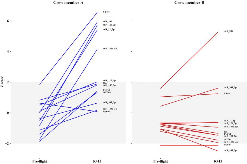
It is known that short- and long-term spaceflights are                       MATERIALS AND METHODS
               associated with physiologic and biologic changes of the
               human body (1–3). Currently, long-term orbiting flights                      Subjects and time series sampling
               are regularly performed to serve the International Space
               Station (ISS) missions, and deep space missions (e.g., to the                Two crewmembers of the same sex and similar age, A and B,
               moon or Mars) are thought to be feasible soon (4). Among                     were tested before and after a 6-mo ISS mission. Ethics com-
                                                                                            mittee approval was obtained in accordance with the ethical
               the many bodily effects, those related to the skeletal-muscle                standards presented in the Declaration of Helsinki and its later
               apparatus and brain appear to be particularly relevant in                    amendments. Accordingly, informed consent was obtained
               terms of possible health risks and difficulty to revert the                  prior to study inclusion and information on in-flight coun-
               changes after landing (5). Many of the space-related changes                 termeasure training was obtained via data sharing with the
               are detrimental to the body, and it has been suggested that                  National Aeronautics and Space Administration as previ-
               microgravity could be seen as a model of ageing (6).                         ously reported (9). Blood/plasma and soleus muscle tis-
                   Access to astronauts is quite limited, which is a con-                   sue samples were obtained between 76–79 d before flight
                                                                                            (preflight) from both astronauts and at 24 h and 15 d after
               siderable impediment to the generation of knowledge in                       return (R+1 and R+15, respectively), in accordance with the
               space medicine. Luckily, the possibility of measuring ad-                    previous work (9). Crewmembers’ data were compared to a
               vanced blood biomarkers, such as microRNAs (miRs), and                       healthy and age-matched control group recruited in Bologna,
               pro- and anti-inflammatory cytokines offer the intriguing                    Italy. Blood and biopsy samples were obtained in the morn-
               opportunity of easily monitoring crew health concerning                      ing, after having food withheld overnight, both in astronauts
               the physiologic and stress-associated challenges of space-                   as well as in the control group. The control group never un-
                                                                                            derwent spaceflight. In particular, 19 plasma samples were
               flight. In addition, circulating (c-)markers are promising
                                                                                            collected from 6 healthy volunteers at 4 different times (up to
               tools for the evaluation of healthy and unhealthy ageing                     7 mo, but some samples were not obtained). Plasma samples
               trajectories (7). Thus, blood is an informative tissue in                    were processed and frozen within 2 h after blood drawing.
               which the presence and the concentration of markers may                      A large set of c-molecules, including miRs, proteasome, mtDNA,
               indicate not only tissue/organ injuries or suffering status                  and cytokines as described in Table 1, was assessed in both
               but also epigenetic changes that may propagate in all the                    crewmembers and control group. In particular, for each mea-
               body, especially in the case of c-miRs. In fact, many of these               surement, time series data of control group have been com-
                                                                                            bined as baseline reference, thus including intraindividual and
               molecules are able to modulate inflammatory signaling                        seasonal variability.
               pathways, in particular the inflamma-miRs (miR-21-5p,
               -126-3p, -146a-5p), which were found to be increased or
               dysregulated in the blood with ageing or pathologic con-                     C-proteasome quantification
               ditions (8).
                   The Sarcolab pilot study has studied the neuromuscular                   C-proteasome analysis was performed in plasma by a self-
               adaptations to long-term space flight in 2 crewmembers                       developed ELISA assay, as previously described (16). Briefly,
               before and after a 6-mo ISS mission and has demonstrated                     ELISA plates were coated with a mouse monoclonal antibody
               substantial muscle wasting and weakness, along with                          toward 20S proteasome-subunit a6 (Enzo Life Sciences, Farm-
                                                                                            ingdale, NY, USA), and 20S purified proteasome in a concen-
               disruption of muscle’s oxidative metabolism, as a result of                  tration range of 0–100 ng/ml was used as calibration standard.
               spaceflight (9). The muscle atrophy observed with space-                     An antiproteasome rabbit pAb (obtained from an expert research
               flight has some analogy with the age-associated loss of                      team) and then a peroxidase-conjugated mouse anti-rabbit IgG
               muscle mass (sarcopenia) (10). In both conditions, the loss                  (Jackson ImmunoResearch Laboratories, West Grove, PA, USA)
               of muscle mass could contribute to the increase of c-                        were applied for antigen detection. OD-values were determined
               markers networking with the stress response and proin-                       at 450 nm. Every sample was tested in triplicate, and the mean of
                                                                                            the values was reported.
               flammatory status as well as inflammageing along with
               life span (11–13). Further support for such a view is pro-
               vided by the recent observation that body core tempera-                      C-miRs relative quantification
               ture is increased in space in a way that is independent of
               impeded heat dissipation and which seems to be linked                        Total RNA was extracted from plasma-EDTA samples (100 ml)
               with an inflammatory response (14).                                          with a Total RNA Purification Kit (Norgen Biotek, Thorold,
                   The driving hypothesis is that spaceflight, as a prolonged               ON, Canada) according to the manufacturer’s protocol. In
               stressor, and recovery may favor a proinflammatory status,                   addition, 20 fmol of spike-in cel-miR-39 (Qiagen, Venlo, The
               increasing the molecular “garbage,” such as misplaced                        Netherlands) was added to the plasma samples at the lysis
                                                                                            step as control for RNA extraction efficiency. Eight miRs were
               molecules (15), which in turn may favor the inflammatory                     chosen, having a crucial and referenced regulatory role (see
               stress conditions. To this purpose, the present work                         Table 1), and were measured by quantitative RT-PCR in plasma
               attempts to evaluate the pro- and anti-inflammatory                          samples: miR-21-5p, -126-3p, -146a-5p, -145-5p, -133a-3p, -206,
               status in the 2 crewmembers (A and B) who spent ;6 mo                        -122-5p, and -363-3p. These miRs were measured by applying
               in space. Blood c-miRs, c-proteasome, c-mitochondrial                        TaqMan technologies (Thermo Fisher Scientific, Waltham, MA,
               DNA (mtDNA), and cytokines were evaluated before                             USA); this method consists of an miR-specific retrotranscription,
               flight and after 1 and 15 d of recovery and correlated with                  in which RNA is first transcribed in cDNA for each miR, then
                                                                                            cDNA is used as a template for the quantitative PCR reaction.
               muscle proteomic analysis. All data were acquired taking                     MiR relative expression was calculated by DCt method using 2
               into account the main question: How similar are the 2                        replicates for each measurement. Ct values were normalized
               crewmembers’ responses to spaceflight and recovery                           with miR-16-5p after validation of its stability along the time
               after 1 and 15 d from landing?                                               series analysis (17).

               BLOOD CIRCULATING MARKERS IN ISS CREWMEMBERS                                                                                                    5169
ebj.org by Universita Degli Studi Padova Ist Fisiologia Umana (147.162.3.235) on November 21, 2019. The FASEB Journal Vol. ${article.issue.getVolume()}, No. ${article.issue.getIssu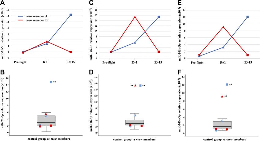
TABLE 1. List of blood c-markers assessed in the current work and related references

                                    Marker                                         Biologic endpoint                                 Reference

                                    miR-206                 Myo-miR, skeletal muscle                                                 53
                                    miR-133a-3p             Myo-miR, skeletal muscle                                                 53
                                    miR-21-5p               Inflamma-miR, proinflammatory and pro-osteogenesis                         8, 40, 54
                                    miR-126-3p              Inflamma-miR, proinflammatory, expressed in                                8, 50
                                                              endothelial cells
                                    miR-146a-5p             Inflamma-miR, proinflammatory and procell senescence                       8, 55
                                    miR-122-5p              Liver integrity and function                                             56, 57
                                    miR-145-5p              Cell proliferation and tumor suppressor                                  58, 59
                                    miR-363-3p              Cell growth and differentiation                                          60
                                    c-proteasome            Tissue injury, pathologic condition                                      16
                                    IL-6                    Systemic proinflammatory citokine                                         61
                                    Leptin                  Adipokine involved in metabolism                                         12
                                    TGF-b1                  Anti-inflammatory cytokine                                                62
                                    mtDNA                   Proinflammatory                                                           63, 46

               C-mtDNA relative quantification                                              ionization–tandem mass spectrometry (MS/MS), as previously
                                                                                            described (19). For further information about PMF and liquid
               Total DNA was isolated from plasma-EDTA samples using                        chromatography–MS/MS, data are listed in Tables 2 and 3. A
               Quick-gDNA MiniPrep Kit (Zymo Research, Irvine, CA, USA).                    representative example of heat shock protein b-1 (HSPB1)
               To quantify the free mtDNA copy number, a real-time quanti-                  analysis with matrix-assisted laser desorption/ionization–
               tative PCR SYBR Green assay was performed using a standard                   ToF PMF and electrospray ionization–MS/MS is reported in
               curve as calibration. Assays were performed in duplicate by                  supporting information (Supplemental Fig. S4). Proteomic
               Rotor-Gene Q 6000 Detector (Qiagen), using SYBR GreenER Mix                  analyses were performed in triplicates.
               (Thermo Fisher Scientific) and forward/reverse primer (specific
               for 69-bp fragment internal to the ND1 mt-gene fragment used
               for calibration). Specificity of PCR products was confirmed by               Bioinformatic analysis
               melting curve analysis. Each run was repeated 3 times. Standard
               curve was set up using 10-log serial dilution of stock solution              Validated miR targets were identified by means of Dianatools
               containing from 1028 to 1024 mtDNA copies/ml. To determine                   (mirPath v.3.0) using TarBase v.7.0 for the union of inflamma-
               mtDNA copies, a 217-bp fragment, corresponding to MT-ND1                     miRs targets and apart, the validated targets of muscle-specific
               gene, was amplified by PCR and loaded on 1% agarose gel. DNA                 (myo)-miR-206. Kyoto Encyclopedia of Genes and Genomes
               corresponding to the 217-bp band was isolated and quantified                 (KEGG) pathways and Gene Ontology (GO) category analyses
               by absorbance and used as a calibrator. mtDNA copy number                    were applied to identify the most significant molecular network
               of calibrator was obtained by the total DNA concentration di-                involving the transcripts regulated by the selected miRs.
               vided by amplicon weight. The latter was estimated as follows:
               (217 bp 3 MWt)/A, where MWt denotes the MW of double-
               stranded DNA (6.6 3 105 g/mole), and A denotes Avogadro’s                    Statistical analysis and modeling
               number (6.02 3 1023 molecules/mole).
                                                                                            Data obtained by astronauts were compared with the control
                                                                                            distribution, and a z score test was applied for each value. Values
               Cytokines/leptin quantification                                              of P , 0.05 were considered significant. Data exploiting was
                                                                                            obtained by considering each molecule as an independent vari-
               IL-6, TGF-b1 and Leptin concentration were measured in plasma                able and was standardized by means of a box-cox transform. The
               samples with commercial ELISA kit (R&D Systems, Minneapolis,                 different ranking of each variable was obtained by graphing the
               MN, USA) according to the manufacturer’s instructions. All                   z score of each molecular target and comparing it with the baseline
               measurements were performed in duplicate, and the average                    population. Proteomic statistical analysis was performed using
               values were used in the statistical analyses.                                the DeCyder 1.0 extended data analysis module. Protein filters
                                                                                            were set to select only those protein spots that matched .90% of
                                                                                            the gel images, and these protein spots were included in data
                                                                                            analysis. Statistically significant differences of 2-D–difference gel
               Muscle proteins
                                                                                            electrophoresis data were computed by paired 1-way ANOVA
                                                                                            (2-sided) coupled to Tukey’s test; the significance level was set
               Protein extraction and minimal labeling with cyanine dyes (Cy3               at a ,0.01. In addition, the false discovery rate was applied as a
               and Cy5), and 2-dimensional (2-D) separation and analyses were               multiple testing correction method to keep the overall error rate as
               performed as previously described (9). Proteins of interest were             low as possible (20). Two independent analyses were performed
               identified by peptide mass fingerprinting (PMF) utilizing a matrix-          for crewmembers A and B by comparing R+1 vs. preflight and
               assisted laser desorption/ionization time-of-flight mass spectrom-           R+15 vs. preflight for each member.
               eter (Ultraflex III-MALDI ToF/ToF Mass Spectrometer; Bruker,
               Billerica, MA, USA), as previously described (18). In particular, a
               search was carried out by correlation of uninterpreted spectra to
               Mammalia entries in the National Center for Biotechnology In-                RESULTS
               formation (Bethesda, MD, USA) database (ID:20090430; 8,483,808
               sequences; 2,914,572,939 residues). When this approach was un-               All results have been reported both in dependence of
               successful additional searches were performed using electrospray             time series (preflight, R+1, and R+15) and in comparison

               5170     Vol. 33   April 2019                           The FASEB Journal x www.fasebj.org                                             CAPRI ET AL.
ebj.org by Universita Degli Studi Padova Ist Fisiologia Umana (147.162.3.235) on November 21, 2019. The FASEB Journal Vol. ${article.issue.getVolume()}, No. ${article.issue.getIssu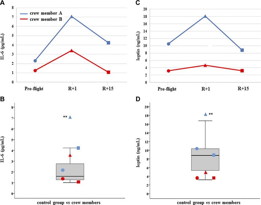
with the control distribution. C-proteasome was found
TABLE 2. The entire list of proteins differentially expressed in all comparisons for both crewmembers (A and B), together with statistical analyses (values reported as P in columns in reference to Tukey’s
% fold change
significantly increased in both crewmembers at R+1, but
R+15 vs. preflight
19
24
15
30
23
subject A had not recovered at R+15 and c-proteasome
resulted in further increase, whereas subject B did recover
(Fig. 1). Myo-miR-206 was found significantly increased
in crewmember A at both R+1 and R+15, whereas was
Tukey’s test
9.47E204
3.93E203
6.99E203
4.44E204
7.06E203
increased only at final recovery time in crewmember B
(Fig. 2). On the contrary, myo-miR-133a-3p showed no
Subject B
significant changes in both crewmembers when com-
pared with control distribution (Supplemental Fig. S1A, B).
% fold change
Inflamma-miR-21-5p was significantly increased in crew-
member A at R+15, whereas the changes revealed in
29
30
227
29
35
231
R+1 vs. preflight
crewmember B were not significant (Fig. 3A, B). Simi-
larly, inflamma-miR-126-3p was significantly increased
in crewmember A at R+1, whereas the significant in-
Tukey’s test
1.58E204
1.55E203
2.13E203
1.61E203
2.57E203
4.05E203
crease in crewmember B (at R+1) was completely re-
covered (Fig. 3C, D). Similar results were obtained when
measuring inflamma-miR-146a-5p in both crewmembers
(Fig. 3E, F). The c-level of miR-122-5p was found signifi-
cantly increased at R+15 in crewmember A only (Sup-
% fold change
plemental Fig. S1C, D). MiR-145-5p was observed to be
218
222
49
81
43
226
R+15 vs. preflight
increased at landing time in both crewmembers, but only
subject B completely recovered (Supplemental Fig. S2A,
B). The different trends of miR-363-3p observed in both
crewmembers were not significant when compared with
Tukey’s test
7.09E203
5.03E203
9.15E203
3.93E203
3.93E203
3.93E203
control group (Supplemental Fig. S2C, D). The changes of
TGF-b1 observed in the 2 crewmembers were not sig-
Subject A
nificant (Supplemental Fig. S3A, B), and similarly, the
c-mtDNA was not found significantly modified in the 2
% fold change
crewmembers (Supplemental Fig. S3C, D), even if values
were at the extreme of control distribution. Dissimilarly,
231
222
66
21
217
219
23
50
225
R+1 vs. preflight
IL-6 and leptin were significantly increased at R+1 and
recovered in crewmember A, whereas they did not
change in crewmember B (Fig. 4A, D).
Tukey’s test
The exploitation of normalized data is reported in Figs.
7.28E204
1.66E203
4.33E203
4.32E203
8.91E203
9.41E203
8.83E203
4.12E203
5.33E203
5 and 6 by graphing the z score of each molecular target
compared with the baseline population (gray bands in-
dicate 95% of the population distribution). Figure 5 shows
the differences between the 2 astronauts at the 3 times,
SERPINA1
TRIM72
Gene
whereas Fig. 6 shows the difference between the 2 astro-
GSTM2
SERPINA1; protein identified by liquid chromatography–MS/MS.
ANXA2
PRDX2
PRDX6
PARK7
HSPD1
HSPA2
HSPA5
HSPB1
HSPB1
SOD2
CAT
CAT
nauts considering only 2 times (i.e., preflight and R+15).
The normalized values of circulating molecules resulted in
the normal distribution at the baseline, and differences
Accession no.
were observed after landing and recovery for each crew-
Q6ZMU5
Q7Z7M6
Q99497
P04792
P04792
P10809
P54652
P11021
P07355
P01009
P32119
P30041
P04040
P04040
P28161
member. Many differences were observed between the
2 subjects at R+1 in terms of inflamma-miRs, myo-miR-
206, c-proteasome, and IL-6/leptin, thus making the 2
crewmembers dissimilar to each other. Examining the
60 kDa heat shock protein, mitochondrial
z scores on the same astronaut before flight and after re-
test), protein Ac number and gene name.
Heat shock–related 70 kDa protein 2
Tripartite motif-containing protein 72
Endoplasmic reticulum chaperone BiP
covery (Fig. 6), the baseline values of most parameters
Protein/nucleic acid deglycase DJ-1
are already inside the limits of the standard distribution
Glutathione S-transferase Mu 2
of the population. The only molecule that significantly
varies in both astronauts after spaceflight is myo-miR-206.
Heat shock protein beta-1
Heat shock protein beta-1
Dissimilarly, c-proteasome, inflamma-miRs, and myo-
Superoxide dismutase
miR-206 were not reverted to the baseline values at R+15
in crewmember A.
Peroxiredoxin-2
Peroxiredoxin-6
a-1-antitrypsina
A bioinformatic approach was applied to investigate
Annexin A2
currently validated and miR-target union of all inflamma-
miRs-21-5p, -126-3p, -146a-5p and separately, myo-
Catalase
Catalase
Protein
a
miR-206. Two lists of 331 and 136 genes/transcripts
were reported in 2 supplemental tables, respectively
BLOOD CIRCULATING MARKERS IN ISS CREWMEMBERS 5171
ebj.org by Universita Degli Studi Padova Ist Fisiologia Umana (147.162.3.235) on November 21, 2019. The FASEB Journal Vol. ${article.issue.getVolume()}, No. ${article.issue.getIssu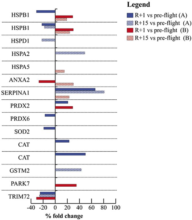
5172
Vol. 33
TABLE 3. The entire list of proteins differentially expressed in reference to protein Ac number, gene name, theoretical molecular mass, isoelectric points, and MS data
April 2019
Matched/searched Protein mascot Sequence MS/MS
Protein Accession no. Gene MW (kDa) pI peptides score coverage (%) MS/MS sequence score m/z z Range (aa)
Heat shock protein beta-1 P04792 HSPB1 22.3 9.1 8/17 116 32.2 LFDQAFGLPR 63 1163.635 1 28–37
Heat shock protein beta-1 P04792 HSPB1 22.3 9.1 9/23 126 44.7 LFDQAFGLPR 88 1163.633 1 28–37
60 kDa heat shock protein, P10809 HSPD1 61.2 5.6 13/15 161 26.5 AAVEEGIVLGGGCALLR 95.6 1684.89 1 430–446
mitochondrial
Heat shock–related 70 kDa P54652 HSPA2 69.9 5.5 15 143 25.8 TTPSYVAFTDTER 104 1487.726 1 38–50
protein 2
Endoplasmic reticulum P11021 HSPA5 72.1 4.9 10/18 114 20.4 EFFNGKEPSR 38 1210.6 1 376–385
chaperone BiP
Annexin A2 P07355 ANXA2 38.6 8.5 14/33 174 36.0 QDIAFAYQR 42.5 1111.53 1 69–77
a-1-antitrypsina P01009 SERPINA1 46.7 5.3 5 257 12.7 SPLFMGK 28 398.297 2 405–411
AVLTIDEK 30 444.823 2 360–367
SVLGQLGITK 70 508.404 2 325–334
LSITGTYDLK 69 555.873 2 315–324
VFSNGADLSGVTEEAPLK 30 917.528 2 335–352
Peroxiredoxin-2 P32119 PRDX2 21.9 5.6 9/13 158.0 44.4 QITVNDLPVGR 66.6 1211.66 1 139–149
Peroxiredoxin-6 P30041 PRDX6 25.0 6.0 12/22 180.0 49.1 LPFPIIDDR 77.9 1085.594 1 96–106
Superoxide dismutase Q7Z7M6 SOD2 22.2 7.0 7/19 99.0 38.4 AIWNVINWENVTER 48.8 1743.882 1 179–192
The FASEB Journal x www.fasebj.org
Catalase P04040 CAT 59.7 7.0 10/36 75 23.9 AFYVNVLNEEQR 59 1481.746 1 445–456
Catalase P04040 CAT 59.7 7.0 11/37 102.0 30.0 AFYVNVLNEEQR 85.3 1481.746 1 445–456
Glutathione S-transferase P28161 GSTM2 25.7 6.0 10/38 103.0 45.0 DCGATWVVLGHSER 87 1586.727 1 85–98
Mu 2
Protein/nucleic acid Q99497 PARK7 19.9 6.4 8/12 105.0 39.2 GAEEMETVIPVDVMR 77.2 1675.811 1 13–27
deglycase DJ-1
Tripartite motif-containing Q6ZMU5 TRIM72 52.6 6.0 9/18 103.0 16.4 LLPAAEAHAR 38 1048.59 1 119–128
protein 72
Ac, ascesion number; pI, isoelectric points. aProtein identified by liquid chromatography–MS/MS.
CAPRI ET AL.
ebj.org by Universita Degli Studi Padova Ist Fisiologia Umana (147.162.3.235) on November 21, 2019. The FASEB Journal Vol. ${article.issue.getVolume()}, No. ${article.issue.getIssu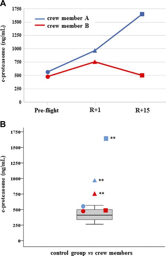
recovery (+81% in A and +23% in B). SERPINA1 is also
                                                                                            a validated target of miR-126-3p (22). Annexin A2
                                                                                            (ANXA2, validated target of miR-146a-5p) was down-
                                                                                            regulated at R+1 (227%) and up-regulated at recovery
                                                                                            (+30%) in crewmember B. Peroxiredoxin-2 (PRDX2) was
                                                                                            more abundant in both crewmembers postflight (R+1)
                                                                                            (+21% in A; +29% in B). Peroxiredoxin-6 (PRDX6) and
                                                                                            superoxide dismutase 2 (SOD2, a validated target of
                                                                                            miR-21-5p and -146a-5p) were decreased (217 and
                                                                                            219%), whereas 2 proteoforms of catalase were increased
                                                                                            (+23 and +50%) in crewmember A after landing. Heat
                                                                                            shock–related 70 kDa protein 2 (HSPA2, +49%) and glu-
                                                                                            tathione S-transferase Mu 2 (GSTM2, +43%, putative tar-
                                                                                            get of miR-21-5p) were increased in abundance, whereas
                                                                                            the mitochondrial 60 kDa heat shock protein (HSPD1,
                                                                                            222%) was down-regulated in crewmember A at recovery
                                                                                            time. Protein/nucleic acid deglycase DJ-1 (PARK7, +35%,
                                                                                            validated target of miR-126-3p) and endoplasmic retic-
                                                                                            ulum chaperone BiP (HSPA5, +15%, validated target of

               Figure 1. C-proteasome. A) Measurements are reported in
               dependence of time in both crewmembers. Circles represent
               preflight, triangles (R+1 d) landing time, and squares de-
               note (R+15 d) recovery time. B) C-proteasome values of
               crewmembers are compared with age-matched control distri-
               bution (19 measurements). Blue: crewmember A; red crew-
               member B. **P # 0.01 (z-score test).

               (Supplemental Tables S1 and S2). In particular, the
               former table is referred to GO: response to stress (P =
               6.78206035218e233), whereas the latter is specifically
               related to miR-206 transcript targets.
                  Muscle proteomic analyses, tested for evaluable miR-
               targets, indicated significant differences in stress and an-
               tioxidant proteins comparing baseline with postflight
               (R+1) in 9 and 6 spots in crewmember A and B, respectively.
               Then, comparing baseline to recovery time (R+15), 6 spots
               changed in crewmember A and 5 spots in crewmember B
               (Fig. 7 and Table 2). In particular, 2 proteoforms (21), dif-
               ferent molecular forms originated from the heat shock
               protein family B (small) member 1 gene (HSPB1) were
               changed in abundance in both crewmembers at R+1 (231
               and 222% in A; +29 and +30% in B) and recovery (218%
                                                                                            Figure 2. C-myo-miR-206. A) Measurements are reported in
               in A; +19 and +24% in B). Tripartite motif-containing
                                                                                            dependence of time in both crewmembers. For explanation of
               protein 72 (TRIM72) was down-regulated after landing                         symbols see Fig. 1. B) C-myo-miRs-206 values of crewmembers
               in both crewmembers (225% in A and 231% in B) and                            are compared with age-matched control distribution (19 mea-
               only in A at recovery (226%). SERPINA1 was more                              surements). Blue: crewmember A; red crewmember B. **P #
               abundant in A after landing (+66%) and in both at                            0.01 (z-score test).

               BLOOD CIRCULATING MARKERS IN ISS CREWMEMBERS                                                                                                    5173
ebj.org by Universita Degli Studi Padova Ist Fisiologia Umana (147.162.3.235) on November 21, 2019. The FASEB Journal Vol. ${article.issue.getVolume()}, No. ${article.issue.getIssu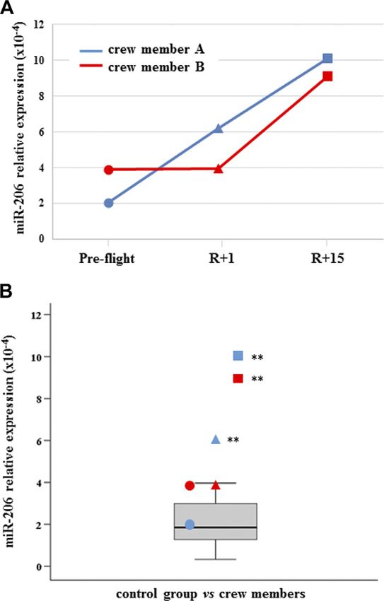
Figure 3. C-inflamma-miRs-21-5p; -126-3p and -146a-5p. A, C, E ) Measurements of circulating inflamma-miRs-21-5p (A); -126-3p
               (C ), and -146a-5p (E ) are reported in dependence of time in both crewmembers. For explanation of symbols see Fig. 1. B, D, F )
               C-inflamma-miRs-21-5p (B); -126-3p (D), and -146a-5p (F ) values of crewmembers are compared with age-matched control
               distribution (19 measurements). Blue: crewmember A; red crewmember B. **P # 0.01 (z-score test).

               miR-21-5p) were increased in crewmember B at R+1                             inflammation initiates by the sympathetic nervous system,
               and recovery, respectively.                                                  resulting in the increase of c-damage associated molecular
                                                                                            patterns, such as mtDNA, and a reduction in immune-
                                                                                            inhibitory miRs, which are carried in the blood circulation
               DISCUSSION                                                                   to tissues throughout the body (32).
                                                                                                To examine at the systemic level possible unbalancing
               Prolonged distress or chronic exposures to stressors, in-                    homeostasis in terms of pro- and anti-inflammatory
               cluding psychologic or physical stresses, are known to                       molecules, we measured relevant blood molecules and
               affect immune system function, which in turn increases                       epigenetic regulators (i.e., c-myo-miRs-206 and -133a-3p,
               inflammatory mediators (23, 24). Furthermore, chronic                        c-inflamma-miRs -21-5p, -126-3p, and -146a-5p, liver c-
               stress throughout the lifespan in absence or modest bodily                   miR-122-5p, cell proliferation regulators c-miR-145-5p
               adaptation and inefficient repair mechanisms may affect                      and -363-3p, c-proteasome, c-mtDNA, proinflammatory
               the ageing process and lifespan (15, 25, 26), favoring the                   IL-6, anti-inflammatory TGF-b1, and leptin). Indeed,
               disease onset (27, 28). Spaceflight may also represent a                     types of various c-shuttles (nano-microextracellular vesi-
               source of prolonged/chronic stress that is due not only to                   cles, proteins, and apolipoproteins) were not the objective
               the psychologic aspect but also environment stressors,                       of the present work, whereas the measurement of the total
               such as adaptation to microgravity, high workload, sleep                     amount of c-miRs/different molecules was supposed to
               deprivation, isolation and confinement, ionizing radiation,                  be more significant.
               and potentially others (29).                                                     Relevant differences between the 2 crewmembers at 1
                  The present work aimed at answering the questions of                      or 15 d, or both points, of recovery were identified, even if
               whether the 2 crewmembers (A and B), beyond the effects                      these findings cannot define the in-flight c-levels of the
               on skeletal muscle (9), had systemic effects in terms of pro-                same molecules. Crewmember A showed more deviations
               and anti-inflammatory c-molecules after about 6 mo of                        from baseline after landing time (R+1) than crewmember
               spaceflight at the ISS and if they recovered at 1 or 15 d after              B. In fact, c-inflamma-miRs -21-5p, -126-3p, and -146a-5p,
               landing.                                                                     and myo-miR-206, c-proteasome, IL-6, and leptin were
                  Both atrophy of skeletal muscle and systemic stress may                   significantly increased after 1 d recovery. At R+15, crew-
               affect the entire body, being that skeletal muscle is the most               member A showed a significant increase of c-proteasome,
               abundant tissue of the human body (about 30–40% of the                       inflamma-miRs, and myo-miR-206. Comparing these
               body) and systemic stress is able to alter metabolism and                    data with crewmember B, the only common molecule
               homeostasis (3, 30, 31). Furthermore, recent evidence                        was myo-miR-206, which was still increased at R+15 in
               supports the hypothesis that systemic stress-evoked sterile                  both crewmembers. It is known that myo-miR-206 is

               5174     Vol. 33   April 2019                           The FASEB Journal x www.fasebj.org                                             CAPRI ET AL.
ebj.org by Universita Degli Studi Padova Ist Fisiologia Umana (147.162.3.235) on November 21, 2019. The FASEB Journal Vol. ${article.issue.getVolume()}, No. ${article.issue.getIssu
Figure 4. IL-6 and leptin. A, C ) Measurements of circulating IL-6 (A) and leptin (C ) are, reported in dependence of time in
both crewmembers. For explanation of symbols see Fig. 1. B, D) IL-6 (B) and leptin (D) values of crewmembers are compared
with age-matched control distribution (19 measurements). Blue: crewmember A; red crewmember B. **P # 0.01 (z-score test).
preferentially expressed in skeletal muscle and com- been found to increase in muscle tissue in catabolic/
pletely absent, or expressed at relatively low levels, in atrophy condition in a mouse model (38). The con-
other tissues. Notably, crewmember B trained more tribution of muscle atrophy/wasting to the pool of
vigorously than A, particularly concerning the loading c-miRs was recently confirmed in exosomes released
forces. In the postflight, crewmember A showed sub- by myofibers, supporting the conclusion that myofiber-
stantial decrements (i.e., muscle volume and architec- derived exosomes modulate protein levels of key factors
ture) in strength and in fiber contractility, which was in myogenic or osteogenic differentiation of mesen-
strongly mitigated in B, as previously reported in a chymal progenitor cells (39). In particular, miR-21-5p
separate work on the same individuals (9). In fact, the was shown to promote the osteogenic differentiation of
increased level of c-miR-206 at landing time in astronaut mouse bone marrow cells by targeting Sprouty homolog
A and in both astronauts at R+15 may also be associated 1 (Spry1), negatively regulating the osteogenic differ-
with the different physical training status of the subjects. entiation of mesenchymal stem cells (40).
This finding suggests a possible role of c-miR-206 as a As far as inflamma-miRs are concerned, crewmember
good candidate for the monitoring of skeletal muscle A showed the highest levels at R+15, whereas crew-
status. Regardless, a consistent literature indicates the member B showed increases of miR-126-3p and -146a-5p
full involvement of myo-miR-206 in different conditions, only at R+1, thus revealing 2 different trends between the 2
such as age, physical training, and type of exercise, such subjects, those being regulators of both stress response and
as acute or prolonged, aerobic or resistance or endurance inflammatory pathway (8).
activity (33–36). Cellular miRs were previously studied in both in
MiR-206 promotes cell differentiation and cell in- vitro microgravity experiments on earth and in vitro
hibition and may influence cell regeneration in the experiments run in the ISS. The former study was
muscle (37). In particular, miR-206 and miR-21 have conducted with g-ray coexposure and many miRs
BLOOD CIRCULATING MARKERS IN ISS CREWMEMBERS 5175
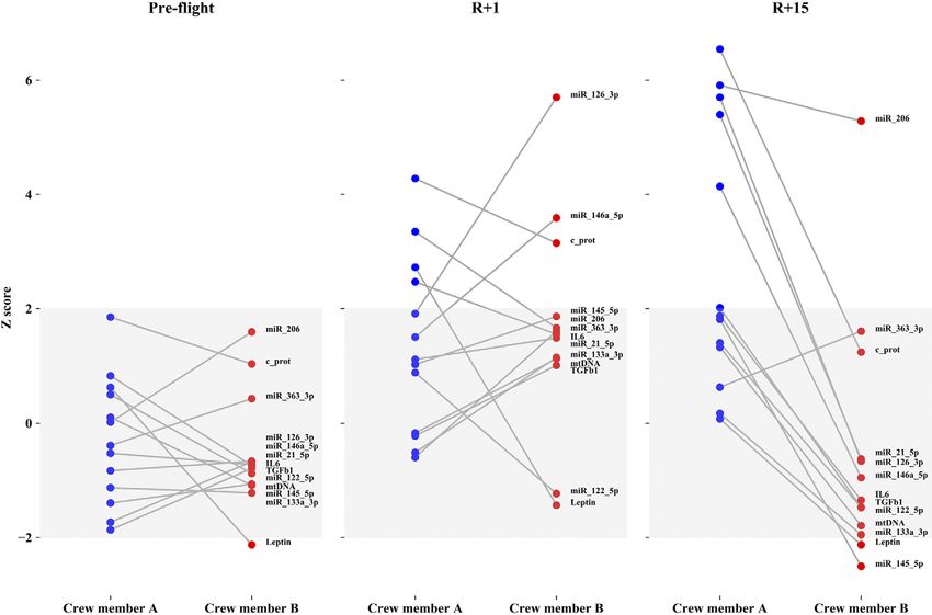
ebj.org by Universita Degli Studi Padova Ist Fisiologia Umana (147.162.3.235) on November 21, 2019. The FASEB Journal Vol. ${article.issue.getVolume()}, No. ${article.issue.getIssuFigure 5. All normalized markers in crewmembers A and B at preflight, R+1, and R+15 times. Y-axis describes z scores and gray
zone contains control group values. Values outside the gray zone are considered significant.
involving cell cycle machinery and DNA repair sys- The inflamma-miR increase may be due to an aug-
tem resulted dysregulated (41). The latter showed the mented exocytosis that can be, at least in part, stress related
dysregulation of miR-21 in a different experiment set- (32), due to an increased tissue and cell injury, or both,
ting (42), thus highlighting its involvement in spaceflight especially in crewmember A. The tissue injury is also
effects. confirmed by the increase of c-proteasome, reaching a
Once c-inflamma-miRs are up-taken by cells and tissue, concentration similar to that of autoimmune disease (45) in
they are able to modulate many genes. Taking into account crewmember A after final recovery. Accordingly, the in-
KEGG pathway analysis, NF-kB pathway had high sig- crease of c-mtDNA was dramatically evident in crew-
nificance (P = 2.516315e208), but p53 signaling (P = member A, even if at the limit of the normal range. Overall,
9.370703e207) and the mechanistic target of rapamycin the in vivo c-proteasome and c-mtDNA levels mediate the
(mTOR) pathway (P = 0.0001298907) also significantly fit inflammatory pathway and represent a general mecha-
the miR-targets. In particular, phosphatase and tensin nism to switch on inflammation, immune cell activities
homolog (PTEN), phosphatidylinositol 3-kinase regula- also being markers of muscle wasting (16, 46). On the other
tory subunit a (PIK3R1), phosphatidylinositol 3-kinase side, the increase of c-proteasome in crewmember A at
regulatory subunit b (PIK3R2), insulin receptor sub- R+15 d as a marker of muscle recovery cannot be com-
strate 1 (IRS1), and serine/threonine kinase 1 (AKT1) are pleted excluded (47). Noteworthy, IL-6 increased only in
inflamma-miR targets and represent the central pathway astronaut A at 1 d recovery concomitantly to leptin, and
involving muscle/tissue anabolism/synthesis. Accord- both are important regulators of inflammation and bone
ingly, some proteins related to mTOR pathway have also turnover (48).
been identified as dysregulated in our previous paper (9). Importantly, various stress-related and antioxidant
Taking into account the GO category analysis, stress re- proteins were found modified in skeleton muscle, and
sponse resulted among those strongly significant (P = many of them are direct targets of c-inflamma-miRs. A
6.78206035218e233). Interestingly, a common target of direct or indirect effect between muscle tissue proteins
both miR-21-5p and miR-146a-5p is CLOCK (43, 44). This and blood c-miRs may only be speculated, which is a
protein plays a central role in the regulation of circadian limitation of the work. However, it is worth noting that
rhythms and could have a systemic role because of its concomitantly with the increased inflamma-miR levels
ubiquitous expression in testis, thyroid, and many other in astronaut A, the soleus muscle tissue showed an in-
tissues. crease of 80% a SERPINA1, a serine protease inhibitor
5176 Vol. 33 April 2019 The FASEB Journal x www.fasebj.org CAPRI ET AL.
ebj.org by Universita Degli Studi Padova Ist Fisiologia Umana (147.162.3.235) on November 21, 2019. The FASEB Journal Vol. ${article.issue.getVolume()}, No. ${article.issue.getIssuFigure 6. The 2 crewmembers, A and B, are compared at preflight vs. R+15. All markers are normalized. Y-axis describes z scores
and gray zone contains control group values. Values outside the gray zone are considered significant.
belonging to acute phase protein and validated target of had different spaceflight recovery effects, in which crew-
miR-126-3p (22), apparently as a tissue-related anti- member A was the most affected. In particular, muscle-
inflammatory response. This effect was also revealed in related stress and proinflammatory status are here
astronaut B but to a lesser extent and at final recovery time highlighted in crewmember A, whereas crewmember B’s
only. Recent data suggest that SERPINA1 is also expressed recovery was almost completed except for myo-miR-206.
by endothelial cells after exposure to simulated micro- Inflamma-miRs seem to mediate the systemic recovery of
gravity (49) and may represent an important marker of crewmember A, and they are expected to reach a complete
tissue-related anti-inflammatory response. SERPINA1 in- recovery beyond 15 d from landing.
crease can be due to the miR-126-3p decrease, especially in In a complex field like space and spaceflight, N of 1
endothelial cells where it is usually expressed (50), as- could be a critical issue. However, population size, usually
suming that a relationship exists with the miR-126-3p in- based on a relatively low number of crewmembers, can be
crease in the blood, as observed in both astronauts even if overwhelmed by time series (or longitudinal) personal-
timing differed. ized studies. In fact, the effects of the 6-mo chronic expo-
In agreement with all data obtained, spaceflight re- sure to such an environment with many variables can have
covery had greater effects on crewmember A than B. In substantially different effects in different individuals, thus
fact, muscle stress–related proteins, such as HSPs, GSTM, the study of intraindividual variability along the time of
PRDX, ANXA2, and PARK7, were largely modified in exposure/recovery becomes more informative (52).
crewmember A rather than in B. These results point to Based on a personalized time series analysis, the present
muscle-stress responses that also involve oxido-reductase data further underpin the importance of countermeasures
enzymes like SOD and catalase as well as the repair aimed at reducing, as much as possible, skeletal muscle
membrane protein TRIM72. The latter was decreased in wasting (9). Moreover, these data further suggest a linkage
muscle tissue at both recovery times in astronaut A and at among muscle wasting, stress response and inflammation
final recovery time in astronaut B. Similar results in terms and potentially affecting systemic metabolism. In this
of stress-related pathway activation were previously ob- perspective, the prolonged or chronic exposure to space/
served in mouse model after 91 d of spaceflight (51). spaceflight may favor the development of metabolic al-
Overall, these results corroborate the view obtained terations, even if additional analyses with later time points
from our previous paper (9), where the 2 crewmembers are necessary.
BLOOD CIRCULATING MARKERS IN ISS CREWMEMBERS 5177
ebj.org by Universita Degli Studi Padova Ist Fisiologia Umana (147.162.3.235) on November 21, 2019. The FASEB Journal Vol. ${article.issue.getVolume()}, No. ${article.issue.getIssuM. Flück., S. Ruoss, L. Brocca, E. Longa, and C. Gelfi
performed laboratory analyses; K. Albracht, M. Canepari,
I. Di Giulio, M. Narici, and J. Rittweger analyzed data and
performed human physiology experiments; R. Bottinelli,
P. Cerretelli, and M. Narici designed the study; J. Rittweger
organized testing sessions; and all authors provided input
and editing.
REFERENCES
1. Carpentier, W. R., Charles, J. B., Shelhamer, M., Hackler, A. S.,
Johnson, T. L., Domingo, C. M. M., Sutton, J. P., Scott, G. B. I., and
Wotring, V. E. (2018) Biomedical findings from NASA’s Project
Mercury: a case series. NPJ Microgravity 4, 6
2. Roberts, D. R., Albrecht, M. H., Collins, H. R., Asemani, D., Chatterjee,
A. R., Spampinato, M. V., Zhu, X., Chimowitz, M. I., and Antonucci,
M. U. (2017) Effects of spaceflight on astronaut brain structure as
indicated on MRI. N. Engl. J. Med. 377, 1746–1753
3. Narici, M. V., and de Boer, M. D. (2011) Disuse of the musculo-skeletal
system in space and on earth. Eur. J. Appl. Physiol. 111, 403–420
4. Schwendner, P., Mahnert, A., Koskinen, K., Moissl-Eichinger, C.,
Barczyk, S., Wirth, R., Berg, G., and Rettberg, P. (2017) Preparing for
the crewed Mars journey: microbiota dynamics in the confined
Mars500 habitat during simulated Mars flight and landing. Microbiome
5, 129
5. Vico, L., and Hargens, A. (2018) Skeletal changes during and after
spaceflight. Nat. Rev. Rheumatol. 14, 229–245
6. Vernikos, J., and Schneider, V. S. (2010) Space, gravity and the
Figure 7. Proteomic analysis in human skeletal muscle. physiology of aging: parallel or convergent disciplines? A mini-review.
Histograms of stress and antioxidant proteins differentially Gerontology 56, 157–166
expressed in the soleus muscle between baseline vs. R+1 7. Olivieri F, Capri M, Bonafè M, Morsiani C, Jung HJ, Spazzafumo L,
(colored bars) and baseline vs. R+15 (striped bars) in Viña J, and Suh Y. (2017) Circulating miRNAs and miRNA shuttles as
crewmember A (blue bars) and B (red bars), as detected by biomarkers: perspective trajectories of healthy and unhealthy aging.
2-D-DIGE analysis. Proteins significantly changed (paired 1-way Mech. Ageing Dev. 165, 162–170
ANOVA and Tukey’s test, a = 0.01) are indicated by their gene 8. Olivieri, F., Rippo, M. R., Monsurrò, V., Salvioli, S., Capri, M.,
Procopio, A. D., and Franceschi, C. (2013) MicroRNAs linking
name and expressed as a percent of spot volume variation. inflamm-aging, cellular senescence and cancer. Ageing Res. Rev. 12,
Statistical details are showed in Table 2. 1056–1068
9. Rittweger, J., Albracht, K., Flück, M., Ruoss, S., Brocca, L., Longa, E.,
Moriggi, M., Seynnes, O., Di Giulio, I., Tenori, L., Vignoli, A., Capri,
M., Gelfi, C., Luchinat, C., Francheschi, C., Bottinelli, R., Cerretelli, P.,
ACKNOWLEDGMENTS and Narici, M. (2018) Sarcolab pilot study into skeletal muscle’s
adaptation to long-term spaceflight. NPJ Microgravity 4, 18; erra-
The authors thank the European Space Agency (ESA) for tum: 23
its support, with great help from Patrik Sundblad, Simone 10. Biolo, G., Heer, M., Narici, M., and Strollo, F. (2003) Microgravity as a
Thomas, and Marine le-Gouic (all from the ESA). Peter model of ageing. Curr. Opin. Clin. Nutr. Metab. Care 6, 31–40
11. Franceschi, C., Capri, M., Monti, D., Giunta, S., Olivieri,
Gauger and Wolfram Sies [German Aerospace Center (DLR), F., Sevini, F., Panourgia, M. P., Invidia, L., Celani, L., Scurti,
Cologne, Germany] were instrumental in the preparation of M., Cevenini, E., Castellani, G. C., and Salvioli, S. (2007)
human testing devices. Asa Beijer (DLR) helped with prepro- Inflammaging and anti-inflammaging: a systemic perspective
cessing of the muscle samples. This study was supported by on aging and longevity emerged from studies in humans. Mech.
internal funding from the DLR (2475 030, Muscle Mechanics Ageing Dev. 128, 92–105
and Metabolism); the Ricerca Fondamentale Orientata (RFO) 12. Bucci, L., Yani, S. L., Fabbri, C., Bijlsma, A. Y., Maier, A. B., Meskers,
(to M. Capri); and by the Project Digital Personalised Medicine– C. G., Narici, M. V., Jones, D. A., McPhee, J. S., Seppet, E., Gapeyeva,
Healthy Ageing: Network Analysis of Big Multiomics Data to H., Pääsuke, M., Sipilä, S., Kovanen, V., Stenroth, L., Musarò, A.,
Search from New Diagnostic, Prognostic, and Therapeutic Targets Hogrel, J. Y., Barnouin, Y., Butler-Browne, G., Capri, M., Franceschi,
(DPM–AGEING) (14.Y26.31.0026 to C. Franceschi); and by C., and Salvioli, S. (2013) Circulating levels of adipokines and IGF-1
are associated with skeletal muscle strength of young and old healthy
the Italian Ministry of Education, University, and Research
subjects. Biogerontology 14, 261–272
(MIUR; Grant PRIN 2015FBNB5Y to C.G.). C.G., C.F., M.N., 13. Calder, P. C., Bosco, N., Bourdet-Sicard, R., Capuron, L., Delzenne,
and J.R. share senior authorship. The authors declare no N., Doré, J., Franceschi, C., Lehtinen, M. J., Recker, T., Salvioli, S., and
conflicts of interest. Visioli, F. (2017) Health relevance of the modification of low grade
inflammation in ageing (inflammageing) and the role of nutrition.
Ageing Res. Rev. 40, 95–119
14. Stahn, A. C., Werner, A., Opatz, O., Maggioni, M. A., Steinach, M.,
AUTHOR CONTRIBUTIONS von Ahlefeld, V. W., Moore, A., Crucian, B. E., Smith, S. M., Zwart,
S. R., Schlabs, T., Mendt, S., Trippel, T., Koralewski, E., Koch, J.,
M. Capri and S. Salvioli provided further critical Choukèr, A., Reitz, G., Shang, P., Röcker, L., Kirsch, K. A., and Gunga,
discussion; M. Capri, C. Franceschi, and J. Rittweger H. C. (2017) Increased core body temperature in astronauts during
long-duration space missions. Sci. Rep. 7, 16180
wrote the manuscript; C. Morsiani and E. Giampieri 15. Franceschi, C., Garagnani, P., Vitale, G., Capri, M., and Salvioli, S.
performed statistical analyses; C. Morsiani, A. Santoro, (2017) Inflammaging and ‘Garb-aging’. Trends Endocrinol. Metab. 28,
M. Moriggi, M. Conte, M. Martucci, E. Bellavista, C. Fabbri, 199–212
5178 Vol. 33 April 2019 The FASEB Journal x www.fasebj.org CAPRI ET AL.
ebj.org by Universita Degli Studi Padova Ist Fisiologia Umana (147.162.3.235) on November 21, 2019. The FASEB Journal Vol. ${article.issue.getVolume()}, No. ${article.issue.getIssu16. Dianzani, C., Bellavista, E., Liepe, J., Verderio, C., Martucci, M., 36. Mooren, F. C., Viereck, J., Krüger, K., and Thum, T. (2014)
Santoro, A., Chiocchetti, A., Gigliotti, C. L., Boggio, E., Ferrara, B., Circulating microRNAs as potential biomarkers of aerobic exercise
Riganti, L., Keller, C., Janek, K., Niewienda, A., Fenoglio, C., Sorosina, capacity. Am. J. Physiol. Heart Circ. Physiol. 306, H557–H563
M., Cantello, R., Kloetzel, P. M., Stumpf, M. P., Paul, F., Ruprecht, K., 37. Simionescu-Bankston, A., and Kumar, A. (2016) Noncoding RNAs in
Galimberti, D., Martinelli Boneschi, F., Comi, C., Dianzani, U., and the regulation of skeletal muscle biology in health and disease. J. Mol.
Mishto, M. (2017) Extracellular proteasome-osteopontin circuit reg- Med. (Berl.) 94, 853–866
ulates cell migration with implications in multiple sclerosis. Sci. Rep. 7, 38. Soares, R. J., Cagnin, S., Chemello, F., Silvestrin, M., Musaro, A.,
43718 De Pitta, C., Lanfranchi, G., and Sandri, M. (2014) Involvement of
17. Schwarzenbach, H., da Silva, A. M., Calin, G., and Pantel, K. (2015) microRNAs in the regulation of muscle wasting during catabolic
Data normalization strategies for microRNA quantification. Clin. conditions. J. Biol. Chem. 289, 21909–21925
Chem. 61, 1333–1342 39. De Gasperi, R., Hamidi, S., Harlow, L. M., Ksiezak-Reding, H.,
18. Viganò, A., Vasso, M., Caretti, A., Bravatà, V., Terraneo, L., Fania, C., Bauman, W. A., and Cardozo, C. P. (2017) Denervation-related al-
Capitanio, D., Samaja, M., and Gelfi, C. (2011) Protein modulation in terations and biological activity of miRNAs contained in exosomes
mouse heart under acute and chronic hypoxia. Proteomics 11, released by skeletal muscle fibers. Sci. Rep. 7, 12888
4202–4217 40. Yang, N., Wang, G., Hu, C., Shi, Y., Liao, L., Shi, S., Cai, Y., Cheng, S.,
19. Capitanio, D., Vasso, M., Fania, C., Moriggi, M., Viganò, A., Procacci, Wang, X., Liu, Y., Tang, L., Ding, Y., and Jin, Y. (2013) Tumor necrosis
P., Magnaghi, V., and Gelfi, C. (2009) Comparative proteomic profile factor a suppresses the mesenchymal stem cell osteogenesis promoter
of rat sciatic nerve and gastrocnemius muscle tissues in ageing by 2-D miR-21 in estrogen deficiency-induced osteoporosis. J. Bone Miner. Res.
DIGE. Proteomics 9, 2004–2020 28, 559–573
20. Hochberg, A. M., Gerhardt, P. N., Cao, T. K., Ocasio, W., Barbour, 41. Girardi, C., De Pittà, C., Casara, S., Sales, G., Lanfranchi, G., Celotti, L.,
W. M., and Mrozinski, P. M. (2000) Sensitivity and specificity of the test and Mognato, M. (2012) Analysis of miRNA and mRNA expression
kit BAX for screening/E. coli O157:H7 in ground beef: independent profiles highlights alterations in ionizing radiation response of
laboratory study. J. AOAC Int. 83, 1349–1356 human lymphocytes under modeled microgravity. PLoS One 7, e31293
21. Smith, L. M., and Kelleher, N. L.; Consortium for Top Down 42. Hughes-Fulford, M., Chang, T. T., Martinez, E. M., and Li, C. F. (2015)
Proteomics. (2013) Proteoform: a single term describing protein Spaceflight alters expression of microRNA during T-cell activation.
complexity. Nat. Methods 10, 186–187 FASEB J. 29, 4893–4900
22. Hassan, T., Smith, S. G., Gaughan, K., Oglesby, I. K., O’Neill, S., 43. Gabriely, G., Wurdinger, T., Kesari, S., Esau, C. C., Burchard, J.,
McElvaney, N. G., and Greene, C. M. (2013) Isolation and Linsley, P. S., and Krichevsky, A. M. (2008) MicroRNA 21 promotes
identification of cell-specific microRNAs targeting a messenger glioma invasion by targeting matrix metalloproteinase regulators.
RNA using a biotinylated anti-sense oligonucleotide capture affinity Mol. Cell. Biol. 28, 5369–5380
technique. Nucleic Acids Res. 41, e71 44. Li, J., Wan, Y., Guo, Q., Zou, L., Zhang, J., Fang, Y., Zhang, J., Zhang, J.,
23. Irwin, M. R., and Opp, M. R. (2017) Sleep health: reciprocal Fu, X., Liu, H., Lu, L., and Wu, Y. (2010) Altered microRNA
regulation of sleep and innate immunity. Neuropsychopharmacology 42, expression profile with miR-146a upregulation in CD4+ T cells from
129–155 patients with rheumatoid arthritis. Arthritis Res. Ther. 12, R81
24. Vitale, G., Salvioli, S., and Franceschi, C. (2013) Oxidative stress and 45. Egerer, K., Kuckelkorn, U., Rudolph, P. E., Rückert, J. C., Dörner, T.,
the ageing endocrine system. Nat. Rev. Endocrinol. 9, 228–240 Burmester, G. R., Kloetzel, P. M., and Feist, E. (2002) Circulating
25. Cevenini, E., Caruso, C., Candore, G., Capri, M., Nuzzo, D., Duro, G., proteasomes are markers of cell damage and immunologic activity in
Rizzo, C., Colonna-Romano, G., Lio, D., Di Carlo, D., Palmas, M. G., autoimmune diseases. J. Rheumatol. 29, 2045–2052
Scurti, M., Pini, E., Franceschi, C., and Vasto, S. (2010) Age-related 46. Picca, A., Lezza, A. M. S., Leeuwenburgh, C., Pesce, V., Calvani, R.,
inflammation: the contribution of different organs, tissues and sys- Bossola, M., Manes-Gravina, E., Landi, F., Bernabei, R., and Marzetti,
tems. How to face it for therapeutic approaches. Curr. Pharm. Des. 16, E. (2018) Circulating mitochondrial DNA at the crossroads of
609–618 mitochondrial dysfunction and inflammation during aging and
26. Bektas, A., Schurman, S. H., Sen, R., and Ferrucci, L. (2017) Human muscle wasting disorders. Rejuvenation Res. 21, 350–359
T cell immunosenescence and inflammation in aging. J. Leukoc. Biol. 47. Ikemoto, M., Nikawa, T., Takeda, S., Watanabe, C., Kitano, T.,
102, 977–988 Baldwin, K. M., Izumi, R., Nonaka, I., Towatari, T., Teshima, S.,
27. Franceschi, C., Garagnani, P., Morsiani, C., Conte, M., Santoro, A., Rokutan, K., and Kishi, K. (2001) Space shuttle flight (STS-90) en-
Grignolio, A., Monti, D., Capri, M., and Salvioli, S. (2018) The hances degradation of rat myosin heavy chain in association with
continuum of aging and age-related diseases: common mechanisms activation of ubiquitin-proteasome pathway. FASEB J. 15, 1279–1281
but different rates. Front. Med. (Lausanne) 5, 61 48. Lombardi, G., Sanchis-Gomar, F., Perego, S., Sansoni, V., and Banfi,
28. Calabrese, V., Santoro, A., Monti, D., Crupi, R., Di Paola, R., Latteri, S., G. (2016) Implications of exercise-induced adipo-myokines in bone
Cuzzocrea, S., Zappia, M., Giordano, J., Calabrese, E. J., and metabolism. Endocrine 54, 284–305
Franceschi, C. (2018) Aging and Parkinson’s disease: inflammaging, 49. Dittrich, A., Grimm, D., Sahana, J., Bauer, J., Krüger, M., Infanger, M.,
neuroinflammation and biological remodeling as key factors in and Magnusson, N. E. (2018) Key proteins involved in spheroid
pathogenesis. Free Radic. Biol. Med. 115, 80–91 formation and angiogenesis in endothelial cells after long-term ex-
29. Cucinotta, F. A. (2014) Space radiation risks for astronauts posure to simulated microgravity. Cell. Physiol. Biochem. 45, 429–445
on multiple International Space Station missions. PLoS One 9, 50. Olivieri, F., Bonafè, M., Spazzafumo, L., Gobbi, M., Prattichizzo, F.,
e96099 Recchioni, R., Marcheselli, F., La Sala, L., Galeazzi, R., Rippo, M. R.,
30. Pedersen, B. K., and Febbraio, M. A. (2012) Muscles, exercise and Fulgenzi, G., Angelini, S., Lazzarini, R., Bonfigli, A. R., Brugè, F.,
obesity: skeletal muscle as a secretory organ. Nat. Rev. Endocrinol. 8, Tiano, L., Genovese, S., Ceriello, A., Boemi, M., Franceschi, C.,
457–465 Procopio, A. D., and Testa, R. (2014) Age- and glycemia-related miR-
31. Naviaux, R. K. (2014) Metabolic features of the cell danger response. 126-3p levels in plasma and endothelial cells. Aging (Albany N.Y.) 6,
Mitochondrion 16, 7–17 771–787
32. Fleshner, M., and Crane, C. R. (2017) Exosomes, DAMPs and miRNA: 51. Sandonà, D., Desaphy, J. F., Camerino, G. M., Bianchini, E., Ciciliot,
features of stress physiology and immune homeostasis. Trends S., Danieli-Betto, D., Dobrowolny, G., Furlan, S., Germinario, E., Goto,
Immunol. 38, 768–776 K., Gutsmann, M., Kawano, F., Nakai, N., Ohira, T., Ohno, Y., Picard,
33. Margolis, L. M., Lessard, S. J., Ezzyat, Y., Fielding, R. A., and Rivas, D. A. A., Salanova, M., Schiffl, G., Blottner, D., Musarò, A., Ohira, Y., Betto,
(2017) Circulating microRNA are predictive of aging and acute R., Conte, D., and Schiaffino, S. (2012) Adaptation of mouse skeletal
adaptive response to resistance exercise in men. J. Gerontol. A Biol. Sci. muscle to long-term microgravity in the MDS mission. PLoS One 7,
Med. Sci. 72, 1319–1326 e33232
34. Wardle, S. L., Bailey, M. E., Kilikevicius, A., Malkova, D., Wilson, R. H., 52. Gabler, N. B., Duan, N., Vohra, S., and Kravitz, R. L. (2011) N-of-1
Venckunas, T., and Moran, C. N. (2015) Plasma microRNA levels trials in the medical literature: a systematic review. Med. Care 49,
differ between endurance and strength athletes. PLoS One 10, 761–768
e0122107 53. Sempere, L. F., Freemantle, S., Pitha-Rowe, I., Moss, E., Dmitrovsky,
35. Gomes, C. P., Oliveira, G. P., Jr., Madrid, B., Almeida, J. A., Franco, E., and Ambros, V. (2004) Expression profiling of mammalian
O. L., and Pereira, R. W. (2014) Circulating miR-1, miR-133a, and microRNAs uncovers a subset of brain-expressed microRNAs with
miR-206 levels are increased after a half-marathon run. Biomarkers 19, possible roles in murine and human neuronal differentiation. Genome
585–589 Biol. 5, R13
BLOOD CIRCULATING MARKERS IN ISS CREWMEMBERS 5179
ebj.org by Universita Degli Studi Padova Ist Fisiologia Umana (147.162.3.235) on November 21, 2019. The FASEB Journal Vol. ${article.issue.getVolume()}, No. ${article.issue.getIssuYou can also read