Reagan Anderson, FAOCD, FAAD, FASMS,
                  CAQ Mohs, MPH, MCS
←
→
Page content transcription
If your browser does not render page correctly, please read the page content below
Reagan Anderson, FAOCD, FAAD, FASMS,
CAQ Mohs, MPH, MCS
Founder of Your Health University
Given published data on the very low
likelihood that incompletely biopsied DN will
recur as melanoma, it does not seem
reasonable to suggest that all DN require re-
excision. Given the fact that some DN turn
out to be invasive melanoma when re-excised
(such as in the article in question), some DN
should clearly be re-excised. As
dermatologists, we all struggle with the
decision regarding observation versus re-
excision, particularly when the atypia has
been characterized as “severe.”
Bowen, Glen. Commentary on Melanoma Diagnosed Following Excision of
“Dysplastic Nevi” Derm Surg 2015: 41:1 (159-161).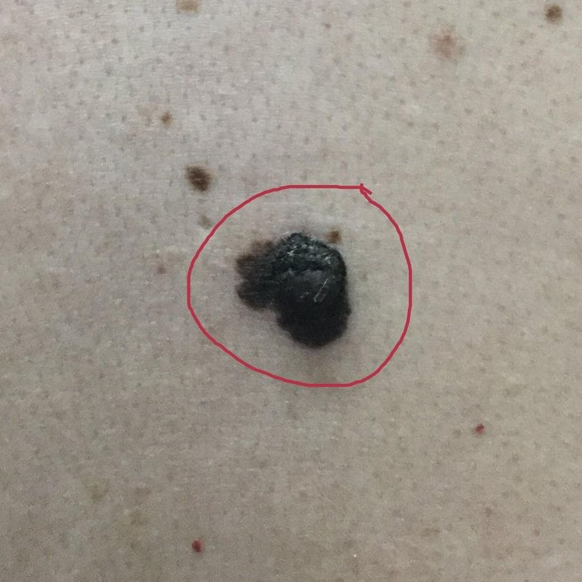
Dysplastic Nevi (DN) were first reported in
1978 by Clark and colleagues.
◦ These were histologic categorizations of nevi found
in patients who were “melanoma prone” due to
family history.
◦ Later they were known as dysplastic nevi as they
had architectural and cytologic atypia (similar
concept to cervical dysplasia)
Dysplastic is more of a histologic term. Some
don’t use it as there is no consensus on how
to grade.
Atypia is more of a clinical term.
However, no term has universal acceptance.
Estimated they occur in about 10% of the
population of Northern European dissent (7-21%)
Pts with a history of melanoma – 34-59% have
DN
Tucker MA, Goldstein AM. Melanoma etiology: where are we? Oncogene
2003;22:3042–52. 22.
Tucker MA. Melanoma epidemiology. Hematol Oncol Clin North Am 2009;23:383–
95.
Melanomas are usually are found in sun-
exposed areas (chronic or intermittent).
Usually in later years of life although due to
tanning beds this is changing.
Dysplastic Nevi are found in sun exposed
AND non sun-exposed areas.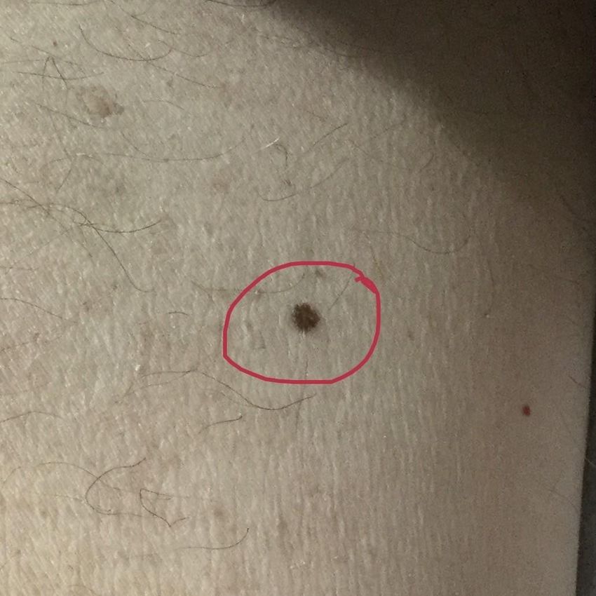
Very difficult question to answer.
There is no agreement on terminology or
grading among Dermatopathologists
Dermatopathologists do not even agree with
themselves on grading
How do you answer the question if we do not
have agreement?
How can we agree to how far something is if
one person measures a mile as 5280 feet and
another as 4500 feet?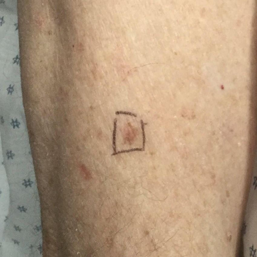
“The reliability of a diagnostic test depends on
the reproducibility of the result.”
8 Expert pathologists convened (published and
well recognized in the community as experts).
Each submitted 5 specimens.
37 of those specimens were used (one slide per
case). Had to be “classic cases.”
Had to say “benign,” “malignant,” or
“indeterminate.”
Farmer et al., Discordance in the Histopathologic Diagnosis of Melanoma and
Melanocytic Nevi Between Expert Pathologists. Hum Pathol 27:528-531, 1995
Given pt history but not diagnosis of slide.
All identifying information removed from
slides.
Same slide went to each expert – rotated.
Sign out was done in the experts “usual
manner.” Agreed with each other 62% of the time!
38% had 2 or more discordant
interpretations.
No expert had more disproportionate
discordance.
K statistic for 8 observers and 3 possible
outcomes was 0.5 with a p value of >0.81 = excellent to almost perfect
agreement
0.61-0.81 = substantial agreement
0.41-0.6 = moderate agreement
0.21-0.4 = fair agreement
0.01-0.2 = slight agreement Similar study found K statistic of 0.34.
False Positives – DN read as MIS 17.6%. DN
read as Invasive Melanomas in 3.2%
False Negatives – Melanomas read as DN in
12%.
Brochez L, Verhaeghe E, Grosshans E, et al. Interobserver variation in the
histopathological diagnosis of clinically suspicious pigmented skin lesions. J
Pathol 2002; 196(4):459-66. “One Dermatolopathologist’s moderately
atypical nevus may be another’s melanoma.”
Elston D, Practical advice regarding problematic pigmented lesions. J Am
Acad Dermatol 2012;67:148-55. “It is not necessary to perform a biopsy of a
dysplastic nevus unless there is clinical
suspicion for melanoma.”
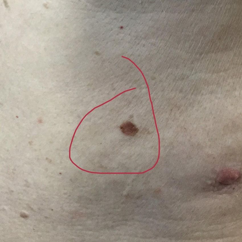
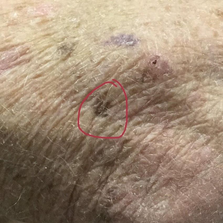
Really??? In my clinic, 4% of shave removals of DN are
actually melanomas!
This does not count things like the last
picture which is an obvious melanoma and
are biopsies, not shave removals
We are projected to treat over 150
melanomas this year in my clinic from shave
removals! Pathologists want excision with 1 foot
margins I think.
When comparing shave to punch, shaves had
95.5% concordance with final diagnosis.
Punches had 70.7% concordance.
Armour K, Mann S, Lee S. Dysplastic naevi: to shave, or not to shave? A
retrospective study of the use of the shave biopsy technique in the initial
management of dysplastic naevi. Australas J Dermatol 2005; 46(2):70-5 DN do relate to a patient’s risk for developing
melanoma (perhaps NOT in an individual
lesion)
Someone with one DN has a RR of 1.6
Someone with five or more DN has a RR of
10.5. Retrospective
Prospective RR of someone developing
melanoma who has DN = 47-92!
Gandini S, Sera F, Cattaruzza MS, Pasquini P, Abeni D, Boyle P,et al. Meta-
analysisofriskfactorsforcutaneousmelanoma:.Commonand atypical naevi. Eur
J Cancer 2005;41:28–44.
MacKie et al. Accelerated Detection with prospective surveillance for
cutaneous malignant melanoma in high-risk groups. Lancet
1993;341(8861):1618-20. Of course there is a lot of debate and
conflicting studies on the above. One thing is
clear, more DN = increased risk of developing
melanoma. Many theories = not exactly clear
Environmental exposures and genetics play a
role.
Genetics – very complicated. More later on this.
de Snoo FA, Hottenga JJ, Gillanders EM, Sandkuijl LA, Jones MP,
BergmanW,etal.Genome-wide linkage scanforatypicalneviinp16Leiden melanoma
families. Eur J Hum Genet 2008;16:1135–41.
Nielsen K, Harbst K, Masback A, Jonsson G, Borg A, Olsson H, et al. Swedish
CDKN2A mutation carriers do not present the atypical mole syndrome phenotype.
Melanoma Res 2010;20:266–72.
Puig S, Ruiz A, Castel T, Volpini V, Malvehy J, Cardellach F, et al. Inherited
susceptibility to several cancers but absence of linkage between dysplastic nevus
syndrome and CDKN2A in a melanoma family with a mutation in the CDKN2A
(P16INK4A) gene. Hum Genet 1997;101:359–64. DN are a clear marker for a patient’s risk of
developing melanoma later in life
Lies, really bad lies, statistics…
20% of melanomas arise from DN.
? % of melanomas arise from regular nevi
Rest of melanomas arise de novo
Duffy K, Grossman D. The dysplastic nevus: from historical
perspectivetomanagementinthemodernera:partI.Historical,histologic,and
clinical aspects. J Am Acad Dermatol 2012;67:1e-16 Estimates are all over the place. Some quoted
as low as 1 in 200,000
These estimates are fraught with so many
problems that we would not allow them in
any other aspect of medicine.
Tsao H, Bevona C, Goggins W, Quinn T. The transformation rate of moles
(melanocytic nevi) into cutaneous melanoma: a populationbased estimate.
Arch Dermatol 2003;139:282–8. “We excise too many DN.” “Most do not turn in melanoma.” Moderate-severe and Severe – excise All others monitor
6177 Dermatologists Surveyed between
2001-2015. 703 responded with data
Margins in 2001 were 1.9mm
Margins in 2015 were 2.3mm Severe – 98% excise!!! Moderate – 67% excise Mild -12% excise 2% do not excise regardless!!!
Severe – 49% Moderate – 10% Mild – 1% 51% do not re-excise
A) Less than 1% B) 1-5% C) 5-15% D) 15-25% E) More than 25% but likely less than 30%
Older Dermatologists do not excise as
frequently and use smaller margins
Average RTC is 6-12 months
Winkelmann R, Rigel D. Management of dysplastic nevi: a 14-year follow-up
survey assessing practice trends among US Dermatologists. J Am Acad Derm
73(6) 1056-59 Shave remove everything that I consider is
likely a DN.
Biopsy anything that I think is a melanoma
Mild, Mild-Moderate – recommend
monitoring. Can have excision if desires.
2mm)
Moderate – recommend excising. Can have
excision if desires. (3mm)
Moderate-severe. Excise (4mm)
Severe. Excise (5 mm) What do you do with a recurrent DN?
Send specimens to the best
Videos to explain to patients
RTC 3 months after initial sampling
Yearly if 1 DN
Q6 months is >1 DN
Scripts we read from. Templates we use
Ophthalmology if DNS. 1 in 200 risk of
ocular melanoma
Questionnaire for melanoma “Until a simple and accurate genetic test can
be applied to tissue specimens that is
characterized by high specificity and
sensitivity, the best the physician can do is to
minimize the potential sources of error.”
Dr. Glen Bowen Mild and Mild-Morderate Dysplastic Nevus-
General consensus among Dermatologists is
that we monitor these lesions. If any pigment
returns we usually recommend excising the
area. These lesions can be excised as primary
form of treatment but this is usually not
necessary. Moderate Dysplastic Nevus- There is some
debate among Dermatologist about the
treatment for these atypical moles. Our
recommendation, due to their unknown biologic
potential, is to excise them. Some Dermatologists
would just monitor these lesions and while this is
not usually our preferred way of addressing these
lesions, it is still acceptable. If you choose this
option it is very important that you look at the
area every month in the mirror to see if it looks
like the mole is returning (darkness or pigment
appearing, change in the scar, …). Moderate- Severe and Severely Dysplastic
Nevus- We recommend excising these
lesions. These lesions can be monitored but
this is against our medical advice and we
highly discourage this approach as these
have a fairly reasonable chance of turning
into the skin cancer called Melanoma. I understand that I need to have routine full
body skin exams, at least yearly, by a
Dermatologist. I understand I should perform
monthly self-skin exams of my skin in order
to help spot concerning lesions early and I
should call immediately for an appointment if
I find a concerning lesion or if anything on
my body is growing, changing, or not healing. I hereby authorize Colorado Dermatology
Institute
providers/residents/associates/assistants to
perform the procedure(s). The procedure, its
purpose, as well as alternative therapeutic
options have been explained to me (including
the option to not having any treatment
performed at all.)Although every attempt will
be made to minimize the chance of
complications, I understand that the following
complications are possible: 1. Allergic reaction to anesthesia, antibiotics, or
bandages.
2. Bleeding from the surgical site.
3. Bruising at or around the surgical site.
4. Scar formation will occur, and on rare occasions
unsightly or thickened scars (keloid, hypertrophic, or
pink/ red scars) can form.
5. Wound infection
6. Ulcerations, necrosis (tissue death), or dehiscence
(separations of the edges of the suture wound).
7. Post-operative discomfort and/or pain.
8. Skin color changes (lightening or darkening), which
may be permanent.
9. Recurrence (regrowth) of the lesion at the surgical
location or elsewhere in the body.
10. Loss of or decreased sensation (feeling), which
may be permanent.
11. In rare instances loss of movement around the
surgical site which may be permanent I have had the opportunity to speak with the
medical/pathology staff at Colorado Dermatology
Institute and have been given the link to
educational videos the Colorado Dermatology
Institute has published. I understand that it is
highly encouraged to watch the videos that
pertain to my diagnosis and treatment so that I
can better understand the diagnosis and
proposed treatment. All of my questions have
been addressed and I understand the diagnosis
and treatment options and recommendations. I understand it is my responsibility to make
sure I take proper care of my treatment site
to ensure the best healing. Post-operative
instructions are provided to minimize the
chance and severity of many of the potential
complications. I acknowledge that no
guarantee or assurance has been given by
anyone as to the end result of the
procedure(s).You can also read