Rare Iatrogenic Oronasal Communication after Superior Canine Odontectomy Attempt
←
→
Page content transcription
If your browser does not render page correctly, please read the page content below
Case Report

Rare Iatrogenic Oronasal Communication after Superior
Canine Odontectomy Attempt
Ciprian Roi 1 , Emilia Ianeș 1 , Diana Nica 1 , Alexandra Roi 2, *, Laura Cristina Rusu 2 ,
Ioana Badea 3 and Mircea Riviș 1
1
  Department of Anaesthesiology and Oral Surgery, “Victor Babes” University of Medicine and Pharmacy Timisoara,
2 Eftimie Murgu Square, 300041 Timisoara, Romania; ciprian.roi@umft.ro (C.R.); emilia.ianes@umft.ro (E.I.);
nica.diana@umft.ro (D.N.); rivis.mircea@umft.ro (M.R.)
2
  Department of Oral Pathology, “Victor Babes” University of Medicine and Pharmacy Timisoara,
2 Eftimie Murgu Square, 300041 Timisoara, Romania; laura.rusu@umft.ro (L.C.R)
3
  PhD Student, “Victor Babes” University of Medicine and Pharmacy Timisoara, 2 Eftimie Murgu Square,
300041 Timisoara, Romania; badeaioanadaniela@yahoo.com (I.B)
* Correspondence: alexandra.moga@umft.ro; Tel.: +40-72608000

Submitted: 15 December 2020; Accepted: 22 January 2021; Published: 26 January 2021

Abstract: (1) Background: Oronasal communication is described in the scientific literature as a common
complication that occurs after a cleft palate surgery. In some cases, it can also be a consequence of
oral surgery procedures; the main problem of this type of rare accident is related to the correct healing
process and treatment option. (2) Case report: A patient with oronasal communication caused by an
unsuccessful attempt of a superior canine odontectomy presented at the Emergency Department of the Oral
and Maxillofacial Surgery Hospital, Timișoara. The case management is described from the first consult to
one-year follow-up. We consider that this pathology was optimally treated surgically, and the results are more
than satisfactory, taking into consideration the high rates of recurrence. (3) Conclusion: This case report can
be a useful to a general dentist who is trying to decide whether to perform the canine odontectomy or refer
to a specialist surgeon due to the accidents and complications of this procedure.

Keywords: oronasal communication; iatrogenic; oral surgery; local flaps; canine odontectomy

How to cite: Roi, C.; Ianeș, E.; Nica, D.; Roi, A.; Rusu, L.C.; Badea, I.; Riviș, M. Rare Iatrogenic
Oronasal Communication after Superior Canine Odontectomy Attempt. Timisoara Med. 2020, 2020(2),
4; doi:10.35995/tmj20200204.

Introduction
Oronasal communication is a rare accident that can occur during oral surgery procedures as exodontia of
superior incisors and canines, odontectomies of impacted tooth in premaxilla area, apical tooth resection,
tumour extirpation or cystectomy [1]. It can also appear as a complication due to trauma, gunshot injuries,
syphilitic gumma, Caldwell-Luc procedure, radiation therapy, orthognathic surgery, cleft palate repair, and
cocaine consumption [2–5].

Timisoara Med. 2020, 2020(2), 4; doi:10.35995/tmj20200204                                     http://www.tmj.ro
Timisoara Med. 2020, 2020(2), 4 2
This abnormal communication is a pathological continuity tract between the oral and nasal cavity with
important functional disorders and repercussions, such as nasal regurgitation of the fluid and food,
hypernasality of speech that may be accompanied by compensatory articulation disorders, whose gravity
depend on the size of the communication [6]. Other symptoms include nasal obstruction, frequent
crusting, accumulation of food particles within the communication, halitosis, leakage of nasal secretions
into the mouth and Eustachian tube dysfunction. Sometimes, these symptoms can cause psychological
problems [5,7].
The surgical closure of the oronasal communication can involve palatal transposition flaps, palatal rotation,
tongue flaps, buccal advancement flaps, and nasolabial flaps [8]. Suture dehiscence, post-operative infection,
avascular necrosis and closure under tension remain major causes of recurrence. Among all complications of
surgical closure, recurrence remains to be most common with rate of 25–37% with a challenging problem to
surgeons. Primary closure in single layer often leads to failure and persists as a major concern to the operative
surgeon [9].
Case Report
The patient, a 48-years-old male, presented at the Emergency Department of Oral and Maxillofacial
Surgery Hospital, Timișoara, one week after an unsuccessful attempt of right superior canine odontectomy,
performed in a dental office. Previously, on the X-ray, the tooth 1.3 was identified in intraosseous impaction.
After plexus anaesthesia, the odontectomy of the canine was intended and the approach was mixed: buccal
flap and palatal flap. Even if the osteotomy of the maxillary bone was performed, the canine could not be
discovered and the decision of the clinician was to suture the flaps, temporize the surgical procedure and refer
the patient further to a department of maxillofacial surgery.
The main complaints of the patient at presentation were persistent pain, halitosis, masticatory perturbances
and minor nasal fluid regurgitation. The general medical history of the patient had nothing relevant to show.
We performed a loco-regional examination of the patient and the written informed consent of regarding the
surgical treatment was obtained.
The intraoral examination revealed the presence of the erupted maxillary teeth 1.7, 1.2, 1.1, 2.1, 2.4, 2.5, and 2.7,
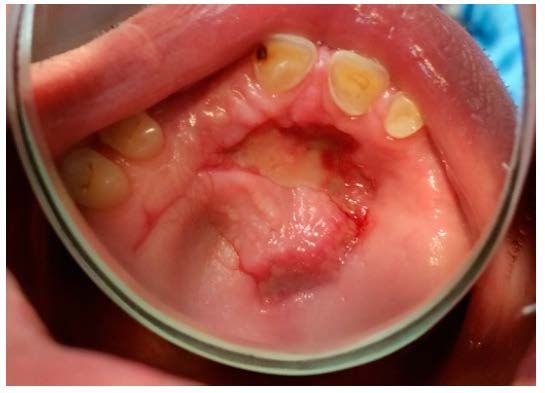
a fact that was noted in the medical chart. In the anterior buccal side, a sutured flap was present. On the
palatal side, the entire flap located in the premaxilla in relation to teeth 1.2, 1.1 and 2.1 was necrotic and
sutured, surrounded by a hyperaemic mucosa (Figure 1). An oro-nasal communication was present, with
and a positive air circulation through the palatal flap (Figure 2).
Figure 1. Necrosis of the palatal flap. The initial oral examination of the patient.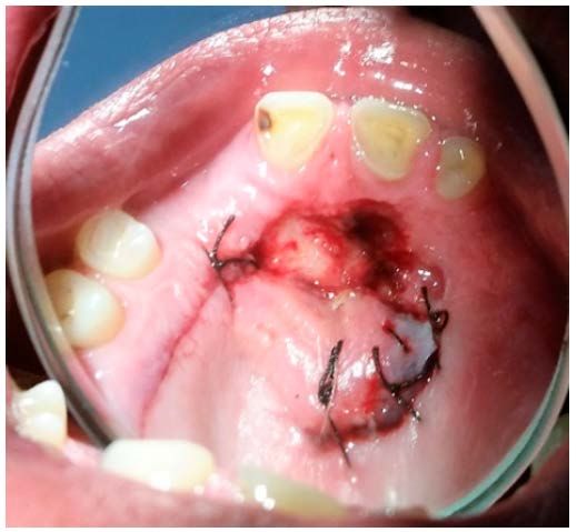
Timisoara Med. 2020, 2020(2), 4 3
Figure 2. Oronasal communication aspect after removing the necrotic debris.
The first therapeutic step was the cleaning of the wound with antiseptic solutions—Povidone-Iodine 10%
and Chlorhexidine Digluconate 0.2%, followed by suture removal and curettage of the necrotic tissues.
The palatal bone could be identified without periosteum. In depth, the nasal cavity was exposed, and nasal
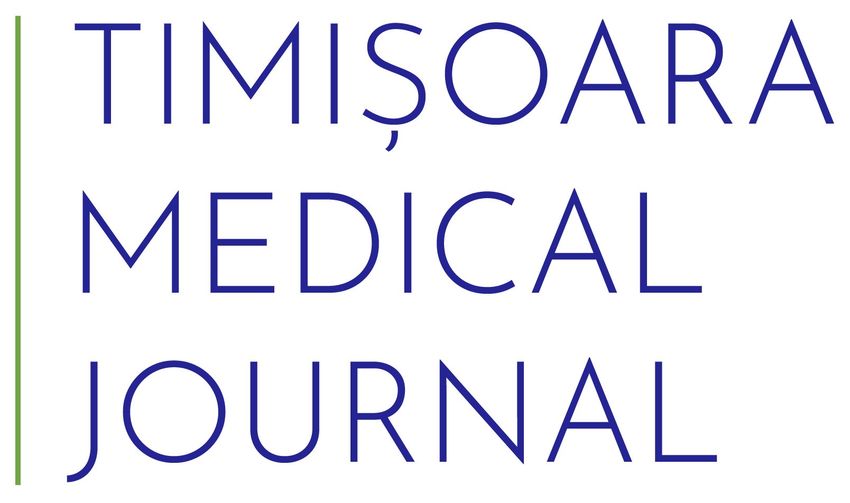
secretions were leaking to the oral cavity. For the certain diagnostic to be confirmed, a full maxillary Cone
Beam Computer Tomography (CBCT) exploration was performed (Figures 3 and 4).
Figure 3. Oronasal communication: Cone Beam Computer Tomography (CBCT) aspect.
Figure 4. Oronasal communication: CBCT virtual 3D reconstruction.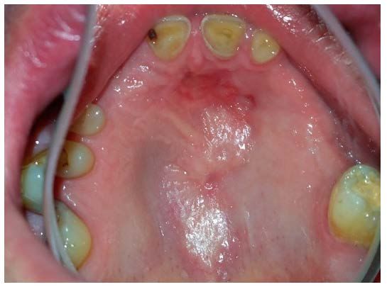
Timisoara Med. 2020, 2020(2), 4 4
The patient was hospitalized and the medication was administrated intravenously: antibiotics, Clindamycin
600 mg 3 times/day; pain killers, Metamizole 1 g/2 mL 3 times/day; and nonsteroidal anti-inflammatory drugs
(NSAID), Ketoprofen 100 mg/2 mL twice daily for 7 days. The patient rinsed his mouth with mouthwash
containing 0.2% Chlorhexidine Digluconate twice a day. Cleaning the wound with Povidone-iodine 10%
was performed twice a day. Nasal saline spray was used in order to prevent maxillary sinusitis.
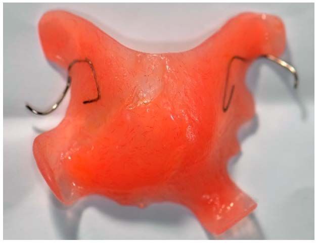
A dental impression was taken for the fabrication of a proper palatal obturator (Figure 5). The improvement
of the symptoms was present and wound healing started to develop. The patient was discharged after 10 days
and twice a week was recalled for a strict local hygiene.
Figure 5. Palatal obturator.
Homecare in this case is very important, the patient being instructed to maintain the personalized palatal
obturator during feeding and drinking and to perform proper oral hygiene. During the periodic check-ups,
the diameter of the oronasal communication decreased and the epithelization was active. After 5 months, the
entire part of the premaxillary fibromucosa was epithelized (where the necrotic area was initially present) and
the palatal bone was covered by fixed oral mucosa. The oronasal communication had a round shape, with
5–6 mm in diameter, clean margins covered by normal oral mucosa, without bleeding or hyperaemic areas,
with nasal secretions that were leaking through the fistula. Anterior from the oronasal communication tract,
the cusp of the impacted canine was revealed (Figure 6).
Figure 6. Oronasal communication after 5 months. The cusp of the impacted canine is revealed.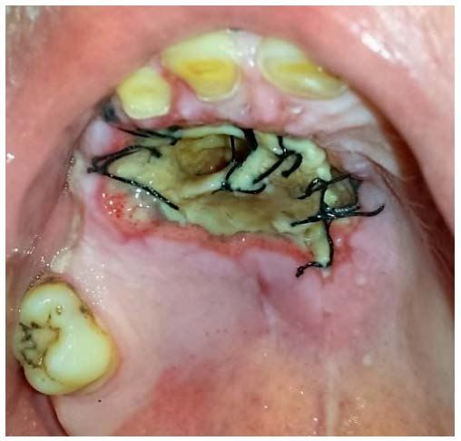
Timisoara Med. 2020, 2020(2), 4 5
Surgical Steps
The surgical approach in this case was considered after the primary healing of the nearby tissue and the
remission of the inflammatory symptoms. After 3 months, the patient was hospitalized for surgical closure
of the oronasal communication. First, the odontectomy of the canine was performed through a palatal
approach (Figure 7). Bilateral greater palatine and maxillary incisive nerve anaesthesia were performed.
The local anaesthetic substances used were Articaine 4% with 1:200,000 Epinephrine.
Figure 7. Oronasal communication after 6 months. The canine was extracted.
With a No. 15 scalpel blade, an incision around the communication was made, about 1.5 cm away from the
margins. An oral mucosa collar was made and detached very close to the nasal side of the communication
and sutured with 4-0 Vicryl suture. This leads to inward movement of palatal mucosa into the nasal cavity,
offering a barrier between the oral nasal cavity and providing a primary closure between these two anatomic
cavities. Inward movement of mucosa exposes the submucosa to the oral cavity and provides a favourable
raw and healthy surface for receiving a second layer closure. The second pedicle flap was based on the greater
palatine artery. A full-thickness incision was performed, and a contralateral pedicled palatal rotation axial flap
based on greater palatine artery was used as a second layer of closure, above the first palatal submucosal layer.
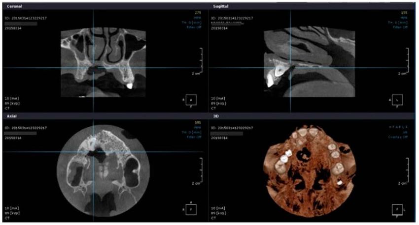
The flap was sutured tension free with 3-0 silk, with alternation of interrupted suture and vertical mattress.
The suture was removed after 7 days (Figure 8).
Figure 8. Local aspect at 24 h after surgery.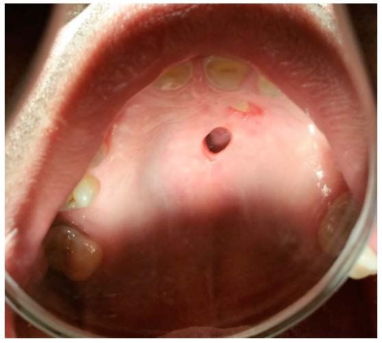
Timisoara Med. 2020, 2020(2), 4                                                                       6

Intravenous therapy was continued: antibiotics, Amoxicillin with clavulanic acid 875 mg/125 mg, twice
daily; pain killers, Metamizole 1 g/2 mL, 3 times/day; and nonsteroidal anti-inflammatory drugs (NSAID),
Ketoprofen 100 mg/2 mL, twice daily for 7 days. The patient rinsed his mouth using a mouthwash with
0.2% Chlorhexidine twice daily. The palatal obturator was used further on for preventing flap dehiscence
(Figures 9 and 10).

                                  Figure 9. Local aspect at 10 days after surgery.

                              Figure 10. Local aspect at one month after surgery.

After one year, the patient was re-examined and no signs of an oronasal fistula were discovered. The OPG
X-ray showed that the maxillary bone was completely healed (Figure 11).

                             Figure 11. X-ray aspect at one year after the surgery.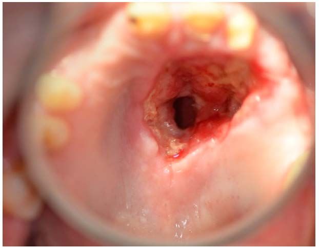
Timisoara Med. 2020, 2020(2), 4                                                                                 7

Discussion
The management of oronasal communication is challenging to perform due to the anatomy of the involved
area: poor vascularization, presence of the nasal secretions in the oral cavity, high chance of the flap
dehiscence, size of the oronasal communication and risk of infection. Although, the spontaneous closure
of the communication is never possible based on the anatomical considerations [10]. Closure techniques
described in the scientific literature varied due to the dimensions of the communication, from one or two
layers mucoperiosteal flap, to buccal advancement flaps, tongue flap, palatal transposition flaps, palatal
rotation, nasolabial flaps or free vascularized tissue [8,11–13].

The transfer of free vascularized tissue is mainly used for the large oronasal communication. It seems to be
the most reliable and logical solution for medium- to large-sized fistulas. The most used flaps are from the
anterolateral thigh, the brachial, the parascapular and the radial forearm [13].

The main advantages of the double-layered surgical closure of oronasal communication are represented by
the perfect coverage of the defect with strength and vascularity improvements, reduced recurrence rate.
This intramucosal purse string suture technique uses local tissue as an alternative option for double-layered
closure. This option is characterized by low donor site morbidity and increased patient comfort [9].

Newer procedures like the use of bone morphogenic protein, acellular dermal matrices, human
amniotic membrane, and distraction osteogenesis have a role to play in the management of oronasal
communication [14].

In our case, the right decision was to wait for the healing of the necrotic maxillary bone and oral mucosa in
order to gain sufficient local tissue resources and avoid the postoperative complications. It is important to
mention that our patient had no medical problems, and the healing potential was exploited.

The main functional disorders caused by the oronasal communication must disappear after the surgery.
These disorders are escaping air from nose to the oral cavity, presence of nasal secretions in oral cavity,
integrity lack of the nasal floor and hard palate, hyper nasality of voice, affected resonance and nasal
regurgitation of fluids [6,10,15]. In the present case report, after the final surgical step none of the mentioned
functional disorders persisted, obtaining complete local healing with no further complications. A complete
documentation of the case and an assessment of the potential intra-operatory accidents and long-term
complications is mandatory in order to perform this type of surgical intervention.

Conclusions
Oronasal communication is a rare pathologic entity with major functional disorders for patient life.
This abnormal communication can occur due to multiple etiologic factors. In our case, a simple canine
odontectomy was transformed in a failure. The management of the case involved the extraction of the
impacted upper canine and surgical sealing the oral cavity of the nasal cavity. The final result of the case
was very satisfactory and all the functions of the oral cavity were restored.

Author Contributions: Conceptualization, C.R., and E.I.; Investigation, D.N.; Writing—Original Draft Preparation,
M.R., L.C.R.; Writing—Review & Editing, M.R. and A.R.; Visualization, I.B.; Supervision, C.R. and A.R.

Funding: This research received no external funding.

Conflicts of Interest: The authors declare no conflicts of interest.
Timisoara Med. 2020, 2020(2), 4                                                                                              8

References
   1. Kaur, P.; Kaur, J. Oronasal Fistula and Complete Edentulism: What to Do? Dent. J. 2014, 2, 142–154.
      [CrossRef]
   2. Eppley, B.; Sclaroff, A. Oronasal fistula secondary to maxillary augmentation. Int. J. Oral Surg. 1984, 13, 535–538.
      [CrossRef]
   3. Emory, R.; Clay, R. Fistula formation and repair after palatal closure: An institutional perspective. Plast.
      Reconstr. Surg. 1997, 99, 1535–1538. [CrossRef] [PubMed]
  4. Jeyaraj, P.E. A Study on Dorsal Pedicled Tongue Flap Closure of Palatal Fistulae and Oronasal Communications.
      Ann. Maxillofac. Surg. 2017, 7, 180–187. [CrossRef] [PubMed]
   5. Hsu, Y.T.; Hao, S.P. Repair of oronasal fistula with silicone button in patients with head and neck cancer. Eur.
      Arch. Otorhinolaryngol. 2015, 272, 1477–1481. [CrossRef] [PubMed]
  6. Jeyaraj, C.P. Techniques to Improve Reliability and Predictability of the Dorsal Pedicled Tongue Flap in Closure
      of Palatal Defects and Oronasal Fistulae. J. Maxillofac. Oral Surg. 2018, 17, 175–181. [CrossRef] [PubMed]
  7. Jeffery, S.L.; Boorman, J.G.; Dive, D.C. Use of cartilage grafts for closure of cleft palate fistulae. Br. J. Plast. Surg.
      2000, 53, 551–554. [CrossRef] [PubMed]
  8. Kumar, V.V.N.P.; Kashyap, S.; Haidry, N.; Singh, M.; Rangaswamy, S.; Ranganatha, N. Buccal Fat Pad for
      Closure of Oroantral and Oronasal Communication: A Study of 16 Cases. Int. J. Oral Care Res. 2018, 6, 27–31.
  9. Singh, V.; Agarwal, S.K.; Jajodia, N.; Garg, A. Purse String Suture Closure: A Useful Double-Layer Technique
      for Closure of an Oronasal Communication. J. Maxillofac. Oral Surg. 2019, 18, 317–319. [CrossRef] [PubMed]
 10. Mohammed, V.A.; Farah, N. Repair of iatrogenic oronasal fistula after periapical surgery. Univ. Res. J. Dent.
      2012, 2, 83–86.
  11. Manimaran, K.; Sureshkannan, P.; Kannan, R. Oro Nasal Fistula Closure by Tongue Flap. JIADS 2011, 2,
      60–62.
 12. Fagan, J. Buccal Fat Pad Flap, Atlas of Otolaryngology, Head & Neck Operative Surgery. Available
      online: https://vula.uct.ac.za/access/content/group/ba5fb1bd-be95-48e5-81be-586fbaeba29d/Buccal%20fat%
      20pad%20flap-1.pdf (Accessed on 12.11.2020).
 13. Colletti, G.; Allevi, F.; Valassina, D.; Bertossi, D.; Biglioli, F. Repair of Cocaine-Related oronasal fistula with
      forearm radial free flap. J. Craniofac. Surg. 2013, 24, 1734–1738. [CrossRef] [PubMed]
 14. Sahoo, N.K.; Desai, A.P.; Roy, I.D.; Kulkarni, V. Oro-nasal communication. J. Craniofac. Surg. 2016, 27,
      e529–e533. [CrossRef] [PubMed]
 15. Henningson, G.; Isberg, A. Oronasal fistulas and speech production. In Multidisciplinary Management of Cleft
      Lip and Palate, 1st ed.; WB Saunders: Philadelphia, PA, USA, 1990; pp. 787–791.

©2021 Copyright by the author. Licensed as an open access article using a CC BY 4.0 license.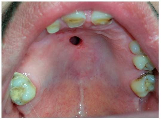
You can also read