Radiographic Measurements of the Cardiac Silhouette and Comparison with Other Radiographic Landmarks in Wild Galahs (Eolophus roseicapilla) - MDPI
←
→
Page content transcription
If your browser does not render page correctly, please read the page content below
animals
Article
Radiographic Measurements of the Cardiac Silhouette and
Comparison with Other Radiographic Landmarks in Wild
Galahs (Eolophus roseicapilla)
Petra Schnitzer *, Shivananden Sawmy and Lorenzo Crosta

                                          Avian, Reptile and Exotic Pet Hospital, Sydney School of Veterinary Science, The University of Sydney,
                                          415 Werombi Road, Camden, NSW 2570, Australia; shivananden.sawmy@sydney.edu.au (S.S.);
                                          lorenzo.crosta@sydney.edu.au (L.C.)
                                          * Correspondence: petra.schnitzer@sydney.edu.au

                                          Simple Summary: Cardiac diseases are a common finding in captive parrots. In this retrospective
                                          study, the cardiac silhouette of 36 wild galahs was measured in the ventrodorsal and laterolateral
                                          projections using radiographic images. The aim of the study was to determine reference values of
                                          the width and the length of the heart of wild galahs in relation to other anatomic landmarks. The
                                          evaluated reference indicates that the width of the heart is 50–65% of the thoracic width and 570–743%
                                          of the coracoid width.

                                          Abstract: Background: Part of the diagnostic workup for cardiac diseases is radiographic imaging.
                                          To determine an enlarged heart, species-specific reference values are necessary. Wild birds are rarely

                                   diagnosed with cardiac disease, and only a few studies have been done to investigate the cardiac
                                          silhouette in wild birds. Methods: In this retrospective study, the cardiac silhouette of 36 wild galahs,
Citation: Schnitzer, P.; Sawmy, S.;
                                          presented at the hospital, was investigated in relation to other anatomic landmarks like the thoracic
Crosta, L. Radiographic
                                          width, clavicula width, synsacrum width, distance between the third and fourth rib, distance of the
Measurements of the Cardiac
Silhouette and Comparison with
                                          clavicula, and length and height of the sternum using a digital DICOM viewer. Results: The cardiac
Other Radiographic Landmarks in           width was significant compared to the thoracic width with a minimum to maximum of 50 to 65%.
Wild Galahs (Eolophus roseicapilla).      The cardiac width compared with the coracoid width also showed significant results with a minimum
Animals 2021, 11, 587. https://           to maximum range of 570 to 743%. A significant correlation was found between the weight and the
doi.org/10.3390/ani11030587               cardiac width and length. Conclusion: The cardiac silhouette in wild galahs is easily measured in
                                          both radiographic views, and the heart size can be compared to other anatomical landmarks.
Academic Editor: Cinzia Benazzi

                                          Keywords: galah; Eolophus roseicapilla; radiographic imaging; cardiac silhouette; heart size;
Received: 27 November 2020
                                          parrot; avian
Accepted: 18 February 2021
Published: 24 February 2021

Publisher’s Note: MDPI stays neutral
                                          1. Introduction
with regard to jurisdictional claims in
published maps and institutional affil-
                                               Radiography is a common diagnostic tool in avian medicine and part of the diagnostic
iations.                                  process for cardiac diseases. Cardiac diseases, whether primary or secondary, are common
                                          in bird species in captivity. In post-mortem findings, Krautwald-Junghanns et al. reported
                                          finding macroscopic changes of the heart in 36% of the cases, with 15% of these cases being
                                          hypertrophic or dilatative cardiomyopathy [1,2]. Another study described 9.7% of pet birds
                                          with cardiomyopathies at post-mortem examination and estimated that 5.6% of these birds
Copyright: © 2021 by the authors.
                                          likely died as a result of primary cardiac disease [3]. Free-living birds, on the other hand,
Licensee MDPI, Basel, Switzerland.
This article is an open access article
                                          have rarely been diagnosed with cardiac failure [2,4].
distributed under the terms and
                                               To improve the accuracy of the diagnosis, many studies have attempted to establish
conditions of the Creative Commons        reference values of the cardiac silhouette in different avian species, including African grey
Attribution (CC BY) license (https://     parrots (Psittacus erithacus), Senegal parrots (Poicephalus senegalus), orange-winged Amazon
creativecommons.org/licenses/by/          parrots (Amazona amazonica), blue-fronted Amazon parrots (Amazona aestiva), budgerigars
4.0/).

Animals 2021, 11, 587. https://doi.org/10.3390/ani11030587                                                    https://www.mdpi.com/journal/animals
Animals 2021, 11, 587 2 of 8
(Melopsittacus undulatus), Spix’s macaws (Cyanopsitta spixii), peregrine falcons (Falco pere-
grinus), Harris’s hawks (Parabuteo unicinctus), saker falcons (Falco cherrug), lanner falcons
(Falco biarmicus), red-tailed hawks (Buteo jamaicensis), bald eagles (Haliaeetus leucocephalus),
ospreys (Pandion haliaetus), screech owls (Otus asio), Canada geese (Branta canadensis), com-
mon kestrels (Falco tinnunculus), Bonelli’s eagles (Aquila fasciata), and Humboldt penguins
(Spheniscus humboldti) [4–15]. The correlations measured between the heart size and stable
points in the body could be used to direct the practitioner and identify alterations in heart
size. A correlation between the width of the cardiac silhouette and the width of the cranial
coelom could be found in most of the studied species [5,6,9,10,14,15]; however, coracoid
and heart width were only correlated in a few species [6,9,15].
In this retrospective study, the cardiac silhouettes of wild galahs (Eolophus roseicapilla)
were investigated in radiographic images. The aim of this study was to measure the cardiac
silhouette of wild galahs in relation to other anatomic landmarks as reference values for
this species.
2. Materials and Methods
The radiographs of 36 adult wild galahs were used to measure the cardiac silhouette.
Medical records from January 2015 to July 2020 were searched using the diagnostic imaging
software Asteris Keystone. All birds were wild and were presented to the Avian Reptile
and Exotic Pet Hospital at the University of Sydney for triage and treatment from public
or wildlife rescue associations. As part of the birds’ diagnostic workup, the animals
underwent radiographic imaging under general anaesthesia with isoflurane (Isothesia
NXT, Henry Schein, 2–4% isoflurane, oxygen 2 L/min) for proper positioning [16]. None
of the patients included in this study showed signs of subclinical cardiac disease according
to the physical examination and showed excellent body condition [17]. Blood (PCV and
TP), taken as a routine examination, showed normal values as compared with reference
values [18]. All birds in this study were judged perfectly fit and could be released after
their hospital check-up. The determination of fitness level included an evaluation of body
condition, plumage, minimal bloodwork, and indoor fly trials and allow for a clinical
assessment of respiratory and heart function [19].
For inclusion in this study, the radiographs had to meet the following criteria: su-
perimposition of the sternum with the spine in the ventrodorsal (VD) projection and
superimposition of the two coracoids in the laterolateral (LL) projection. Animals that
presented traumatic injury that might have altered the anatomy and location of the inner
organs as well as birds with infectious diseases or fractures of the shoulder girdle were
excluded from the study. The survey digital radiographs were all taken on a commer-
cial X-ray machine (Cuattro DR, Golden, CO, USA) using exposure factors of 70 kV and
2.5 mAs. Each measurement was taken after accurate calibration on a Digital Imaging and
Communications in Medicine (DICOM) viewer (Asteris Keystone) using a left/right mark
as standard.
Measurements: In the VD projection, the width of the heart (cardiac silhouette width,
CW) was measured at the widest point using the system ruler on the Asteris Keystone
in mm (Figure 1). The width of the cranial coelom (thorax width, TW) was measured
at the same height as the width of the heart (Figure 1). The width of the coracoid bones
(coracoid width, CoW) was measured directly under the scapulohumeral joints to the
nearest mm. The distance between the third and fourth ribs (DR) was measured parallel to
the spine. The width of the synsacrum (synsacrum width, SynW) and the distance between
the clavicles (distance clavicle, DC) and from the cranial to scapulohumeral joints were
measured as described by Geerinckx et al. [20] (Figure 1). In the LL projection, the length
of the heart (cardiac silhouette length, CL) starting from the aorta to the cardiac apex in
addition to the length (sternum length in lateral, SL) and height of the sternum (sternum
height, SH) were measured. The insertion of the coracoid to the sternum was used as a
landmark to measure the length of the sternum and at 90◦ the height to the nearest mmAnimals 2021, 11, 587 3 of 8
Animals 2021, 11, 587 3 of 8
Animals 2021, 11, 587 3 of 8
(Figure 2). Data for bird weight were incomplete; in 12 out of 36 birds, no weight could be
for bird weight were incomplete; in 12 out of 36 birds, no weight could be evaluated, pre-
for bird weight
evaluated, were incomplete;
precluding statistical in 12 out of with
comparison 36 birds, heart
no weight could
onlybe
24evaluated, pre-
cluding statistical comparison with the heart size inthe size in
only 24 birds. birds.
cluding statistical comparison with the heart size in only 24 birds.
Figure 1. Ventrodorsal (VD) projection and measurements in a radiograph of a wild galah: (a) dis-
Figure 1. Ventrodorsal (VD) projection and measurements in a radiograph of a wild galah: (a)
tance of the clavicle cranial to the shoulder joint (blue); (b) coracoid width immediately caudal to
Figure
distance 1.ofVentrodorsal
the shoulder thejoint
clavicle (VD)(c)projection
cranial
(orange); to andheart
the shoulder
width of the measurements
joint (blue);
at the in point
(b)
widest a radiograph
coracoid
(lightwidthof immediately
blue); a(d)
wild galah:
width (a) dis-to
of caudal
the
tance
the of the
shoulder
thorax clavicle
at the joint cranial
same (orange); to the shoulder
(c) width
height as the joint
of the(white);
heart width (blue);
heart at(e)the(b) coracoid
widestbetween
distance width
point (light immediately
blue);
the third and(d) caudal
width
fourth oftothe
ribs
the shoulder
thorax attothe
parallel joint
thesame (orange);
spineheight (c)
as the
(yellow); width of the
heart width
(f) synsacrum heart at
at(white); the
the widest widest
(e)point point
distance (light blue); (d) width of
between the third and fourth ribs
(black). the
thorax at the same height as the heart width (white); (e) distance between the third and fourth ribs
parallel to the spine (yellow); (f) synsacrum at the widest point (black).
parallel to the spine (yellow); (f) synsacrum at the widest point (black).
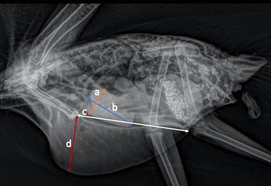
Figure 2. Laterolateral (LL) projection and measurements in a radiograph of a wild galah: (a) wid-
est point of the heart (orange); (b) length of the heart at height of the aorta and pulmonary artery
to the apex
Figure (light blue); (LL)
2. Laterolateral (c) length of theand
projection sternum from the insertion
measurements of the coracoid
in a radiograph to the
of a wild caudal
galah: (a) wid-
Figure 2. Laterolateral
edge (white); (d) depth(LL) projection
of the sternum,and measurements
90° from the junctioninof
a radiograph
the coracoidof
toathe
wild galah:(red).
sternum (a) widest
est point of the heart (orange); (b) length of the heart at height of the aorta and pulmonary artery
point of the heart (orange); (b) length of the heart at height of the aorta and pulmonary artery to
to the apex (light blue); (c) length of the sternum from the insertion of the coracoid to the caudal
the apex
edge (light(d)
(white); blue); (c) of
depth length of the sternum
the sternum, from
90° from the the insertion
junction ofcoracoid
of the the coracoid
to thetosternum
the caudal edge
(red).
(white); (d) depth of the sternum, 90◦ from the junction of the coracoid to the sternum (red).Animals 2021, 11, 587 4 of 8
Data analysis: The statistical analysis was performed using a commercially available
statistics program (SPSS, IBM® , Armonk, NY, USA). The Shapiro–Wilk test was performed
to investigate normality and the Pearson correlation coefficient was used to test for correla-
tion. Homoscedasticity and linearity were evaluated graphically. The heart width and its
relationship with the widths of the cranial coelom, coracoid, and synsacrum as well as the
distance between clavicles and between the third and fourth rib were evaluated by linear
regression. On the LL projection, the relationship between the length of the heart and the
length and depth of the sternum was also evaluated by linear regression. The confidence
interval was set at 90% due to the small sample size. The cardiac width and length were
considered dependent variables, and each radiographic index was an independent variable.
The minimum, maximum, median, mean, and standard deviation were also calculated and
expressed as mean ± SD. Significance was considered with values p < 0.05.
3. Results
The results show that the ratio of the cardiac silhouette is 50–65% of the width of the
cranial coelom. The width of the cardiac silhouette is 570–743% of the coracoid width, as
measured caudally to the shoulder joint. The distance between clavicles is 83–124% of
the cardiac width. The mean values ± SD, minimum, maximum, median, and confidence
intervals are listed in Tables 1 and 2. All variables were normally distributed, except for
the clavicle measurement, which included three outliers.
Table 1. Measurements from the VD radiographic projection. The mean value with standard deviation, minimum, maximum,
and confidence intervals of the variables are reported.
Mean ± SD Median Minimum Maximum 90% Confidence Interval
Cardiac width (mm) CW (N = 36) 21.9 ± 1.3 21.5 17.8 23.6 21.0–21.8
Thoracic width (mm) TW (N = 36) 36.9 ± 2.1 36.6 32.5 42.1 36.3–37.5
Coracoid width (mm) CoW (N = 36) 3.3 ± 0.2 3.3 2.8 3.7 3.2–3.4
Distance between third and fourth
DR (N = 36) 6.2 ± 0.6 6.3 5.00 7.3 6.0–6.4
rib (mm)
Synsacrum width (mm) SynW (N = 36) 5.4 ± 0.4 5.4 4.4 6 5.3–5.5
Distance clavicle (mm) DC (N = 33) 20.6 ± 1.9 20.4 16.4 27.6 20.1–21.1
Table 2. Measurements from the LL radiographic projection. The mean value with standard deviation, minimum, maximum,
and confidence intervals of the variables are reported.
Mean ± SD Median Minimum Maximum 90% Confidence Interval
Cardiac length (mm) CL (N = 36) 27.6 ± 3.1 27.1 22.7 37.2 26.7–28.5
Sternum length (mm) SL (N = 36) 51.1 ± 2.3 51.0 46.7 56.3 50.4–51.7
Sternum height (mm) SH (N = 36) 23.5 ± 2.6 24.1 15.9 27.9 22.8–24.2
A significant weak positive correlation was found between the CW and TW (R2 = 0.196;
p = 0.003) with a regression formula as follows:
CW (mm) = 10.755 + (TW (mm) ∗ 0.288).
Between CW and CoW (R2 = 0.133; p = 0.029), a significant weak positive correlation
was also found with a regression formula as follows:
CW (mm) = 13.778 + (CoW (mm) ∗ 2.309).
The correlation between CW and TW and CW and CoW is demonstrated in Figures 3 and 4
in scatter plots.Animals 2021, 11, 587 5 of 8
Animals 2021, 11, 587 5 of 8
Animals 2021, 11, 587 5 of 8
Figure 3. Scatterplot showing the relation between the cardiac width (mm) and the cranial coelom
Figure 3. Scatterplot showing the relation between the cardiac width (mm) and the cranial coelom
width
Figure(mm) in the VD
3. Scatterplot radiographic
showing projection
the relation (N =the
between 36).cardiac width (mm) and the cranial coelom
width (mm) in the VD radiographic projection (N = 36).
width (mm) in the VD radiographic projection (N = 36).
Figure 4. Scatterplot showing the relation between the cardiac silhouette width (mm) and the cora-
coid width
Figure (mm) in the VD radiographic projectionthe
(N cardiac
= 36). silhouette width (mm) and the cora-
Figure 4.4. Scatterplot
Scatterplotshowing
showingthe
therelation
relationbetween
between the cardiac silhouette width (mm) and the
coid width (mm) in the VD radiographic projection (N = 36).
coracoid width (mm) in the VD radiographic projection (N = 36).
A significant correlation is also present between the heart size and the body weight.
In 24AAoutsignificant
of 36 birds,
significant correlation
the body
correlation is is also
weight
also present
present between
was between
reported the
over
the heart
the
heart size
course
size and
andof the
the the body
physical
body weight.
exam-
weight. In
24Inout
24 out
ination.of 36 ofbirds,
The 36 birds, the body
significance
the body and weight
correlation
weight wasare
was reported reported
reported over
over the as the course
follows:
course of the of the
body physical
weight
physical and exam-
CW, r
examination.
=ination.
The 0.459, pThe
significance significance
= 0.012; body
and and correlation
weight
correlation and
are CL, r are
reported reported
= 0.452, as body
follows:
p = 0.007.
as follows: bodybody
Theweight
mean weight
and and
weight
CW, CW,
in ther
r = 0.459,
= 0.459,
group p
was = 0.012;
275 ± body
52.43 weight
g. No and
other CL, r = 0.452,
significant p = 0.007.
correlations The
p = 0.012; body weight and CL, r = 0.452, p = 0.007. The mean body weight in the group mean
between body weight
variables in the
could be
group
identifiedwas (CW275 ±
and 52.43
DR, g.
r No
= other
0.088, p =significant
0.306; CW correlations
and SynW, between
r =
was 275 ± 52.43 g. No other significant correlations between variables could be identified0.098, variables
p = 0.285; could
CW be
and
identified
DC r = 0.129,(CW p = and DR,
0.227). Onr = 0.088,
the LL p = 0.306;
projection, CW
the and
length SynW,
of the r = 0.098,
heart
(CW and DR, r = 0.088, p = 0.306; CW and SynW, r = 0.098, p = 0.285; CW and DC, r = 0.129, was p = 0.285;
between CW
43% and
DC
p70% rof
= 0.129,
= 0.227).theOn pthe
= 0.227).
sternum On the
LL length. LL
Although
projection, projection,
the of the
thelength
not significant,
length of the
heartthere
was heart
was
between was
43%between
a trend for
andthe70% 43%
lengthand
of theof
70%heart
the
sternum of the to sternum
increase
length. length.
with the
Although Although
not length (r =not
significant, significant,
0.232,
there p was
= 0.087)there was
andfor
a trend heighta trend
the for
(r = 0.211,
length of the
p =length
the 0.108)toof
heart
heart
the sternum.
increase to increase
with the length with
(r =the length
0.232, p = (r = 0.232,
0.087) andpheight
= 0.087) and
(r = height
0.211, p = (r = 0.211,
0.108) p =sternum.
of the 0.108) of
the sternum.Animals 2021, 11, 587 6 of 8
4. Discussion
The results of the present study show that the cardiac silhouette of wild galahs in
the VD projection is 21.38 ± 1.34 mm wide, 50–65% of the width of the cranial coelom,
and 570–743% of the width of the coracoid. The cardiac silhouette in the LL projection
is 27.60 ± 3.12 mm long and between 43% and 70% of the sternum length. During the
study, it was easy to measure the width of the heart in the VD projection as well the length
of the heart in the LL view. Unfortunately, the length of the heart in the VD projection
and the width of the heart in the LL projections could not be measured in all images due
to superimposition of the liver and proventriculus. The decision to measure the other
anatomical structures (TW, CoW, DC, DR, SynW, SL, and SH) was based on other studies
measuring the cardiac silhouette. In other studies, the heart length was not measured.
Among all Psittaciformes, only in cockatoos is the apex of the heart clearly visible, and no
superimposition of the liver is present [20,21]. Like in other species of Psittaciformes, the
heart in cockatoos is superimposed by the proventriculus and, thus, its height and other
parameters cannot be measured precisely in the LL projection [20], which was also the case
in our study.
The main indication for performing radiography in the wild galahs presented to our
hospital was trauma. Birds with fractures of the shoulder girdle, inner coelomic lesions,
or any other traumatic or pathologic lesions that could have altered the measurements in
the radiography image were excluded from the study. A total number of 27 birds were
excluded from the study, due to severe lesions or disease.
In addition, it was not possible to calibrate the DICOM viewer in three cases, and this
resulted in the exclusion of these patients from the study.
One of the selection criteria was the superimposition of the sternum with the spine
in the VD projection plus superimposition of the insertion point of the coracoid in the
sternum. In cases where the carina was distinctly separated from the spine (VD view),
the animal was excluded from the study. These indications resulted in the exclusion of
11 animals, which limited this study.
In the present study, the relation between weight and heart size showed a moderate
significant correlation with r = 0.459 and p = 0.012. The sex/weight ratio does not appear
statistically relevant in budgerigars according to one study [14], but it was suggested to be
relevant in other species [5]. In our study, a correlation between the weight and the cardiac
width and length could be verified. The sex of our birds was not known as determination
of sex is not part of the common procedure in our hospital. Further studies are necessary
to identify a possible correlation between the sex and the heart width and length.
The results of the present study suggest that in wild galahs, the width of the cardiac
silhouette is approximately 50–65% of the width of the cranial coelom and 570–743% of
the width of the coracoid. In studies performed in other species, similar values were
measured, although our study showed the highest maximum range with 65%. Straub
et al. [6] described the heart width to be within 51–61% of the cranial coelom width and
545–672% of the width of the coracoid in captive African grey parrots, Senegal parrots,
and orange-winged Amazon parrots. In captive blue-fronted Amazon parrots, the cardiac
width compared to the cranial coelomic width ranged between 40% and 45%.
In budgerigars, the cardiac width is reported to be the 62% of the thoracic width [14],
and in Spix’s macaws it ranges from 46 to 60% [10].
In non-parrot species, the cardiac/thoracic width ratio showed considerable variation.
It is reported to be 45–59% in Humboldt penguins [8], 66–72% in saker falcons [9], 54–61%
in Harris’s hawks [9], 66–74% in peregrine falcons [9], 65–72% in lanner falcons [9], 48–57%
in Bonelli’s eagle [15], 67–69% in ospreys, 44–52% in bald eagles and 51–75% in common
kestrels [4].
In these previous studies, only the common kestrels and the ospreys were wild ani-
mals and showed the widest ratio, with a maximum value of 75% [4] of the thoracic width.
Migrating birds and birds performing exercise have a larger heart than non-exercising
birds [17,22]. Beaufrère and Fitzgerald describe the larger hearts of wild cockatoos com-Animals 2021, 11, 587 7 of 8
pared with captive species as sign of their cardiac fitness [19]. The falcon species studied
in previous studies showed generally larger cardiac width than parrot species [6,9,13]. In
Mirshahi et al. [4] the width of the heart in wild common kestrels was larger compared to
other falcon species, and in our study, our measured heart width was larger compared to
that in other parrot species. Further investigations are necessary for a similar comparison
of cardiac silhouette in captive galahs.
However, in Pees et al. [2], the heart size of wild galahs (rose-breasted cockatoos—
Eolophus roseicapilla) was investigated, after euthanasia, to evaluate anatomy and pathology.
In the study, the heart width was determined to be 17.7–27.3 mm (20 ± 1.8), the heart length
19.5–30.3 mm (26 ± 2.2), and the heart height 14.1–18 mm (16 ± 0.9) [2]. In the present
study, the width was within the same range presented by Pees et al., 17.77–23.63 mm
(21.38 ± 1.34). The heart length in our study was longer at 22.73–37.15 mm (27.60 ± 3.12).
The difference in the length is possibly due to differences in the method and landmarks
used for measurements in the two studies; in our study, we measured the heart from a ra-
diographic image, while Pees et al. measured the heart following explant from the cadaver.
Cardiac diseases with different aetiology are common in captive birds, but still it is
not easy to diagnose heart problems in living patients [19,23–27].
Krautwald-Junghanns (2004) [1] reports that 15% of 36% birds with macroscopic
changes of the heart had a hypertrophic or dilatative cardiomyopathy. Furthermore, the
same paper reports that 6% of 36% of birds with macroscopic cardiac changes had a
pericardial effusion. It is reported that 21% of birds with a heart problem have a cardiac
disease that leads to a cardiomegaly and, hence, this could be observed with the application
of radiography. Further studies are necessary to obtain reference values for possible
application in diagnosis.
5. Conclusions
This report is the first to measure the cardiac silhouette of wild galahs and of wild
Psittaciformes, in general, using radiographs and providing so reference values for wild
galahs. These findings indicate that cardiac measurements are mostly similar between the
birds but that differences do exist within the species.
About 21% of birds with a cardiac issue have a heart disease that will cause car-
diomegaly and, therefore, can be identified with radiographs [1].
Because of this, species-specific measurements are necessary to help guide the clinician
when using radiographs to assess cardiac size and plan for cardiac workup and, hence,
signify the importance of studies that establish reference values for wild and captive birds,
which can also help to understand the causes of cardiac diseases that develop in birds in
captivity but are almost non-existent in the wild [2].
Author Contributions: Conceptualization: L.C., P.S.; methodology: L.C.; investigation: P.S., S.S.; data
curation: L.C., P.S., S.S.; writing—original draft preparation: P.S.; writing—review and editing: L.C.,
S.S.; supervision: L.C. All authors have read and agreed to the published version of the manuscript.
Funding: This research received no external funding.
Institutional Review Board Statement: Ethical review and approval was waived for this study, due
to the retrospective nature.
Informed Consent Statement: Not applicable.
Data Availability Statement: Database University of Sydney.
Conflicts of Interest: The authors declare no conflict of interest.Animals 2021, 11, 587 8 of 8
References
1. Krautwald-Junghanns, M.-E.; Braun, S.; Pees, M.; Straub, J.; Valerius, H.-P. Research on the Anatomy and Pathology of the
Psittacine Heart. J. Avian Med. Surg. 2004, 18, 2–11. [CrossRef]
2. Pees, M.; Zeh, C.; Filippich, L.J.; Krautwald-Junghanns, M.-E. Pathologisch-anatomische und morphometrische Untersuchungen
am Herzen von wildlebenden Kakadus. Tierärztl. Prax. Ausg. K Kleintiere Heimtiere 2014, 42, 390–396. [CrossRef] [PubMed]
3. Oglesbee, B.L.; Oglesbee, M.J. Results of Portmortem Examination of Psittacine Birds with Cardiac Diseases: 26 Cases (1991–1995).
J. Am. Vet. Med. Assoc. 1998, 212, 1737–1742.
4. Mirshahi, A.; Shariatzadeh, M.; Razmyar, J.; Azizzadeh, M. Evaluation of Cardiac Size in the Common Kestrel (Falco tinnunculus)
Based on Radiographic Measurements. J. Avian Med. Surg. 2016, 30, 345–349. [CrossRef]
5. Hanley, C.S.; Murray, H.G.; Torrey, S.; Pokras, M.A. Establishing Cardiac Measurement Standards in Three Avian Species. J. Avian
Med. Surg. 1997, 11, 15–19.
6. Straub, J.; Pees, M.; Krautwald-Junghanns, M.-E. Measurement of the Cardiac Silhouette in Psittacines. J. Am. Vet. Med. Assoc.
2002, 221, 76–79. [CrossRef]
7. Woo, K.M.T.; Barron, G.H.; Daugherty, A.L. Measurements of the Radiographic Cardiac Silhouette of Ospreys. J. Avian Med. Surg.
2020, 34, 101–102.
8. Yunker, K.A.; Hostnik, E.T.; Johnson, J.G.; Giatis, I.Z. Radiographic evaluation of cardiac silhouette in clinically healthy humboldt
penguins (Spheniscus humboldti). J. Zoo Wildl. Med. 2018, 49, 573–580. [CrossRef]
9. Barbon, A.R.; Smith, S.; Forbes, N. Radiographic Evaluation of Cardiac Size in Four Falconiform Species. J. Avian Med. Surg. 2010,
24, 222–226. [CrossRef]
10. Rettmer, H.; Deb, A.; Watson, R.; Hatt, J.-M.; Hammer, S. Radiographic Measurement of Internal Organs in Spix’s Macaws
(Cyanopsitta spixii). J. Avian Med. Surg. 2011, 25, 254–258. [CrossRef]
11. Silva, J.P.; Castiglioni, M.C.R.; Doiche, D.P.; Vettorato, M.C.; Fogaça, J.L.; Mamprim, M.J.; Rahal, S.C.; Vulcano, L.C. Radiographic
Measurements of the Cardiac Silhouette in Healthy Blue-Fronted Amazon Parrots (Amazona aestiva). J. Avian Med. Surg. 2020, 34,
26–31. [CrossRef] [PubMed]
12. Locke, S.; Johnson, D.; Shimp, J.; Pridgen, T.J. Radiographic Reference Intervals of the Cardiac Silhouette Width in the Bald Eagle
(Haliaeetus leucocephalus). J. Avian Med. Surg. 2020, 34, 260–267. [CrossRef] [PubMed]
13. Lumeij, J.T.; Shaik, M.A.S.; Ali, M. Radiographic Reference Limits for Cardiac Width in Peregrine Falcons (Falco peregrinus).
J. Am. Vet. Med. Assoc. 2011, 238, 1459–1463. [CrossRef]
14. Velayati, M.; Mirshahi, A.; Razmyar, J.; Azizzadeh, M. Radiographic reference limits for cardiac width of budgerigars (Melopsittacus
undulatus). J. Zoo Wildl. Med. 2015, 46, 34–38. [CrossRef]
15. Lopes, F.; Jesus, S.; Med, L.V.; Márquez, I.L.; Fernández, V.M.; Nunes, T.; Regalado, L.S.; González, F.G. Radiographic Reference
Values for the Cardiac Silhouette in Bonelli’s Eagle (Aquila fasciata). J. Avian Med. Surg. 2019, 33, 53–58. [CrossRef] [PubMed]
16. Crosta, L.; Melillo, A.; Schnitzer, P. Basic Radiography; BSAVA Library: Gloucester, UK, 2018; pp. 269–285. ISBN 978-1-910443-32-3.
17. Strunk, A.; Wilson, G.H. Avian Cardiology. Vet. Clin. Exot. Anim. Pract. 2003, 6, 1–28. [CrossRef]
18. Fudge, A.M. Laboratory Medicine: Avian and Exotic Pets; Saunders: Philadelphia, PA, USA, 2000; ISBN 0-7216-7679-0.
19. Fitzgerald, B.C.; Beaufrère, H. Chapter 6—Cardiology. In Current Therapy in Avian Medicine and Surgery; Speer, B.L., Ed.; W.B.
Saunders: St. Louis, MO, USA, 2016; pp. 252–328. ISBN 978-1-4557-4671-2.
20. Geerinckx, L.; Van der Vekens, E.; Saunders, J.H.; Lautenschläger, I.; Van Caelenberg, A.I.L. Literature review of radiographic
measurements of internal organs in psittaciformes. J. Exot. Pet Med. 2019, 28, 60–68. [CrossRef]
21. Pees, M.; Krautwald-Junghanns, M.-E. Cardiovascular Physiology and Diseases of Pet Birds. Vet. Clin. N. Am. Exot. Anim. Pract.
2009, 12, 81–97. [CrossRef]
22. King, A.S.; McLelland, J. Cardiovascular System. In Birds—Their Structure and Function, 2nd ed.; Baillière Tindall: Eastbourne,
UK, 1984.
23. Beaufrère, H.; Ammersbach, M.; Reavill, D.R.; Garner, M.M.; Heatley, J.J.; Wakamatsu, N.; Nevarez, J.G.; Tully, T.N. Prevalence of
and Risk Factors Associated with Atherosclerosis in Psittacine Birds. J. Am. Vet. Med. Assoc. 2013, 242, 1696–1704. [CrossRef]
24. Phalen, D.N.; Hays, H.B.; Filippich, L.J.; Silverman, S.; Walker, M. Heart Failure in a Macaw with Atherosclerosis of the Aorta and
Brachiocephalic Arteries. J. Am. Vet. Med. Assoc. 1996, 209, 1435–1440.
25. McCleery, B.; Jones, M.P.; Manasse, J.; Johns, S.; Gompf, R.E.; Newman, S. Pericardial Mesothelioma in a Yellow-Naped Amazon
Parrot (Amazona auropalliata). J. Avian Med. Surg. 2015, 29, 55–62. [CrossRef] [PubMed]
26. Niemeyer, C.; Favero, C.M.; Kolesnikovas, C.K.M.; Bhering, R.C.C.; Brandão, P.; Catão-Dias, J.L. Two Different Avipoxviruses
Associated with Pox Disease in Magellanic Penguins (Spheniscus magellanicus) along the Brazilian Coast. Avian Pathol. J. WVPA
2013, 42, 546–551. [CrossRef] [PubMed]
27. Maria-Elisabeth, K.-J.; Sven, R.; Thomas, T.; Michael, P. Diagnostic Imaging of Exotic Pets: Birds—Small Mammals—Reptiles;
Schlütersche: Hannover, Germany; CRC Press LLC: London, UK, 2010; ISBN 978-3-89993-049-8.You can also read


