Proteomic Signatures Reveal Differences in Stress Response, Antioxidant Defense and Proteasomal Activity in Fertile Men with High Seminal ROS ...
←
→
Page content transcription
If your browser does not render page correctly, please read the page content below
International Journal of
           Molecular Sciences

Article
Proteomic Signatures Reveal Differences in Stress
Response, Antioxidant Defense and Proteasomal
Activity in Fertile Men with High Seminal
ROS Levels
Tânia R. Dias 1,2,3,† , Luna Samanta 1,4,† , Ashok Agarwal 1, * , Peter N. Pushparaj 5 ,
Manesh Kumar Panner Selvam 1 and Rakesh Sharma 1
 1    American Center for Reproductive Medicine, Cleveland Clinic, Cleveland, OH 44195, USA;
      taniadias89@gmail.com (T.R.D.); lsamanta@ravenshawuniversity.ac.in (L.S.);
      manesh.balu@gmail.com (M.K.P.S.); sharmar@ccf.org (R.S.)
 2    Universidade da Beira Interior, 6201-001 Covilhã, Portugal
 3    Department of Microscopy, Laboratory of Cell Biology, Institute of Biomedical Sciences Abel Salazar and
      Unit for Multidisciplinary Research in Biomedicine, University of Porto, 4050-313 Porto, Portugal
 4    Redox Biology Laboratory, Department of Zoology, School of Life Sciences, Ravenshaw University,
      Cuttack 753003, Odisha, India
 5    Center of Excellence in Genomic Medicine Research, Jeddah 21589, Saudi Arabia;
      peter.n.pushparaj@gmail.com
 *    Correspondence: agarwaa@ccf.org; Tel.: +1-216-444-9485
 †    These authors contributed equally to this work.

 Received: 9 November 2018; Accepted: 29 December 2018; Published: 8 January 2019

 Abstract: Elevated levels of reactive oxygen species (ROS) are a major cause of male infertility.
 However, some men with high seminal ROS levels are still fertile. The main objective of this
 study was to understand the molecular mechanism(s) responsible for the preservation of fertility
 in those men. Semen samples from fertile men were divided into two groups: control (n = 10,
 ROS < 102.2 RLU/s/106 sperm) and ROS+ (n = 10, ROS > 102.2 RLU/s/106 sperm). Proteomic
 analysis of seminal plasma and spermatozoa was used to identify the differentially expressed proteins
 (DEPs) between the experimental groups, from which some proteins were validated by Western blot
 (WB). A total of 44 and 371 DEPs were identified between the study groups in the seminal plasma
 and spermatozoa, respectively. The identified DEPs were primarily involved in oxidoreductase,
 endopeptidase inhibitor, and antioxidant activities. We validated by WB the underexpression of
 NADH:ubiquinone oxidoreductase core subunit S1 (p = 0.01), as well as the overexpression of
 superoxide dismutase 1 (p = 0.03) and peroxiredoxin 4 (p = 0.04) in spermatozoa of ROS+ group.
 Our data suggest that fertile men with high ROS levels possess an effective antioxidant defense
 system that protects sperm proteins, as well as an active proteasomal system for degradation of
 defective proteins.

 Keywords: seminal plasma; spermatozoa; reactive oxygen species; antioxidants; chemiluminescence;
 proteomics; bioinformatics; differentially expressed proteins; Western blot

1. Introduction
     A common end to numerous pathways that lead to defective sperm function is the increase in
reactive oxygen species (ROS) levels in semen [1,2]. Physiological levels of ROS in the semen are
essential for an optimal sperm function and fertilization, as they participate in motility acquisition,
capacitation, and acrosome reaction [3,4]. However, when the rate of ROS generation exceeds the

Int. J. Mol. Sci. 2019, 20, 203; doi:10.3390/ijms20010203                           www.mdpi.com/journal/ijms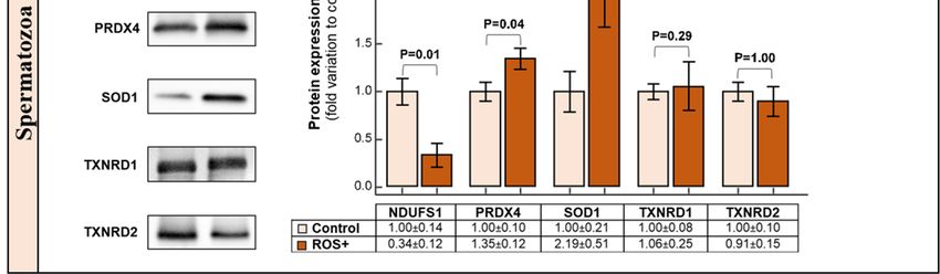
Int. J. Mol. Sci. 2019, 20, 203                                                                                        2 of 14

cells’ antioxidant defense capacity, it leads to oxidative stress (OS), which may damage sperm DNA,
lipids and proteins, thus compromising sperm fertilizing potential [3]. Spermatozoa possess a limited
intrinsic antioxidant machinery that make them dependent on seminal plasma defense system [5].
This characteristic increases the interest regarding the clinical utility of seminal OS testing in infertility
clinics [6,7].
      Besides routine semen analysis, advanced sperm function tests for the assessment of ROS levels,
total antioxidant capacity, sperm DNA fragmentation and compaction, as well as genetic testing are
currently used for the evaluation of male fertility status [8]. Nevertheless, these tests are unable to
establish the etiology of infertility, leading to the classification of many cases as idiopathic [8]. Even
though the chances of conception are increased by assisted reproductive technology (ART) in these
patients, the genomic stability of the embryo is not guaranteed [9]. OS-induced sperm DNA damage
is the cause of infertility in many men [10,11]. In fact, many infertile men with high ROS levels
show sperm DNA fragmentation and poor chromatin packaging [9]. This is associated with lower
fertilization and pregnancy rates in ART, impaired embryo development and quality; and increased
risk of spontaneous abortions, birth defects and childhood diseases such as cancer [10–12]. In recent
years, proteomic analysis of the semen has helped in understanding the biological pathways associated
with male infertility [13]. Our group has extensively studied the proteomic profile of both seminal
plasma and spermatozoa from men with different fertility-related conditions, giving attention to ROS
levels [14–16]. During these investigations, we noticed that some healthy men who presented high
ROS levels in their ejaculates were able to father children. The cutoff to classify a semen sample
as containing high ROS levels was 102.2 relative light units per second per million of spermatozoa
(RLU/s/106 sperm), as previously established [17]. Therefore, we decided to explore the molecular
mechanisms by which these men preserve their fertility. The goal of this study was to compare the
proteome of seminal plasma and spermatozoa from fertile men with high ROS levels with that of
fertile men with physiological ROS levels. We aimed to identify possible alterations in the expression
levels of key antioxidant proteins, as well as the underlying pathways responsible for the protection of
spermatozoa from ROS attack.

2. Results

2.1. Semen Analysis and ROS Levels
     All samples in both the groups were normozoospermic according to World Health Organization
(WHO) 2010 criteria [18] (Table 1). There were no significant differences in semen parameters between
the control and the ROS+ groups. ROS levels were higher (p = 0.0001) in ROS+ group compared to the
control group (Table 1).

                      Table 1. Semen parameters of fertile donors from control and ROS+ groups.

                       Parameter                 WHO 1            Control                ROS+              p-Value
                  Volume (mL)                      >1.5         4.24 ± 0.67           3.76 ± 0.72           0.4384
                       pH                          7.6–8        7.66 ± 0.07           7.60 ± 0.05           0.3333
               Sperm motility (%)                   >40            55 ± 3                58 ± 6             0.5035
          Sperm concentration (106 /mL)             >15        90.95 ± 15.59         75.02 ± 12.87          0.6221
             Total sperm count (106 )               >39       359.50 ± 63.86        254.95 ± 49.67          0.1809
              Round cells (106 /mL)
Int. J. Mol. Sci. 2019, 20, 203 3 of 14
2.2.Int.Global
J. Mol. Proteomic
Sci. 2018, 19,Profile
x FOR of Seminal
PEER Plasma and Spermatozoa
REVIEW 3 of 14
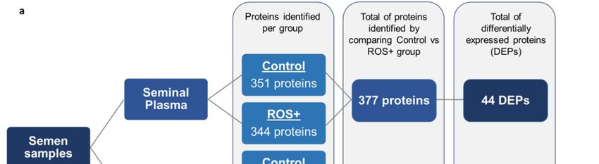
Proteomic analysis of seminal plasma resulted in the identification of 351 proteins in the control
Proteomic analysis of seminal plasma resulted in the identification of 351 proteins in the control
group and 344 proteins in ROS+ group. From a total of 377 proteins in both groups, 44 were
group and 344 proteins in ROS+ group. From a total of 377 proteins in both groups, 44 were
differentially expressed proteins (DEPs) (Figure 1a). One of the seminal plasma DEPs was unique to
differentially expressed proteins (DEPs) (Figure 1a). One of the seminal plasma DEPs was unique to
the control group (2%), while 29 were overexpressed (66%), and 14 underexpressed (32%) in ROS+
the control group (2%), while 29 were overexpressed (66%), and 14 underexpressed (32%) in ROS+
group (Figure 1b).
group (Figure 1b).
Figure
Figure 1. Schematic
1. Schematic representation
representation of theof the results
results obtainedobtained by proteomic
by proteomic analysis:
analysis: (a) number(a)
of number
proteins of
proteinsinidentified
identified in the
the seminal seminal
plasma andplasma and spermatozoa
spermatozoa of fertile
of fertile men men and
(control) (control)
men and
withmen
highwith high
levels
of reactive
levels ofoxygen
reactivespecies
oxygen(ROS+),
speciesas well asasthe
(ROS+), number
well as theofnumber
differentially expressedexpressed
of differentially proteins (DEPs)
proteins
between
(DEPs)the experimental
between groups; (b) groups;
the experimental expression
(b) profile of seminal
expression profileplasma DEPs;plasma
of seminal and (c)DEPs;
expression
and (c)
profile of spermatozoa
expression profile ofDEPs.
spermatozoa DEPs.
In In
spermatozoa, 885 and
spermatozoa, 885 567
andproteins were identified
567 proteins in the control
were identified and ROS+
in the controlgroups, respectively.
and ROS+ groups,
A total of 1144 proteins where identified after the comparison between both groups, from
respectively. A total of 1144 proteins where identified after the comparison between both groups, which 371
proteins were differentially
from which 371 proteinsexpressed (Figure 1a). The
were differentially majority
expressed (45%) of
(Figure theThe
1a). spermatozoa
majority DEPs
(45%) were
of the
unique to the control group (168 proteins), while only 16 proteins were unique to the ROS+
spermatozoa DEPs were unique to the control group (168 proteins), while only 16 proteins were group (4%).
Besides,
unique95toDEPs were group
the ROS+ underexpressed (26%)
(4%). Besides, 95and
DEPs92 were
overexpressed (25%) in
underexpressed ROS+
(26%) group
and (Figure 1c).
92 overexpressed
(25%) in ROS+ group (Figure 1c).
2.3. Functional Annotations and Pathway Analysis
2.3.Protein
Functional Annotationsrevealed
annotations and Pathway
thatAnalysis
the DEPs identified in seminal plasma belong to
exosomes,Protein annotations revealed that the DEPsand
different vesicles, secretory granules, extracellular
identified proteins
in seminal (Figure
plasma 2a). toHowever,
belong exosomes,
membrane-bound organelle
different vesicles, proteins
secretory wereand
granules, alsoextracellular
detected in seminal plasma
proteins (Figure
(Figure 2a). In spermatozoa,
2a). However, membrane-
thebound
identified DEPsproteins
organelle belong to various
were subcellular
also detected locationsplasma
in seminal such as(Figure
mitochondria and flagellumthe
2a). In spermatozoa,
cytoskeleton (Figure 2b).
identified DEPs belong to various subcellular locations such as mitochondria and flagellum
cytoskeleton (Figure 2b).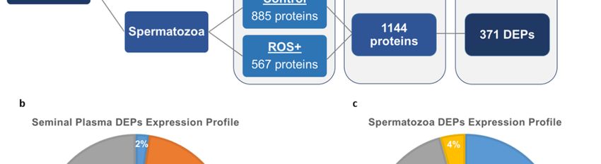
Int. J. Mol. Sci. 2019, 20, 203                                                                             4 of 14
 Int. J. Mol. Sci. 2018, 19, x FOR PEER REVIEW                                                               4 of 14

     Figure 2. Localization of differentially expressed proteins (DEPs) in: (a) seminal plasma; and (b)
      Figure 2. Localization of differentially expressed proteins (DEPs) in: (a) seminal plasma; and (b)
     spermatozoa. The number of DEPs that were overexpressed (grey), underexpressed (orange), unique
      spermatozoa. The number of DEPs that were overexpressed (grey), underexpressed (orange), unique
     to control (blue), and unique to ROS+ (yellow) are shown for seminal plasma and spermatozoa.
      to control (blue), and unique to ROS+ (yellow) are shown for seminal plasma and spermatozoa.
      Functional enrichment analysis of seminal plasma DEPs using STRING online software showed
      Functional enrichment analysis of seminal plasma DEPs using STRING online software showed
the biological processes and molecular functions in which they were involved. According to the
 the biological processes and molecular functions in which they were involved. According to the
biological processes, 4 DEPs were involved in acute phase response, 6 in protein folding and 18 in
 biological processes, 4 DEPs were involved in acute phase response, 6 in protein folding and 18 in
regulation of biological quality. Regarding the molecular functions, 4 DEPs were associated with
 regulation of biological quality. Regarding the molecular functions, 4 DEPs were associated with
antioxidant activity and 7 with endopeptidase inhibitor activity. Haptoglobin (HP), peroxiredoxin 4
 antioxidant activity and 7 with endopeptidase inhibitor activity. Haptoglobin (HP), peroxiredoxin 4
(PRDX4) and S100 calcium-binding protein A9 (S100A9) were the main proteins involved in antioxidant
 (PRDX4) and S100 calcium-binding protein A9 (S100A9) were the main proteins involved in
activity, while serpin B6 (SERPINB6) and complement C3 (C3) were among the proteins involved in
 antioxidant activity, while serpin B6 (SERPINB6) and complement C3 (C3) were among the proteins
endopeptidase inhibitor activity. According to the Ingenuity Pathway Analysis (IPA) semenogelins
 involved in endopeptidase inhibitor activity. According to the Ingenuity Pathway Analysis (IPA)
I (SEMG1) and II (SEMG2) were in the top list of downregulated proteins in seminal plasma with a
 semenogelins I (SEMG1) and II (SEMG2) were in the top list of downregulated proteins in seminal
higher fold change between the groups. On the other hand, HP and C3 were among the top list of
 plasma with a higher fold change between the groups. On the other hand, HP and C3 were among
upregulated proteins with a higher fold change in ROS+ relative to control group. These two proteins
 the top list of upregulated proteins with a higher fold change in ROS+ relative to control group. These
were also classified as positive acute phase response proteins, which was one of the toxicity functions
 two proteins were also classified as positive acute phase response proteins, which was one of the
identified by the Tox lists tool (Supplementary Figure S1a). PRDX4 and S100A9 were associated with
 toxicity functions identified by the Tox lists tool (Supplementary Figure S1a). PRDX4 and S100A9
OS as identified by the IPA Tox lists tool (Supplementary Figure S1a). These seven DEPs were selected
 were associated with OS as identified by the IPA Tox lists tool (Supplementary Figure S1a). These
for validation by Western blot (WB) and compared with the results obtained by the proteomic results
 seven DEPs were selected for validation by Western blot (WB) and compared with the results
(Table 2).
 obtained by the proteomic results (Table 2).
     Table 2. Proteomic data of the differentially expressed proteins identified in seminal plasma samples
      Table 2. Proteomic data of the differentially expressed proteins identified in seminal plasma samples
     from fertile donors from control and ROS+ groups, selected for validation by Western blot.
      from fertile donors from control and ROS+ groups, selected for validation by Western blot.
                              Abundance
            Protein            Abundance             NSAF Ratio      Expression Profile       p-Value
             Protein      Control       ROS+          NSAF ratio Expression Profile p-Value
                              Control            ROS+
            SEMG1
             SEMG1           High
                               High    High
                                        High             0.26
                                                          0.26           UE
                                                                         UEininROS+
                                                                                ROS+          0.00074
                                                                                              0.00074
            SEMG2            High      High              0.26            UE in ROS+           0.00023
             SEMG2
              HP
                               High
                           Very Low
                                        High
                                       Low
                                                          0.26
                                                         9.03
                                                                         UE in ROS+
                                                                         OE in ROS+
                                                                                              0.00023
                                                                                              0.00349
               HP
            PRDX4           Very  Low
                           Very Low     Low
                                       Low                9.03
                                                         3.39            OEininROS+
                                                                         OE     ROS+          0.00349
                                                                                              0.00099
             PRDX4
           SERPINB6         Very Low
                             Low        Low
                                       Low                3.39
                                                         2.68            OE in ROS+
                                                                         OE in ROS+           0.00099
                                                                                              0.00424
            S100A9
            SERPINB6       VeryLow
                                Low   Medium
                                        Low             3.77
                                                          2.68           OE
                                                                         OEininROS+
                                                                                ROS+          0.01707
                                                                                              0.00424
              C3           Very Low   Medium            17.22            OE in ROS+           0.00210
               S100A9        Very Low       Medium        3.77            OE in ROS+           0.01707
     C3, Complement C3; HP, Haptoglobin; NSAF, Normalized spectral abundance factor; OE, overexpressed; PRDX4,
                C3          Very Low Medium               17.22          OE in ROS+             0.00210
     Peroxiredoxin 4; S100A9, S100 calcium-binding protein A9; SEMG1, Semenogelin I; SEMG2, Semenogelin II;
      C3, Complement
     SERPINB6,             C3; underexpressed.
                Serpin B6; UE,  HP, Haptoglobin; NSAF, Normalized spectral abundance factor; OE,
      overexpressed; PRDX4, Peroxiredoxin 4; S100A9, S100 calcium-binding protein A9; SEMG1,
      Semenogelin I; SEMG2, Semenogelin II; SERPINB6, Serpin B6; UE, underexpressed.

    In spermatozoa, the functional enrichment analysis of Search Tool for the Retrieval of Interacting
Genes/Proteins (STRING) software showed that, among the biological processes, 76 proteins were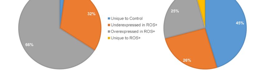
Int. J. Mol. Sci. 2019, 20, 203                                                                               5 of 14

     In spermatozoa, the functional enrichment analysis of Search Tool for the Retrieval of Interacting
Genes/Proteins (STRING) software showed that, among the biological processes, 76 proteins were
associated with response to stress, 19 with protein folding, 37 were involved in oxidation-reduction
processes, and 42 in the regulation of response to stress. Regarding the molecular functions, 11 proteins
presented antioxidant activity, including superoxide dismutase 1 (SOD1), PRDX4, thioredoxin
reductase 1 and 2 (TXNRD1 and TXNRD2). Moreover, 28 proteins were associated with oxidoreductase
activity, comprising NADH-ubiquinone oxidoreductase core subunit S1 (NDUFS1), TXNRD2, SOD1
and PRDX4. After performing the IPA analysis, similar results were observed by the IPA Tox lists tool
(Supplementary Figure S1b). PRDX4, SOD1 and TXNRD2 were associated with OS, while NDUFS1
and TXNRD2 were related to mitochondrial dysfunction. Besides, SOD1 and TXNRD1 were also
associated with NRF2-mediated OS response. 5 proteins were selected for validation by WB and
compared with the results obtained by the proteomic analysis (Table 3).

      Table 3. Proteomic data of the differentially expressed proteins identified in spermatozoa samples from
      fertile donors from control and ROS+ groups, selected for validation by Western blot.

                                      Abundance
               Protein                                  NSAF Ratio     Expression Profile      p-Value
                                  Control     ROS+
              NDUFS1              Medium     Very Low     0.02           UE in ROS+             0.00004
               PRDX4                Low      Medium        4.48          OE in ROS+             0.00134
                SOD1                Low      Medium       3.99           OE in ROS+             0.02830
              TXNRD1                  -      Very Low        -          Unique to ROS+          0.00006
              TXNRD2              Very Low   Medium       10.95          OE in ROS+             0.03640
      NDUFS1, NADH:Ubiquinone Oxidoreductase Core Subunit S1; NSAF, Normalized spectral abundance factor;
      OE, overexpressed; PRDX4, Peroxiredoxin 4; SOD1, superoxide dismutase 1; TXNRD1, Thioredoxin reductase 1;
      TXNRD2, Thioredoxin reductase 2; UE, underexpressed.

2.4. Upstream Regulators
     Using the upstream analysis tool of IPA, several cytokines were predicted to be responsible
for the altered expression levels of seminal plasma proteins in the dataset. Interleukin-1 alpha and
beta (IL1A and IL1B), interleukin-6 (IL6), Interleukin-22 (IL22), and tumor necrosis factor (TNF) were
predicted to be activated, explaining the overexpression of DEPs such as S100A9, C3 and HP. They
may also be responsible for the underexpression of prostate-specific antigen (KLK3), lipoprotein lipase
(LPL) and chaperone heat shock protein HSP 90-beta (HSP90AB1) (Supplementary Figure S2).
     In spermatozoa, two upstream regulators were predicted to be activated in this dataset: nuclear
factor erythroid 2-related factor 2 (NFE2L2) and TNF. The transcription regulator NFE2L2 was
shown to regulate the overexpression of proteins involved in oxidation-reduction processes, such
as SOD1, SOD2 and 6-phosphogluconate dehydrogenase, decarboxylating (PGD) (Supplementary
Figure S3). Its activation may also explain the overexpression of some proteasomes (PSMB2 and
PSMB5). The cytokine TNF was also predicted to be activated and regulate the overexpression of
SOD2, fibronectin (FN1), ion-binding proteins (GPD2, HSPG2, LCN2), as well as the underexpression
of prohibitin (PHB) (Figure S3).

2.5. Western Blot
     All the selected seminal plasma proteins (SEMG1, SEMG2, HP, SERPINB6 and PRDX4) were
identified by WB, however, there were no significant alterations in their expression levels between the
control and the ROS+ groups (Figure 3a).
Int. J. Mol. Sci. 2019, 20, 203 6 of 14
Int. J. Mol. Sci. 2018, 19, x FOR PEER REVIEW 6 of 14
Figure 3. Graphical representation of Western blot results and respective representative blots for: (a)
Figure 3. Graphical representation of Western blot results and respective representative blots for:
seminal plasma; and (b) spermatozoa proteins. Results are expressed as mean ± SEM and were
(a) seminal plasma; and (b) spermatozoa proteins. Results are expressed as mean ± SEM and were
considered significant for
considered significant for pp <
< 0.05.
0.05.
In sperm proteins, there was a decrease in NDUFS1 (p = 0.01) protein expression levels in the
ROS+
ROS+ group
grouprelative
relativetotothe
thecontrol
control(Figure
(Figure3b).
3b).AnAn
overexpression
overexpressionof PRDX4
of PRDX4(p =(p0.04) and and
= 0.04) SOD1 (p =
SOD1
0.03) was observed in ROS+ group when compared to the control group. There were no
(p = 0.03) was observed in ROS+ group when compared to the control group. There were no significantsignificant
alterations in the protein expression of TXNRD1 and TXNRD2 (Figure (Figure 3b).
3b).
3. Discussion
seminal ROS
High seminal ROS levels
levels have
have been
been widely
widely debated
debated as as aa major
major cause
cause of
of male
male infertility
infertility [19,20].
[19,20].
Nevertheless, the role of ROS at physiological concentrations in regulation of sperm function cannot
be ignored [3,21]. In In the
the present
present study,
study, we report a comparative
comparative proteomic analysis of seminal plasma
and spermatozoa
spermatozoafrom fromfertile
fertilemen
menexhibiting
exhibitinghigher ROS
higher ROSlevels thanthan
levels the pre-established reference
the pre-established level
reference
with respect to fertile men with basal ROS levels. This is important to gain a better insight
level with respect to fertile men with basal ROS levels. This is important to gain a better insight into into the role
of ROS
the rolein of
sperm
ROSfunction
in sperm in general
function andinto general
understandandsperm dysfunctionsperm
to understand under dysfunction
pathophysiological
under
conditions with elevated
pathophysiological ROS level.
conditions with elevated ROS level.
In semen, the principal source of ROS are morphologically abnormal, immature immature spermatozoa,
spermatozoa,
leukocytes[22].
and leukocytes [22].As Asboth
both groups
groups were
were negative
negative for for leukocytes
leukocytes (Endtz
(Endtz negative),
negative), the elevated
the elevated ROS
ROS generation
generation may be may be attributed
attributed to the presence
to the presence of immature
of immature cells in
cells in these these samples.
samples. Recently, Recently,
we have
we have reported
reported the presence the presence
of immature of immature
cells withcells with proteome
different different proteome
profile in profile in the ejaculated
the ejaculated semen of
semenmen
fertile of fertile men [23]. Therefore,
[23]. Therefore, the in
the difference difference in theprofile
the proteome proteome profile of spermatozoa
of spermatozoa in the control inand
the
control and ROS+ groups may be due to the presence of comparatively
ROS+ groups may be due to the presence of comparatively more number of immature spermatozoa more number of immature
spermatozoa
in in theThis
the latter group. latter
wasgroup. This was
corroborated bycorroborated
our proteomic byresults
our proteomic
that showedresults that showed an
an underexpression
of sperm surface protein Sp17 (SPA17) in ROS+ group. This protein is weakly expressed in
spermatocytes, while a high expression was reported in early and late spermatids, which suggestsInt. J. Mol. Sci. 2019, 20, 203 7 of 14
underexpression of sperm surface protein Sp17 (SPA17) in ROS+ group. This protein is weakly
expressed in spermatocytes, while a high expression was reported in early and late spermatids, which
suggests that most of the ejaculated spermatozoa express SPA17 protein. This also supports its role in
the sperm differentiation [24,25]. Similarly, underexpression of annexins (1–6) points towards failure
of apoptosis in these samples, resulting in the increase in immature/or undifferentiated spermatozoa.
After bioinformatic analysis of the seminal plasma DEPs, we focused on SEMG1, SEMG2,
SERPINB6, HP, PRDX4, S100A9 and C3. SEMGI and SEMGII are highly abundant in seminal plasma
and are responsible for the formation of the characteristic gel-like coagulum after ejaculation [26].
They play an important role in protecting the spermatozoa and in the fertilization process [27].
The underexpression of SEMG1 and SEMG2 in ROS+ men was accompanied by the underexpression
of KLK3, which is one of the trypsin-like serine proteases responsible for semenogelins digestion to
attain semen liquefaction [28]. Moreover, an overexpression of SERPINB6 was observed in ROS+ men.
This protein is a member of the serpins protein family that is involved in the regulation of trypsin-like
serine proteases activity [29]. The alterations in the expression profile of these proteins resulted in
normal liquefaction of semen samples in ROS+ group, an important factor for the preservation of
sperm fertilizing potential.
HP, PRDX4 and S100A9 were identified as the main seminal plasma proteins involved in
antioxidant activity, which were overexpressed in ROS+ samples. HP in human fluids binds to
hemoglobin to inhibit its oxidative potential as a free molecule [30]. In the presence of hydrogen
peroxide (H2 O2 ), one of the main ROS in semen, hemoglobin can act as a peroxidase [31], thus
generating more ROS. Overexpression of HP in the seminal plasma of ROS+ men can prevent an
oxidative chain reaction. PRDX4 belongs to the family of peroxiredoxins, which are major players
of the antioxidant defense system in semen. This protein was previously identified in both seminal
plasma and spermatozoa of human semen samples [32]. PRDX4 contain two cysteine residues in its
active site, which are major targets for ROS [33]. As ROS are neutralized after binding to PRDX4,
the overexpression of this protein in the seminal plasma of ROS+ men confers higher protection against
increased ROS levels.
S100A9 is a calcium- and zinc-binding protein associated with stress response [34]. It is considered
a danger- or damage-associated molecular pattern (DAMP) molecule, as, in response to various stimuli,
it can bind to pro-inflammatory receptors and initiate an inflammatory reaction [35]. In this particular
study, the stimuli for the overexpression of this protein was the high ROS levels in semen of ROS+ men.
In fact, there is a direct link between high ROS levels and inflammation [36]. A previous proteomic
study also identified the overexpression of S100A9 in the seminal plasma of smoking men [37], which
also reflects an environment with high ROS levels. Overexpression of S100A9 was associated with
the activation of NADPH oxidase [38], which may be one of the reasons for the accumulation of ROS
in semen. S100A9 pro-inflammatory activity starts with the activation of the nuclear factor-kappa B
(NF-κB), which consequently induces cytokine secretion [38]. This may explain why many interleukins
were predicted to be active in the seminal plasma of ROS+ men, including IL1A, IL1B, IL6, IL22,
and TNF (Supplementary Figure S2). These inflammatory factors were identified as the upstream
regulators of many proteins in the dataset and are implicated in the regulation of sperm fertilization
processes during sperm transit through the female reproductive tract [39]. Accordingly, Tox lists
showed that many positive acute phase response proteins were upregulated in ROS+ men. This may
also be related to the observed overexpression of protein C3, which is a mediator of local inflammatory
processes and immune responses [40]. For instance, it has been demonstrated that cytokines IL1A,
IL1B, IL6 and TNF can lead to increased C3 secretion [41]. In human seminal plasma, C3 complement
system is regulated by complement-inhibiting factors to protect spermatozoa from damage by chronic
inflammation [42]. Although all the selected proteins were identified by WB, the results were not
concordant with the proteomic data (Figure 3a).Int. J. Mol. Sci. 2019, 20, 203 8 of 14
Spermatozoa proteomic data showed 371 DEPs, from which 5 were selected for validation by WB:
NDUFS1, SOD1, PRDX4, TXNRD1, and TXNRD2. NDUFS1 is one of the subunits of the mitochondrial
complex I, which is the starting point of oxidative phosphorylation (OXPHOS). Complex I is responsible
for NADH oxidation, thus providing electrons for the respiratory chain [43]. Mitochondrial function
is crucial for sperm fertilization, not only for ATP production to obtain energy, but also for the
physiological production of ROS. NDUFS1 is the largest subunit of complex I and is essential for the
proper assembly of the complex required for its function [44]. The underexpression of NDUFS1 in the
spermatozoa of ROS+ men may impair complex I assembly and result in its dysfunction, which is
one of the most common mitochondrial dysfunctions observed in humans [44]. Moreover, subunits of
complex IV (COX4I1 and COX5A) and complex V (ATP5H) were also underexpressed in ROS+ group.
These alterations contribute to the higher production of ROS levels in this group. We were able to
validate the underexpression of NDUFS1 by WB. Mitochondrial dysfunction in mature spermatozoa
may contribute to the high ROS levels in ROS+ group.
The preponderance for OS in spermatozoa of ROS+ group is counteracted by the increased
antioxidant defense. Both cytosolic and mitochondrial superoxide dismutase (SOD1 and SOD2,
respectively), mitochondrial thioredoxin reductase 2 (TXNRD2), and PRDX4 were overexpressed in
spermatozoa of ROS+ group. Moreover, cytosolic thioredoxin reductase 1 (TXNRD1) was uniquely
expressed in ROS+ group providing additional defense. SOD1 belongs to the superoxide dismutase
family and is one of the first line of antioxidant defense enzymes against ROS attack in spermatozoa [45].
The overexpression of SOD1, which was further confirmed by the WB analysis (Figure 3b), may explain
the higher antioxidant protection in spermatozoa of ROS+ men. This protein provides protection
against the attack from superoxide anion radicals. SOD1 and SOD2 increased activity was predicted
to be regulated by NFE2L2 and TNF, which were identified as their activated upstream regulators
(Supplementary Figure S3). These transcription factors were described as important regulators of
antioxidant responses [46].
PRDX4 is one of the main proteins responsible for reduction of peroxides in spermatozoa [33].
It can be found in sperm plasma membrane, acrosome, nucleus, and cytosol [33]. The binding
of ROS to the active site of PRDX4 leads to the oxidation of its cysteine residues and the enzyme
becomes inactive [47]. Without an active thioredoxins system, PRDX4 would remain permanently
inactive in an environment with high ROS levels, thus being unable to scavenge other forms of ROS.
The thioredoxin system is constituted by thioredoxins, thioredoxins reductases and NADPH [48].
Thioredoxins reductases, including TXNRD1 (cytosolic) and TXNRD2 (mitochondrial), play a key role
in maintaining the cyclicity of this system; they are responsible for maintaining thioredoxins in their
reduced (active) state in a NADPH-dependent manner [33]. Subsequently, thioredoxins act as electron
donors for peroxiredoxins, facilitating their reduction and reactivation [47]. Based on our proteomic
data, PRDX4 and TXNRD2 were overexpressed, while TXNRD1 was unique in the spermatozoa of
ROS+ men. This indicates that this ROS-scavenging system is highly enhanced and responsible for
the redox homeostasis in fertile men. In fact, lower levels of peroxiredoxins have been reported in the
spermatozoa of infertile men [49]. Through WB, we were able to validate the overexpression of PRDX4
in ROS+ men (Figure 3b), although no differences were found for TXNRD1 and TXNRD2 between the
experimental groups.
ROS can also cause oxidative modification of proteins leading to loss of structure and function or
gain in undesirable function. These proteins result in structural changes by oxidative modification, and
expose the hydrophobic interior of the protein, which is recognized by 20S proteasome for its effective
clearance [50]. IPA pathway analysis of DEPs identified the overexpression of 11 proteasome subunits,
namely, PSMA1, PSMA2, PSMA3, PSMA4, PSMA5, PSMA6, PSMA7, PSMB1, PSMB2, PSMB3, PSMB5
in ROS+ group, which indicate an efficient regulation of the protein turnover [51]. Future studies need
to be done to validate the proteasomal pathway in fertile ROS+ men.
The discrepancies between the proteomic and WB results may be related to the differences in the
specificity and sensitivity of the two techniques. In shotgun proteomics, liquid chromatography–tandemInt. J. Mol. Sci. 2019, 20, 203 9 of 14
mass spectrometry (LC-MS/MS) data recognizes a protein when at least two peptide fragments are
detected for the protein of interest. However, in WB, the detection of protein is based on the epitope against
which the primary antibody is generated. As in LC-MS/MS only tryptic digestion is considered, it was
easy to match the peptide sequence and identify this from the database. In the case of seminal plasma,
various mucolytic and proteolytic enzymes often cleave the matrix proteins to release the spermatozoa after
liquefaction. In our study, we used completely liquefied semen samples, therefore, the peptide fragments
may acquire different molecular masses than the predicted ones, making the detection by WB difficult. For
example, semenogelins, which are highly abundant proteins in seminal plasma, are cleaved into smaller
peptides during the process of liquefaction and show multiple bands in WB. This makes the quantitation
at a specific molecular weight unpractical. A limitation of this study was the small sample size due to the
difficulty to enroll sufficient number of men who are fertile and positive for ROS and willing to participate
in a study.
This study represents an important step towards the understanding of the molecular dynamics
of sperm and seminal plasma involved in fertility preservation. We confirmed our hypothesis
by demonstrating the overexpression of several antioxidant proteins in both seminal plasma and
spermatozoa of proven fertile men with high ROS levels. These results indicate that in an environment
of higher ROS production, some men possess the molecular machinery essential to modulate the
expression of several seminal proteins to control ROS deleterious effects. Our findings suggest that the
DEPs involved in proteasomal pathway and antioxidant defense may be targeted for development of
new antioxidant therapies for infertile men with high seminal ROS levels.
4. Materials and Methods
4.1. Ethical Approval
This study (14-235) was conducted after approval by the Institutional Review Board (IRB) from
the Cleveland Clinic.
4.2. Semen Analysis
A total of 20 semen samples from healthy volunteers with proven fertility were used in this
study after informed written consent. The inclusion criteria were: normozoospermic men according
to the WHO 2010 guidelines [18], who fathered a child in the last two years. Semen samples were
collected by masturbation into a sterile container after 2–5 days of sexual abstinence and immediately
incubated at 37 ◦ C for 30 min to allow liquefaction. After complete liquefaction, the volume, pH,
viscosity and color were evaluated. For hyperviscous samples, the viscosity was broken down by
repeated pipetting to avoid interference of proteolytic enzymes in proteomic analysis [5]. Microscopic
evaluation of the samples including sperm motility, concentration, and presence of round cells was
performed using a disposable Leja counting chamber (Spectrum Technologies, Healdsburg, CA). Endtz
test [52] was performed for samples with round cells >1 × 10⁶ /mL and samples with leukocytospermia
were excluded.
4.3. Measurement of Reactive Oxygen Species
The ROS levels in the semen samples were measured by a luminol-based chemiluminescence
assay as previously described [53] using a Berthold luminometer (Autolumat Plus 953, Oakridge,
TN, USA). ROS levels were taken into consideration to segregate the samples into: control (n = 10;
ROS < 102.2 RLU/s/106 sperm) or ROS+ (n = 10; ROS > 102.2 RLU/s/106 sperm) groups [17].
4.4. Protein Extraction and Quantification
Spermatozoa were separated from the seminal plasma by centrifugation at 400× g for 20 min,
washed 3 times in phosphate buffer saline (PBS) and finally re-suspended in radio-immunoprecipitation
assay buffer (RIPA) supplemented with EDTA-free protease inhibitor cocktail (cOmplete ULTRA Tablets;Int. J. Mol. Sci. 2019, 20, 203 10 of 14
Roche, Indianapolis, IN, USA) and digested overnight at 4 ◦ C. The sperm lysates were centrifuged at
14,000× g for 30 min at 4 ◦ C and the supernatant was taken for the experiments. Seminal plasma was
further centrifuged at 10,000× g for 10 min to eliminate possible remaining cells or debris, checked under
microscope for presence of spermatozoa, if any, and centrifuged again to get clear seminal plasma devoid
of spermatozoa. PBS supplemented with protease inhibitor was added to seminal plasma and it was again
centrifuged at 10,000× g for 10 min. Total protein content of both the fractions i.e., seminal plasma and
spermatozoa were estimated by bicinchoninic acid method using Pierce BCA Protein Assay kit (Thermo
Fisher Scientific, Waltham, MA, USA) according to the manufacturer’s instructions.
4.5. Quantitative Proteomic Analysis
From the 20 semen samples collected, ten were used for the quantitative proteomic analysis. Five
protein samples of seminal plasma and spermatozoa were randomly selected from experimental group
(control and ROS+) to maintain the biological variability. After extraction of proteins, the proteomic
analysis of seminal plasma and spermatozoa fractions was carried out by LC-MS/MS. Four pooled
samples were prepared: (i) spermatozoa proteins (n = 5) from control group; (ii) seminal plasma
proteins (n = 5) from control group; (iii) spermatozoa proteins (n = 5) from ROS+ group; and (iv) seminal
plasma proteins (n = 5) from ROS+ group. Each pool was regarded as an individual sample for the
proteomic analysis. To maintain the technical variability, each of these four pooled samples were run in
triplicate during LC-MS/MS analysis. Proteins were analyzed in a Finnigan LTQ-Obitrap Elite hybrid
mass spectrometer system using the previously described conditions [4,54]. The resulting spectra
were analyzed by the Proteome Discoverer (Thermo Fisher Scientific, Waltham, MA, USA; version
1.4.1.288) software. Database-searching algorithms from Mascot, SEQUEST and X!Tandem software
were used to identify peptides/proteins from the mass spectra. The search was defined to the human
protein reference database. Search results were then uploaded into the program Scaffold (Proteome
Software Inc., Portland, OR, USA; version 4.0.6.1), which uses probability and statistical methods for
label-free quantitation and identification of DEPs. Only protein identifications with a 99.0% probability
to achieve a false discovery rate less than 1.0% and containing at least two identified peptides were
considered. The abundance of each protein (very low, low, medium or high) was determined by the
spectral counts. The expression profile of the DEPs between the experimental groups is based on the
normalized spectral abundance factor (NSAF) ratio, which allows the identification of the proteins
that are unique, underexpressed or overexpressed. The categorization of overall abundance and the
identification of DEPs between the experimental groups was performed with the previously described
criteria [54].
4.6. Bioinformatic Analysis
Publicly available bioinformatics annotation tools and databases such as GO Term Finder, GO
Term Mapper, UniProt, and Software Tools for Researching Annotations of Proteins (STRAP) were
used for functional annotation and enrichment analysis [55,56]. For the large list of proteins derived
from proteomic study, Database for Annotation, Visualization and Integrated Discovery (DAVID)
(http://david.niaid.nih.gov), and proprietary software package such as IPA from Ingenuity® Systems
were used to obtain consensus based, comprehensive functional context, and to conduct Tox lists and
upstream analysis related to the identified DEPs. Tox lists provide a list of processes that may be
affected by the altered proteomic profile, while upstream analysis tool allows the identification of
the upstream regulators that may be responsible for the expression changes observed in the dataset.
STRING (https://string-db.org/) was used for protein–protein interaction analysis. Based on the
bioinformatic analysis, key proteins were selected for validation by WB for both seminal plasma and
spermatozoa. The proteins were selected based on their involvement in ROS-related mechanisms,
including in the antioxidant defense system and mitochondrial function. Besides, we focused on
proteins already described in the literature as important for spermatozoa or seminal plasma functions.Int. J. Mol. Sci. 2019, 20, 203 11 of 14
4.7. Western Blot
The remaining 10 semen samples were used for validation of proteomic data by WB. Five protein
samples from each experimental group (control and ROS+) were used individually to validate the
selected proteins of seminal plasma (n = 5) and spermatozoa (n = 5). 25 µgof each spermatozoa protein
sample and 50 µg of each seminal plasma protein sample were mixed with 4× Laemmli sample buffer
(BioRad, Hercules, CA, USA) in a ratio 1:3 and completed up to 25 µL with PBS. Polyvinylidene
difluoride (PVDF) membranes were incubated overnight (4 ◦ C) with specific primary antibodies
followed by the respective secondary antibodies at room temperature, for 90 min (Supplementary
Table S1). Membranes were reacted with enhanced chemiluminescence (ECL) reagent (GE Healthcare,
Marlborough, MA, USA) for 5 min and read with the ChemiDoc™ MP Imaging System (BioRad,
Hercules, CA, USA) to detect the chemiluminescence signals. Densities from each band were obtained
with Image Lab™ Software (BioRad, Hercules, CA, USA) according to standard methods and divided
by the corresponding total protein lane density. Results were expressed as fold change relative to
the control group.
4.8. Statistical Analysis
Semen parameters and WB results were tested for normality using the Kolmogorov–Smirnov
test. As data did not present a normal distribution, results were analyzed by a non-parametric
Mann–Whitney test for independent samples, using the MedCalc Software (V. 17.8; MedCalc Software,
Ostend, Belgium). All data are presented as mean ± SEM and differences with p < 0.05 were considered
statistically significant.
Supplementary Materials: The following are available online at http://www.mdpi.com/1422-0067/20/1/203/s1.
Author Contributions: Project administration and study design, A.A. and R.S.; methodology, data curation,
validation, writing—original draft preparation, review and editing, T.R.D. and L.S.; writing—review and editing,
M.K.P.S.; software, P.N.P. All authors read and approved the final manuscript.
Funding: Financial support for this study was provided by the American Center for Reproductive Medicine,
Cleveland Clinic, Ohio, USA. Tania R. Dias was supported by “Fundação para a Ciência e a Tecnologia”
(FCT, SFRH/BD/109284/2015) and Fulbright Program (E0585639).
Acknowledgments: The authors would like to thank to Saradha Baskaran (honorary research scientist) and Ana
D. Martins, from American Center for Reproductive Medicine, for their help in revising the manuscript and
providing helpful suggestions.
Conflicts of Interest: The authors declare no conflict of interest.
References
1. Agarwal, A.; Saleh, R.A.; Bedaiwy, M.A. Role of reactive oxygen species in the pathophysiology of human
reproduction. Fertil. Steril. 2003, 79, 829–843. [CrossRef]
2. Lavranos, G.; Balla, M.; Tzortzopoulou, A.; Syriou, V.; Angelopoulou, R. Investigating ROS sources in male
infertility: A common end for numerous pathways. Reprod. Toxicol. 2012, 34, 298–307. [CrossRef] [PubMed]
3. Sharma, R.K.; Agarwal, A. Role of reactive oxygen species in male infertility. Urology 1996, 48, 835–850.
[CrossRef]
4. Agarwal, A.; Sharma, R.K.; Nallella, K.P.; Thomas, A.J.; Alvarez, J.G.; Sikka, S.C. Reactive oxygen species as
an independent marker of male factor infertility. Fertil. Steril. 2006, 86, 878–885. [CrossRef]
5. Panner Selvam, M.K.; Agarwal, A.; Sharma, R.; Samanta, L. Treatment of semen samples with α-chymotrypsin
alters the expression pattern of sperm functional proteins—A pilot study. Andrology 2018, 6, 345–350. [CrossRef]
6. Ko, E.Y.; Sabanegh, E.S., Jr.; Agarwal, A. Male infertility testing: Reactive oxygen species and antioxidant
capacity. Fertil. Steril. 2014, 102, 1518–1527. [CrossRef]
7. Wang, X.; Grammatikakis, N.; Siganou, A.; Calderwood, S.K. Regulation of molecular chaperone gene
transcription involves the serine phosphorylation, 14-3-3ε binding, and cytoplasmic sequestration of heat
shock factor 1. Mol. Cell. Biol. 2003, 23, 6013–6026. [CrossRef] [PubMed]Int. J. Mol. Sci. 2019, 20, 203 12 of 14
8. Kovac, J.R.; Pastuszak, A.W.; Lamb, D.J. The use of genomics, proteomics, and metabolomics in identifying
biomarkers of male infertility. Fertil. Steril. 2013, 99, 998–1007. [CrossRef] [PubMed]
9. Tremellen, K. Oxidative stress and male infertility—A Clinical Perspective. Hum. Reprod. Update 2008, 14,
243–258. [CrossRef]
10. Lewis, S.E. Sperm DNA fragmentation and base oxidation. In Advances in Experimental Medicine and Biology;
Springer: New York, NY, USA, 2014; Volume 791, pp. 103–116.
11. Simon, L.; Proutski, I.; Stevenson, M.; Jennings, D.; McManus, J.; Lutton, D.; Lewis, S. Sperm DNA damage
has a negative association with live-birth rates after IVF. Reprod. Biomed. Online 2013, 26, 68–78. [CrossRef]
12. Aitken, R.; Bronson, R.; Smith, T.; De Iuliis, G. The source and significance of DNA damage in human
spermatozoa; a commentary on diagnostic strategies and straw man fallacies. Mol. Hum. Reprod. 2013, 19,
475–485. [CrossRef] [PubMed]
13. Agarwal, A.; Bertolla, R.P.; Samanta, L. Sperm proteomics: Potential impact on male infertility treatment.
Expert Rev. Proteom. 2016, 13, 285–296. [CrossRef] [PubMed]
14. Hamada, A.; Sharma, R.; Du Plessis, S.S.; Willard, B.; Yadav, S.P.; Sabanegh, E.; Agarwal, A. Two-dimensional
differential in-gel electrophoresis–based proteomics of male gametes in relation to oxidative stress.
Fertil. Steril. 2013, 99, 1216–1226.e2. [CrossRef] [PubMed]
15. Sharma, R.; Agarwal, A.; Mohanty, G.; Hamada, A.J.; Gopalan, B.; Willard, B.; Yadav, S.; Du Plessis, S. Proteomic
analysis of human spermatozoa proteins with oxidative stress. Reprod. Biol. Endocrinol. 2013, 11, 48. [CrossRef]
[PubMed]
16. Ayaz, A.; Agarwal, A.; Sharma, R.; Arafa, M.; Elbardisi, H.; Cui, Z. Impact of precise modulation of reactive
oxygen species levels on spermatozoa proteins in infertile men. Clin. Proteom. 2015, 12. [CrossRef]
17. Agarwal, A.; Ahmad, G.; Sharma, R. Reference values of reactive oxygen species in seminal ejaculates using
chemiluminescence assay. J. Assist. Reprod. Genet. 2015, 32, 1721–1729. [CrossRef]
18. WHO. WHO Laboratory Manual for the Examination and Processing of Human Semen, 5th ed.; World Health
Organization: Geneva, Switzerland, 2010.
19. Aitken, R. Oxidative stress and the etiology of male infertility. J. Assist. Reprod. Genet. 2016, 33, 1691–1692.
[CrossRef]
20. Agarwal, A.; Virk, G.; Ong, C.; du Plessis, S.S. Effect of oxidative stress on male reproduction. World J.
Mens Health 2014, 32, 1–17. [CrossRef]
21. Agarwal, A.; Sharma, R.K.; Sharma, R.; Assidi, M.; Abuzenadah, A.M.; Alshahrani, S.; Durairajanayagam, D.;
Sabanegh, E. Characterizing semen parameters and their association with reactive oxygen species in infertile
men. Reprod. Biol. Endocrinol. 2014, 12, 33. [CrossRef]
22. Moustafa, M.H.; Sharma, R.K.; Thornton, J.; Mascha, E.; Abdel-Hafez, M.A.; Thomas, A.J.; Agarwal, A.
Relationship between ROS production, apoptosis and DNA denaturation in spermatozoa from patients
examined for infertility. Hum. Reprod. 2004, 19, 129–138. [CrossRef]
23. Cui, Z.; Sharma, R.; Agarwal, A. Proteomic analysis of mature and immature ejaculated spermatozoa from
fertile men. Asian J. Androl. 2016, 18, 735–746. [CrossRef] [PubMed]
24. Chiriva-Internati, M.; Gagliano, N.; Donetti, E.; Costa, F.; Grizzi, F.; Franceschini, B.; Albani, E.; Levi-Setti, P.E.;
Gioia, M.; Jenkins, M. Sperm protein 17 is expressed in the sperm fibrous sheath. J. Transl. Med. 2009, 7, 61.
[CrossRef] [PubMed]
25. Grizzi, F.; Chiriva–Internati, M.; Franceschini, B.; Hermonat, P.L.; Soda, G.; Lim, S.H.; Dioguardi, N.
Immunolocalization of sperm protein 17 in human testis and ejaculated spermatozoa. J. Histochem. Cytochem.
2003, 51, 1245–1248. [CrossRef] [PubMed]
26. Jonsson, M.; Lundwall, Å.; Malm, J. The semenogelins: Proteins with functions beyond reproduction?
Cell. Mol. Life Sci. 2006, 63, 2886–2888. [CrossRef] [PubMed]
27. Jonsson, M.; Linse, S.; Frohm, B.; Lundwall, Å.; Johan, M. Semenogelins I and II bind zinc and regulate the
activity of prostate-specific antigen. Biochem. J. 2005, 387, 447–453. [CrossRef]
28. Prassas, I.; Eissa, A.; Poda, G.; Diamandis, E.P. Unleashing the therapeutic potential of human
kallikrein-related serine proteases. Nat. Rev. Drug Discov. 2015, 14, 183–202. [CrossRef] [PubMed]
29. Silverman, G.A.; Whisstock, J.; Askew, D.; Pak, S.; Luke, C.; Cataltepe, S.; Irving, J.; Bird, P. Human clade B
serpins (ov-serpins) belong to a cohort of evolutionarily dispersed intracellular proteinase inhibitor clades
that protect cells from promiscuous proteolysis. Cell. Mol. Life Sci. 2004, 61, 301–325. [CrossRef]Int. J. Mol. Sci. 2019, 20, 203 13 of 14
30. Melamed-Frank, M.; Lache, O.; Enav, B.I.; Szafranek, T.; Levy, N.S.; Ricklis, R.M.; Levy, A.P. Structure-function
analysis of the antioxidant properties of haptoglobin. Blood 2001, 98, 3693–3698. [CrossRef]
31. Kapralov, A.A.; Vlasova, I.I.; Feng, W.; Maeda, A.; Walson, K.; Tyurin, V.A.; Huang, Z.; Aneja, R.K.; Carcillo, J.;
Bayir, H. Peroxidase activity of hemoglobin/haptoglobin complexes: Covalent aggregation and oxidative
stress in plasma and macrophages. J. Biol. Chem. 2009, 284, 30395–30407. [CrossRef]
32. O’Flaherty, C.; Rico de Souza, A. Hydrogen peroxide modifies human sperm peroxiredoxins in a
dose-dependent manner. Biol. Reprod. 2011, 84, 238–247. [CrossRef]
33. O’Flaherty, C. The enzymatic antioxidant system of human spermatozoa. Adv. Androl. 2014, 2014, 1–15.
34. Srikrishna, G. S100A8 and S100A9: New insights into their roles in malignancy. J. Innate Immun. 2012, 4,
31–40. [CrossRef] [PubMed]
35. Foell, D.; Wittkowski, H.; Vogl, T.; Roth, J. S100 proteins expressed in phagocytes: A novel group of
damage-associated molecular pattern molecules. J. Leukoc. Biol. 2007, 81, 28–37. [CrossRef] [PubMed]
36. Ingram, S.; Diotallevi, M. Reactive oxygen species: Rapid fire in inflammation. Biochemist 2017, 39, 30–33.
37. Antoniassi, M.P.; Intasqui, P.; Camargo, M.; Zylbersztejn, D.S.; Carvalho, V.M.; Cardozo, K.H.; Bertolla, R.P.
Analysis of the functional aspects and seminal plasma proteomic profile of sperm from smokers. BJU Int.
2016, 118, 814–822. [CrossRef] [PubMed]
38. Benedyk, M.; Sopalla, C.; Nacken, W.; Bode, G.; Melkonyan, H.; Banfi, B.; Kerkhoff, C. HaCaT keratinocytes
overexpressing the S100 proteins S100A8 and S100A9 show increased NADPH oxidase and NF-κB activities.
J. Investig. Dermatol. 2007, 127, 2001–2011. [CrossRef] [PubMed]
39. Samanta, L.; Parida, R.; Dias, T.R.; Agarwal, A. The enigmatic seminal plasma: A proteomics insight from
ejaculation to fertilization. Reprod. Biol. Endocrinol. 2018, 16, 41. [CrossRef]
40. Mastellos, D.; Lambris, J.D. Complement: More than a ‘guard’against invading pathogens? Trends Immunol.
2002, 23, 485–491. [CrossRef]
41. Andrews, E.; Feldhoff, P.; Feldhoff, R.; Lassiter, H. Comparative effects of cytokines and cytokine
combinations on complement component C3 secretion by HepG2 cells. Cytokine 2003, 23, 164–169. [CrossRef]
42. Chowdhury, N.; Kamada, M.; Takikawa, M.; Mori, H.; Gima, H.; Aono, T. Complement-inhibiting activity of
human seminal plasma and semen quality. Arch. Androl. 1996, 36, 109–118. [CrossRef]
43. Janssen, R.J.; Nijtmans, L.G.; Van Den Heuvel, L.P.; Smeitink, J.A. Mitochondrial complex I: Structure,
function and pathology. J. Inherit. Metab. Dis. 2006, 29, 499–515. [CrossRef] [PubMed]
44. Mimaki, M.; Wang, X.; McKenzie, M.; Thorburn, D.R.; Ryan, M.T. Understanding mitochondrial complex I
assembly in health and disease. Biochim. Biophys. Acta Bioenerg. 2012, 1817, 851–862. [CrossRef] [PubMed]
45. Zelko, I.N.; Mariani, T.J.; Folz, R.J. Superoxide dismutase multigene family: A comparison of the CuZn-SOD
(SOD1), Mn-SOD (SOD2), and EC-SOD (SOD3) gene structures, evolution, and expression. Free Radic.
Biol. Med. 2002, 33, 337–349. [CrossRef]
46. Sampath, V.; Garland, J.S.; Helbling, D.; Dimmock, D.; Mulrooney, N.P.; Simpson, P.M.; Murray, J.C.;
Dagle, J.M. Antioxidant response genes sequence variants and BPD susceptibility in VLBW infants.
Pediatr. Res. 2015, 77, 477. [CrossRef] [PubMed]
47. Rhee, S.G.; Kil, I.S. Multiple functions and regulation of mammalian peroxiredoxins. Annu. Rev. Biochem.
2017, 86, 749–775. [CrossRef] [PubMed]
48. Holmgren, A.; Lu, J. Thioredoxin and thioredoxin reductase: Current research with special reference to
human disease. Biochem. Biophys. Res. Commun. 2010, 396, 120–124. [CrossRef] [PubMed]
49. Gong, S.; Gabriel, M.C.S.; Zini, A.; Chan, P.; O’Flaherty, C. Low amounts and high thiol oxidation of
peroxiredoxins in spermatozoa from infertile men. J. Androl. 2012, 33, 1342–1351. [CrossRef]
50. Grune, T.; Merker, K.; Sandig, G.; Davies, K.J. Selective degradation of oxidatively modified protein substrates
by the proteasome. Biochem. Biophys. Res. Commun. 2003, 305, 709–718. [CrossRef]
51. Hirano, Y.; Hendil, K.B.; Yashiroda, H.; Iemura, S.-I.; Nagane, R.; Hioki, Y.; Natsume, T.; Tanaka, K.; Murata, S.
A heterodimeric complex that promotes the assembly of mammalian 20S proteasomes. Nature 2005, 437,
1381–1385. [CrossRef]
52. Agarwal, A.; Gupta, S.; Sharma, R. Leukocytospermia Quantitation (ENDTZ) Test. In Andrological Evaluation
of Male Infertility; Springer: Cham, Switzerland, 2016; pp. 69–72.
53. Agarwal, A.; Gupta, S.; Sharma, R. Reactive oxygen species (ROS) measurement. In Andrological Evaluation of
Male Infertility; Springer: Cham, Switzerland, 2016; pp. 155–163.Int. J. Mol. Sci. 2019, 20, 203 14 of 14
54. Agarwal, A.; Ayaz, A.; Samanta, L.; Sharma, R.; Assidi, M.; Abuzenadah, A.M.; Sabanegh, E. Comparative
proteomic network signatures in seminal plasma of infertile men as a function of reactive oxygen species.
Clin. Proteom. 2015, 12, 23. [CrossRef]
55. Boyle, E.I.; Weng, S.; Gollub, J.; Jin, H.; Botstein, D.; Cherry, J.M.; Sherlock, G. GO::TermFinder—Open source
software for accessing Gene Ontology information and finding significantly enriched Gene Ontology terms
associated with a list of genes. Bioinformatics 2004, 20, 3710–3715. [CrossRef] [PubMed]
56. Bhatia, V.N.; Perlman, D.H.; Costello, C.E.; McComb, M.E. Software tool for researching annotations of
proteins: Open-source protein annotation software with data visualization. Anal. Chem. 2009, 81, 9819–9823.
[CrossRef] [PubMed]
© 2019 by the authors. Licensee MDPI, Basel, Switzerland. This article is an open access
article distributed under the terms and conditions of the Creative Commons Attribution
(CC BY) license (http://creativecommons.org/licenses/by/4.0/).You can also read