Protein Nucleotidylylation in +ssRNA Viruses - MDPI
←
→
Page content transcription
If your browser does not render page correctly, please read the page content below
viruses
Review
Protein Nucleotidylylation in +ssRNA Viruses
Alice-Roza Eruera † , Alice M. McSweeney † , Geena M. McKenzie-Goldsmith and Vernon K. Ward *

                                          Department of Microbiology & Immunology, School of Biomedical Sciences, University of Otago, PO Box 56,
                                          Dunedin 9054, New Zealand; alice.eruera@postgrad.otago.ac.nz (A.-R.E.);
                                          alice.mcsweeney@otago.ac.nz (A.M.M.); geena.mckenzie-goldsmith@postgrad.otago.ac.nz (G.M.M.-G.)
                                          * Correspondence: vernon.ward@otago.ac.nz; Tel.: +64-(0)-3-4799028
                                          † These authors contributed equally to the manuscript.

                                          Abstract: Nucleotidylylation is a post-transcriptional modification important for replication in the
                                          picornavirus supergroup of RNA viruses, including members of the Caliciviridae, Coronaviridae,
                                          Picornaviridae and Potyviridae virus families. This modification occurs when the RNA-dependent
                                          RNA polymerase (RdRp) attaches one or more nucleotides to a target protein through a nucleotidyl-
                                          transferase reaction. The most characterized nucleotidylylation target is VPg (viral protein genome-
                                          linked), a protein linked to the 50 end of the genome in Caliciviridae, Picornaviridae and Potyviridae.
                                          The nucleotidylylation of VPg by RdRp is a critical step for the VPg protein to act as a primer
                                          for genome replication and, in Caliciviridae and Potyviridae, for the initiation of translation. In
                                          contrast, Coronaviridae do not express a VPg protein, but the nucleotidylylation of proteins involved in
                                          replication initiation is critical for genome replication. Furthermore, the RdRp proteins of the viruses
                                          that perform nucleotidylylation are themselves nucleotidylylated, and in the case of coronavirus, this
                                          has been shown to be essential for viral replication. This review focuses on nucleotidylylation within
                                          the picornavirus supergroup of viruses, including the proteins that are modified, what is known

                                   about the nucleotidylylation process and the roles that these modifications have in the viral life cycle.

Citation: Eruera, A.-R.; McSweeney,
                                          Keywords: nucleotidylylation; VPg; RdRp; calicivirus; picornavirus; potyvirus; coronavirus
A.M.; McKenzie-Goldsmith, G.M.;
Ward, V.K. Protein Nucleotidylylation
in +ssRNA Viruses. Viruses 2021, 13,
1549. https://doi.org/10.3390/
v13081549                                 1. Introduction
                                               Positive-sense single-stranded (+ss) RNA viruses are an enormously diverse class of
Academic Editor: John S. L. Parker        animal, plant and bacterial viruses. Despite this diversity, +ssRNA viruses share a number
                                          of key features which facilitate their successful propagation. Notably, the ubiquitous RNA-
Received: 1 June 2021                     dependent RNA polymerase is conserved across all +ssRNA viruses, as are its associated
Accepted: 2 August 2021                   functions, targets, main structural architecture and seven highly conserved functional
Published: 5 August 2021
                                          motifs [1–3]. RNA polymerases are multifunctional proteins primarily involved in RNA
                                          synthesis and transcription of the viral genome (and in some cases the sub-genome), as
Publisher’s Note: MDPI stays neutral
                                          directed by an RNA template. In addition to transcription, RNA polymerases play key roles
with regard to jurisdictional claims in
                                          in modulating the activity of other molecules, including post-translational modification
published maps and institutional affil-
                                          by nucleotidylylation.
iations.
                                               The attachment of one or more oligonucleotides, such as a GTP (guanylylation) or UTP
                                          (uridylylation) to a target protein, can influence the function of the recipient enzyme or
                                          protein. Nucleotidylylation of viral proteins by RNA polymerases is therefore a mechanism
                                          by which viruses can regulate protein functions during the viral lifecycle. In addition, RNA
Copyright: © 2021 by the authors.         polymerases implicated in the nucleotidylylation of other viral proteins have also been
Licensee MDPI, Basel, Switzerland.
                                          shown to be nucleotidylylated.
This article is an open access article
                                               Analysis of positive-strand genomic RNA synthesis by a range of +ssRNA viruses
distributed under the terms and
                                          shows that initiation can occur via a protein primer-dependent mechanism regulated by the
conditions of the Creative Commons
                                          nucleotidylylation of a VPg protein (viral protein genome-linked) [4–7]. While picornavirus
Attribution (CC BY) license (https://
                                          VPg is only involved in genome replication, the nucleotidylylation of calicivirus and potyvirus
creativecommons.org/licenses/by/
                                          VPg and its consequent association with the 50 end of +ssRNA viral genomes—and in some
4.0/).

Viruses 2021, 13, 1549. https://doi.org/10.3390/v13081549                                                  https://www.mdpi.com/journal/viruses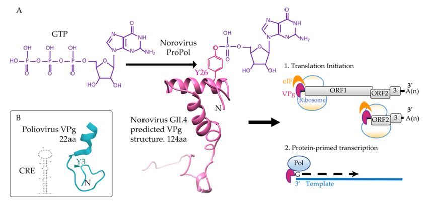
Viruses 2021, 13, 1549                                                                                                             2 of 19

                                  cases, subgenomic RNA—is an essential step for viral protein synthesis, playing a critical role
                                  in translation initiation through interaction with cellular translation initiation factors [8–12].
                                        The biochemical modification of VPg that precedes VPg-mediated priming of viral
                                  genome replication, and to a lesser extent the nucleotidylylation of the polymerase itself,
                                  has been the basis of a body of research across a number of +ssRNA viral families and
                                  is the focus of this review. The picornaviruses represent the most widely studied viral
                                  family for nucleotidylylation of VPg proteins, with extensive information for both the
                                  biochemical nature of the reaction and the accessory components required for efficient
                                  nucleotidylylation. In contrast, less is known about VPg nucleotidylylation in the Cali-
                                  civiridae and Potyviridae families, and the details of nucleotidylylation in astroviruses and
                                  sobemoviruses remain to be elucidated. Viruses within the order Nidovirales also undertake
                                  protein nucleotidylylation; however, they do not encode a VPg protein, nor has a direct
                                  role for the nucleotidylylated proteins in priming genome replication been described.
                                        Regardless of the exact mechanism, nucleotidylylation is essential to the replication
                                  of a wide range of +ssRNA viruses, and this review will focus upon our understanding
                                  of the function and importance of nucleotidylylation during the life cycle of RNA viruses
                                  belonging primarily to the Caliciviridae, Coronaviridae, Picornaviridae and Potyviridae viral
                                  families (Figure 1).

      Figure 1. Genome structure of representative members of the Caliciviridae, Potyviridae, Picornaviridae and Coronaviridae.
      The presence of a covalently attached (nucleotidylylated) 50 VPg is shown as a yellow hexagon. The coding region for
      VPg proteins is shown in yellow and proteins known to be nucleotidylylated are indicated by a yellow star. Proteolytic
      processing of viral polyproteins are indicated by arrowheads. Potyvirus P1-Pro and HC-Pro self-cleave from the polyprotein
      (arrows). Viral capsid proteins are shown in green and the RNA-dependent RNA polymerases (RdRp) are shown in blue.
      Non-structural proteins (nsp) other than RdRp and VPg are shown in pink with the common name and/or nsp designation
      indicated. A second reading frame (uORF) produced by poliovirus is shown in grey. The PIPO coding region expressed as a
      fusion to P3 via a polymerase slippage mechanism is shown in grey for tobacco etch virus. In caliciviruses, the protease (pro)
      and polymerase (RdRp) are found as unprocessed precursors, analogous to 3CD in picornaviruses. The NS and VP1 genes
      of some calicivirus genera (e.g., lagoviruses) are a single ORF. Nucleotide lengths for the genomes depicted are indicated
      and genomes are not to scale.
Viruses 2021, 13, 1549                                                                                                             3 of 19

                                  2. RNA Polymerases and Nucleotidylylation
                                        Viral RNA polymerases primarily function to catalyze the transcription of RNA during
                                  viral replication. The RNA polymerases of picornaviruses, caliciviruses, potyviruses,
                                  sobemoviruses and coronaviruses are supergroup I RdRps [13] and all cluster within branch
                                  2 of the ‘picornavirus supergroup’ based on RdRp phylogeny as defined by Wolf et al. [14].
                                  The common and unique features of RNA polymerases are reviewed in Velthuis [2] and
                                  RdRp structures are reviewed by Ferrero et al. [15]. We refer the reader to these reviews for
                                  complementary information.
                                        Nucleotidyl-transferases are a diverse group of enzymes that transfer nucleotides to
                                  a protein, nucleic acid or other molecule. Enzymes with nucleotidyl-transferase activity
                                  are involved in antibiotic resistance [16], polyadenylation of RNA [17], DNA repair [18],
                                  regulation of protein activity [19,20] and viral replication [21].
                                        Nucleotidyl-transferase or nucleotidylylation reactions, catalyzed by the viral poly-
                                  merase, have been shown to be essential for the replication of certain classes of virus.
                                  These viruses use a protein, VPg, covalently linked to a nucleotide monophosphate (NMP)
                                  to prime replication of the viral genome. A tyrosine residue in VPg provides the free
                                  hydroxyl for the nucleotidylylation reaction in the Astroviridae, Caliciviridae, Picornaviridae
                                  and Potyviridae families, while the Sobemovirus genus uses a threonine or a serine amino
                                  acid for nucleotidylylation [22–28]. The role of VPg in priming RNA synthesis has not been
                                  confirmed for sobemoviruses [29].
                                        Catalysis of NTPs to NMPs by polymerases occurs via a two-metal-ion mechanism
                                  that is reliant on the presence of divalent cations in the form of Mg2+ or Mn2+ [30–32].
                                  In the first step of the reaction, a nucleotide, VPg and two divalent cations bind to the
                                  polymerase. The polymerase then undergoes a conformational change to orientate and
                                  retain the NTP for catalysis. In the nucleotidylylation reaction, one metal ion functions to
                                  lower the pKa of the hydroxyl group (in this instance, on the tyrosine or threonine of VPg),
                                  making it an effective nucleophile. The hydroxyl group then performs a nucleophilic attack
                                  on the alpha phosphate of an NTP. The result is the formation of a phosphodiester bond
                                  between the nucleophile and the alpha phosphate and protonation of a pyrophosphate
                                  molecule, a by-product of the reaction (Figure 2).

      Figure 2. Guanylylation of a calicivirus VPg. (A) For the nucleotidylylation reaction, ProPol catalyzes a nucleophilic
      attack by the hydroxyl of Y26 of VPg (pink) on the alpha phosphate of GTP (purple). This produces a phosphodiester
      bond covalently linking GMP onto Y26 of VPg. Guanylylated human norovirus VPg functions to recruit host eukaryotic
      initiation factors (eIF) for translation and interacts with Pol or ProPol to stimulate protein-primed transcription of the RNA
      genome. Structural predictions for human norovirus GII.4 VPg (KY905331) were performed with Phyre and imaged with
      Chimera [33,34]. (B) In addition to polymerase, a CRE RNA (AY184219) is required for nucleotidylylation of the poliovirus
      VPg (blue) on tyrosine 3 (Y3). The poliovirus 1 VPg structure (2BBL) was obtained from PDB and the representation of the
      CRE RNA stem-loop predicted using mFold [35]. Nucleotidylylation of poliovirus VPg does not contribute to translation
      initiation, only protein-primed transcription. Amino acid (aa) length of the VPg proteins is indicated.
Viruses 2021, 13, 1549 4 of 19
Nucleotidylylation reactions utilizing asparagine, lysine and possibly serine have
also been described for viruses in the Coronaviridae family [36,37]. Lysine and asparagine
contain a free amino group rather than a hydroxyl group, and therefore, the resulting
bond is a phosphoramide rather than a phosphodiester. In the Coronaviridae family, nu-
cleotidylylation occurs on selected proteins in the replication complex (nsp 7, 8, 9 and 12
for SARS-CoV-2), although the exact biological role is not known [36,37].
3. Picornaviruses
The Picornaviridae family comprises a large group of non-enveloped positive-sense
single-stranded RNA viruses. The genome is translated using an internal ribosomal entry
site (IRES) and produces a large polyprotein that is cleaved to its constituent proteins
(Figure 1). For enteroviruses, a second ORF (termed uORF) has been identified [38].
Currently, the family contains 63 genera which is subsequently divided into 147 species [39].
Viruses in this family are primarily transmitted by fecal–oral or respiratory routes and infect
mammals, birds, reptiles, amphibians and fish [39]. Of these viruses, the Enterovirus genus
contains a number of important human pathogens including coxsackieviruses, polioviruses,
echoviruses, enteroviruses and rhinoviruses. Although severe disease (such as meningitis
and paralysis) does occur, a vast majority of infections are asymptomatic or only cause
mild infection [40]. The abundance of these viruses in the environment means that they
present a significant annual burden to society in the forms of morbidity, mortality, health
care costs and economic losses [40–42]. Aside from humans, picornaviruses also cause
significant infections in livestock with substantial economic burdens. For example, foot
and mouth disease virus (FMDV) has a significant impact in Asian and African countries
and is highly contagious among cloven-hoofed animals [43].
3.1. Picornavirus Nucleotidylylation and Replication
The replication of picornaviruses has been reviewed recently [40], and for a com-
prehensive review of protein-primed replication, the reader is referred to [44]. As with
caliciviruses and potyviruses, the VPg (3B) protein is a central player in replication of
picornaviruses; it was first described for poliovirus in 1977 upon discovery of a protein
covalently attached to the 50 end of the viral genome [45]. VPg proteins have subsequently
been detected in all viruses in the family and are a key determinant for classing new
viruses into Picornaviridae [39]. Both positive-sense and negative-sense RNA can be isolated
with VPg (3B) covalently attached [46,47], and the nucleotidylylation of VPg by the viral
polymerase (3D) is an essential step leading to the incorporation of VPg onto the viral
genome.
Picornaviruses have been shown to hijack a host protein, TDP2, to cleave the phos-
phodiester bond linking VPg to the genomic RNA, with this RNA becoming the template
for translation [48–50]. In addition to a polymerase, viruses in the picornavirus family
require cis-acting RNA elements for VPg nucleotidylylation and replication of the viral
genome. These include the 50 clover leaf (50 CL) [51–54]; the 30 untranslated region (30 UTR),
including the poly (A) tail [55,56]; and a cis replication element (CRE) in the open reading
frame [46,57].
Viruses in the Picornaviridae encode one VPg protein, with the exception of FMDV,
which encodes three, all of which can be uridylylated [58,59]. VPg proteins in this family
are small (~19–26 amino acids in length) with a conserved tyrosine at position three of the
amino acid sequence [60] (Figure 2). This tyrosine is used for the covalent linkage of VPg
to the RNA, through formation of the VPgpUpU primer. Structural analysis of poliovirus
1 VPg by NMR shows a flexible protein which, in the presence of trimethylamine N-oxide,
forms a single conformer with an N-terminal loop containing the tyrosine for uridylylation
and a C-terminal helix [61].Viruses 2021, 13, 1549 5 of 19
3.1.1. Precursor Proteins Associated with Nucleotidylylation
The nucleotidylylation of VPg in picornaviruses involves a complex interplay of a
range of viral components. Partially processed precursor proteins from the viral polyprotein
play important roles in nucleotidylylation, including different forms of the VPg (3B) and
the 3CD protein (protease and polymerase precursor protein).
Precursors of VPg include the 3AB and 3BC proteins, both of which have been pro-
posed as sources of VPg for uridylylation [62–64]. In addition to being a target for uridy-
lylation, 3AB stimulates the polymerase activity of 3D [65–67] and also functions as an
RNA binding protein with RNA chaperone and helix destabilizing activities [66–68]. The
RNA binding activity of 3AB alone is non-specific, but when complexed with 3CD, the
binding becomes specific for the 50 CL and 30 UTR of picornaviruses [66,69]. Interaction
with the 50 CL and 30 UTR helps to promote circularization of the genome and is important
for replication of the positive-sense and negative-sense RNA [54,70–74].
Poliovirus 3CD lacks polymerase activity but retains protease activity, and interest-
ingly, this protein can stimulate uridylylation by 100-fold in in vitro assays [58,75,76]. The
ability of 3CD to bind either the 50 CL or CRE RNA is required for this stimulation and it
has been hypothesized that 3CD might provide specificity to the uridylylation reaction by
enhancing the binding of 3D and/or VPg to these RNA structures [66,67,75,77,78].
3.1.2. Uridylylation of VPg
In picornaviruses, formation of VPgpUpU is reliant on the CRE, which functions
as a template for the uridylylation reaction [57,79,80]. CREs have been identified within
the open reading frames of cardioviruses [81], polioviruses [82], hepatovirus [83], pare-
chovirus [84], and sapelovirus [85]. The CRE of FMDV was identified in the 50 UTR [86],
whereas all others are within the first open reading frame. The 3D polymerase, VPg and
uridine triphosphate (UTP) interact with the CRE, thus bringing all three components into
close association for uridylylation [75,78,80,87]. 3D polymerase then catalyzes a reaction
between the free hydroxyl of tyrosine three of VPg and UTP to form a phosphodiester
bond [88].
Currently, a “slide-back” mechanism for uridylylation of VPg on the CRE RNA is
proposed, which utilizes a conserved 50 AAACA 30 motif, representing nucleotides five
through nine of the CRE RNA loop [79,87]. For poliovirus, this model proposes that
the linkage of UMP to tyrosine three of VPg is templated by adenine five (A5, the first
nucleotide of the conserved motif) of the CRE loop. VPgpU then “slides back” and the
covalently linked uracil hydrogen bonds with adenine six (the second nucleotide in the
conserved motif), followed by the addition of a second uracil, again using A5 as the
template [87]. Following the formation of VPgpUpU, elongation of the VPg primer by Pol
ceases [87,89]. The precise mechanism for how the polymerase knows to abort has not been
determined, but it has been hypothesized that it is due to a structural characteristic of Pol
during the reaction.
Two binding sites have been described for VPg on the polymerase, leading to two
different models for the uridylylation reaction. In the first of these, VPg binds to the ‘front’
of the polymerase and presents the conserved tyrosine for nucleotidylylation into the active
site of Pol [90–92]. This has been observed in the FMDV crystal structure showing VPg
bound to 3Dpol with tyrosine three in the active site of Pol, and suggests a cis mechanism
for FMDV uridylylation [92].
In contrast, uridylylation of enterovirus 71 has been proposed to occur in trans as VPg
binds to the polymerase at the base of the palm domain, termed site-311 [93]. Substitutions
to amino acids within this site reduce uridylylation activity by more than 90% but do
not affect RNA elongation activity, indicating that this site is independent from the RNA
polymerization activity [93]. A trans-uridylylation mechanism for coxsackie B virus has
also been proposed [94] and similar results have also been seen for poliovirus through
structure predictions and biochemical experiments [95].Viruses 2021, 13, 1549 6 of 19
3.1.3. Uridylylation of Non-VPg Proteins
Aside from VPg, other viral proteins and protein precursors are able to be uridy-
lylated. In the presence of a 15 nucleotide adenylate template, 3Dpol and 3CD become
uridylylated [96]. This uridylylation can be further stimulated by the addition of the 3AB
precursor and can also be detected in poliovirus-infected HeLa cells [96]. The reaction
occurs intermolecularly with a short template but switches to an intramolecular mechanism
in the presence of poly (A) RNA. As with VPg, the linkage is thought to be through a
phosphodiester bond, but the site(s) of uridylylation have not been able to be identified [96].
Whether there is a functional role in uridylylation of 3Dpol remains to be determined;
however, phosphate chirality experiments showed that the 3Dpol uridylylates are not
obligate intermediates in the formation of uridylylated VPg [96]. However, it should be
noted that all of the RNA polymerases implicated in nucleotidylylation (Figure 1) are
themselves nucleotidylylated, implying a yet to be determined role for this modification.
Uridylylation of 3BC and 3BCD can also be detected in in vitro reactions [64]. Mutation
of the 3BC cleavage site results in viruses that are still able to replicate and forms 3BC-
linked RNA, showing that this can be used to prime RNA synthesis [64,97]. In this system,
uridylylation of 3BC showed a 10-fold greater efficiency than VPg alone and has led to
the suggestion that 3BC is the precursor protein that is uridylylated on the CRE and
subsequently processed to VPgpUpU [64]. 3BC uridylylation has also been observed for
FMDV [58]. However, the 3BC protein is expressed at low levels during infection and
normally cannot be observed in infected cells. Uridylylation of the VPg precursor 3AB
has been detected in the presence of a short adenylate template with Mn2+ [96], but in
the presence of CRE RNA and Mg2+ , no uridylylation is detectable [64]. Whether 3AB is
uridylylated during an infection is not clear.
3.1.4. Replication of the Negative-Sense and Positive-Sense RNA
The overall picture that is emerging for the replication of picornavirus genomes is
outlined below. The VPgpUpU formed on the CRE of the positive-sense RNA is transferred
through an unknown mechanism to the 30 end of the poly (A) tail of the positive-sense RNA.
The polymerase can then extend the RNA using uridylylated VPg as the primer to produce
the negative-sense viral RNA with VPg covalently attached to the 30 end [98]. However, it is
worth noting that there is some disagreement in the literature as to whether a CRE is required
for the uridylylation of VPg, specifically for negative-sense RNA synthesis [46,57,99,100]. The
poly (A) tail has been proposed as an alternative RNA structure to stimulate uridylylation
in this instance [46,89].
For synthesis of the positive-sense RNA, the double-stranded replicative form of the
RNA must be partially unwound or destabilized to allow uridylylated VPg to transfer to
the 30 end of the negative-sense RNA. As the negative-sense RNA begins with AA bases,
uridylylated VPg base pairs with these and the polymerase again elongates the RNA using
the negative-sense as a template, thus yielding positive-sense genomic RNA with VPg
linked at the 50 end.
Overall, this paints a complex picture of nucleotidylylation in vivo, encompassing both
viral and host proteins in the presence of RNA to achieve efficient replication. Dissecting
the role of individual components in relation to RNA replication remains an ongoing
objective in the understanding of this complex process.
4. Caliciviruses
Caliciviridae is a family of +ssRNA viruses, comprising 11 viral genera that infect
mammals (Lagovirus, Norovirus, Nebovirus, Recovirus, Sapovirus, Valovirus and Vesivirus
genera), birds (Bavovirus and Nacovirus genera) and fish (Minovirus and Salovirus gen-
era) [101], causing diseases which encompass respiratory, hemorrhagic and gastrointestinal
pathologies. Unclassified caliciviruses have also been identified in amphibians, reptiles
and lampreys. Representative caliciviruses include human noroviruses (HuNV; Norovirus),
rabbit hemorrhagic disease virus (RHDV; Lagovirus), and feline calicivirus (FCV; Vesivirus).Viruses 2021, 13, 1549 7 of 19
Notably, noroviruses became the most common cause of viral gastroenteritis following
the global introduction of rotavirus vaccines [102–105]. Causing an estimated 685 million
cases per annum, a report published by the Center for Disease Control and Prevention in 2016
revealed that human noroviruses have an estimated medical and socioeconomic burden of
$60 billion USD each year [106]. Norovirus is highly infectious and is responsible for around
~800,000 hospitalizations globally each year and more than 50% of all viral gastroenteritis
outbreaks in the world [107–109]. Despite this, there remain no approved antiviral therapeutics
or vaccines available for the treatment or prevention of noroviral infections.
4.1. Calicivirus Nucleotidylylation
Calicivirus genomes carry a VPg covalently bound to the 50 end, whilst the 30 end
has a poly (A) tail. The genome usually encodes three open reading frames (ORF1–3),
the first of which encodes all the non-structural proteins, whilst ORF2 and ORF3 encode
the major and minor capsid proteins. The ORF1 non-structural gene is translated to a
polyprotein which is post-translationally cleaved into precursor and mature proteins by a
3C-like protease (NS6) (Figure 1).
Within caliciviruses, the length of VPg proteins can vary from 65 amino acids (e.g.,
bovine nebovirus VPg) to 138 amino acids (e.g., Norwalk virus VPg) [26]. The structure
of feline calicivirus and murine norovirus (MNV) VPg proteins were solved by nuclear
magnetic resonance spectroscopy, revealing that the core of the calicivirus VPg protein is
the only ordered domain, and is flanked by disordered N- and C-termini [110]. Regions of
intrinsic disorder allow viral proteins to interact with a range of potential binding partners.
Unlike picornavirus VPg proteins, calicivirus VPg proteins are known to recruit host cell
translation machinery, namely eIF4E and eIF4G [9,111]. Recruitment of these initiation
factors to the viral nucleic acid by VPg initiates translation. In addition to this role, VPg
can also bind RNA polymerases and their precursors, induce a G1/S cell cycle arrest and
has been proposed to serve as a protective cap for the viral genome against detection by
the host immune system [8,27,112–117].
Nucleotidylylation is an essential step in the viral lifecycle of caliciviruses, with
nucleotidylylated VPg acting to prime transcription of the positive-sense genome by viral
RdRp in vitro [5,113] and initiate translation [5,26,111,113,118]. Experimental evidence
for MNV, for which a robust cell model exists [119], demonstrated that all infectious
MNV virions have VPg caps on the 50 end of the genome [26], confirming the essential
requirement of nucleotidylylated VPg in viral replication.
Nucleotidylylation of VPg by RNA polymerases, specifically the addition of GTP or
UTP [27], facilitates the role of VPg in priming RNA synthesis, leading to covalent attachment
of VPg at the 50 end of the viral genome and/or sub-genome [120]. A mass spectrometry-
based approach identified that VPg from FCV and MNV are covalently linked to guanosine
via a tyrosine at position 24 and 26, respectively [26], that lies within a highly conserved
acidic amino acid motif (DEEYD/EE). Guanylylation of VPg provides the 50 G nucleotide
of the viral genome, and hence, mutation of the tyrosine in this motif is detrimental to viral
replication and results in a non-infectious virus [26]. The calicivirus VPg N-terminus adjacent
to the nucleotidylylated tyrosine is rich in lysine/arginine amino acids and has been shown
to have both nucleotide binding capability and a role in nucleotidylylation [121]. Deletion
of this region, specifically the first 3, 8 or 10 amino acids of the N-terminus of HuNV VPg,
resulted in a progressive reduction of nucleotidylylation at Tyr27 [27].
4.1.1. Proteins That Catalyze the Nucleotidyl-Transferase Reaction
The biomolecular mechanisms behind nucleotidylylation in caliciviruses are not well
understood. In contrast to poliovirus nucleotidylylation, where 3Dpol is the active form of
the polymerase responsible for catalyzing the reaction, it has been shown for the Caliciviri-
dae family that this is more variable. In MNV and HuNV, nucleotidylylation is catalyzed
by both forms of polymerase, the mature RdRp and the ORF1-derived precursor pro-
tein comprised of unprocessed protease-polymerase (ProPol, analogous to picornavirusViruses 2021, 13, 1549 8 of 19
3CD) [27,121]. In contrast, for FCV, the protease and polymerase remain as a single protein,
hence nucleotidylylation relies solely on the 3CD equivalent [122,123]. ProPol was also
found to be the predominant form of polymerase in RHDV infected cells [124,125]. In
HuNV, ProPol has been demonstrated to be 100-fold more efficient at nucleotidylylation
than the mature polymerase, and a similar finding has been shown for RHDV VPg [121].
This suggests that ProPol may be the primary form of polymerase performing protein
nucleotidylylation in caliciviruses.
The ability of calicivirus RdRps to nucleotidylylate target proteins from other viruses
varies. HuNV RdRp specifically nucleotidylylates only HuNV VPg; in contrast, MNV RdRp
is able to nucleotidylylate both HuNV VPg and MNV VPg [126]. Interestingly, the MNV
polymerase was more efficient at nucleotidylylating HuNV VPg than the HuNV RdRp,
and both HuNV and MNV RdRps nucleotidylylated themselves in vitro [126]. Whether
this self-nucleotidylylation mechanism is similar to that identified in coronaviruses and
picornaviruses is not known, nor have the target residue(s) been identified and nor has it
been determined whether this is an essential function for viral replication.
4.1.2. The Role of RNA in Nucleotidylylation
The evidence for RNA structures contributing to the nucleotidylylation of calicivirus
VPg proteins is not obvious. Noroviruses contain evolutionarily conserved RNA struc-
tures, which when disrupted, reduce or completely destroy replication of MNV [127,128].
Evidence suggests HuNV ProPol can catalyze nucleotidylylation independently of a poly
(A) RNA template in the presence of Mn2+ as a divalent metal cation, whereas mature
polymerase is more active in the presence of an RNA template, regardless of the cation
available [4,27,129]. Uridylylation by the HuNV ProPol, which does not strictly require
an RNA template, can nevertheless be enhanced upon addition of an ORF3-30 UTR poly
(A) template [27]. A similar stimulatory effect on nucleotidylylation activity has also been
shown for the MNV polymerase [130]. FCV ProPol, unlike HuNV ProPol, is strictly depen-
dent on the presence of an RNA template as a cofactor for nucleotidyl-transfer [26,123].
4.1.3. Nucleotide Selection for Nucleotidylylation
The HuNV MD145 strain VPg has been shown to be preferentially nucleotidylylated
with GTP and UTP over other nucleotides, exhibiting a 2-fold preference for GTP over
UTP [27]. A similar finding was found by Medvedev et al. [121], and has also been shown
for MNV VPg, which is preferentially guanylylated compared to incorporation of the
other NTPs [130]. As all calicivirus genomic and subgenomic RNAs begin with a guanine
nucleotide, this supports the priming theory of positive-sense viral RNA replication by a
guanylylated VPg.
Both UTP and GTP may be added to VPg, and some evidence suggests that two
nucleotides can be added to calicivirus VPg proteins [27], similar to the VPgpUpU formed
during picornavirus replication. The VPg of RHDV was originally proposed to be uridyly-
lated by the RNA polymerase [7], based on analogies to picornavirus VPg. Belliot et al. [27]
proposed that uridylylated VPg may prime initiation of RNA synthesis only on the anti-
sense genome or anti-sense subgenome on the poly (A) tail. Using recombinant polymerase
and subgenomic RNA as a surrogate for the norovirus genome, Rohayem et al. showed that
uridylylated HuNV VPg primed the subgenome in vitro [4], but not the anti-subgenome.
This was consistent with experimental observations in FCV and MNV, which did not find
that the antigenome was VPg-linked [26,131], and with findings that initiation of replication
of the antigenome occurs via a primer-independent de novo mechanism [4]. If VPg does
prime antisense genomic RNA synthesis, then a role for uridylylated VPg can be proposed
for this process. That GTP is preferred over UTP would be expected in this case, given that
the antisense genome is >1000-fold less abundant than the positive-sense genome during
viral replication [131].
To date, the mechanisms behind nucleotide discrimination in nucleotidylylation re-
main unclear. An NTP-binding sequence has been identified in calicivirus VPgs [121],Viruses 2021, 13, 1549 9 of 19
suggesting VPg itself, as opposed to the polymerase, may bind NTPs prior to nucleotidyly-
lation, and the nucleotide addition cycle of nucleotidylylation begins with the binding of
VPg-NTP by the polymerase. However, despite the fact that VPg is preferentially guany-
lylated, VPg was shown to bind all NTPs with equal efficiency [121], suggesting that the
polymerase may have a discriminatory role in preferential NTP addition. It may be possible
that VPg is both guanylylated and then uridylylated, as the second nucleotide of norovirus
genomes is a uracil. However, how a specific VPgpGpU might be synthesized is unclear,
although it is interesting to speculate on the role an RNA template might have in this
process, and whether the nucleotides might be added by different forms of polymerase.
5. Potyviruses
Potyviruses are a genus of plant viruses belonging to the Potyviridae viral family
and have a wide range of plant hosts, including many vegetables and legume species
which constitute important parts of the global food supply. Potyviruses are some of
the most common plant viruses in the world, accounting for approximately ~30% of
known plant pathogens. Potyviruses are known for causing extensive economic damage
to important crops such as potatoes, turnips, tomatoes, beans and peas. Potyviruses are
primarily transmitted from plant to plant by arthropods [132]. The viral particles are fil-
amentous and almost all members of the Potyviridae have a monopartite +ssRNA genome of
8–11 kb in length. Potyvirus genomes encode an ORF which is translated to a
340–370 kDa polyprotein, which is then co- and/or post-translationally processed by
three forms of internally encoded protease into cleavage intermediates or mature protein
products (Figure 1). A fusion protein, P3N-PIPO, is read off an alternative open read-
ing frame as a result of a frameshift caused by RNA polymerase slippage on the first
ORF [133,134].
Potyvirus Nucleotidylylation
Potyvirus VPg proteins range from 20–22 kDa and exist in both a mature form as well
as a number of uncleaved precursor forms. The potyvirus turnip mosaic virus appears to
possess VPg exclusively in precursor forms, as one study focusing on subcellular localiza-
tion could not detect the mature form of VPg [135]. Potyvirus VPgs, like other viral VPgs,
have an ordered central domain flanked by disordered N- and C-termini [136,137]. VPgs
from Potyviridae share roughly 50% sequence identity, mostly in the conserved regions of
the NTP binding motif, and a bipartite nuclear localization signal [138].
Potyvirus VPgs are genome-linked to the 50 end of the viral nucleic acid and prime
the synthesis of the viral genome [139,140]. As with caliciviruses, potyvirus VPg proteins
are covalently bound to genomic RNA by a conserved tyrosine residue. In tobacco vein
mottling virus (TVMV) VPg is linked via Tyr60 to the viral nucleic acid, whilst pepper vein
banding virus (PVBV) VPg is linked via Tyr66 and potato virus A (PVA) VPg is linked by
Tyr63 and possibly by an alternative residue Tyr119 [6,141,142]. In addition to functioning
as a genome cap, potyvirus VPgs share a number of other functional similarities to VPgs
from picornaviruses and caliciviruses, including binding to host factors such as eIF4E [143].
Potyvirus VPgs are also uridylylated by their respective RNA polymerases [6,138,141].
Co-purification of TVMV VPg with a GST-labelled polymerase fusion protein demon-
strated that VPg associates with the polymerase in vitro [144]. Tyr60 of the VPg was shown
to be essential for this interaction, as mutation of this residue ablated the association
between the polymerase and the VPg entirely [145], preventing viral replication in pro-
toplasts [28,142]. Further, TVMV VPg stimulates inherent polymerase activity, acting as
a co-factor in RNA synthesis in addition to the role of VPg as a primer of the viral RNA
genome [144].
Studies of PVA and PVBV VPg uridylylation showed that RNA polymerase uridy-
lylates VPg in the presence of a divalent metal cation independently of a poly (A) RNA
template [6,141]. A conserved tyrosine residue in PVBV VPg, specifically Tyr42, is part of
the nucleotide binding motif AYTTKKGK [141], analogous to a nucleotide binding motifViruses 2021, 13, 1549 10 of 19
in closely related PVA VPg [6]. However, deletion of this nucleotide binding motif in
PVA VPg did not result in complete loss of uridylylation [6], indicating that Tyr42 was not
the nucleotidylylation target residue of the PVA polymerase. Tyr66 was identified as the
target residue when mutation of Tyr66 to threonine prevented uridylylation by PVBV RNA
polymerase [141]. It is not clear if potyvirus VPgs are nucleotidylylated with one residue or
two, whether there is nucleotide preference, and whether potyvirus precursor polymerases
outperform mature polymerases in this function.
6. Coronaviruses and Other Nidoviruses
The order Nidovirales encompasses 14 families of positive-sense RNA viruses, within
which 109 species have currently been identified [146]. Of this order, three families of bio-
logical and economic value are Arteriviridae, Mesoniviridae and Coronaviridae. Arteriviruses
are known to infect a diverse group of mammals including pigs (porcine reproductive and
respiratory syndrome virus; PRRSV), horses (equine arteritis virus; EAV) and monkeys
(simian hemorrhagic fever virus) [147]. PRRSV is estimated to cause $550 million in losses
in the US alone, making it a pathogen significant to the agricultural industry [148]. Viruses
within the Mesoniviridae family infect mosquitos, and due to the importance of mosquitos
in vector transmission, it is an area that warrants further research [149]. Coronaviruses
are the causative agents for three major disease outbreaks: SARS (severe acute respiratory
syndrome) caused by the SARS virus, MERS (Middle East respiratory syndrome) caused
by the MERS virus and COVID-19 (coronavirus disease 2019) caused by SARS-CoV-2, all of
which have arisen from animal reservoirs [150]. General pathologies include pneumonia,
fever and dry cough, with other symptoms being more strain specific. Four other species
of coronavirus have been found in humans (HCoV229E, HCoV-OC43, HCoV-NL63 and
HCoV-HKU1) and are estimated to be responsible for 15–30% of all cases of the com-
mon cold worldwide [151]. Other viruses of interest in the Coronaviridae family include
toroviruses, which primarily infect livestock, although some strains have been identified to
cause disease in humans [152,153].
6.1. Nidovirus RdRp and Nucleotidylylation
The RdRps of coronaviruses (nsp9 for EAV or nsp12 for HCoV-229E, SARS or SARS-
CoV-2) are made up of three main domains: the RdRp domain, the nidovirus RdRp-
associated nucleotidyltransferase (NiRAN) domain and an uncharacterized third domain
that connects the NiRAN to the RdRp domain [36]. In vitro studies using the SARS nsp12
show that it can perform primer-dependent and primer-independent RNA synthesis [154].
Separately, nsp12 has poor polymerase activity, but when in a complex with nsp7 and nsp8,
the processivity of RNA synthesis is increased [155].
While coronaviruses do not possess a VPg protein as described for other viruses in this
review, they perform nucleotidylylation as a critical component of the viral replication cycle.
Currently, there is only experimental data on nidovirus nucleotidylylation from four viruses:
EAV, HCoV-229E, SARS and SARS-CoV-2. As with the other ‘branch 20 /picornavirus
supergroup of viruses [14] described earlier in this review, nucleotidylylation involves the
polymerase linking a nucleotide to a separate viral protein or to itself.
The preference of the incorporated nucleotide during nucleotidylylation by SARS-CoV-
2 RdRp and HCoV-229E RdRp is UTP over GTP with the HCoV-229E activity being 2–3-fold
higher using UTP [156]. However, for EAV, this preference for UTP appears to be pH-
dependent, with the nucleotide preference switching to GTP above pH 8.5. Furthermore,
characterization of the EAV RdRp activity identified that GTP was able to outcompete UTP
for nucleotide preference in a competition assay [37]. The modified residue was shown to
be K380 via mass spectrometry, and this was validated by mutation of the site resulting in
no nucleotidylylation.Viruses 2021, 13, 1549 11 of 19
The NiRAN Motif
Analysis of nidovirus polymerase sequences has identified a unique domain in the N-
terminal portion of nidovirus RdRp, termed a NiRAN domain. Alignments of this domain
between viruses from Coronaviridae, Toroviridae, Mesonivirdae and Arterivirdae families [36]
showed several areas of conservation indicating that the NiRAN domain is found in these
different viral lineages. Closer analysis of this N-terminal domain of the RNA polymerase
identified eight invariant residues, which were further mapped to three motifs denoted
An , Bn and Cn (Figure 3). Mutational analysis of these invariant residues has shown either
severe growth impairment or a nonviable virus [36,156]. The NiRAN domain is considered
to be a hallmark of Nidovirales, with no homologs identified elsewhere [36,157]. Whether
the other viral RdRps that perform protein nucleotidylylation have alternate conserved
motifs critical for this function is unknown.
Figure 3. Sequence alignment of the NiRAN domains from viruses within the order Nidovirales. Schematic diagram of nsp12
(nsp9 for Arterivirus). Sequence alignment of the N-terminal region of the RdRps from EAV (Arterivirus NP_705590.1), Dak
Nong virus (Mesonivirus YP_009505590.1), Breda virus (Torovirus YP_337905.2), HCoV229E (QNT54752.1), MERS virus
(YP_009047223.1), SARS-CoV-2 (QHD43415.1) and SARS virus (AER30332.1) was generated by Clustal Omega [158]. The
three NiRAN motifs are designated An , Bn and Cn . Bolded resides denote invariant residues and a colon (:) indicates a
residue that is conserved in more than 50% of the viruses aligned. The filled circles indicate positions that are lethal when
mutated, and open circles indicate residues that result in impaired virus when mutated.
6.2. Nucleotidylylation of Nidovirus Proteins
6.2.1. EAV nsp7
Each of the RdRps described above were identified to nucleotidylylate other non-
structural proteins. The nucleotidylylation activity of EAV RdRp was notably enhanced
when in the presence of nsp7 [37]. Nsp7 is unique to the Arteriviridae family [159] and is
processed by the EAV protease into two proteins, nsp7α and nsp7β [160]. The role in the
viral life cycle has not been elucidated for EAV; however, studies in PRRSV show that nsp7
is essential for virus survival and indicate that it may have a role in RNA synthesis [161].
Three sites in nsp7β (K143, K156 and K172) were shown to be guanylylated by the RdRp.
Two of these sites (K156 and K172) were seen to be conserved in PRSSV [37]. The role these
modifications have in EAV is unknown.
6.2.2. Coronavirus nsp9
The nsp9 of both HCoV-229E and SARS-CoV-2 are nucleotidylylated by their respec-
tive RdRps. Both viruses preferentially use UTP as a substrate; however, GTP can be
utilized [37,156]. Nsp9 is a small dimerized protein [162] that has been shown to be criticalViruses 2021, 13, 1549 12 of 19
for growth in SARS [163]. Sequence analysis has shown nsp9 is likely to be unique to the
coronavirus family, with large regions of conservation between coronaviruses, and is able
to bind to ssRNA and ssDNA [164]. Characterization of the interaction between RdRp and
nsp9 showed that for nucleotidylylation to occur, the N-terminal end of nsp9 had to be
free from the pp1ab polyprotein, and it is proposed that the interaction between the nsp12
and nsp9 is transient [156]. Tandem mass spectrometry identified Asn1 of the HCoV-229E
nsp9 to be modified, and it is proposed that the linkage occurs on the primary amine of
the amino acid. Of particular note, mutational analysis showed that the nucleotidylylation
activity was independent of the RdRp activity [156].
6.2.3. Coronavirus nsp7 and nsp8
While HCoV-229E RdRp was not seen to nucleotidylylate nsp7 in an in vitro as-
say [156], the SARS-CoV-2 RdRp was able to link a GTP to K2 of the nsp7 via a phospho-
ramide bond [37]. Further mutation of K2 significantly reduced the amount of radiolabeling
seen on nsp7, indicating that this is likely to be the preferred nucleotidylylation site [37].
Both GTP and UTP are linked to nsp7, with GTP being preferred over UTP during nu-
cleotidylylation; however, UTP was able to label nsp7 and nsp8 more effectively, possibly
due to UMP being more stable than GMP. Nsp8 was also seen to be labelled with UTP and
GTP; however, the site of modification is not known [37]. Nsp7 and nsp8 of SARS-CoV-2
are able to form a heterotetramer structure proposed to have primase activity [165], which
has been seen to act as a co-factor for the RdRp, increasing its polymerase activity [166].
The role of nucleotidylylation in SARS-CoV-2 is unknown.
6.3. Role of Nucleotidylylation
Nucleotidylylation activity is essential for coronavirus replication, with nucleotidyly-
lation knockout mutations resulting in a nonviable virus. Slanina et al. [156] proposed that
the nucleotidylylation of the coronavirus nsp9 would allow it to act as a protein primer
for RNA synthesis. This is supported by the RdRp from both the SARS virus and EAV
being able to perform de novo and protein-primed RNA synthesis [2,36]. The RNA binding
activity of nsp9 lends credibility to this proposal; however, since no nsp9 linked to RNA
has been identified, there would have to be an additional mechanism that releases the nsp9
from the RNA. It has also been suggested that UMP linked to the coronavirus nsp9 interact
with cis-acting RNA elements identified at the 30 end on the coronavirus genome [156].
Yan et al. showed that the NiRAN domain of nsp12 is able to catalyze the transfer
of GMP to ppA, which forms the GpppA cap core structure. They suggested that the
association of the nsp9 to the NiRAN domain may stabilize the 50 end of GpppA-RNA due
to nsp9 being an RNA-binding protein before the GpppA-RNA is further processed during
cap synthesis [167].
7. Conclusions
Protein nucleotidylylation is a critical component of the replication of a range of
viruses, with this modification commonly found in +ssRNA viruses of the picornavirus
supergroup [14], and highlights a common feature of this group of viruses. These viruses
are important pathogens of humans, other animals and plants with a significant impact
on health and well-being, as evidenced by the current SARS-CoV-2 global pandemic.
Nucleotidylylation is central to the initiation of viral genome replication, an essential step
in the viral life cycle, and the placement of caliciviral and potyviral VPg proteins at the
50 end of the viral genome is critical for the recruitment of translational initiation factors,
and hence, viral protein synthesis, for these two viral families. While nucleotidylylation
plays a central role in the replication of viruses that perform this function, the underlying
mechanisms and processes largely remain to be determined.
Author Contributions: All authors contributed to conceptualization, writing—original draft prepa-
ration, writing—review and editing, and visualization. All authors have read and agreed to the
published version of the manuscript.Viruses 2021, 13, 1549 13 of 19
Funding: This research was funded by the New Zealand Ministry of Business, Innovation & Employ-
ment, grant number UOOX1904, the Maurice Wilkins Centre for Molecular Biodiscovery and the
University of Otago.
Institutional Review Board Statement: Not applicable.
Informed Consent Statement: Not applicable.
Acknowledgments: V.L. Young for critical review of the manuscript.
Conflicts of Interest: The authors declare no conflict of interest. The funders had no role in the design
of the study; in the collection, analyses, or interpretation of data; in the writing of the manuscript; or
in the decision to publish the results.
References
1. Gorbalenya, A.E.; Pringle, F.M.; Zeddam, J.L.; Luke, B.T.; Cameron, C.E.; Kalmakoff, J.; Hanzlik, T.N.; Gordon, K.H.; Ward, V.K.
The palm subdomain-based active site is internally permuted in viral RNA-dependent RNA polymerases of an ancient lineage. J.
Mol. Biol. 2002, 324, 47–62. [CrossRef]
2. Te Velthuis, A.J. Common and unique features of viral RNA-dependent polymerases. Cell Mol. Life Sci. 2014, 71, 4403–4420.
[CrossRef] [PubMed]
3. Bruenn, J.A. A structural and primary sequence comparison of the viral RNA-dependent RNA polymerases. Nucleic Acids Res.
2003, 31, 1821–1829. [CrossRef]
4. Rohayem, J.; Robel, I.; Jager, K.; Scheffler, U.; Rudolph, W. Protein-primed and de novo initiation of RNA synthesis by norovirus
3Dpol. J. Virol. 2006, 80, 7060–7069. [CrossRef] [PubMed]
5. Subba-Reddy, C.V.; Goodfellow, I.; Kao, C.C. VPg-primed RNA synthesis of norovirus RNA-dependent RNA polymerases by
using a novel cell-based assay. J. Virol. 2011, 85, 13027–13037. [CrossRef]
6. Puustinen, P.; Makinen, K. Uridylylation of the potyvirus VPg by viral replicase NIb correlates with the nucleotide binding
capacity of VPg. J. Biol. Chem. 2004, 279, 38103–38110. [CrossRef] [PubMed]
7. Machin, A.; Martin Alonso, J.M.; Parra, F. Identification of the amino acid residue involved in rabbit hemorrhagic disease virus
VPg uridylylation. J. Biol. Chem. 2001, 276, 27787–27792. [CrossRef] [PubMed]
8. Daughenbaugh, K.F.; Wobus, C.E.; Hardy, M.E. VPg of murine norovirus binds translation initiation factors in infected cells. Virol.
J. 2006, 3, 33. [CrossRef]
9. Chung, L.; Bailey, D.; Leen, E.N.; Emmott, E.P.; Chaudhry, Y.; Roberts, L.O.; Curry, S.; Locker, N.; Goodfellow, I.G. Norovirus
translation requires an interaction between the C Terminus of the genome-linked viral protein VPg and eukaryotic translation
initiation factor 4G. J. Biol. Chem. 2014, 289, 21738–21750. [CrossRef]
10. Tavert-Roudet, G.; Anne, A.; Barra, A.; Chovin, A.; Demaille, C.; Michon, T. The Potyvirus Particle Recruits the Plant Translation
Initiation Factor eIF4E by Means of the VPg covalently Linked to the Viral RNA. Mol. Plant. Microbe Interact. 2017, 30, 754–762.
[CrossRef]
11. Walter, J.; Barra, A.; Charon, J.; Tavert-Roudet, G.; Michon, T. Spectroscopic Investigation of the Kinetic Mechanism Involved in
the Association of Potyviral VPg with the Host Plant Translation Initiation Factor eIF4E. Int. J. Mol. Sci. 2020, 21, 5618. [CrossRef]
12. Khan, M.A.; Goss, D.J. Poly (A) binding protein enhances the binding affinity of potyvirus VPg to eukaryotic initiation factor
eIF4F and activates in vitro translation. Int. J. Biol. Macromol. 2019, 121, 947–955. [CrossRef]
13. Aravind, L.; Koonin, E.V. DNA polymerase beta-like nucleotidyltransferase superfamily: Identification of three new families,
classification and evolutionary history. Nucleic Acids Res. 1999, 27, 1609–1618. [CrossRef]
14. Wolf, Y.I.; Kazlauskas, D.; Iranzo, J.; Lucia-Sanz, A.; Kuhn, J.H.; Krupovic, M.; Dolja, V.V.; Koonin, E.V. Origins and Evolution of
the Global RNA Virome. mBio 2018, 9, e02329-18. [CrossRef]
15. Ferrero, D.; Ferrer-Orta, C.; Verdaguer, N. Viral RNA-Dependent RNA Polymerases: A Structural Overview. Subcell Biochem.
2018, 88, 39–71. [CrossRef]
16. Morar, M.; Bhullar, K.; Hughes, D.W.; Junop, M.; Wright, G.D. Structure and mechanism of the lincosamide antibiotic adenylyl-
transferase LinB. Structure 2009, 17, 1649–1659. [CrossRef]
17. Wang, L.; Eckmann, C.R.; Kadyk, L.C.; Wickens, M.; Kimble, J. A regulatory cytoplasmic poly(A) polymerase in Caenorhabditis
elegans. Nature 2002, 419, 312–316. [CrossRef] [PubMed]
18. Della, M.; Palmbos, P.L.; Tseng, H.M.; Tonkin, L.M.; Daley, J.M.; Topper, L.M.; Pitcher, R.S.; Tomkinson, A.E.; Wilson, T.E.;
Doherty, A.J. Mycobacterial Ku and ligase proteins constitute a two-component NHEJ repair machine. Science 2004, 306, 683–685.
[CrossRef]
19. Stadtman, E.R. The story of glutamine synthetase regulation. J. Biol. Chem. 2001, 276, 44357–44364. [CrossRef] [PubMed]
20. Yarbrough, M.L.; Li, Y.; Kinch, L.N.; Grishin, N.V.; Ball, H.L.; Orth, K. AMPylation of Rho GTPases by Vibrio VopS disrupts
effector binding and downstream signaling. Science 2009, 323, 269–272. [CrossRef] [PubMed]
21. Wu, W.; Wang, Z.; Xia, H.; Liu, Y.; Qiu, Y.; Liu, Y.; Hu, Y.; Zhou, X. Flock house virus RNA polymerase initiates RNA synthesis de
novo and possesses a terminal nucleotidyl transferase activity. PLoS ONE 2014, 9, e86876. [CrossRef] [PubMed]Viruses 2021, 13, 1549 14 of 19
22. Ambros, V.; Baltimore, D. Protein is linked to the 50 end of poliovirus RNA by a phosphodiester linkage to tyrosine. J. Biol. Chem.
1978, 253, 5263–5266. [CrossRef]
23. Olspert, A.; Peil, L.; Hebrard, E.; Fargette, D.; Truve, E. Protein-RNA linkage and post-translational modifications of two
sobemovirus VPgs. J. Gen. Virol. 2011, 92, 445–452. [CrossRef]
24. Olspert, A.; Arike, L.; Peil, L.; Truve, E. Sobemovirus RNA linked to VPg over a threonine residue. FEBS Lett. 2011, 585, 2979–2985.
[CrossRef] [PubMed]
25. Fuentes, C.; Bosch, A.; Pinto, R.M.; Guix, S. Identification of human astrovirus genome-linked protein (VPg) essential for virus
infectivity. J. Virol. 2012, 86, 10070–10078. [CrossRef] [PubMed]
26. Olspert, A.; Hosmillo, M.; Chaudhry, Y.; Peil, L.; Truve, E.; Goodfellow, I. Protein-RNA linkage and posttranslational modifications
of feline calicivirus and murine norovirus VPg proteins. PeerJ 2016, 4, e2134. [CrossRef]
27. Belliot, G.; Sosnovtsev, S.V.; Chang, K.O.; McPhie, P.; Green, K.Y. Nucleotidylylation of the VPg protein of a human norovirus by
its proteinase-polymerase precursor protein. Virology 2008, 374, 33–49. [CrossRef]
28. Murphy, J.F.; Klein, P.G.; Hunt, A.G.; Shaw, J.G. Replacement of the tyrosine residue that links a potyviral VPg to the viral RNA is
lethal. Virology 1996, 220, 535–538. [CrossRef]
29. Somera, M.; Sarmiento, C.; Truve, E. Overview on Sobemoviruses and a Proposal for the Creation of the Family Sobemoviridae.
Viruses 2015, 7, 3076–3115. [CrossRef]
30. Carvalho, A.T.; Fernandes, P.A.; Ramos, M.J. The Catalytic Mechanism of RNA Polymerase II. J. Chem Theory Comput. 2011, 7,
1177–1188. [CrossRef] [PubMed]
31. Castro, C.; Smidansky, E.; Maksimchuk, K.R.; Arnold, J.J.; Korneeva, V.S.; Gotte, M.; Konigsberg, W.; Cameron, C.E. Two proton
transfers in the transition state for nucleotidyl transfer catalyzed by RNA- and DNA-dependent RNA and DNA polymerases.
Proc. Natl. Acad. Sci. USA 2007, 104, 4267–4272. [CrossRef]
32. Steitz, T.A.; Steitz, J.A. A general two-metal-ion mechanism for catalytic RNA. Proc. Natl. Acad. Sci. USA 1993, 90, 6498–6502.
[CrossRef]
33. Pettersen, E.F.; Goddard, T.D.; Huang, C.C.; Couch, G.S.; Greenblatt, D.M.; Meng, E.C.; Ferrin, T.E. UCSF Chimera—A visualiza-
tion system for exploratory research and analysis. J. Comput. Chem. 2004, 25, 1605–1612. [CrossRef]
34. Kelley, L.A.; Mezulis, S.; Yates, C.M.; Wass, M.N.; Sternberg, M.J. The Phyre2 web portal for protein modeling, prediction and
analysis. Nat. Protoc. 2015, 10, 845–858. [CrossRef]
35. Zuker, M. Mfold web server for nucleic acid folding and hybridization prediction. Nucleic Acids Res. 2003, 31, 3406–3415.
[CrossRef]
36. Lehmann, K.C.; Gulyaeva, A.; Zevenhoven-Dobbe, J.C.; Janssen, G.M.; Ruben, M.; Overkleeft, H.S.; van Veelen, P.A.; Samborskiy,
D.V.; Kravchenko, A.A.; Leontovich, A.M.; et al. Discovery of an essential nucleotidylating activity associated with a newly
delineated conserved domain in the RNA polymerase-containing protein of all nidoviruses. Nucleic Acids Res. 2015, 43, 8416–8434.
[CrossRef] [PubMed]
37. Conti, B.J.; Leicht, A.S.; Kirchdoerfer, R.N.; Sussman, M.R. Mass spectrometric based detection of protein nucleotidylation in the
RNA polymerase of SARS-CoV-2. Commun. Chem. 2021, 4, 41. [CrossRef]
38. Lulla, V.; Dinan, A.M.; Hosmillo, M.; Chaudhry, Y.; Sherry, L.; Irigoyen, N.; Nayak, K.M.; Stonehouse, N.J.; Zilbauer, M.;
Goodfellow, I.; et al. An upstream protein-coding region in enteroviruses modulates virus infection in gut epithelial cells. Nat.
MicroBiol. 2019, 4, 280–292. [CrossRef] [PubMed]
39. Zell, R.; Delwart, E.; Gorbalenya, A.E.; Hovi, T.; King, A.M.Q.; Knowles, N.J.; Lindberg, A.M.; Pallansch, M.A.; Palmenberg, A.C.;
Reuter, G.; et al. ICTV Virus Taxonomy Profile: Picornaviridae. J. Gen. Virol. 2017, 98, 2421–2422. [CrossRef]
40. Zell, R. Picornaviridae-the ever-growing virus family. Arch. Virol. 2018, 163, 299–317. [CrossRef] [PubMed]
41. Fendrick, A.M.; Monto, A.S.; Nightengale, B.; Sarnes, M. The economic burden of non-influenza-related viral respiratory tract
infection in the United States. Arch. Intern. Med. 2003, 163, 487–494. [CrossRef] [PubMed]
42. Fine, J.; Bray-Aschenbrenner, A.; Williams, H.; Buchanan, P.; Werner, J. The Resource Burden of Infections with Rhi-
novirus/Enterovirus, Influenza, and Respiratory Syncytial Virus in Children. Clin. Pediatr. 2019, 58, 177–184. [CrossRef]
43. Knight-Jones, T.J.; Rushton, J. The economic impacts of foot and mouth disease—What are they, how big are they and where do
they occur? Prev Vet. Med. 2013, 112, 161–173. [CrossRef] [PubMed]
44. Paul, A.V.; Wimmer, E. Initiation of protein-primed picornavirus RNA synthesis. Virus Res. 2015, 206, 12–26. [CrossRef]
45. Lee, Y.F.; Nomoto, A.; Detjen, B.M.; Wimmer, E. A protein covalently linked to poliovirus genome RNA. Proc. Natl. Acad. Sci.
USA 1977, 74, 59–63. [CrossRef]
46. Morasco, B.J.; Sharma, N.; Parilla, J.; Flanegan, J.B. Poliovirus cre(2C)-dependent synthesis of VPgpUpU is required for positive-
but not negative-strand RNA synthesis. J. Virol. 2003, 77, 5136–5144. [CrossRef]
47. Gamarnik, A.V.; Andino, R. Switch from translation to RNA replication in a positive-stranded RNA virus. Genes Dev. 1998, 12,
2293–2304. [CrossRef] [PubMed]
48. Holmes, A.C.; Zagnoli-Vieira, G.; Caldecott, K.W.; Semler, B.L. Effects of TDP2/VPg Unlinkase Activity on Picornavirus Infections
Downstream of Virus Translation. Viruses 2020, 12, 166. [CrossRef]
49. Virgen-Slane, R.; Rozovics, J.M.; Fitzgerald, K.D.; Ngo, T.; Chou, W.; van der Heden van Noort, G.J.; Filippov, D.V.; Gershon,
P.D.; Semler, B.L. An RNA virus hijacks an incognito function of a DNA repair enzyme. Proc. Natl. Acad. Sci. USA 2012, 109,
14634–14639. [CrossRef] [PubMed]You can also read