Professional-Grade TCA-Lactic Acid Chemical Peel: Elucidating Mode of Action to Treat Photoaging and Hyperpigmentation - PCA SKIN ...
←
→
Page content transcription
If your browser does not render page correctly, please read the page content below
BRIEF RESEARCH REPORT
                                                                                                                                        published: 12 February 2021
                                                                                                                                    doi: 10.3389/fmed.2021.617068

                                              Professional-Grade TCA-Lactic Acid
                                              Chemical Peel: Elucidating Mode of
                                              Action to Treat Photoaging and
                                              Hyperpigmentation
                                              Vinay Bhardwaj 1*, Krati Sharma 2 , Srdjan Maksimovic 1 , Aixing Fan 1 ,
                                              Alison Adams-Woodford 3 and Junhong Mao 1
                                              1
                                               Department of Global Personal Care and Skin Health R&D, Colgate-Palmolive Company, Piscataway, NJ, United States,
                                              2
                                               Independent Researcher, Philadelphia, PA, United States, 3 Physicians Care Alliance (PCA) SKIN, Scottsdale, AZ,
                                              United States

                                              Chemical peeling is usually performed by dermatologists, plastic surgeons, and
                                              aestheticians for the treatment of photo-aged skin, dyspigmented skin, skin prone to
                                              acne eruption, and pre-cancerous skin lesions, etc. In this research paper, we report
                                              our investigative findings to understand the mode of action of a commercial professional
                                              chemical peel to treat hyperpigmented and photoaged skin. In the in-vitro experiments,
                                              we found that the peel inhibits enzymes that are responsible for degradation of collagen
                                              and elastin, and the production of melanin pigment. It was surprising to observe that
                           Edited by:         trichloroacetic acid (TCA), which is considered a workhorse of chemical peels for its
                       Ivan V. Litvinov,
                                              cauterant action, could synergistically promote the inhibitory action of lactic acid. The
             McGill University, Canada
                                              rationale behind this synergistic effect could be the conformational change in TCA from
                        Reviewed by:
                          Ilya Shoimer,       linear structure to ring-like structure, which was elucidated through sequential docking
        University of Calgary, Canada         using Rosetta software. The in-vitro results on collagen and elastin were corroborated
                      Elzbieta Budzisz,
    Medical University of Lodz, Poland        by up-regulation of COL1A, COL3B, fibronectin, and elastin gene expression from 3D
                  *Correspondence:            human skin equivalents treated with the peel. The findings were further validated through
                      Vinay Bhardwaj          ex-vivo testing on human skin biopsy. The peel significantly inhibits the production
          vinay_bhardwaj@colpal.com
                                              of total melanin, and ameliorates photo-damage that was evident through repair of
                    Specialty section:
                                              the collagen in the skin exposed to a biological effective dose of UV daily light (6
          This article was submitted to       J/cm2 ). These research findings have implications for product developers and users
                           Dermatology,
                                              (dermatologists, plastic surgeons, and aestheticians) in improving safety and efficacy of
                a section of the journal
                   Frontiers in Medicine      chemical peels/peeling.
         Received: 13 October 2020            Keywords: chemical peeling, molecular docking, melanin, collagen, anti-pigmentation, photo-aging
         Accepted: 20 January 2021
        Published: 12 February 2021

                             Citation:        INTRODUCTION
Bhardwaj V, Sharma K, Maksimovic S,
Fan A, Adams-Woodford A and Mao J             Chemical peeling is a non-surgical dermatology procedure that is most often performed by a
(2021) Professional-Grade TCA-Lactic
                                              licensed skincare professional in a medical spa or a physician in their clinic. According to the
Acid Chemical Peel: Elucidating Mode
    of Action to Treat Photoaging and
                                              year 2019 surveys, the American Society of Plastic Surgeons (1) and the American Society for
                   Hyperpigmentation.         Aesthetic Plastic Surgery (2) reports chemical peeling among the most popular minimally-invasive
                Front. Med. 8:617068.         non-surgical procedures in the United States. The procedure involves use of chemicals to exfoliate
     doi: 10.3389/fmed.2021.617068            “chemoexfoliation” dead or damaged cells from the skin surface to reveal new and healthy cells, in a

Frontiers in Medicine | www.frontiersin.org                                        1                                       February 2021 | Volume 8 | Article 617068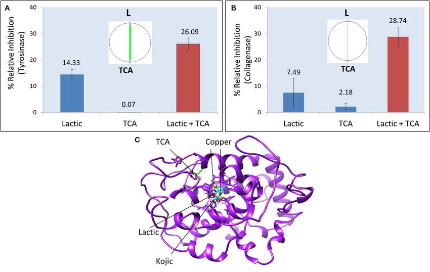
Bhardwaj et al.                                                                                                Professional TCA-Lactic Chemical Peel

process called cell turnover or skin rejuvenation. Although                professional and physician’s peels is prescription level and hence
the user will not necessarily experience visible exfoliation after         not available for retail sale or at-home use. Depending on skin
peeling, the active ingredients in peels are still working at a            conditions and sensitivity, a dermatologist will pick professional
cellular level. The treatment of sun damage (photo-aging) and              or physician’s peels from one of these three depths: (1) Superficial
unevenly pigmented skin (dyspigmentation) are two primary                  peels that usually do not penetrate beyond the papillary dermis
indications of chemical peeling intervention (2, 3). Although in           and usually contain active ingredients, such as TCA at
Bhardwaj et al.                                                                                              Professional TCA-Lactic Chemical Peel

collagenase, and 400Ex/505Em for neutrophil elastase. Readings            remove DNA contaminant, followed by passing through RNeasy
were taken every 5 min for at least 30 min to plot a kinetic              Minielute spin column to separate RNA that was eventually
curve and obtain slope of the curve. The percent relative                 eluted in water for the next step of cDNA synthesis. cDNA was
inhibition with respect to enzyme control was calculated using            synthesized using Maxima First Strand cDNA Synthesis Kit with
this equation: [(Slope of Enzyme Control – Slope of Test                  dsDNase (Thermo Scientific, USA) following the manufacturer’s
Samples)/slope of Enzyme Control] × 100. A dose-dependent                 protocol. Briefly, it was a two-step procedure. First, RNA samples
study (serial dilution) and positive controls were run to validate        were incubated with dsDNase (supplied in the kit) to remove
the results. The pH of all the samples including test agents,             any genomic DNA. Second, enzyme mix (Maxima Reverse
positive and negative controls was measured at the end of the             Transcriptase) and Reaction Mix [dNTPs, oligo (dt)18 and
final reading (pH was 7.2 ± 0.2) to rule out any effect of                random hexamer primers] were added to the purified RNA from
acidity on the enzymes activity. We used the positive controls            step 1. The mixture is centrifuged and incubated for 10 min at
agents that were well-studied: kojic acid for tyrosinase (13), 1,10       25◦ C, followed by 15 min incubation at 50◦ C, and finally the
phenanthroline for collagenase and N-(Methoxysuccinyl)-Ala-               reaction was terminated by heating the mixture at 85◦ C for
Ala-Pro-Val-Chloromethyl Ketone (SPCK) for elastase (14).                 5 min. The cDNA was stored at −80◦ C unless used for Real-time
                                                                          qPCR (RT-qPCR). Rt-qPCR was performed using Quant Studio
Synergy Investigation and Computation                                     7 Flex Real-Time PCR System (Applied Biosystems, USA) to
A mixture of TCA and lactic acid was tested at ratio 1:2 (as              quantify differential expression (1Ct) of genetic markers for
it exists in the test peel product), for above-mentioned enzyme           inflammation (Interleukins 1a, 6, and 8), hydration/barrier
inhibition activities. The synergy and dose reduction index (DRI)         (Filagrin and Claudin1), regeneration (Connective tissue growth
was investigated following constant ratio serial dilution method          factor and insulin-like growth factor 1), and extracellular matrix
and CompuSyn software developed by Chou (15, 16).                         (Collagen 1A, Collagen 3A, Elastin, and Fibronectin 1).

In-silico Molecular Docking                                               Ex-vivo: Tissue Culture and Product
To elucidate the mechanism of tyrosinase inhibition by
lactic and trichloroacetic acids, Rosetta software was used for
                                                                          Treatment
                                                                          Human skin biopsies were cut into 8 × 3 mm (diameter ×
molecular docking, and the binding energy was reported as
                                                                          thickness) skin samples. Skin samples were weighed to select
Rosetta Energy Unit (REU). Chimera was used for energy
                                                                          samples with approximately similar weight. Skin samples were
minimization, clash removal, and structure visualization. Active
                                                                          cultured on top of the perforated stainless-steel rings that were
Site Prediction Server from IIT, Delhi was used to compute
                                                                          in contact with modified William’s E culture media. Each skin
binding pockets/cavities in the protein. Before application of the
                                                                          sample was covered with a 6 mm diameter membrane, and
docking model on lactic acid and TCA, the robustness of the
                                                                          the commercial peel solution (liquid) was applied on top of
model was first tested on kojic acid, the positive control, and
                                                                          the membrane to achieve uniform product application on the
a benchmark compound in molecular docking for tyrosinase.
                                                                          skin. Product was applied every day, and the culture media was
Sequential docking of TCA and lactic acid was performed to gain
                                                                          changed every 3rd day until the 6th day when skin samples were
insights on their synergistic interactions.
                                                                          harvested for viability and histochemical measurements.
In-vitro: 3D Human Skin Equivalents and
qPCR                                                                      Skin Viability
EpiDermFT 3D full thickness human skin equivalents                        Skin samples were tested for viability using 3-(4,5-
(MatTek Corp, USA) were cultured in EFT400 culture                        dimethylthiazol-2-yl)-2,5-diphenyl tetrazolium bromide (MTT)
media supplemented with growth factors, hormones, and                     assay. The liquid peel solution was tested in serial dilution: 1:1
lipid precursors (MatTek Corp, USA). This human skin model                (100% stock product), 1:2, 1:4, and 1:8 to identify non-toxic dose
is bioengineered using epidermal keratinocytes and dermal                 of the peel for next step, that is, modulation of melanin and
fibroblasts from a single normal human donor to form a                    collagen by peel.
multilayered model for human epidermis and dermis. The
model is characterized for metabolic and mitotic activities, and          Photo-Damaged Collagen Ex-vivo Skin
other in-vivo-like morphological and growth characteristics               Model
required to study anti-aging, wound healing, skin irritation,             To assess the efficacy of the peel in treating photo-damaged
and many other properties of cosmetic and medical products                skin, UV-damaged ex-vivo skin model was prepared following
(17–19). The peel was diluted to 10% solution before testing;             individual typology angle (ITA)-based method to predict
this dose was found to be non-toxic to tissue as investigated             biological effective dose of UV (BED-UV) (20). Briefly, BIO-
by Alamar Blue assay. The skin equivalents were treated with              SUN from Vilber Lourmat was used to expose skin biopsies
buffer (control) or 10% peel solution for 1 min. RNA was                  (donor aged 62 years and intermediate skin type with ITA 37◦ )
isolated from the skin using RNeasy Plus Mini Kit (Qiagen                 with 6 J/cm2 UV. To mimic real-world conditions UVA (5.76
Inc., USA) following manufacturer’s protocol. Briefly, skin               J/cm2 ) and UVB (0.24 J/cm2 ) were used in 96:4% (21), which
samples were lysed using lysis buffer supplied in the kit. The            corresponds to sun burn but without any DNA damage (20).
lysate was passed through gDNA eliminator spin column to                  This 6 J/cm2 BED-UV was found to induce only the collagen

Frontiers in Medicine | www.frontiersin.org                           3                                   February 2021 | Volume 8 | Article 617068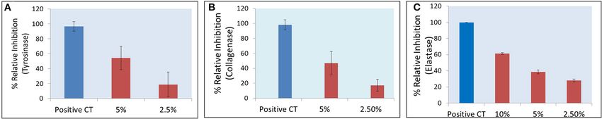
Bhardwaj et al.                                                                                                                  Professional TCA-Lactic Chemical Peel

damage (but not melanin pigmentation), and hence only the                              RESULTS
photo-damaged collagen skin model was used for the study.
                                                                                       In-vitro Enzyme Inhibition Activities
                                                                                       The commercial peel was found to significantly inhibit tyrosinase
Histochemical Imaging and                                                              (Figure 1A), collagenase (Figure 1B), and elastase (Figure 1C)
Semi-quantification                                                                    in dose-dependent manner. Compared to collagenase and
For each treatment group (untreated, positive control, and                             tyrosinase, the elastase assay had relatively higher reproducibility
test product “peel” after 2× dilution), skin biopsies (N =                             (evident from tight error bars), as the latter was fluorescence
6) were treated every day and then harvested on day 6 for                              assay, while the other two were based on absorbance. TCA
histochemical processing: sectioning followed by staining and                          and lactic acid (the active ingredients in the peel) were further
imaging. Two random sections from each biopsy were imaged                              tested as neat solutions to investigate any possible synergy.
and processed to obtain statistical information from N = 12                            TCA:lactic at ratio 1:2 were found to exhibit strong synergy
images or data points for each treatment group. For melanin                            for anti-tyrosinase (Figure 2A) and anti-collagenase activities
imaging, sections were stained with Fontana Masson (melanin                            (Figure 2B). This is the optimum ratio to achieve maximum
granules in black/brown/dark gray color), and for collagen                             synergy as we observed a decrease in synergy when deviated
sections were stained with Picrosirius Red (collagen fibers in                         from this ratio. The results using CompuSyn further confirmed
purple-red color). Proprietary algorithm and process was used                          that it is not an additive effect but a true synergy between
to quantify melanin and collagen content in sectioned biopsies.                        TCA:lactic at 1:2 for anti-tyrosinase (Figure 2A, inset) and anti-
Briefly, images were de-convoluted to separate signals from                            collagenase (Figure 2B, inset). The intensity of the green line
background, for example, collagen in red/purple from non-                              in CompuSyn polygonograms (insets) indicates strength of the
collagen background in blue/green color. The raw RGB colored                           synergy. No synergy was observed for anti-elastase activity.
images were converted into 8-bit L∗ a∗ b color space using Image                       Figure 2A and the computational results from CompuSyn
J software from National Institute of Health. The transformed L∗                       (Supplementary Table 1) insinuate that TCA has little to no
values were normalized on the ratio between selected area and the                      tyrosinase inhibition activity, however, TCA is critical for synergy
area of the slide to produce pigmentation and collagen scores.                         and it can reduce dose of the TCA and lactic acids (denoted
                                                                                       by DRI “dose reduction index” in Supplementary Table 1) by
                                                                                       several folds to achieve the same level of efficacy (denoted by Fa
Positive Controls                                                                      “fractional activity” in the Supplementary Table 1).
Kojic acid for anti-pigmentation activity (in-silico molecular
docking, in-vitro anti-tyrosinase assay, and ex-vivo melanin                           In-silico Molecular Docking to Elucidate
inhibition), 1,10 phenanthroline for in-vitro anti-collagenase
assay, retinol for ex-vivo collagen synthesis, and SPCK for in-vitro                   Mechanism of Tyrosinase Inhibition
anti-elastase assay.                                                                   Tyrosinase is the key enzyme responsible for pigmentation. The
                                                                                       structure of tyrosinase enzyme and its mechanisms of inhibition
                                                                                       by benchmarks ingredients, such as kojic acid and hydroquinone
Statistics                                                                             for hyper- and dyspigmented skin are well-characterized (22).
For statistics at least two independent experiments were done                             Figure 2C shows the crystal structure of tyrosinase. The
for all experiments. Student t-test was done for significance                          catalytic site of tyrosinase (active site responsible for production
testing on in-vitro data. One-way ANOVA with permutation                               of melanin from tyrosinase) is characterized by the presence
test followed by Tukey’s or Dunnett’s and t-test followed by                           of two copper atoms that are essential for the catalytic activity
permutation was done on ex-vivo data to evaluate significant                           of the enzyme. The superimposition of native kojic acid
differences between different groups. P-value
Bhardwaj et al. Professional TCA-Lactic Chemical Peel
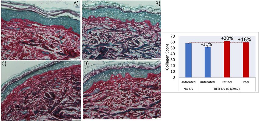
FIGURE 2 | Synergistic effect of TCA:lactic at ratio 1:2 for inhibitory effect on tyrosinase (A) and collagenase (B). Insets show polgonograms computed by CompuSyn
showing synergy. Crystal structure of tyrosinase showing docked structures of lactic, kojic and TCA (C). The concentration of TCA and lactic acid in tyrosinase
synergy testing is 0.156 and 0.312%, respectively and for collagenase synergy testing the concentration is 0.625% TCA and 1.25% lactic acid.
with previous reports validate the robustness of our model IGF1 (data not shown). An increase in these ECM components
(13, 22). This validated model was used to dock TCA, lactic (collagen, elastin, and fibronectin) and growth factors (CTGF and
acid, and their sequential docking. Kojic acid shows highest IGF1) supports the peel’s mode of action behind anti-aging and
affinity for tyrosinase inhibition (−6.686 REU), followed by skin regeneration.
lactic acid (−5.496 REU), and TCA (−2.149 REU) (Table 1).
Compared to kojic and lactic acid which bind at the catalytic Ex-vivo Human Biopsy
site (Figure 2C), and share the same hydrophobic cavity The commercial peel product was tested at 50% (2× dilution of
(Supplementary Figure 1B), TCA showed maximum affinity commercial product) as it was found to induce no inhibition of
away from the catalytic site (Figure 2C). When TCA and metabolic activity of the human biopsies at this concentration
lactic acid were docked sequentially, surprisingly, a change in (Supplementary Figure 2). Figure 3 shows images of skin
structure from linear to ring-like structure was observed in sections after Fontana Masson staining for melanin imaging
TCA (Supplementary Figures 1C,D). This change to ring-like and semi-quantification of the melanin content (black/dark gray
structure could be the rationale behind the synergistic effect granules) in the skin: Untreated vs. treated with product or
between TCA and lactic acid, and needs further investigation. positive control (kojic acid). The integrity of skin structure
and abundance of melanin-containing cells in the basal layer
In-vitro 3D Human Skin Model of epidermis is clearly visible. There is a clear decrease in
The commercial peel solution was found to exhibit no significant black/dark gray intensity of melanin granules after treatment of
effect on metabolic inhibition of bioengineered 3D human skin the skin with peel (50% solution) and positive control (0.1% kojic
model. There was no significant change (Fold changeBhardwaj et al. Professional TCA-Lactic Chemical Peel
TABLE 1 | Binding or inhibition properties of Kojic acid (positive control), lactic acid, and trichloroacetic acid (TCA) to tyrosinase enzyme, docked using Rosetta software.
Ligand or ingredient Type of Binding energy Binding residues H bonds
inhibitor (REU)
Kojic acid Catalytic −6.686 ARG 205,209, ASN 201,205, HIS 208,204,200, 56, 60, MET 211, 215, ALA217,221 HIS 56, MET 211
Lactic acid Catalytic −5.496 ARG 205,209, ASN 201,205, HIS 208,204, GLY 212,216, VAl213,217 ARG 205, ASN 201
Trichloroacetic acid Allosteric −2.149 GLY 42, 46, MET 57,61, ASN 53,57, VAL 214, 218 0
The common binding residues of kojic acid and lactic acid are highlighted bold.
FIGURE 3 | Histochemical images (A–C) and quantification (D) showing melanin granules in gray and melanin content of skin sections before (untreated) and after
treatment with kojic acid (positive control, 0.1%) or peel (after 2× dilution). P-valueBhardwaj et al. Professional TCA-Lactic Chemical Peel FIGURE 4 | Histochemical images of skin biopsies showing collagen in purple-red color, before treatment “no UV and no product” (A), after exposure to BED-UV (B), treatment of BED-UV exposed skin with retinol as positive control (C) and peel (D). The graph shows significant change (recovery) of BED-UV damaged collagen (−11%) after treatment with retinol (positive control, 0.05%) or peel (after 2× dilution). P-value
Bhardwaj et al.                                                                                                                         Professional TCA-Lactic Chemical Peel

DATA AVAILABILITY STATEMENT                                                                SUPPLEMENTARY MATERIAL
The original contributions presented in the study are included                             The Supplementary Material for this article can be found
in the article/Supplementary Material, further inquiries can be                            online at: https://www.frontiersin.org/articles/10.3389/fmed.
directed to the corresponding author/s.                                                    2021.617068/full#supplementary-material
                                                                                           Supplementary Figure 1 | Crystal structure of Tyrosinase showing catalytic site
AUTHOR CONTRIBUTIONS                                                                       (indicated by presence of two copper atoms in brown), and the binding of native
                                                                                           (cyan) and retrieved (elemental color) structures of kojic acid to the catalytic site
                                                                                           (A). Kojic (cyan) and lactic acid bind in the same hydrophobic cavity, while TCA
VB did planning and required collaborations for the study. AF                              binds into a separate cavity (B). Lactic (green) and TCA (elemental) when docked
supervised the study. VB, KS, and SM performed experiments.                                independently are linear structures (C), but when docked sequentially a
JM and AA-W revised the manuscript. All authors reviewed and                               conformational change to the ring-like new structure appears (D).
approved the final draft of the manuscript.                                                Supplementary Figure 2 | MTT assay showing effect of peel concentration on
                                                                                           metabolic activity of the skin biopsies.
FUNDING                                                                                    Supplementary Table 1 | Dose reduction index (DRI) of lactic (L) and TCA
                                                                                           simulated by CompuSyn. Fa, fractional activity (efficacy).
This study was fully funded by Colgate-Palmolive Company.                                  Supplementary Table 2 | Mean melanin scores of treatment conditions after
                                                                                           proprietary image processing (10 images for each treatment condition) to
ACKNOWLEDGMENTS                                                                            compare melanin inhibition efficacy of untreated vs. positive control and peel.

                                                                                           Supplementary Table 3 | Mean collagen scores of treatment conditions
We acknowledge Cutech (now Symrise S.r.l.) and Jin                                         calculated after proprietary image processing (12 images for each treatment
Namkoong for assistance in ex-vivo and 3D human skin                                       condition) to compare collagen repair efficacy of positive control and peel vs.
                                                                                           untreated UVDL (after UV daily light damage).
equivalents studies.

REFERENCES                                                                                 14. Pientaweeratch S, Panapisal V, Tansirikongkol A. Antioxidant, anti-
                                                                                               collagenase and anti-elastase activities of Phyllanthus embilica,
 1. The American Society of Plastic Surgeons. Plastic Surgery Statistics Report.               Manilkara zapota and silymarin: an in-vitro comparative study
    (2019). Available online at: https://www.plasticsurgery.org/documents/News/                for anti-aging applications. Pharm Biol. (2016) 54:1865–72.
    Statistics/2019/plastic-surgery-statistics-full-report-2019.pdf       (accessed            doi: 10.3109/13880209.2015.1133658
    August 11, 2020).                                                                      15. Chou TC. Drug combination studies and their synergy quantification
 2. The American Society for Aesthetic Plastic Surgery. Aesthetic Plastic Surgery              using the Chou-Talalay method. Cancer Res. (2010) 70:440–6.
    National Databank Statistics. (2019). Available online at: https://www.                    doi: 10.1158/0008-5472.CAN-09-1947
    surgery.org/sites/default/files/Aesthetic-Society_Stats2019Book_FINAL.pdf              16. Zhang N, Fu JN, Chou TC. Synergistic combination of microtubule targeting
    (accessed August 11, 2020).                                                                anticancer fludelone with cytoprotective panaxytrio derived from panax
 3. Soleymani T, Lanoue J, Rahman Z. A practical approach to chemical peels. J.                ginseng against MX-1 cells in-vitro: experimental design and data analysis
    Clin. Aesthet. Dermatol. (2018) 11:21–8.                                                   using the combination index method. Am J Cancer Res. (2016) 6:97–104.
 4. Obagi S, Niamtu J. Chemical peel. In: Niamtu J, editor.                                17. Xiong ZM, O’Donovan M, Sun L, Choi JY, Ren M, Cao K. Anti-aging
    Cosmetic Facial Surgery, 2nd ed. Elsevier (2018). p. 732–55.                               potentials of methylene blue for human skin longevity. Sci Rep. (2017) 7:2475.
    doi: 10.1016/B978-0-323-39393-5.00014-5                                                    doi: 10.1038/s41598-017-02419-3
 5. Tang SC, Yang JH. Dual effects of alpha hydroxy acids on the skin. Molecules.          18. Lee ES, Ahn Y, Bae IH, Min D, Park NH, Jung W, et al. Synthetic retinoid
    (2018) 23:863. doi: 10.3390/molecules23040863                                              seletinoid G improves skin barrier function through wound healing and
 6. The Food and Drug Administration. Alpha Hydroxy Acids. (2019).                             collagen realignment in human skin equivalents. Int J Mol Sci. (2020) 21:3198.
    Available online at: https://www.fda.gov/cosmetics/cosmetic-ingredients/                   doi: 10.3390/ijms21093198
    alpha-hydroxy-acids (accessed June 25, 2019).                                          19. Mallampati R, Patlolla RR, Agarwal S, Babu RJ, Hayden P, Klausner M, et al.
 7. Liu H, Khachemoune A, Rashid RM. Chemical burn following 50%                               Evaluation of Epiderm full thickness 300 (EFT-300) as an in-vitro model for
    trichloroacetic acid for acne: presentation of a case and a focused review. J              skin irritation: studies on aliphatic hydrocarbons. Toxicol In Vitro. (2010)
    Dermatol Dermatol Surg. (2016) 20:71–4. doi: 10.1016/j.jdds.2015.06.001                    24:669–76. doi: 10.1016/j.tiv.2009.08.019
 8. Dayan E, Rohrich RJ. Jessner’s solution with trichloroacetic acid chemical peel:       20. Bino SD, Sok J, Bessac E, Bernerd F. Relationship between skin response to
    optimizing outcomes and safety. Int Open Access J Am Soc Plast Surg. (2019)                ultraviolet exposure and skin color type. Pigment Cell Res. (2006) 19:606–14.
    7:e2250. doi: 10.1097/GOX.0000000000002250                                                 doi: 10.1111/j.1600-0749.2006.00338.x
 9. Nguyen TH, Rooney JA. Trichloroacetic acid peels. Dermatol Ther. (2000)                21. D’Orazio J, Jarrett S, Ortiz AA, Scott T. UV radiation and the skin. Int J Mol
    13:173–82. doi: 10.1046/j.1529-8019.2000.00020.x                                           Sci. (2013) 14:12222–48. doi: 10.3390/ijms140612222
10. Kligman DE, Draelos ZD. Combination superficial peels with salicylic acid              22. Deri B, Kanteev M, Goldfeder M, Lecina D, Guallar V, Adir N, et al. The
    post peel retinoids. J Drugs Dermatol. (2016) 15:442–50.                                   unraveling of the complex pattern of tyrosinase inhibition. Sci Rep. (2016)
11. Briden ME. Alpha hydroxyacids chemical peeling agents: case studies and                    6:34993. doi: 10.1038/srep34993
    rationale for safe and effective use. Cutis. (2000) 73:18–24.                          23. Rakic L, Lapiere CM, Nusgens BV. Comparative caustic and biological activity
12. Babilas P, Knie U, Abels C. Cosmetic and dermatologic use of                               of trichloroacetic and glycolic acids on keratinocytes and fibroblasts in-
    alpha hydroxyl acids. J German Soc Dermatol. (2012) 10:488–91.                             vitro. Skin Pharmacol Appl Skin Physiol. (2000) 13:52–9. doi: 10.1159/0000
    doi: 10.1111/j.1610-0387.2012.07939.x                                                      29908
13. Jung HJ, Noh SJ, Park Y, Kang D, Chun P, Chung HY, et al. In-                          24. Dayal S, Sahu P, Yadav M, Jain VK. Clinical efficacy and safety on combining
    vitro and in-silico insights into tyrosinase inhibitors with (E)-benzylidene-              20% trichloroacetic acid peel with topical 5% ascorbic acid for melasma.
    1-indanone derivatives. Comput Struct Biotechnol J. (2019) 17:1255–64.                     J Clin Diagn Res. (2017) 11:WC08–11. doi: 10.7860/JCDR/2017/26078.
    doi: 10.1016/j.csbj.2019.07.017                                                            10685

Frontiers in Medicine | www.frontiersin.org                                            8                                            February 2021 | Volume 8 | Article 617068
Bhardwaj et al. Professional TCA-Lactic Chemical Peel
25. Wang Y, Hao MM, Sun Y, Wang LF, Wang H, Zhang YJ, et al. Synergistic Conflict of Interest: VB, SM, AF, and JM was employed by the company
promotion on tyrosinase inhibition by antioxidants. Molecules. (2018) 23:1– Colgate Palmolive. AA-W was employed by the company PCA Skin (brand of
13. doi: 10.3390/molecules23010106 Colgate-Palmolive).
26. Deo KS, Dash KN, Sharma YK, Virmani NC, Oberai C. Kojic acid vis-à-vis
its combinations with hydroquinone and betamethasone valerate in melasma: The remaining author declares that the research was conducted in the absence of
a randomized, single blind, comparative study of safety and efficacy. Indian J any commercial or financial relationships that could be construed as a potential
Dermatol. (2013) 58:281–5. doi: 10.4103/0019-5154.113940 conflict of interest.
27. Trunet A, Meyer I, Maire ML, Vielhaber GL. A Skin and/or Hair
Whitening Mixture. Patent application number 13182603.4. European Patent Copyright © 2021 Bhardwaj, Sharma, Maksimovic, Fan, Adams-Woodford
Office, (2015). and Mao. This is an open-access article distributed under the terms of the
28. Kong BY, Sheu SL, Kundu RV. Assessment of consumer knowledge Creative Commons Attribution License (CC BY). The use, distribution or
of new sunscreen labels. JAMA Dermatol. (2015) 151:1028–30. reproduction in other forums is permitted, provided the original author(s)
doi: 10.1001/jamadermatol.2015.1253 and the copyright owner(s) are credited and that the original publication in
29. Kim HK. Garlic supplementation ameliorates UV-induced photoaging in this journal is cited, in accordance with accepted academic practice. No use,
hairless mice by regulating antioxidative activity and MMPs expression. distribution or reproduction is permitted which does not comply with these
Molecules. (2015) 21:70. doi: 10.3390/molecules21010070 terms.
Frontiers in Medicine | www.frontiersin.org 9 February 2021 | Volume 8 | Article 617068You can also read