Prediction of Death After Multiple Wasp Stings with a Clinical Prognostic Model
←
→
Page content transcription
If your browser does not render page correctly, please read the page content below
Prediction of Death After Multiple Wasp Stings with
a Clinical Prognostic Model
Maohe Wang
Suining Central Hospital
Mei Qin
Suining Central Hospital
Amanda Y Wang
University of New South Wales
Jia Wei Zhao
Bond University
Fei Deng
Sichuan Provincial People's Hospital: Sichuan Academy of Medical Sciences and Sichuan People's
Hospital
Guisen Li
Sichuan Provincial People's Hospital: Sichuan Academy of Medical Sciences and Sichuan People's
Hospital
Wei Wang ( wangweisz@med.uestc.edu.cn )
Sichuan Provincial People's Hospital: Sichuan Academy of Medical Sciences and Sichuan People's
Hospital https://orcid.org/0000-0001-5664-7725
Research
Keywords: wasp sting, poisoning severity score, death, prognostic, nomogram
Posted Date: September 13th, 2021
DOI: https://doi.org/10.21203/rs.3.rs-877961/v1
License: This work is licensed under a Creative Commons Attribution 4.0 International License.
Read Full License
Page 1/17
Abstract
Background: We aimed to assess the utility of the poisoning severity score (PSS) as early prognostic
predictors in patients with wasp stings, and to explore a reliable and simple predictive tool for short-term
outcomes.

Methods: From January 2016 to December 2018, 363 patients with wasp stings in Suining Central
Hospital were taken as research subjects. In the first 24h of hospital admission, the PSS and Chinese
expert consensus on standardized diagnosis and treatment of wasp stings (CECC) were used as the
criterion for severity classification, and their correlation was analyzed. The patients were divided into
survival and death groups according to the state of discharge. The factors that affect outcome were
analyzed by logistic regression analysis. A clinical prognostic model of death was constructed according
to the risk factors, and 1000 times repeated sampling was done to include the data to verify the model
internally.

Results: The mortality of wasp sting patients was 3.9%. There was a correlation between PSS and CECC
(r=0.435, P
severity of poisoning patients (including environmental toxins)[9]. Stay in observation, hospitalization,
admission to ICU or general ward and nursing grade were decided according to the assessment
results[10]. However, to the authors' knowledge, no previous studies have specifically addressed the use
of PSS for evaluating the severity of wasp sting patients. Therefore, we undertook this study in which we
looked at 363 patients with wasp stings from January 2016 to December 2018 in Suining Central
Hospital, to assess the utility of the PSS as early prognostic predictors of short-term outcomes. This
study also explored a reliable and simple predictive tool to identify patients at a high risk of death, to
optimise patient management to reduce the fatality rate.

Methods
Research subjects
This was a retrospective study of patients with wasp stings who presented to the nephrology department
and ICU of Suining Central Hospital of Sichuan Province, China between January 2016 to December
2018. Suining Central Hospital of Sichuan Province, China, is the only tertiary grade A general hospital in
the interior areas of Sichuan Province that has more than 94,000 annual hospitalized patients and more
than 100 wasp sting patients each year.

The inclusion criteria were: 1) Patients with a definite diagnosis of wasp stings; 2) age ≧ 14 years old; 3)
the clinical data were complete. The exclusion criteria were as follows: 1) age < 14years; 2) re-hospitalized
patients with wasp stings; 3) wasp sting patients who died before admission. We categorized them into
survival group (n = 349) and death group (n = 14) according to the state of discharge. This study was
approved by the Ethics Committee of Suining Central Hospital (Serial number: LLSNCH20200022).
Clinical data collection
We collected information on the patients' demographics (age, sex), the time interval between sting and
admission (admission time), number of stings, signs and symptoms (allergic rash, hypotension,
macroscopic hematuria, and oliguria or anuria), severe complications (rhabdomyolysis, acute kidney
injury (AKI), coagulation disorders, hemolysis, liver dysfunction, acute respiratory distress syndrome
(ARDS) and multiple organ dysfunction syndrome (MODS)), inpatient days, and short-term outcomes
(death or survival).

We recorded laboratory data on admission, including: white blood cells (WBC), activated partial
thromboplastin time (APTT), prothrombin time (PT), alanine aminotransferase (ALT), aspartate
aminotransferase (AST), indirect bilirubin (I-BIL), creatine kinase (CK), lactate dehydrogenase (LDH), the
serum creatinine (SCr).

At admission, PSS and CECC were used as the criterion for
severity classification respectively

                                                   Page 3/17
The PSS grades severity as (0) none, (1) minor, (2) moderate, (3) severe, and (4) fatal poisoning. The
severity grading takes into account only the observed clinical symptoms and signs, but does not estimate
risks or hazards on the basis of information such as the amount ingested or serum concentrations of the
toxic agent[11]. According to the admission criteria, patients with no symptoms and those who died before
admission were excluded. Thus the PSS graded the patients into minor, moderate, and severe poisoning
levels.
The CECC grades severity as: (1) minor: the number of stings was less than 10, with only local allergic
reactions and no organ function involvement, (2) moderate: the number of stings was between 10 and 30;
allergic reaction was classified as Ⅰ-Ⅱ, only 1 organ was involved; sequential organ failure score (SOFA) ≥
2 points; there were macroscopic hematuria in an early stage, (3) severe: the number of stings was
greater than 30; allergic reaction was classified as Ⅲ-Ⅳ or at least 2 organs involved; SOFA ≥ 2 points for
each organ[6].
Statistical analysis
Continuous variables with normal distribution were expressed as means and standard deviations.
Categorical variables without normal distribution were expressed as medians and interquartile ranges.
Variables of the two groups were compared by Mann-Whitney U test. Spearman analysis was performed
for the correlation between PSS and CECC. ROC curve analysis of PSS and CECC was performed
respectively, and Z-test was used to analyze the difference of AUC between them.
Univariate and multivariate logistics regression were used to analyze the risk factors of death in wasp
sting patients. On the basis of the final model, a nomogram was built using R software with the rms
package. A concordance index (C-index) was used to determine the discrimination of the nomogram, and
it was calculated by a bootstrap approach with 1,000 resamples for internal validation. Receiver
operating characteristic (ROC) curves were plotted, and the areas under the ROC curve were calculated.
A p-value less than 0.05 was considered statistically significant.
Results
Descriptive Results
Between January 2016 to December 2018, a total of 390 patients were identified with wasp stings. Of
these patients, 27 patients were excluded, including three who died on admission and 24 who refused to
be hospitalized. Finally, 363 patients were included in this study, which included 219 (60.3%) males and
144 (39.7%) females. The mean age was 55.9 ± 16.3 years. Fourteen(3.9%) patients died during
hospitalization, including 1 in (PSS) grade 1, 9 in (PSS) grade 2, and 4 in (PSS) grade 3. Over the three-
year period of this study, the deaths only occurred from September to November. (Fig. 1, Table 3)
Page 4/17PSS and CECC
The median PSS was 1(1,1) upon admission. grade 1 (minor) PSS upon admission were seen in 290
(79.9%) cases, grade 2 (moderate) in 59 (16.2%) cases and grade 3 (severe) in 14 (3.9%) cases. (Table 1)
Table 1
Spearman analysis between PSS and CECC
PSS Sum
Grade 1 Grade 2 Grade 3
CECC Minor 124 7 0 131
Moderate 147 26 3 176
Severe 19 26 11 56
Sum 290 59 14 363
Spearman analysis, r = 0.435 P < 0.001.
Regarding the CECC assessment upon admission, minor condition was found in 131 (36.1%) cases,
moderate in 176 (48.5%) cases, severe in 56 (15.4%) cases. (Table 1)
Spearman analysis between PSS and CECC
The Spearman analysis showed the correlation between PSS and CECC (r = 0.435, P < 0.001), which
indicated that there was a correlation between PSS and CECC in assessing the severity of wasp sting
patients. (Table 1)
ROC curve analysis of PSS and CECC
The AUC of the PSS and CECC in predicting death in wasp sting patients was 0.890 and 0.845,
respectively, and they showed a certain predictive power. Although the AUC of the PSS was better than
that of CECC, the difference between them was not statistically significant (Z = 0.7230, P = 0.4697).
Table 2 showed the optimal cut off value, sensitivity, specificity, positive predictive value, and negative
predictive values of PSS and CECC. (Fig. 2, Table 2).
Page 5/17Table 2
The ROC analysis of PSS and CECC in the optimal cut off scores
Score Value The comparison of AUC
Sensitivity Specificity +PV -PV AUC Z P
(%) (%) (%) (%)
PSS > 1 92.86 82.81 17.8 99.7 0.890 0.7230 0.4697
CECC > 71.43 86.82 17.9 98.7 0.845
2
+PV: Positive predictive value -PV: Negative predictive value
Comparison of clinical data between the survival and the
death group
The death group had a greater proportion of females (85.7% vs. 37.8%, P < 0.001), and was significantly
older (71.1 ± 9.8years vs. 55.3 ± 16.2years, P < 0.001) than that of the survival group. The number of
stings (30 vs. 8, P < 0.001) and the time from stings to admission (7h vs. 3h, P = 0.004) in the death group
were higher than those in the survival group (P < 0.001). The PSS of the death group was significantly
higher than that of the survival group (2 vs. 1, p < 0.001). The length of hospital stay was significantly
shorter in the death group (1 day vs. 4 days, P < 0.001). (Table 3)
Page 6/17Table 3
Comparison of clinical data between the survival and the death group
Variable Survival group (n = 349) Death group (n = 14) P
Age (years) 55.3 ± 16.2 71.1 ± 9.8 < 0.001
Gender (M: F) 217:132 2:12 < 0.001
Number of stings 8(4,15) 30(20,45) < 0.001
Admission time (h) 3(2,5) 7(3.7,10) 0.004
Poisoning severity score 1(1,1) 2(2,3) < 0.001
Grade 1 (n) 289 1
Grade 2 (n) 50 9
Grade 3 (n) 10 4
Inpatient days (day) 4(3,7) 1(1,2.2) < 0.001
Allergic rash n (%) 50(14.3) 0(0) 0.232
Hypotension n (%) 19(5.4) 1(7.1) 0.554
AKI* n (%) 28(8%) 11(78.6) < 0.001
Rhabdomyolysis n (%) 92(26.4) 13(92.9) < 0.001
Hemolysis n (%) 49(14) 12(85.7) < 0.001
Oliguria or anuria n (%) 18(5.2) 9(64.3) < 0.001
Macroscopic hematuria n (%) 38(10.9) 13(92.9) < 0.001
Coagulation abnormalities n (%) 126(36.1) 13(92.9) < 0.001
Liver damage n (%) 69(19.8) 13(92.9) < 0.001
Dialysis n (%) 35(10) 10(71.4) < 0.001
MODS** n (%) 43(12.3) 13(92.9) < 0.001
ARDS # n (%) 4(1.1) 9(64.3) < 0.001
ICU## n (%) 4(1.1) 3(21.4) < 0.001
*Acute Kidney Injury, **Multiple organ dysfunction syndrome, ***Acute respiratory distress syndrome,
##
Intensive care unit.
Comparisons of complications between the survival and the
death group
Page 7/17No allergic rash was seen in the death group (0% vs. 14.3%, P = 0.232). One patient in the death group
developed hypotension compared to 19 in the survival group (P = 0.554). The incidences of
rhabdomyolysis, hemolysis, liver dysfunction, coagulation disorder, ARDS, MODS, oliguria (or anuria) and
macroscopic hematuria in the death group were significantly higher than those in the survival group (P <
0.001). Seven (1.9%) patients were admitted to ICU, including three in the death group and four in the
survivor group. The incidence of AKI in the death group was significantly higher (78.6%) than that in the
survival group (8%) (P < 0.001). A larger proportion of patients in the death group (ten patients (71.4%))
received hemodialysis compared to that in the survival group (35 patients (10%)) (p < 0.001). (Table 3)
Comparisons of biochemical parameters between the
survival and the death group
In the first 24h of hospital admission, the laboratory parameters including WBC, APTT, PT, ALT, AST, IBIL,
CK, LDH, LDH, SCr values in the death group were significantly higher than those in the survival group (p
< 0.05). (Table 4)
Table 4
Comparison of biochemical parameters between the survival and the death group
Biochemical parameters Survival group (n = 349) Death group (n = 14) P
SCr* (59-104umol/l) 66(55,77) 78(68.5,140) 0.004
CK**(40-200U/l) 199(117.5,415.5) 2321(407.7,5342.5) < 0.001
AST***(13-35U/l) 35.5(26,55.2) 557(255,1505.5) < 0.001
IBIL**** (0-18umol/l) 8.1(5.2,16.8) 39.4(19.8,83.4) < 0.001
ALT#(7-40U/l) 22(17,37) 286(58.5,1369.5) < 0.001
PT##(11-14.5s) 13.7(13,14.6) 15(14,17) 0.002
APTT###(26-40s) 51.3(36.8,91.2) 120.5(94.1,180) < 0.001
LDH (120-250U/l) 215(179,277.5) 1671(1195.5,2795) < 0.001
WBC#### (3.5–9.5*109/l) 11.8(8.4,15.8) 26.3(21.2,33.6) < 0.001
*
Serum creatinine, **Creatine kinase, ***Aspartate aminotransferase, ****Indirect bilirubin, #Alanine
transaminase, ##Prothrombin time, ###Activated partial thromboplastin time, ####white blood cells
Univariate logistic regression analysis
Based on the pre-specified hypothesis that season was one risk factor for death in wasp sting patients, as
such deaths only occur in summer and autumn, there were significant statistical differences in age, sex,
number of stings, admission time, PSS and biochemical parameters between the death group and the
Page 8/17survival group. However, the evaluation criteria of PSS included cardiovascular system, respiratory
system, liver, kidney, muscular system and so on. Therefore, we took the patients' age, sex, number of
stings, admission time, PSS and season as independent variables to perform univariate logistic
regression. The results were shown in Table 5.
Multivariate logistic regression analysis
The backward multivariate logistic regression analysis showed that female sex, age, number of stings
and PSS were independent risk factors (OR = 8.651, 1.103, 1.033, 6.768 respectively). (Table 6)
Table 5
Univariate logistic regression analysis of risk factors of death
Variable β Wald χ2 P OR 95% C.I.
Age 0.102 12.099 0.001 1.108 1.046–1.173
Female 2.289 8.797 0.003 9.864 2.174–44.762
Admission time 0.004 0.071 0.790 1.004 0.974–1.035
Season 1.694 5.759 0.016 5.440 1.364–21.699
Number of stings 0.043 14.808 < 0.001 1.044 1.022–1.068
PSS * 2.135 29.403 < 0.001 8.460 3.910-18.305
*
Poisoning severity score
Table 6. Multivariate logistic regression analysis of risk factors of death
Variable β Wald χ2 P OR 95%C.I.
Age 0.098 5.029 0.025 1.103 1.012–1.201
Female 2.158 4.885 0.027 8.651 1.277–58.629
Number of stings 0.033 4.852 0.028 1.033 1.004–1.064
PSS * 1.912 9.306 0.002 6.768 1.981–23.120
Hosmer-Lemeshow analysis: χ2 = 0.826 P = 0.999, *Poisoning severity score
A nomogram was built using R software with the rms
package
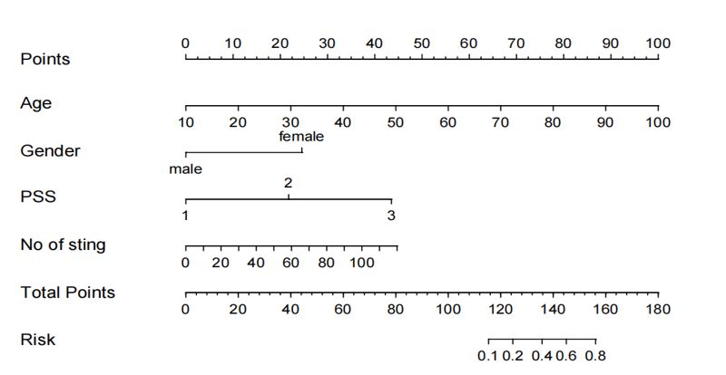
Page 9/17Based on the 4 independent risk factors screened by the above regression analysis, a nomogram model
was built using R software with the rms package to predict the risk of death in wasp sting patients, and
the risk prediction ability of the model was verified. (Fig. 3)
The predicted value C-index was 0.962, indicating that the clinical prognostic model had a good
prediction accuracy and discrimination. (Fig. 4)
The internally verified AUC was 0.962(95%C.I. 0.936–0.988, P < 0.001), indicating that the nomogram had
a high predictive value. (Fig. 5)
Discussion
In China, Nepal, Vietnam and other Asian countries, systemic organ damage or even death due to wasp
venom is not rare[1, 5, 12]. This retrospective study found that the mortality rate in wasp sting patients
was 3.9%, and death was only seen in the summer and autumn months from September through
November among this cohort. Of the 14 deceased patients, all died between the 1st-3rd day after
admission. The PSS in the death group was significantly higher than that of the survival group.
Multivariate logistic regression analysis showed that female sex, increased age, higher number of stings
and greater PSS grade were independent risk factors for death in wasp sting patients.
Wasp venom contains a variety of bioactive components, such as peptides, enzymes and amines[3, 13].
Severe wasp stings can lead to systemic allergic reaction, rhabdomyolysis, shock, hemolysis, acute
kidney injury (AKI), and even death[2, 14, 15]. Unfortunately, there is no specific antidote for wasp venom
at present. It is especially important to classify the severity of wasp sting patients at an early stage, and
to carry out the corresponding treatment. Patients with severe wasp stings should be promptly referred to
a medical institution that can perform blood purification treatment, which can reduce mortality[6, 16, 17].
Chinese Society of Toxicology had prepared a consensus statement on the standardized diagnosis and
treatment of wasp stings (CECC) in 2018[6]. Nevertheless, a wider application of this consensus criteria is
likely limited by the complex evaluation criteria. In Mong's literature, the use of poisoning severity score
(PSS) for the assessment of the severity of the poisoning patients (including wasp sting patients) in the
emergency department was reported[10]. In the present research, there was a correlation between PSS
and CECC in assessing the severity of wasp stings. The severity of wasp stings was evaluated by PSS,
including 59(16.2%) patients with grade 2 and 14(3.9%) patients with grade 3, where a severity poisoning
≥ grade 2 (PSS) might lead to death. The severity of wasp stings was evaluated by CECC, including
56(15.4%) patients with severe wasp stings, where a severity poisoning ≥ severe (CECC) might lead to
death. PSS had higher accuracy and sensitivity value (89.0%, 92.9% respectively), whilst CECC had a
higher specificity value (86.8%) when predicting the short-term clinical outcome in wasp sting patients.
The results revealed that these two criteria determined the severity of poisoning and were able to predict
the short‑term clinical outcomes due to wasp stings. As the PSS was more predictive and simpler, it is
recommended to poisoning centers as effective criteria for classify the severity of wasp sting patients.
Page 10/17Wasp stings were the main cause of human death caused by animal injuries, and it was the main cause
of community-acquired AKI in Asia[16]. In this study, the mortality of our wasp sting patients was 3.9%,
which was consistent with Xie, etc.'s report[1]. When the severity was graded according to the PSS, the
mortality was as high as 28.5% in the severe group and 15.2% in the moderate group. PSS was an
independent risk factor for death in wasp sting patients. The severity was also related to the number of
stings. In Xie and Liu, etc.'s literature, the overall incidence of severe complications was higher in the
group with more than 10 stings[1, 18]. In our patients, the mean number of stings in the death group was
30. Females accounted for 70% of patient deaths. Multivariate logistics regression showed that the
number of stings and female sex were also independent risk factors for death in wasp sting patients. The
average age in the death group was significantly higher than that in the survival group. As such, age was
also one of the risk factors for death, which may be related to underlying diseases in elderly patients. The
four indexes of female sex, age, the number of stings, and PSS were combined as prediction criteria, and
predicting the death of wasp sting patients had high accuracy (AUC = 0.962, 95%C.I. 0.936–0.988, P <
0.001), which was more powerful than using PSS (AUC = 0.890) or CECC (AUC = 0.845) alone. Therefore,
a nomogram prediction model was built on the base of independent risk factors determined through
logistic analyses. The predicted and observed values were found to be similar, which indicates that this
prediction model demonstrates a good degree of discrimination and calibration. Clinicians can identify
the patients at high risk of death for patient management and reduce the fatality rate according to this
nomogram. It is recommended to poisoning centers as effective criteria.
Limitations Of This Study
Our study has some limitations. First, as this was a single-center retrospective study, there may be
selection bias in addition to possible confounding. Second, our prediction model only takes into account
the relevant clinical data on admission, and does not consider the impact of previous diseases and
treatment on the prognosis. Multi-center prospective studies will be needed to verify the accuracy of this
model in future research.
Conclusions
This retrospective cohort study demonstrated that PSS is helpful in early classification the severity of
wasp sting patients. Female sex, age, number of stings, and PSS were independent risk factors for the
death of wasp stings. The nomogram model established in this study can accurately predict the
occurrence risk of death.
Abbreviations
PSS
poisoning severity score; CECC:Chinese expert consensus on standardized diagnosis and treatment of
wasp stings; AKI:acute kidney injury; ARDS:acute respiratory distress syndrome; MODS:multiple organ
dysfunction syndrome; WBC:white blood cells; APTT:activated partial thromboplastin time;
Page 11/17PT:prothrombin time (),ALT:alanine aminotransferase; AST:aspartate aminotransferase; I-BIL:indirect
bilirubin; CK:creatine kinase; LDH:lactate dehydrogenase; SCr:the serum creatinine.
Declarations
Ethics approval and consent to participate
The study was approved by the Ethics Committee of Suining Central Hospital (Suining, China). and all the
patients signed informed consents to participate in this study. (LLSNCH20200022)
Consent for publication
Not applicable.
Availability of data and material
All data and material were obtained from Suining Central Hospital.
Competing interests
The authors report no conflicts of interest.
Funding
This work was supported by a grant from the University of Electronic Science and Technology of China
Central University Research Fund (ZYGX2019J104), the Science and Technology Project of Sichuan
Province (2020YJ0447), the National Natural Science Foundation of China (No. 81970641), Sichuan
Medical Research Project(S18040), the Science and Technology project of the heath planning Committee(
19PJ132) and the Renal Department and Institute of Nephrology, Sichuan Provincial People's Hospital,
University of Electronic Science and Technology of China, Sichuan Clinical Research Center for Kidney
Diseases(2019YFS0538). Dr Amanda Y Wang is supported by the National Heart Foundation Post-
Doctoral Fellowship and RACP jacquot Research Establishment Fellowship Australia.
Authors′ contributions
Data collection (MHW, MQ, FD, WW), study design (WW, GSL,FD), statistical analyses (MHW, WW), writing
(MHW,AYW, JWZ,WW),language modification (AYW, JWZ). All authors have read and approved the
manuscript.
Acknowledgments
We are grateful to all the subjects who participated in this work.
References
Page 12/171. Xie C, Xu S, Ding F, Xie M, Lv J, Yao J, Pan D, Sun Q, Liu C, Chen T, et al. Clinical features of severe
wasp sting patients with dominantly toxic reaction: analysis of 1091 cases. PLoS One.
2013;8(12):e83164.
2. Vikrant S, Parashar A. Wasp venom-induced acute kidney injury: a serious health hazard. Kidney Int.
2017;92(5):1288.
3. Gong J, Yuan H, Gao Z, Hu F. Wasp venom and acute kidney injury: The mechanisms and therapeutic
role of renal replacement therapy. Toxicon. 2019;163:1–7.
4. Zhang L, Yang Y, Tang Y, Zhao Y, Cao Y, Su B, Fu P. Recovery from AKI following multiple wasp stings:
a case series. Clin J Am Soc Nephrol. 2013;8(11):1850–6.
5. Sigdel MR, Raut KB. Wasp bite in a referral hospital in Nepal. J Nepal Health Res Counc.
2013;11(25):244–50.
6. Chinese Society Of. Toxicology P, Treatment Of Specialized C, Hubei Emergency Medicine Committee
Of Chinese Medical Hubei Provincial A, Occupational Disease P, Yang U, Xiao X. M: Expert consensus
statement on standardized diagnosis and treatment of wasp sting in China. Zhonghua Wei Zhong
Bing Ji Jiu Yi Xue 2018, 30(9):819–23.
7. Wijerathne BT, Rathnayake GK, Agampodi SB. Hornet stings presenting to a primary care hospital in
Anuradhapura District, Sri Lanka. Wilderness Environ Med. 2014;25(1):122–6.
8. Yuan H, Chen S, Hu F, Zhang Q. Efficacy of Two Combinations of Blood Purification Techniques for
the Treatment of Multiple Organ Failure Induced by Wasp Stings. Blood Purif. 2016;42(1):49–55.
9. Persson HE, Sjoberg GK, Haines JA, Pronczuk de Garbino J. Poisoning severity score. Grading of
acute poisoning. J Toxicol Clin Toxicol. 1998;36(3):205–13.
10. Mong R, Arciaga GJ, Tan HH. Use of a 23-hour emergency department observation unit for the
management of patients with toxic exposures. Emerg Med J. 2017;34(11):755–60.
11. Casey PB, Dexter EM, Michell J, Vale JA. The prospective value of the IPCS/EC/EAPCCT poisoning
severity score in cases of poisoning. J Toxicol Clin Toxicol. 1998;36(3):215–7.
12. Xuan BH, Mai HL, Thi TX, Thi MT, Nguyen HN, Rabenou RA. Swarming hornet attacks: shock and
acute kidney injury–a large case series from Vietnam. Nephrol Dial Transplant. 2010;25(4):1146–50.
13. Habermann E. Bee and wasp venoms. Science. 1972;177(4046):314–22.
14. Warrell DA. Venomous Bites, Stings, and Poisoning: An Update. Infect Dis Clin North Am.
2019;33(1):17–38.
15. Forrester JD, Forrester JA, Tennakoon L, Staudenmayer K. Mortality, hospital admission, and
healthcare cost due to injury from venomous and non-venomous animal encounters in the USA: 5-
year analysis of the National Emergency Department Sample. Trauma Surg Acute Care Open.
2018;3(1):e000250.
16. Yang L. Acute Kidney Injury in Asia. Kidney Dis (Basel). 2016;2(3):95–102.
17. Zuk A, Bonventre JV. Acute Kidney Injury. Annu Rev Med. 2016;67:293–307.
Page 13/1718. Liu Z, Li XD, Guo BH, Li Y, Zhao M, Shen HY, Zhai Y, Wang XL, Liu T. Acute interstitial nephritis, toxic
hepatitis and toxic myocarditis following multiple Asian giant hornet stings in Shaanxi Province,
China. Environ Health Prev Med. 2016;21(4):231–6.
Figures
Figure 1
Monthly distribution of wasp sting patients
Page 14/17Figure 2
ROC curve analysis of PSS and CECC in predicting death
Page 15/17Figure 3
the nomogram model for predicting the death risk
Figure 4
verification of the nomogram model
Page 16/17Figure 5
ROC curve analysis of the nomogram model
Page 17/17You can also read