Phospho-Ku70 induced by DNA damage interacts with RNA Pol II and promotes the formation of phospho-53BP1 foci to ensure optimal cNHEJ
←
→
Page content transcription
If your browser does not render page correctly, please read the page content below
11728–11745 Nucleic Acids Research, 2021, Vol. 49, No. 20                                                           Published online 30 October 2021
https://doi.org/10.1093/nar/gkab980

Phospho-Ku70 induced by DNA damage interacts with
RNA Pol II and promotes the formation of
phospho-53BP1 foci to ensure optimal cNHEJ
Amelie Schellenbauer1 , Marie-Noelle Guilly1 , Romain Grall1 , Romain Le Bars2 ,
Vincent Paget3 , Thierry Kortulewski4 , Haser Sutcu5 , Cécile Mathé1 , Marie Hullo1 ,
Denis Biard6 , François Leteurtre1 , Vilma Barroca7 , Youenn Corre1 , Lamya Irbah8 ,
Emilie Rass9 , Benoit Theze9 , Pascale Bertrand9 , Jeroen A. A. Demmers10 ,

                                                                                                                                                                  Downloaded from https://academic.oup.com/nar/article/49/20/11728/6414600 by guest on 25 December 2021
Josée Guirouilh-Barbat11 , Bernard S. Lopez 11 , Sylvie Chevillard1 and Jozo Delic                                                             1,4,*

1
  Laboratoire de Cancérologie Expérimentale, Commissariat à l’Energie Atomique et aux Energies Alternatives
(CEA), Université Paris-Saclay, DRF, Institut de Biologie François Jacob (IBFJ), IRCM, 18, Av. du Panorama, 92265
Fontenay aux Roses, *Université Paris Descartes, 75006 Paris, France, 2 Light Microscopy Facility, Imagerie-Gif,
Institute for Integrative Biology of the Cell (I2BC), CEA, CNRS, Univ. Paris-Sud, Université Paris-Saclay,
91198 Gif-sur-Yvette cedex, France, 3 IRS[N]/PSE-SANTE/SERAMED/LRMed, 31, Av. De la Division Leclerc, 92260
Fontenay aux Roses, France, 4 Laboratoire de Radiopathologie, UMR Stabilité Génétique Cellules Souches et
Radiations, Commissariat à l’Energie Atomique et aux Energies Alternatives (CEA), Université Paris-Saclay, DRF,
Institut de Biologie François Jacob (IBFJ), IRCM, UMRE008-U1274, 18 Av. du Panorama, 92265 Fontenay aux
Roses, France, 5 IRS[N]/PSE-SANTE/SERAMED/LRAcc, 31, Av. De la Division Leclerc, 92260 Fontenay aux Roses,
France, 6 Service d’étude des prions et maladies atypiques (SEPIA), DRF, Institut de Biologie François Jacob (IBFJ),
IRCM, 18, Av. du Panorama, 92265 Fontenay aux Roses, France, 7 Laboratoire Réparation et Transcription dans les
cellules Souches, Commissariat à l’Energie Atomique et aux Energies Alternatives (CEA), Université Paris-Saclay,
DRF, Institut de Biologie François Jacob (IBFJ), IRCM, UMRE008-U1274, 18, Av. du Panorama, 92265 Fontenay aux
Roses, France, 8 Plateforme de Microscopie, Commissariat à l’Energie Atomique et aux Energies Alternatives (CEA),
Université Paris-Saclay, DRF, Institut de Biologie François Jacob (IBFJ), IRCM, UMRE008-U12745, 18, Av. du
Panorama, 92265 Fontenay aux Roses, France, 9 Laboratoire de Réparation et Vieillissement; Commissariat à
l’Energie Atomique et aux Energies Alternatives (CEA), Université Paris-Saclay, DRF, Institut de Biologie François
Jacob (IBFJ), IRCM, UMRE008-U1274, 18, Av. du Panorama, 92265 Fontenay aux Roses, France, 10 Proteomics
Center, Room Ee-679A | Faculty Building, Erasmus University Medical Center Wytemaweg 80, 3015 CN Rotterdam,
The Netherlands and 11 Université de Paris, INSERM U1016, UMR 8104 CNRS, Institut Cochin, Equipe Labellisée
Ligue Contre le Cancer, 24 rue du Faubourg St Jacques, 75014 Paris, France

Received April 23, 2021; Revised September 15, 2021; Editorial Decision October 02, 2021; Accepted October 26, 2021

ABSTRACT                                                                          pKu70 are evidenced through the recruitment of RNA
                                                                                  Pol II and concomitant formation of phospho-53BP1
Canonical non-homologous end-joining (cNHEJ)
                                                                                  foci. Phosphorylation is also a prerequisite for the
is the prominent mammalian DNA double-strand
                                                                                  dynamic release of Ku70 from the repair complex
breaks (DSBs) repair pathway operative through-
                                                                                  through neddylation-dependent ubiquitylation. Al-
out the cell cycle. Phosphorylation of Ku70 at
                                                                                  though the non-phosphorylable ala-Ku70 form does
ser27-ser33 (pKu70) is induced by DNA DSBs
                                                                                  not compromise the formation of the NHEJ core com-
and has been shown to regulate cNHEJ activity,
                                                                                  plex per se, cells expressing this form displayed con-
but the underlying mechanism remained unknown.
                                                                                  stitutive and stress-inducible chromosomal instabil-
Here, we established that following DNA damage
                                                                                  ity. Consistently, upon targeted induction of DSBs by
induction, Ku70 moves from nucleoli to the sites
                                                                                  the I-SceI meganuclease into an intrachromosomal
of damage, and once linked to DNA, it is phospho-
                                                                                  reporter substrate, cells expressing pKu70, rather
rylated. Notably, the novel emanating functions of

* To   whom correspondence should be addressed. Tel: +33 1 4654 7552; Email: jozo.delic@cea.fr

C The Author(s) 2021. Published by Oxford University Press on behalf of Nucleic Acids Research.
This is an Open Access article distributed under the terms of the Creative Commons Attribution-NonCommercial License
(http://creativecommons.org/licenses/by-nc/4.0/), which permits non-commercial re-use, distribution, and reproduction in any medium, provided the original work
is properly cited. For commercial re-use, please contact journals.permissions@oup.com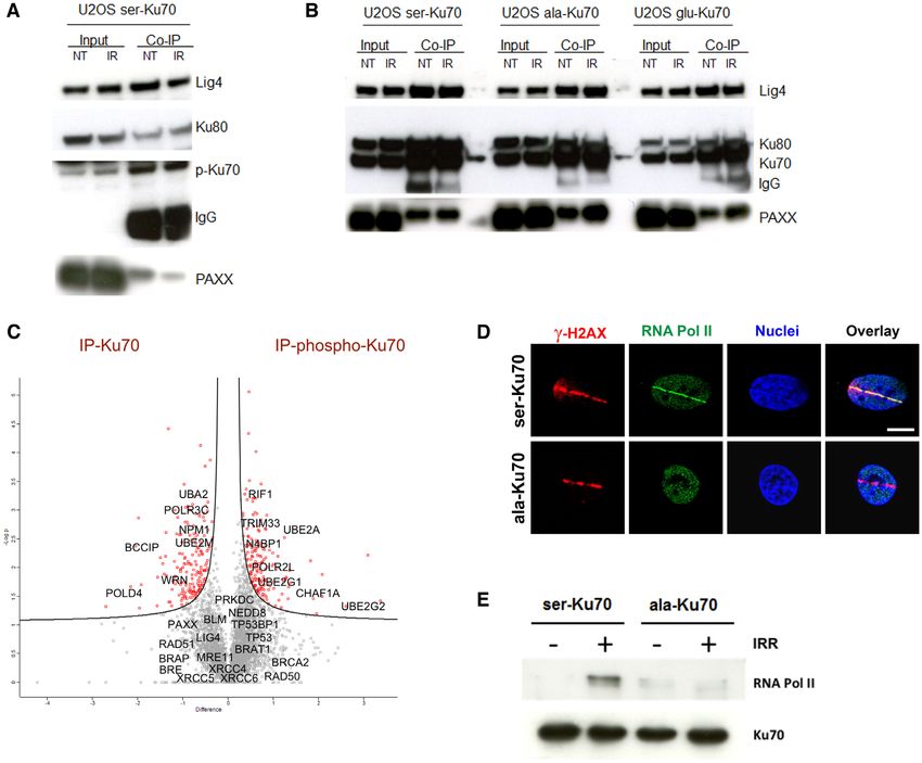
Nucleic Acids Research, 2021, Vol. 49, No. 20 11729
than ala-Ku70, are protected against the joining of as cofactors XLF (Cernunnos) and Paxx (9), follows the
distal DNA ends. Collectively, our results underpin processing step. Thus, the recruitment and activation of all
the essential role of pKu70 in the orchestration of cNHEJ proteins are essentially ensured by the highly abun-
DNA repair execution in living cells and substanti- dant (∼4 × 10⁵ molecules/cell) Ku heterodimer through the
ated the way it paves the maintenance of genome Ku-binding motif (24) and DNA-PKcs kinase activity (25).
End-joining may result in inaccurate repair when non-
stability.
cohesive DNA ends with 5’ or 3’ overhangs should be re-
solved by a search of sequence microhomology or by sim-
GRAPHICAL ABSTRACT ple annealing before ligation, thus resulting in mutations
and/or nucleotide loss (7,21,26). Subset of microhomology-
directed DNA end-joining may be Ku independent [alter-
native NHEJ (aNHEJ)], and it represents powerful mech-
anisms of genome instability (rev. 27). The aNHEJ path-
Downloaded from https://academic.oup.com/nar/article/49/20/11728/6414600 by guest on 25 December 2021
way is dependent on the MRN complex (27–30) and may be
suppressed by the versatile protein WRN (regulating both,
HR and cNHEJ) through the inhibition of Mre11/CtIP-
mediated resection and subsequent large DNA deletions
(31). Notably, the resection-dependent slow kinetics of end-
joining during G1 may be of particular importance for
genome stability, which involves the same initial compo-
nents as HR. Effectively, this kind of end resection in DNA
repair requires CtIP/Brca1 interaction and the exonuclease
Exo1, as well as the endonuclease Artemis (32), which func-
tions in both HR and NHEJ. This process is highly mu-
INTRODUCTION
tagenic, as it results in nucleotide loss and translocation
DNA damage must be repaired rapidly and accurately to events, thus representing a potential source of the illegiti-
prevent genome instability that can lead to cancer devel- mate, disordered cancer genome. Therefore, depending on
opment. Hence, it is not only the cellular DNA repair ca- the DNA-end configuration at sites of damage, the avail-
pacity but also simultaneous repair fidelity that needs to be ability of necessary factors, and the cell cycle phase, the
orchestrated in a time-dependent manner at each DNA le- end-processing step engages common proteins of different
sion. To avoid cell death and ensure legitimate survival, cells DNA repair pathways in tight dynamic interactions. This
have developed multiple DNA repair systems that are spe- concept may also favour interplay and complementarity be-
cific for the type of DNA damage (1). DNA double strand tween the actors of different repair pathways in all phases
breaks (DBs) are the most deleterious damages regarding of the cell cycle, rather than their competition (33). While
possible cell outcomes (2,3). In response to DSBs, the cel- irradiation induces multiple types of DNA DSB ends, an
lular DNA damage response (DDR) is activated through adaptability of DNA repair pathways, especially of the cN-
three phosphatidylinositol-3-related family kinases, ATM HEJ pathway, must be crucial for the final genome outcome.
and DNA-PKcs, which regulate the activities of proteins in- In this context, the engagement of 53BP1 at DNA DSBs ap-
volved in non-homologous end joining (NHEJ), homology- pears to be a critical point of DDR fulfilment. 53BP1 is re-
directed recombination (HR) (3–10), and cell cycle check- cruited by the methylated histone H4-K20 in the vicinity of
points (11–16). In this context, BRCA1 and 53BP1, assisted DNA damage (34). It impedes BRCA1 activity, thus antag-
by auxiliary factors, are two principal deciders for initiating onising HR (35) and promoting cNHEJ through the down-
HR and cNHEJ, respectively (17–20). stream recruitment of RIF1 and shielding complex (36,37).
HR is considered accurate when it relies on homologous In parallel to the inhibition of end resection and the pro-
sister chromatid strands for finding identical DNA tem- tection of DNA ends’ degradation, including distant DNA
plates in late-S/G2 phases of the cell cycle. In contrast, ends (26), 53BP1 create[s] the environment nanodomains,
NHEJ which is active throughout the cell cycle, has re- facilitating access of repair factors to DNA damage (36).
mained for a longtime as an error-prone pathway of DSBs One new member of factors acceding DNA repair is RNA
repair. Nowadays, new data are precising that the accuracy polymerase II (RNA Pol II). Indeed, recruitment of RNA
of NHEJ repair is mainly depending on DNA end sequence Pol II at sites of DNA damage in actively transcribed genes
features (21). Increasing evidences favour cNHEJ as a repair (38), but also elsewhere in the genome (39,40), argued the
system that maintains the stability of the genome. Ku70 and accuracy of cNHEJ repair. Furthermore, these non-coding
Ku80 form the Ku heterodimer which displays high DNA RNAs synthesised at DSBs by RNA Pol II were shown to
end-binding affinity and forms a trimer with DNA-PKcs, be necessary for proper DNA damage response foci forma-
referred to as the DNA-PK holoenzyme, which launches tion, as evidenced by 53BP1 recruitment (40).
cNHEJ (22,23). The next step in NHEJ, which may re- Here, we have addressed the operative mode of action of
quire DNA end processing, involves the Artemis nuclease, phospho-ser27-ser33-Ku70 (named thereafter pKu70). In-
polynucleotide kinase (PNK), DNA polymerases Pol and deed, we have previously identified pKu70 in resistant pri-
Pol , aprataxin, and polynucleotide kinase/phosphatase- mary leukemic cells (41), displaying upregulated cNHEJ
like factor (APLF), MRN, and WRN helicase. The ligation (42) and short telomeres recognised as DSBs (43). Here we
step, which necessitates DNA ligase IV and XRCC4, as well deepen the molecular analysis of the impact of pKu70 on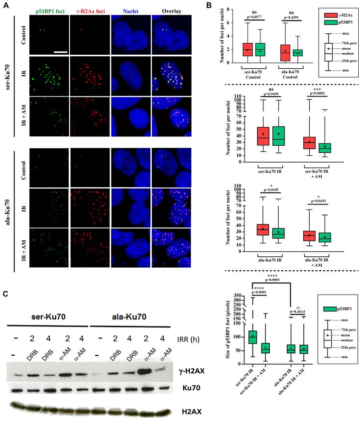
11730 Nucleic Acids Research, 2021, Vol. 49, No. 20
DSB repair; we established that following DNA damage in- Successful transfection was monitored in the different Ku70
duced by irradiation, the final accuracy of cNHEJ is depen- mutants using SDS-PAGE and western blot analysis. Us-
dent on the Ku70 phosphorylation status. Effectively, Ku70 ing the specific monoclonal anti-phospho-S27-Ku70 anti-
phosphorylation occurring at the sites of DNA DSBs ap- body, we showed that there was no expression of endoge-
pears necessary for both the interaction with RNA Pol II nous phospho-Ku70 in ala-Ku70 (non-phosphorylable) or
and the proper formation of phospho-53BP1 foci as well as glu-Ku70 (phosphomimetic)-expressing cells, while the ex-
its release from DNA. We showed that the lack of these last ogenous expression of total Ku70 was the same in all cases.
events compromises chromosomal stability. All stably transfected cell lines were frozen and stored in
liquid nitrogen. eGFP-Ku70 and mEos-Ku70-expressing
MATERIALS AND METHODS cells were freshly transfected before each live cell acquisi-
tion. All cell lines were kept under constant antibiotic se-
Cell lines and cell culture conditions lection pressure. Human POLR2D gene ORF cDNA clone
C-GFPSpark tag and Human POLR2B gene ORF cDNA
Downloaded from https://academic.oup.com/nar/article/49/20/11728/6414600 by guest on 25 December 2021
U2OS cancer cell lines (osteosarcoma) were purchased from
the ATCC. U2OS-HR cells contain the truncated-GFP re- C-GFPSpark tag were purcahssed from Sino Biological.
porter gene and GFP gene harbouring the I-SceI cleavage
site that allows the measurementof homology-directed re- Antibodies
pair (44). The human fibroblast cell line GC92 contains the
CD4 reporter gene and two I-SceI restriction sites allow- Anti-pKu70 was generated in mouse hybridoma cells by
ing distant end-joining assesment (30,45). These cells were BioGenes GmbH (Berlin, Germany); mouse anti-Ku70,
cultured in Dulbecco’s modified Eagle’s medium (DMEM clone N3H10 (ThermoFisher Scientific); rabbit anti-Ku70,
GlutaMAX, Life Technologies, Thermo Scientific, France) ARG57851 (Arigo Biolaboratories); mouse anti-phospho-
supplemented with 10% (v/v) foetal bovine serum and 1x histone H2AX, clone JBW301 (Merck-Millipore), rab-
non-essential amino acids (Life Technologies, Thermo Sci- bit anti phospho-53BP1 (ser1778, #2675, Cell Signalling);
entific, France). The non-cancerous human mammary ep- rabbit anti-ubiquitin (ab137031, Abcam); mouse anti-
ithelium derived HME cell line (46) was a kind gift from the ubiquitin, clone P4D1 (Cell Signalling); mouse anti-
R.A. Weinberg lab (Whitehead Institute for Biomedical Re- ubiquitinated proteins, clone FK2 and clone FK1 (Merck-
search, MIT). The HME cell line medium was additionally Millipore); rat anti-RNA polymerase 2, CTD Ser5ph
supplemented with insulin, epidermal growth factor (EGF), (Cosmo Bio Co. LTD); rabbit anti-phospho RNA Poly-
and hydrocortisone (purchased from Sigma-Aldrich). RNA merase II (S5) (A304-408A-M-2, Bethyl); rabbit anti-
Pol II inhibition by ␣-amanitin and 5,6-dichloro-1-b- NEDD8 (Cell Signalling); and rabbit anti-Rad6 (ab31917,
D-ribofuranosylbenzimidazole (Sigma-Aldrich) was per- Abcam).
formed as previously described (40). To arrest GC92 cells
in the G1 phase, cells were treated with 300 M mimo- ␥-Irradiation, laser micro-irradiation, and real-time live cell
sine (Sigma-Aldrich, USA) overnight, followed by 2 h of imaging
mimosine-free cell culture before irradiation. For the in-
hibition of neddylation, cells were pre-treated with 3 M Gamma irradiation was performed using the ␥ -irradiator
MLN4924 (Interchim) for 1 h as previously described (47). IBL-637 (Cs137). The dose rate was 4.96 Gy/min for
For Rad6 inhibition, cells were treated with 10 M TZ9 all experiments. For laser micro-irradiation and live cell
(Merck-Millipore) for 24 h. imaging experiments, U2OS cells expressing eGFP-Ku70
and mEos2-Ku70 fusion proteins (ser-Ku70Ser, ala-Ku70)
were transfected one week before each experiment. To
Ku70 shRNA/cDNA vectors and cell transfection
stain nuclei, 1 g/ml Hoescht 33342 (Sigma-Aldrich) was
We constructed EBV-based shRNA double-cassette vectors added 30 min before laser irradiation with a 405 nm laser
that enabled simultaneous inhibition of the expression of beam (2 seconds of 10% laser intensity with NIS Photo-
endogenous Ku70 (by shRNA) and episomal expression of Activation software, Nikon) in a line of 5 m in the nu-
different forms of exogenous proteins. Encoded mRNAs are clear region (avoiding nucleoli). The background eGFP-
resistant to shRNA due to codon changes in the cDNA fluorescence intensity was measured before irradiation for
(i.e. codons for aa54 (D) and aa57 (T) were converted from each cell. The cells were followed at intervals of 10 min,
GAT to GAC and from ACA to ACG, respectively) (41). for up to 4 h after irradiation. Data were acquired using
By this approach, the vector serine-Ku70 enables an ex- a video-confocal inverted Nikon A1 microscope with an in-
pression of the wild-type form, alanine-Ku70 of a mutant tegrated chamber that maintained a 5% CO2 atmosphere
form, and glutamic acid-Ku70 of a mutant phosphomimetic and 37◦ C temperature. Data analysis was conducted using
form. The same vectors were used to generate eGFP-Ku70 NIS Elements software (Nikon). Because of the observed
(pcDNA-eGFP, Addgene, UK) and mEos-Ku70 (mEos-2 interference of fluorescences, for DNA damage induction
pcDNA, Addgene, UK) fusion proteins. Cell transfection and subsequent colocalisation analysis of RNA Pol II and
for all vectors was performed on the same day using the ␥ -H2AX, we have irradiated cells by Chameleon Vision II
same JetPrime DNA transfection kit (Polyplus, Transfec- (Coherent) biphotonic laser at 800 nm and 30% of laser
tion SA, France) and antibiotic selection conditions (hy- power. Images were acqisited and analysed by LSM 780
gromycin or puromycin). As a control for antibiotic selec- confocal microscopy (Zeiss). For cell localisation analysis
tion, non-transfected cells were treated with antibiotics, and of Ku70, PALM acquisitions were performed with an N-
cell death was monitored over time by light microscopy. STORM (Nikon) system and an SR Apo TIRF 100x (1.49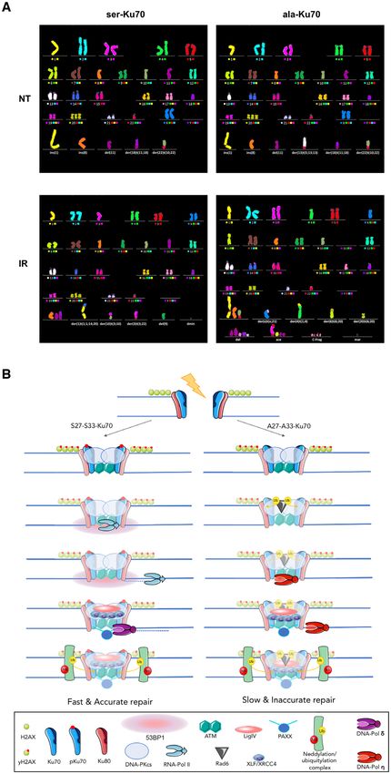
Nucleic Acids Research, 2021, Vol. 49, No. 20 11731
numerical aperture) oil objective. Images were obtained was used for the detection and scoring of foci in p53BP1 and
using a TIRF inverted Nikon Eclipse Ti-E microscope ␥ -H2AX images. Representation of the data as boxplot was
equipped with a quad band emission filter (450/60–525/50– performed using GraphPad Prism 7. For statistical analy-
605/50–730/120, Chroma) coupled to an EMCCD cam- sis, to compare the number of ␥ -H2AX and p53BP1 foci in
era (iXon DU897, Andor). Fixed cells expressing mEos2 each condition, a Mann-Whitney rank test based on at least
constructs were used with sequential activation at 405 nm 100 observations was performed. Western blot analysis was
(Cube laser, 100 mW, Coherent), followed by excitation at performed as previously described (41).
561 nm (Sapphir laser, 150 mW, Coherent). NIS-elements
AR software (Nikon, v. 4.30.01) and the STORM analy-
Chromatin-binding assay
sis module (Nikon) were used to control the system and
perform molecule detection, respectively. To prevent axial Cells were seeded at approximately 70% confluency. For
drift, we used a perfect focus system (Nikon); to correct protein extraction, pre-extraction (PEB) buffer [10 mM
for any lateral or axial drift during acquisition, we applied PIPES (pH 7.0), 100 mM NaCl, 300 mM sucrose, 3 mM
Downloaded from https://academic.oup.com/nar/article/49/20/11728/6414600 by guest on 25 December 2021
the auto-correlation algorithm of NIS-elements AR soft- MgCl, 0.7% (vol/vol) Triton X-100, H2 O] was prepared
ware (Nikon, v. 4.30.01). Local density analysis was per- on ice. Cells were washed three times with PBS without
formed using SR-Tesseler software. The local density of the Ca2+/ Mg2+ and then incubated in PEB-R buffer (PEB
mEos2 molecules was calculated using Voronoi tessellation buffer, 7% RNAse A) for 3 min. The supernatant was col-
and then displayed according to a colour scale that was nor- lected as Fraction 1. The same procedure was repeated to
malised to the mean molecule density of each nucleus. To obtain Fraction 2. After two PBS washes, Laemmli buffer
examine the kinetics of mEos2-Ku recruitment after laser (2x) [100 mM Tris–HCl (pH 6.8), 10% SDS, 20% glycerol,
irradiation, time-lapse experiments were performed on an 100 mM DTT, 1% bromophenol blue, H2 O] was added to
inverted Nikon Ti Eclipse Eclipse-E microscope coupled obtain Fraction 3 (chromatin fraction). DNA was then di-
with a spinning disk (Yokogawa, CSU-X1-A1), a 60× plan gested with benzonase (Merck Millipore). To prepare the
apo objective (Nikon, NA 1.49, oil immersion) and an sC- samples for SDS-PAGE, Laemmli 2× was added to Frac-
MOS camera (Photometrics, Prime 95B). Cells express- tions 1 and 2, and all samples were heated for 5 min at 95◦ C.
ing mEos2-Ku constructs grown in 35 mm Ibidi -dishes
were imaged in a controlled atmosphere (37◦ C, 5% CO2 ).
Anti-pKu70/anti-Ku70 protein immunopurification
Before imaging, the cells were incubated for 30 min with
1 g/ml Hoechst 33342. Hoechst fluorescence was detected Proteins from at least 15 × 106 cells were extracted on ice
with 405 nm excitation (Vortran, 100 mW laser) and a for 1 h in hypertonic buffer containing 50 mM NaF, 450
450/50 bandpass filter (Chroma). mEos2 was excited at ei- mM NaCl, 20 mM HEPES, 0.2 mM EDTA, 0.5 mM DTT,
ther 488 nm (Vortran, 150 mW laser) or 561 nm (Coherent, 5 mM iodoacetamide, and a complete cocktail (Roche Diag-
100 mW laser), and a 525/45 bandpass filter (Semrock) or a nostic) inhibitor of phosphatases (100 l extraction buffer
607/36 bandpass filter (Semrock) was used to detect its na- per 107 cells). The cell lysates were centrifuged at 67 000 rpm
tive or photoconverted form, respectively. Irradiation was for 1 h at 4◦ C. Next, 10% glycerol and -mercaptoethanol
performed by scanning a line of 10 m with the 405 nm (1:1250) were added to the supernatant, which was divided
laser for 60 s. For each field of view, a time-lapse series was into three parts (two parts for immunoprecipitation and one
recorded with a pre-irradiation sequence of 60 s and a post- for the input control). To equilibrate the salt content, equi-
irradiation sequence of 600 seconds, acquiring an image ev- libration buffer (EQ-buffer) [10 mM KCl, 10 mM MgCl,
ery 20 s. A perfect focus system (Nikon) was used to keep 20 mM HEPES, 2.5 mM EDTA, 0.5 mM DTT, 5 mM
the focus constant during the time-lapse series; the whole iodoacetamide, complete cocktail 1×, 10% (vol/vol) glyc-
system was driven by Metamorph (version 7.7, Molecular erol] was added at a ratio of 1:3, and the protein con-
Devices). tent was determined (Bradford method). Dynabeads (BE-
M01/03, EMD Millipore) (1 l per 10 g total protein)
were washed twice with PBS without Ca2+ /Ca2+ , and the
␥-H2AX and phospho-ser1778-53BP1 foci assessments
cell lysate was added to the beads. To obtain a total volume
Cells were grown on 8-well covered slides (Millicell EZ of 500 l, EQ buffer was added. Antibodies against Ku70
Slides, Merck-Millipore) for 2 days until 70–80% conflu- (NeoMarkers, Thermo-Fisher) and p-Ku70 (42) were mixed
ency. The slides were irradiated at 2 or 4 Gy and left in the with the lysate/Dynabead mixture at a concentration of 1%
incubator for the indicated times (0.5–24 h). After anti-␥ - of the total protein content and incubated overnight at 4◦ C
H2AX and/or p53BP1 labelling, data acquisition was per- with agitation. The supernatants were discarded, and the
formed using a microscope (Zeiss, Imager Z2) as previously beads were washed three times in PBS before adding EQ
described (41). Each analysis was performed on at least 100 buffer. Laemmli-buffer (5×) (0.255 M Tris–HCl (pH 6.8),
cells, and at least 10 images of each condition were anal- 50% glycerol, 5% SDS, 0.05% bromophenol blue, 0.1 M
ysed. Confocal microscopy optical slice sections of 8–20 m DTT) was added, and the samples were heated at 95◦ C
were recorded from the apical to the basal pole of the cells, for 10 min. The immunoprecipitated proteins were resolved
with each acquisition containing 26 stacks. Images were pre- by SDS-PAGE for orbital-based shotgun mass spectrome-
pared and stacked with ImageJ software (Bethesda, MD) try according to the standardised protocol (Proteomic Cen-
(48) by using the stacks tool. Then, TIFF images were con- ter, Rotterdam). Analysis was performed with Perseus soft-
verted to 8 bits before performing ␥ -H2AX and p53BP1 ware by varying FDR and S0 simultaneously. The S0 value
foci counts. Cell Profiler software (Cambridge, MA) (49) is a statistical factor based on a paper referred to by the
11732 Nucleic Acids Research, 2021, Vol. 49, No. 20
Max Quant developers and the t-test performed in SAM idated Ku70 vectors that enabled the expression of the pho-
(from the microarray field). We chose values of S0 = 0.1 and toconvertible molecule mEos2 fused to the N-terminal re-
FDR = 0.05 in the present study, which are the commonly gion of different forms of Ku70. These vectors were used
used values for this type of analysis. to characterise the nuclear repartitioning of different forms
of Ku70 in cells under non-stressed conditions. Figure 1B
Anti-eGFP-Ku70 immunoprecipitation shows that ser-Ku70 (wild-type) filled the nucleolar struc-
ture and densely accumulated in a few nuclear subregions,
eGFP-ser-Ku70 and eGFP-ala-Ku70 fusion proteins were while structural homologue glu-Ku70 was completely ex-
immunoprecipitated using the ChromoTek GFP-Trap kit cluded from the nucleoli and showed homogenous nuclear
according to the supplier’s protocol, except for the cell lysis localisation. Non-phosphorylable ala-Ku70 cells exhibited
step. Briefly, cells were lysed in the buffer specified above (see two different patterns: nucleolar with less dense regions in
anti-Ku70 immunoprecipitation) supplemented with ben- most cases or, less frequently, empty nucleoli with homoge-
zoase (15 U/assay). The final protein extracts at 1 mg per nous nuclear localisation (as observed for glu-Ku70). As
Downloaded from https://academic.oup.com/nar/article/49/20/11728/6414600 by guest on 25 December 2021
assay were adjusted to 0.5 M NaCl. Equilibrated GFPTrap a control, Supplemental Figure S1 shows enlarged micro-
beads (25 l) were added to the lysates, followed by incu- scopic fields of cells expressing eGFP-Ku70 fusion proteins,
bation for 1 h at 4◦ C. Magnetically separated beads were supporting the notion that the mEos2 fusion ligand does
washed three times in lysis buffer adjusted to 1 M NaCl. The not affect Ku70 cellular localisation, which was also demon-
immunoprecipitated proteins were eluted from the beads in strated using eGFP-Ku70 fusion proteins via a laser micro-
100 l 2× SDS–Laemmli buffer by boiling for 10 min at irradiation approach and live cell imaging (see below).
95◦ C. The supernatants were used for SDS-PAGE followed Next, we sought to determine whether pKu70 may con-
by western blot analysis. trol clonogenic survival and cell growth following genotoxic
stress. Effectively, both cell growth and clonogenic survival
Multi-FISH analysis of chromosomal translocations. Hu- were affected in cells expressing ala-Ku70 or glu-Ku70 (Sup-
man mammary epithelial cells, which were also used in plementarl Figure 2). Further, we addressed if the phospho-
the proteomic approach for the analysis of the Ku70 in- rylation of Ku70 may occur when it was linked to DNA-
teractome, were used to assess chromosomal anomalies ends at the sites of DNA damage. We modified above vec-
following irradiation. Cells were irradiated at 2 Gy, and tors, enabling the expression of mEos2-Ku70 fusion pro-
metaphase cells were prepared after 24 h as described previ- teins, by introducing cDNA Ku70-mut6E (kind gift by S.
ously (50). Briefly, the slides were pre-treated with RNAse Britton). In this vector, a positive charge in the Ku70 cav-
A (120 l per slide; stock solution: 10 g.ml–1 ) in 2× SSC ity domain was reversed by introducing six glutamic acids,
buffer (AM9763, ThermoFisher Scientific) for 45 min at giving rise to a defective Ku70 protein to bind DNA ends
37◦ C to eliminate RNA. To remove the remaining cyto- (51). Therefore, we generated a vector encoding the mEos2-
plasm, pre-treatment with pepsin (P6887, Sigma Aldrich) Ku70-mut6E fusion protein. As shown in the lower panels
was performed. The slides were incubated for 30 min at of Figure 1C, a, this form was unable to co-localise with
37◦ C in 0.01 M HCl (pH 2) with 2 g/ml pepsin. The laser-induced DNA damage compared to mEos2-Ku70wt
slides were washed with PBS without Ca2+ /Mg2+ (2×) (upper panel). In parallel, western blots, shown in Figure
and then with PBS with Ca2+ /Mg2+ for 5 min. To fix 1C, b, validated that mEos2-Ku70-mut6E was not phospho-
pepsin-treated metaphases, the slides were incubated in 1% rylated following irradiation (missing band at ∼95 kDa).
formaldehyde/PBS with Ca2+ /Mg2+ for 10 min at RT. The Importantly, this form was highly toxic to cells (even in
slides were then washed with PBS without Ca2+ /Mg2+ for the absence of irradiation stress), impeding the complete
5 min. Dehydration was performed in three steps with 70, hygromycin selection of transfected cells. This toxicity ex-
90 and 100% ethanol at RT for 3 min, and the slides were plains why there were two forms of Ku70, endogenous (wt)
left to dry for 2–3 min. mFISH probe denaturation and and exogenous, fused to mEos2 (corresponding to the ∼95
hybridisation were performed according to the manufac- kDa band). Nevertheless, this construction allowed us to
turer’s protocol (24XCyte, D-0125-120-DI, MetaSystems). demonstrate that this form cannot undergo phosphoryla-
Metaphase cells were mounted with aqueous Flouromount- tion due to the absence of DNA binding, in contrast to the
G (F4680, Sigma Aldrich). Data were acquired using a Zeiss mEos2-Ku70 fusion protein.
Imager Z2 microscope. Data analysis was performed with In parallel, demonstrating that pKu70 colocalized with
ISIS software (Metasystem). DNA damage sites (Supplemental Figure S3, Supplemental
Figure S4A), we have established an accelerated dynamic of
RESULTS disappearance of the ␥ -H2AX foci in both unsynchronised
U2OS cells and in G1-arrested SV40-transformed human fi-
Phosphorylation of Ku70 occurs at the sites of DNA damage
broblast GC92 (30), expressing ser-Ku70 as compared with
and fosters its dissociation
cells expressing ala-Ku70 (Supplemental Figure S4B–F).
To establish how pKu70 may regulate the response to DNA We next performed real-time live-cell imaging to monitor
damage, we decided to track it in living cells expressing three the dynamics of Ku70 recruitment and release at DSBs. For
forms of Ku70 (ser-, ala- or glu-Ku70, Figure 1A), after this purpose, we transfected cells with vectors, enabling the
DNA damage induction. At first instance, to explore the expression of eGFP-Ku70 fusion proteins. DSBs were in-
cell localisation of different forms of Ku70 at the molec- duced by micro-irradiation (405 nm UV in the presence
ular scale, we performed PALM (photoactivated localisa- of 1 g ml–1 Hoechst 33342) under the same experimen-
tion microscopy) experiments. We introduced cDNA in val- tal conditions as applied in the experiments done for as-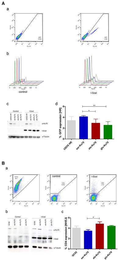
Nucleic Acids Research, 2021, Vol. 49, No. 20 11733
Downloaded from https://academic.oup.com/nar/article/49/20/11728/6414600 by guest on 25 December 2021
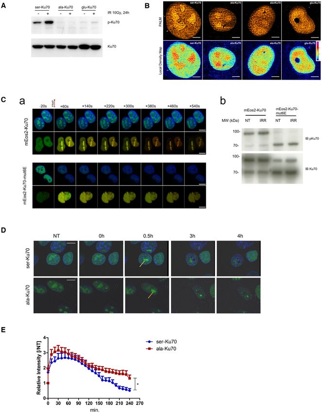
Figure 1. Ser-Ku70 preferentially localises in nucleoli and translocates to the sites of DNA damage where phosphorylation occurs; delayed kinetics of
ala-Ku70 dissociation from the sites of damage. (A) Cells used in this study were transfected by the shRNA vectors double-cassette, and the extinction
of endogenous Ku70 and re-expression of vectors-encoded Ku70 was verified by Western blots. (B, upper panel), mEos2 cDNA was fused into the above
vectors to generate the mEos2-ser-Ku70, mEos2-ala-Ku70, or mEos2-glu-Ku70 vectors, and after 48 h, florescence was examined by photoactivated lo-
calisation microscopy (PALM). Scale bar = 5 m. (B, Lower panel), Local density map analysis was also performed on PALM data to characterise the
different patterns of the Ku70 nuclear distribution. (C, a) Localisation of mEos2-Ku70 (upper panels) and mEos2-Ku70-mut6E (lower panels) to DSBs
after laser irradiation. Time-lapse observation of Hoechst (blue)-labelled cells expressing Ku70 or Ku70-mut6E fused to mEos2. Both native (green) and
photoconverted (red) forms of mEos2 are recruited to the DSBs after 405 nm laser irradiation in the case of cells expressing mEos2-Ku70, but cells ex-
pressing mEos2-Ku70-mut6E exhibited no recruitment to the DSBs. Scale bar: 10 m (b) Western blot analysis of protein extracts from cells expressing
mEos2-Ku70 or mEos2-Ku70-mut6E. After SDS-PAGE, the membranes were probed with an anti-phospho-Ku70 antibody. An anti-total Ku70 antibody
was used as a control. (D) Kinetics of the dissociation of Ku70 from the sites of DNA DSBs. Representative images of the dissociation kinetics in eGFP-ser-
Ku70- and eGFP-ala-Ku70-expressing cells. Images depict cells before (NT), immediately after (t0), and at three time points following micro-irradiation
(405 nm). Yellow arrows indicate the damage sites. Scale bar: 10 M. (E) Relative intensity fold change based on the initial fluorescence intensity in NT
cells. n = 20 (ser-Ku70); n = 27 (ala-Ku70). Unpaired t-test, *P < 0.01 .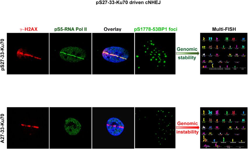
11734 Nucleic Acids Research, 2021, Vol. 49, No. 20
Downloaded from https://academic.oup.com/nar/article/49/20/11728/6414600 by guest on 25 December 2021
Figure 2. Phospho-Ku70 causes rapid dissociation from sites of DNA damage; inhibition of neddylation compromises both pKu70 release and DNA
repair following laser micro-irradiation. (A) Ku70 release from DNA damage sites after the inhibition of neddylation by MLN4924 (3 M, 1 h), followed
by laser micro-irradiation as in Figure 1C. Relative fluorescence intensity fold change (established as in Figure 1D). Unpaired t-test, NS = non-significant.
(B) Western blot of ␥ -H2AX after irradiation alone (a) or after combined cell treatment (b) with MLN4924 (3 M, 1 h), followed by irradiation (4Gy).
Anti-Ku70 (clone N3H10) was used as the loading control. (C, a) Western blot analyses of the neddylation patterns of untreated, irradiated (4 Gy), or
MLN4924 (3 M, 1 h) pre-treated and irradiated (4Gy) cells expressing eGFP-ser- or -ala-Ku70-fusion proteins. Cells were lysed 2 h post-irradiation. After
SDS-PAGE, the membranes were probed with anti-NEDD8 or anti-Ku70 antibodies. The band below the 100 KDa-MW marker with decreased intensity
upon MLN4924 treatment indicates decreased neddylation of cullin. (b) The ubiquitylation pattern analysed in protein extracts from cells expressing
eGFP-Ku70-fusion proteins. Cells expressing eGFP-ser-Ku70 or eGFP-ala-Ku70 were irradiated with or without MLN4924 or TZ9 pre-treatment and
lysed 2 h post-irradiation. Protein extracts were immunoprecitpitated by using a GFP-Trap MA kit (Chromotek). After SDS-PAGE, the membranes were
probed with an anti-ubiquitin, anti-Ku70, or anti-GFP antibody.Nucleic Acids Research, 2021, Vol. 49, No. 20 11735
sessing phospho-Ku70 co-localisation with ␥ -H2AX. After (UBE2M) and NEDD4-binding protein 1 (N4BP1), as
micro-irradiation, fluorescence signals were followed for up well as the ubiquitin-conjugating enzyme UBE2G2 and
to 4 h, and images were obtained every 10 min (Figure 1D the ubiquitin-ligase protein TRIM33, are new partners of
and Supplemental ‘ser-Ku70’ and ‘ala-Ku70’ movies). The Ku70, according to its phosphorylation status (see below
recruitment kinetics were rapid and quite similar, indepen- Figure 3D). Notably, proteomic data also indicated an
dent of introduced mutations in Ku70 cDNA vectors. Sup- interaction between pKu70 and UBE2A, also known as
plemental Figure S5 shows the micro-irradiation-induced the Rad6A ubiquitin-conjugating protein. Therefore, we
laser strips at 2- and 12-s post-irradiation in cells express- tested possible neddylation-independent ubiquitylation of
ing ser-Ku70 or glu-Ku70. eGFP-ser-Ku70- and ala-Ku70- Ku70 through Rad6. We treated cells expressing eGFP-ser-
expressing cells exhibited a maximal fluorescence signal be- Ku70 and eGFP-ala-Ku70 in parallel with the TZ9 Rad6-
tween 30 and 50 min after irradiation. The quantified in- inhibitor or MLN4924 inhibitor. Immunopurification was
tensities indicated that ala-Ku70 provided a stronger sig- performed by using the ChromoTek GFP-Trap kit. Figure
nal at DSBs (i.e. 3.1-fold induction for ala-Ku70, whereas 2C, b shows that MLN4924 inhibition affected the ubiq-
Downloaded from https://academic.oup.com/nar/article/49/20/11728/6414600 by guest on 25 December 2021
the maximal recruitment was 2.6-fold for ser-Ku70; Fig- uitination pattern of both the ser- and ala-Ku70 forms. In
ure 1E; however, this difference is not statistically signifi- contrast, Rad6 inhibition seems to affect the ubiquitylation
cant). The dissociation of eGFP-serKu70 from DNA dam- of the ala-Ku70 form only. This may raise the possibility
age sites occurred much faster and reached 50% at 150 min. that in the absence of any inhibition, Rad6 may exert an ef-
The basal levels in the ser-Ku70-expressing cells occurred fect in cells expressing ala-Ku70, but not in cells expressing
between 190 and 200 min. In contrast, the level in ala-Ku70 pKu70, since this should be dependent on completed repair
cells remained over 50% after 220 min and did not reach complex formation.
basal levels within 240 min. These data indicated that the
phosphorylation of Ku70 favoured Ku70 release from sites
pKu70 is essential for the recruitment of RNA Pol II
of DNA damage and might implicate faster completion of
the NHEJ process in these cells compared to ala-Ku70- Following the above kinetic studies, we performed co-
expressing cells; these data are in accordance with data re- immunopurification assays to verify that pKu70 makes part
garding ␥ -H2AX foci (Supplemental Figure S4). of the cNHEJ complex. Using monoclonal anti-phospho-
Ku70 and anti-Ku70 antibodies for immunoprecipitation,
Figure 3A a shows the interactions of phospho-Ku70 with
Inhibition of neddylation-dependent ubiquitylation impairs
Ku80, Ligase 4, and PAXX, validating that phospho-Ku70
pKu70 dissociation from DSBs
is one component of the cNHEJ complex required to repair
Neddylation has been reported to be an important prereq- DNA DSBs. However, ala-Ku70 and glu-Ku70 interacted
uisite for Ku70 ubiquitylation and subsequent Ku70 release equally with Ku80, Ligase 4, and PAXX (Figure 3B, where
from DNA mediated by VCP/p97 3A + ATPase (47,52). Ku70 antibodies recognaising ala- and glu-Ku70 forms,
Therefore, we asked whether the inhibition of neddylation were used for protein immunoprecipitation). Furthermore,
by MLN4924 might also impair the release of phospho- we applied a proteomics approach to analyse the proteins
Ku70 from the repair complex. Figure 2A shows the dis- immunopurified with the same antibodies as in above co-
sociation kinetics of eGFP-ser-Ku70 and eGFP-ala-Ku70 immunoprecipitation assays, anti-phospho-ser27-Ku70 or
after treating cells with 3 M MLN4924 for 1 h prior to anti-Ku70 (clone N3H10, NeoMarkers) antibodies. This
micro-irradiation. The maximal recruitment of ser-Ku70 approach allowed us to validate that there was no significant
(6.4-fold) and ala-Ku70 (6.1-fold) after MLN 4924 treat- difference between the binding of Ku70 or phospho-Ku70
ment was ∼2-fold higher in comparison to the conditions to the NHEJ core factors XRCC5 (Ku80), XRCC6 (Ku70),
without MLN 4924 (Figure 2A). The dissociation of ser- PRKDC (DNA- PKcs), LIG4 (Ligase 4) or XRCC4 (Figure
Ku70 was severely impaired, with only 20–25% of serKu70 3B). These data indicated that phospho-Ku70 is not neces-
having dissociated at 240 min, showing kinetics similar to sary for the formation and recruitment of cNHEJ factors to
that of ala-Ku70. These data indicated that ubiquitylation the break site but that it is a part of the cNHEJ complex and
of Ku70 might act downstream of Ku70 phosphorylation interacts with key partners. However, this interactome ap-
to induce Ku70 release. Figure 2B, b shows the efficacy of proach indicated the presence of other factors (most of them
MLN4924 inhibition on ␥ -H2AX protein levels at indicated have been already reported in the literature to interact with
times post-treatment. The level was sustained at 4 and 6 Ku70). In addition the abowe results showing that Ku70
h after combined treatment with MLN4924 and irradia- mutated in its DNA-binding domain was not phosphory-
tion (as compared to the irradiation treatment alone; Fig- lated, the observation that the core nucleolar factor nucle-
ure 2B, a), in ser-Ku70-expressing cells. Cells expressing ala- ophosmin (NPM1) interacted less strongly with pKu70 fur-
Ku70 exhibited similar protein levels to ␥ -H2AX, regard- ther supported that phosphorylation occurs outside of nu-
less of MLN4924 treatment, further supporting the neces- cleoli. Known auxiliary factors affecting cNHEJ were also
sity of neddylation-dependent ubiquitylation (Figure 2C, b) found to be interactors with unphosphorylated (WRN and
prior to pKu70 release from DSBs. Figure 2C,a shows the DNA-Pol␦ subunit 4) or phosphorylated Ku70 (CHAF1,
efficacy of MLN4924 inhibition of neddylation pattern. RIF1; Figure 3B).
In parallel, we performed interactome approach (see be- Of particular interest are the factors that may affect the fi-
low) which revealed that several factors of the ubiquitin delity of DNA repair (38–40), such as DNA-directed RNA
system interact with Ku70 depending on its phosphoryla- polymerase I, II, and III subunit RPABC5, encoded by the
tion status. Thus, the NEDD8-conjugating enzyme Ubc12 POLR2L gene, which was indicated to be a specific part-11736 Nucleic Acids Research, 2021, Vol. 49, No. 20
Downloaded from https://academic.oup.com/nar/article/49/20/11728/6414600 by guest on 25 December 2021
Figure 3. Phospho-Ku70 and mutant forms of Ku70 interact with core components of cNHEJ but only pKu70 recruits RNA Pol II in the repair complex.
(A) Representative western blot of co-immunoprecipitation using anti-phospho-Ku70 shows that pKu70 interacts with the Ku80, ligase 4 and Paxx. (B)
Similarly, using the anti-Ku70 antibody shows that all Ku70 forms (wild-type and mutants) interact with Ku80, ligase 4 and Paxx. The membranes were
cut according to the corresponding molecular weights and probed with antibodies as previously described (see the Materials and Methods section). (C)
Whole-cell protein extracts of transfected HME cells (46), expressing ser-Ku70 at 2 h post-irradiation (2 Gy), were co-immunoprecitpitated with anti-Ku70
or anti-pKu70 antibodies, resolved by SDS-PAGE and proteolyzed with trypsin. Peptides were analysed using a label-free-quantification (LFQ) approach
with an orbitrap-based mass spectrometry analyser. The analysis was performed with Perseus software by varying FDR and S0 simultaneously. We chose
values of S0 = 0.1 and FDR = 0.05, as the most commonly used values for this type of analysis. The corresponding genes, indicated by grey squares,
are considered to exhibit non-significant differences in their affinity toward Ku70 and/or pKu70, whereas those in red squares are presumed to be more
specific partners of total Ku70 or pKu70. (D) RNA Pol II localizes with ␥ -H2AX at laser microirradiation-induced DNA damage only in cells expressing
phosphorylable ser-Ku70. Cells esxpressing ser-Ku70 or ala-Ku70 were irradiated by laser Chaameleon Vision II system and 1 min postirradiation cells
were fixed and probed with anti-␥ -H2AX (red) and anti-phospho-ser5-RNA Pol II (green). Hoechst 33342 was used to stain chromatin DNA. Scale
bar = 5 m. (E) RNA Pol II is recruited by pKu70 in the repair complex. Western blot analysis of immunopurified proteins from cells expressing ser-Ku70
or ala-Ku70. Cells were irradiated at 4Gy or left unirradiated, and following 30 min of post-irradiation culture, cells were lysed. The protein extracts were
immunopurified by using a monoclonal anti-Ku70 antibody (clone N3H10) and magnetic beads coated with anti-mouse IgG (Estapor, Merck-Millipore).
After SDS-PAGE, the membranes were probed with anti-RNA Pol II or anti-Ku70 antibodies.
ner of phospho-Ku70. While the other subunits specific of whether transcriptionally active RNA Pol II localised at
RNA Pol II have been found (in less significant manner) to microirradiation-induced DNA damage. Because of the en-
be partners of pKu70 (data not shown), we have addressed countered interferences of fluorescences by laser at 405nm
whether RNA Pol II localizes at laser microirradiation- in presence of Hoechst 33342 as a photosensibilising agent
induced DNA DSBs. Thus, we have expressed POLR2B- and subsequent Alexaflour labellings, we have used the
eGFP and POLR2D-eGFP vectors in transient transfec- biphotonic laser Chameleon Vision II at 800nm to induce
tion assay. As shown in Supplemental Figure S7A both DNA DSBs without need of Hoechst treatment. At first, by
of these subunits localazed at microirradiation stretches using cells expressing eGFP-ser-Ku70 and eGFP-ala-Ku70,
in 10 seconds following irradiation. Next, we addressed we established optimal laser power of 30% to recruit Ku70Nucleic Acids Research, 2021, Vol. 49, No. 20 11737
at DNA damage (Supplemental Figure S7B). Following values of 24.5 ␥ -H2AX vs 19,5 p53BP1 foci, *P = 0.024).
irradiation of cells expressing ser-Ku70 or ala-Ku70 in these Untreated cells displayed no significant differences in foci
conditions, cells were fixed at 1min or 10 min postirradia- number, regardless of the Ku70 expression status. Differ-
tion and probed with anti-␥ -H2AX and anti phospho-S5- ences in cell cycle phases should be excluded as a possible
RNA Pol II antibodies. The results shown in Figure 3D and bias in above discrepancies, as all cell types exhibited simi-
Supplemental Figure S7C clearly evidenced that the tran- lar cell cycle profiles post-irradiation (Supplemental Figure
scriptionally active RNA Pol II was recruited to DNA dam- S6). In addition to their altered number, the size of p53BP1
age site only in cells expressing phosphorylable ser-Ku70 foci also decreased in cells expressing ala-Ku70 in statis-
but not in cells expressing ala-Ku70. This was further vali- tically significant maner; ***P = 0.0001, median values
datyed by co-immunoprecipitation assays followed by west- of 100 versus 52 for ser-Ku70- versus ala-Ku70-expressing
ern blot analysis of protein fractions from cells expressing cells, respectively. Of note, the p53BP1 foci size decreased in
ser-Ku70 or ala-Ku70 before and after ␥ -irradiation stress. ser-Ku70-expressing cells following irradiation and aman-
As shown in Figure 3E, an interaction between Ku70 and itin treatments (median values of 100 for irradiation only
Downloaded from https://academic.oup.com/nar/article/49/20/11728/6414600 by guest on 25 December 2021
RNA Pol II was observed only after irradiation stress in versus 55 after double treatment; ****P = 0.0001). This
extracts from U2OS cells expressing phosphorylable ser- was not observed in ala-Ku70-expressing cells (P = 0.3614)
Ku70. Indeed, unphosphorylated ser-Ku70 did not inter- (Figure 4B).
act with RNA Pol II before irradiation. This interaction Following the above results evidencing a defect in p53BP1
was also completely abolished in cells expressing the ala- foci formation in ala-Ku70-expressing cells, we further
Ku70 form, regardless of irradiation stress. These results sought to validate the involvement of RNA Pol II as the spe-
indicated that even though the above experiments showed cific partner of pKu70 in dynamics of the ␥ -H2AX level in
ala-Ku70 recruitment at the site of DNA damage and inter- two types of cells. We opted to perform western blots to val-
action with core elements of cNHEJ quasi-identical to those idate the dynamic of ␥ -H2AX protein level because the ki-
of ser-Ku70, the final repair complex was not the same for netics were similar to the number of foci by the immunoflu-
these two forms of Ku70. orescence approach. The timing was based on the results
presented in Supplemental Figure S4, showing the signif-
icant differences in the remaining ␥ -H2AX foci between
Formation of phospho-ser1778-53BP1 foci depends on the
ser- and ala-Ku70-expressing cells at 4 h post-irradiation.
phosphorylation status of Ku70
Therefore, we again inhibited RNA Pol II by exposing cells
Small non-coding RNAs, generated at the DSBs (called to ␣-amanitin, a specific inhibitor, and 5,6-dichloro-1-b-D-
DDRNAs) by RNA Pol II, have been reported as criti- ribofuranosylbenzimidazole (DRB), an inhibitor of RNA
cal factors involved for proper activation of DDR. This Pol II elongation (40). Cells pre-treated by ␣-amanitin (50
involvement was evidenced by a defect in the formation g ml–1 ) for 2 h before irradiation slowed down the ki-
of 53BP1 foci after ionising irradiation when RNA Pol netics of DNA damage repair only in ser-Ku70-expressing
II was inhibited (40). Proteomic data indicated no signif- cells. Effectively, Figure 4C shows a persistence of the ␥ -
icant difference between 53BP1’s affinity towards Ku70- H2AX protein level at 4 h post-irradiation (4 Gy) in cells ex-
or pKu70-antibodies. However, RIF1, a downstream ef- pressing ser-Ku70, whereas this did not occur in ala-Ku70-
fector of 53BP1, was found in the group of proteins im- expressing cells. The lower concentrations of ␣-amanitin (25
munopurified by the anti-pKu70 antibody (Figure 3C). We g ml–1 , not shown), as well as those of DRB, had no ef-
sought to determine whether a defect in the formation of fect on the level of the protein ␥ -H2AX and DNA repair
53BP1 foci following irradiation stress could be discrimina- kinetics in two types of cells. Therefore, these results fur-
tory between cells expressing ser-Ku70 or ala-Ku70. We per- ther strengthened the requirement of the phosphorylation
formed double immunofluorescence staining of ␥ -H2AX of Ku70 in the proper assembly of the repair complex.
and p53BP1. The results shown in Figure 4A and B val-
idated this hypothesis. Indeed, while ser-Ku70-expressing
Phospho-Ku70 protects against distal DNA DSB end junc-
cells displayed no significant differences between ␥ -H2AX
tions
and p53BP1 number of foci following 1 h of 2 Gy post-
irradiation culture (median values of 37 versus 35 foci, re- Considering that the joining of distant ends inevitably re-
spectively; P = 0.997 ns), cells expressing ala-Ku70 dis- sults in genomic rearrangements and the fact that 53BP1
played a significant discrepancy between the number of foci can affect the end joining of distant DNA DSBs (26), we
of ␥ -H2AX and p53BP1 (median values of 33.5 versus 26.5, used two specifically dedicated cell models, U2OS-HR and
respectively; **P = 0.009. In agreement with the involve- GC92 cells bearing intrachromosomal substrate monitor-
ment of DDRNAs in 53BP1 foci formation, we performed ing HR or end-joining activities, respectively This approach
assays for the inhibition of RNA Pol II by ␣-amanitin (50 allowed the targeting into the intrachromosomal substrates
M, 2 h) prior to irradiation treatment (2Gy) and per- of DSBs by the I-SceI meganuclease. The DR-GFP sub-
formed the same immunostainings. In cells expressing ser- strate (44) contains a single I-SceI site inside of the GFP
Ku70, the number of p53BP1 foci severely decreased com- gene located downstream of the truncated GFP gene, while
pared to the number of ␥ -H2AX foci (Figure 4, median val- the NHEJ CD4 gene substrate (26,30,45) is located down-
ues 21 versus 30; ***P < 0.0001). Considering the defect in stream of an insertion of 3.2 Kbp between the 5 and 3 I-
p53BP1 foci formation in the irradiation only treated cells SceI sites. The pCMV promoter was integrated upstream
expressing ala-Ku70, additional amanitin treatment, had a of this insertion (for detailed description see ref. 44 and
weak, if any, effect on the decrease in foci number (median 26,30). Expression of these substrates enabled a measure11738 Nucleic Acids Research, 2021, Vol. 49, No. 20
Downloaded from https://academic.oup.com/nar/article/49/20/11728/6414600 by guest on 25 December 2021
Figure 4. Phospho-53BP1 foci formation is impaired following irradiation in cells expressing unphosphorylable ala-Ku70; inhibition of RNA Poll II impairs
foci formation in cells expressing the phosphorylable ser-Ku70. (A) Immunofluorescence labelling of ␥ -H2AX and p-53BP1 (S1778) foci in cells expressing
ser-Ku70 or ala-Ku70. Foci were assessed in the untreated control (Control) or at 1 h post-irradiation (2Gy) without (IR) or with ␣-amanitin pre-treatment
(2 h prior irradiation at 50 g ml–1 ). Scale bar (white) corresponds to 10 m. (B) Each analysis was performed on at least 100 cells, and at least 10 images of
each condition were analysed. Confocal microscopy optical slice sections of 8–20 m were recorded from the apical to the basal pole of the cells, with each
acquisition containing 26 stacks. Images were prepared and stacked with ImageJ software (Bethesda, MD) (48) by using the stacks tool. Then TIFF images
were converted into 8 bits before performing ␥ -H2AX and p53BP1 foci counts. Cell Profiler software (Cambridge, MA) (49) was used for the detection and
scoring of foci in p53BP1 and ␥ -H2AX images. The representation of data as a box plot was performed using GraphPad Prism 7. For statistical analysis,
to compare the number of ␥ -H2AX and p53BP1 foci as well as the size of p53BP1 foci, in each condition, a Mann–Whitney rank test based on at least
100 observations was performed. (C) The specific inhibition of RNA Pol II by ␣-amanitin delays the level of ␥ -H2AX in ser-Ku70-expressing cells. Cells
were untreated (–) or pre-treated with ␣-amanitin (AM) at 50 g ml–1 or with 5,6-dichloro-1b-D-ribofuransylbenzimidazole (DRB) at 50 M for 2 h and
then irradiated at 4 Gy. Total protein extracts were done at 2 or 4 h post-irradiation of the cell culture. After SDS-PAGE, membranes were probed with
anti-␥ -H2AX (ser-139) antibodies. Anti-Ku70 (clone N3H10), and anti-H2AX were probed as controls.Nucleic Acids Research, 2021, Vol. 49, No. 20 11739
of efficacy of DNA break repair upon the expression of FISH analyses indicated that the karyotype of untrans-
I-SceI. Seventy-two hours after I-SceI transfection, cells fected HME cells was found in all metaphases of ser- and
were probed for GFP, CD4-, and H2Kd expression by ala-Ku70-expressing cells indicating that these two cell
FACS analysis. lines were isogenic. Interestingly, ala-Ku70 but not ser-
HR activity was based on the expression of GFP after a Ku70-expressing cells, showed the presence of additional
gene conversion event (Figure 5A, a). For U2OS-HR cells, clonal aberrations, as represented by der(13)t(5;13;13)
equal expression of I-SceI was evaluated, and an empty and der(13)t(13;13) in 26 and 24% of metaphase cells,
transfection plasmid was used as a negative control (Fig- respectively. After exposure to 2Gy irradiation and 24
ure 5A, c). To exclude any cell cycle-dependent events, the h of post-irradiation cell culture, 34% of ser-Ku70 cells
cell cycle was evaluated before and after transfection. The retained the initial non-treated cell karyotype, while only
cell cycle was similar in all cell lines (Figure 5A, b). The 20% of ala-Ku70 cells displayed the initial karyotype.
percentage of cells expressing GFP increased in ser-Ku70- These percentages decreased to 12% in ser-Ku70 cells
expressing cells compared with ala-Ku70-expressing cells, after exposure to 4Gy irradiation, and no ala-Ku70 cells
Downloaded from https://academic.oup.com/nar/article/49/20/11728/6414600 by guest on 25 December 2021
indicating that ser-Ku70-expressing cells had elevated HR displayed this karyotype after exposure to 4Gy irradiation.
activity under experimental conditions that was not due Supplementary major chromosomal aberrations were
to an increased proportion of these cells in S/G2. Thus, represented by translocations and deletions in 23 and 62%
these data indicated that phosphorylation of Ku70 can af- of ala-Ku70 cells compared with 12 and 47% of ser-Ku70
fect the HR repair pathway (commented in Discussion sec- cells, respectively, after exposure to 2Gy irradiation. These
tion). NHEJ activity was measured via the expression of results clearly highlighted the genomic instability within
CD4, which was induced upon I-SceI transfection and dele- cells expressing ala-Ku70 compared with those expressing
tion of the internal fragment containing the H2Kd and CD8 ser-Ku70. This instability was observed both with and
genes. As a control, H2Kd expression before transfection without exogenous irradiation stress.
was monitored to be ∼97–99% in all cell lines (Figure 5A
and B). As for U2OS cells, equal expression of I-SceI was
DISCUSSION
verified by western blotting (Figure 5B, b). As a negative
control, an empty transfection plasmid was used. More el- Identified in primary leukemic cells disclosing several,
evated CD4 expression, which was observed in ala-Ku70- somewhat conflicting, aspects reflecting an upregulated
compared with ser-Ku70-expressing cells (Figure 5B, c), in- NHEJ and multiple chromosomal/telomeric aberrations
dicated that ala-Ku70 cells exhibited higher NHEJ activity (41–43), pKu70 remained for us a stumbling block. Previ-
that ligate two distals (3.2 kb) DNA ends. Thus, these data ously, we showed accelerated repair kinetics due to pKu70
indicated that the phosphorylation of Ku70 can affect both in a breast cancer cell line. Here, we sought to obtain
the HR and end-joining DNA repair efficiency. mechanistic insights into how the regulation of cNHEJ
by phospho-Ku70 may proceed. To perform this study,
we again sought to exploit an advantageous experimen-
pKu70 promotes genome stability after genotoxic stress
tal approach by constructing the EBV-based/shRNA vec-
Following the above results showing that pKu70 prevents tors, enabling the simultaneous inhibition of endogenous
distant DNA end junctions, we hypothesised that it should Ku70 and expression of different forms of exogenous Ku70
play a direct role in maintaining chromosomal stability. In in both cancer- and non-cancer-derived cell lines. Laser
addition to the established defect in RNA Pol II/53BP1 re- micro-irradiation and chromatin-binding approaches and
cruitment, starting from day 3 post-irradiation, U2OS cells live cell microscopy allowed us to demonstrate pKu70 co-
expressing ala-Ku70 displayed significantly higher levels of localisation with DNA-damage sites/␥ -H2AX (Figure 1C,
hyperploid cells (data not shown). To address genome sta- D, Supplemental Figures S3, 4A). Moreover, we show that
bility issues, we performed multi-FISH analysis. Because the phosphorylation of Ku70 occurred at the sites of DNA
the U2OS and GC92 cell lines are inherently polyploid and damage (Figure 1C, b). Most importantly, this phosphory-
harbour multiple chromosomal aberrations, we chose to lation appeared to be a crucial prerequisite for RNA poly-
use a human mammary epithelial diploid cell line (HME) merase II interaction which was shown to be recruited to
(45), exhibiting a quasi normal karyotype. This approach DNA damage sites (Figure 3D, Supplemental Figure S7A,
allowed us to assess more precisely the type of chromoso- C). Effectively, the interaction Ku70-RNA Pol II was ob-
mal aberrations that may be indicative of both chromoso- served only after DNA damage induction in cells express-
mal translocations and loss (Figure 6A). ing ser-Ku70 but not in cells expressing ala-Ku70 (Figure
We performed two independent transfection assays with 3E). This substantiates the evidence of Ku70 phosphory-
the previously described Ku70 vectors and analysed the lation as an event occurring at DNA damage. Thus, con-
resulting chromosomal aberrations via the multi-FISH comitance of the above events should be regulated precisely
technique. The transfections were performed using the and transiently. In consequence, inhibition of RNA Pol II
same batch of HME cells with the same experimental by ␣-amanitin delayed DNA damage repair only in cells ex-
protocols as previously described (50). The transfected pressing ser-Ku70 (Figure 4C). These data agree with re-
cell lines exhibited the same proliferation rate as the cent reports on the involvement of RNA Pol II in error-
parental (untransfected) HME cells (not shown). The free DNA repair by cNHEJ (38,53,54). Coherently, the ab-
mainline karyotype of parental HME (untransfected) sence of RNA Pol II in the repair complex in cells express-
cells was as follows: 46,XX,der(1)t(1;1),der(8)t(8;8), ing ala-Ku70 causes chromosomal rearrangements and the
10,del(11),der(18)t(11;18),+20,der(22)t(10;22). Multi- appearance of chromosomal instability in these cells (Fig-You can also read