Pediatric Dentistry - MSPCA-Angell
←
→
Page content transcription
If your browser does not render page correctly, please read the page content below
Pediatric Dentistry
                           By Jessica Riehl, DVM, DAVDC
                           angell.org/dentistry
                           dentistry@angell.org
                           617-522-7282
                           March 2021

Comparative to humans, our canine and feline patients have
a significantly shorter interval of time when the deciduous
dentition is present and growth is occurring. However, this
period of tooth development, tooth exfoliation, and jaw
growth can have a huge impact on these pets over a much
greater length of time. The key to pediatric dentistry is
being able to recognize normal from abnormal – although it
may not be an immediate problem, we can often assist in
helping prevent a future issue.

Dental Formula and Eruption Times

If we know the dental formulas and eruption times, it will help us identify abnormal pathology including missing
teeth, delayed eruption, persistent/retained teeth, and supernumerary teeth. Neither the first premolar nor any of the
molars have deciduous teeth (they are nonsuccessional teeth). When differentiating between deciduous or
permanent dentition, the deciduous teeth can often be identified based on the tooth shape and smaller size. It is
important to remember that the deciduous third and fourth premolars will look like the adult fourth premolar and
first molar in shape.
        Dental formula for deciduous teeth – Canine
           o I3 – C1 – P3 // I3 – C1 – P3
        Dental formula for deciduous teeth – Feline
           o I3 – C1 – P3 // I3 – C1 – P2
        Eruption times for deciduous dentition – Canine
           o Incisors 3-4 weeks
           o Canines 3 weeks
           o Premolars 4-12 weeks
           o Molars – no deciduous

 Angell Animal Medical Center • 350 S. Huntington Ave., Boston, MA 02130 • 617-522-7282 • fax: 617-989-1635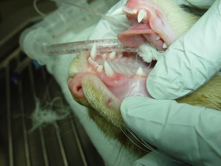
Eruption times for deciduous dentition – Feline
o Incisors 2-3 weeks
o Canines 3-4 weeks
o Premolars 3-6 weeks
o Molars – no deciduous
Dental formula for permanent teeth – Canine
o I3 – C1 – P4 – M2 // I3 – C1 – P4 – M3
Dental formula for permanent teeth – Feline
o I3 – C1 – P3 – M1 // I3 – C1 – P2 – M1
Eruption times for permanent dentition – Canine
o Incisors 3-5 months
o Canines 4-6 months
o Premolars 4-6 months
o Molars 5-7 months
Eruption times for permanent dentition – Feline
o Incisors 3-4 months
o Canines 4-5 months
o Premolars 4-6 months
o Molars 4-5 months
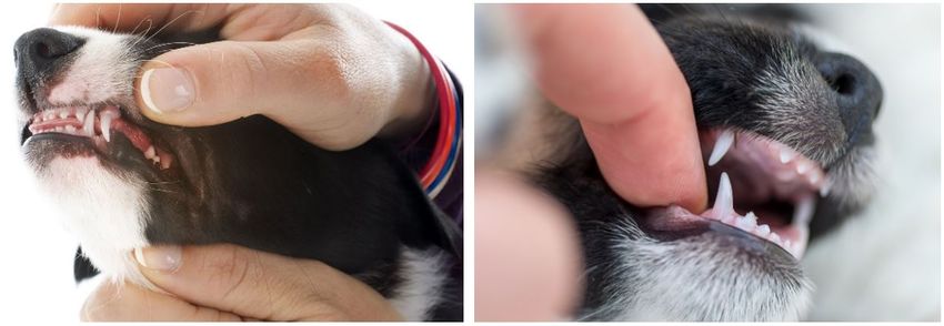
Deciduous Teeth – Persistent, (Retained), or Fractured
Common pathology associated with deciduous teeth include persistent or retained teeth. These two terms are often
used interchangeably, however, this is not appropriate as each term is specific. Persistent deciduous teeth is the term
that should be used for a deciduous tooth which remains present when the adult tooth is erupted. It persists, despite
the presence of the adult tooth. The deciduous tooth in this case should be extracted. No two teeth should occupy
the same space. The presence of the deciduous tooth can lead to malocclusion and will predispose the adult tooth to
periodontal disease. The term retained is better suited for dental structure that is present underneath the gums (i.e.
retained tooth root) or can sometimes be used when describing a deciduous tooth which is present due to the lack of
an adult counterpart. I most commonly see this with second premolars in dogs. I let owners know that a dental
radiograph should be performed to ensure there is no adult tooth. The deciduous tooth can remain if there is no
indication of pathology associated with it. Owners are warned that the tooth roots may resorb and there is the
potential the tooth can fracture – both situations requiring extraction in the future.
With adult patients we have more treatment options for a fractured tooth with pulp exposure (complicated fracture,
endodontic disease) including extraction, root canal therapy, or vital pulpotomy given the right circumstances. If a
deciduous tooth is fractured, treatment recommendation should be for extraction. Not only is the fractured tooth a
source of pain and infection, but the inflammation associated with endodontic disease can affect the developing
adult tooth bud. The apex of the deciduous tooth root sits in close proximity to the permanent tooth bud and changes
in the local environment can lead to disruption of the ameloblasts forming enamel on the adult tooth.
Angell Animal Medical Center • 350 S. Huntington Ave., Boston, MA 02130 • 617-522-7282 • fax: 617-989-1635
Malocclusions

Most times patients presenting for malocclusion
will be pediatric or young juvenile when this issue
is noticed. When assessing occlusion, keeping in
mind the standards of normal occlusion are most
important. Normal or ideal occlusion (canine)
consists of interdigitation of the upper and lower
teeth, maxillary incisor teeth are slightly rostral to
the mandibular incisor teeth with the mandibular
incisor teeth contacting the cingulum of the
maxillary incisors, the mandibular canine teeth
should be slightly inclined labially and sit within
the interdental space between the maxillary third
incisor and canine tooth, the crown cusps of the
mandibular premolars should be lingual to the
maxillary premolar teeth, the premolars should         Lance (mesioverted) canine
have a pinking shears relationship with the
mandibular first premolar rostral to the maxillary premolars, the mesial cusp tip of the maxillary fourth premolar
should be lateral to the space between the mandibular fourth premolar and first molar. Normal occlusion in cats
follows the same basic guidelines with minor adjustments.
Malocclusions can be a result of abnormal tooth positioning or due to abnormal jaw length relationship. Many
animals with malocclusion may have more than one deviation from normal and thus multiple concurrent
malocclusions (i.e. mandibular distoclusion with linguoverted mandibular canine teeth). Class 1 malocclusion
consists of a normal jaw length relationship with malpositioning of one or more teeth. Generally these
malocclusions are described by the physical direction that the tooth is angled or deviated (i.e. mesioverted). Another
version of class one malocclusion is a crossbite, where the mandibular teeth are more buccal or labial to the
opposing maxillary tooth. Crossbites can be described as rostral crossbite – referring to the incisor teeth, or caudal
crossbite – when the malocclusion is associated with the premolars or molars. Other classes of malocclusion deal
with skeletal malocclusion.
Class 2 malocclusion, or mandibular distoclusion, is when the mandible occludes caudal to the normal position
relative to the maxilla. Colloquial terms for this malocclusion include parrot mouth, overbite, overshot jaw. On the
other hand, class 3 malocclusion, mandibular mesioclusion, the mandible will be rostral to the maxillary arch. In
some breeds this occlusion is normal for the breed (i.e. brachycephalics). Other terms often used are underbite or
monkey mouth. Class 4 malocclusions involve asymmetry and can be in a rostro-caudal, side-to-side, dorso-ventral
direction, or a combination of these. We try to steer away from the term wry bite, as this is non-specific and does
not accurately provide an image of a patient’s malocclusion. An example of class 4 malocclusion in a rostrocaudal
direction would be when either the right or left side of the face has mandibular mesioclusion or distoclusion. A side-
to-side malocclusion would describe when the midline alignment of the maxilla and mandible is shifted. And
finally, a dorsoventral malocclusion would correspond with an open bite in which there is abnormal vertical space
between the maxilla and mandible when the mouth is in a closed position.
Treating malocclusion cases can be more of an art than a science, and frequently we are dealing with evolving
circumstances. Expectations of malocclusion treatment should be relayed to the owner and procedures should be
performed to provide pets with a comfortable bite rather than cosmetic outcome. Options for treatment include
interceptive orthodontics (extractions), corrective orthodontics, or alteration to a tooth shape/structure (crown
reduction with vital pulpotomy).

 Angell Animal Medical Center • 350 S. Huntington Ave., Boston, MA 02130 • 617-522-7282 • fax: 617-989-1635

Our role as veterinarians in helping pets have a mouth free of discomfort and disease should begin when our
patients are very young. Issues with deciduous dentition should not be overlooked due to the short timespan that
these teeth are present, since these issues may contribute to problems when the patient gets older. The stage when
adult teeth are erupting is a crucial time to ensure that teeth are accounted for, structurally normal, and positioned
appropriately.

 Angell Animal Medical Center • 350 S. Huntington Ave., Boston, MA 02130 • 617-522-7282 • fax: 617-989-1635
You can also read